Embed Size (px)
Citation preview
Neurobiology of Disease
p75NTR and Its Ligand ProNGF Activate ParacrineMechanisms Etiological to the Vascular, Inflammatory, andNeurodegenerative Pathologies of Diabetic Retinopathy
Pablo F. Barcelona,1,2* X Nicholas Sitaras,3* Alba Galan,1 X Gema Esquiva,4 Sean Jmaeff,1,2 Yifan Jian,5
Marinko V. Sarunic,5 X Nicolas Cuenca,4 Przemyslaw Sapieha,3,6,7* and H. Uri Saragovi1,2,8*1Lady Davis Institute-Jewish General Hospital, Center for Translational Research, McGill University, Montreal, Quebec H3T 1E2, Canada, 2Department ofPharmacology and Therapeutics, McGill University, Montreal, Quebec H3G 1Y6, Canada, 3Department of Ophthalmology, Maisonneuve-Rosemont HospitalResearch Centre, University of Montreal, Montreal, Quebec H1T 2M4, Canada, 4Department of Physiology, Genetics and Microbiology, University ofAlicante, Alicante CP 03690, Spain, 5School of Engineering Science, Simon Fraser University, Burnaby, British Columbia V5A 1S6, Canada, 6Department ofBiochemistry, Maisonneuve-Rosemont Hospital Research Centre, University of Montreal, Montreal, Quebec H1T 2M4, Canada, 7Department of Neurology-Neurosurgery, McGill University, Montreal, Quebec H3A 2B4, Canada, and 8McGill Cancer Center, McGill University, Montreal, Quebec H3A 1A3, Canada
In many diseases, expression and ligand-dependent activity of the p75 NTR receptor can promote pericyte and vascular dysfunction,inflammation, glial activation, and neurodegeneration. Diabetic retinopathy (DR) is characterized by all of these pathological events.However, the mechanisms by which p75 NTR may be implicated at each stage of DR pathology remain poorly understood. Using astreptozotocin mouse model of diabetic retinopathy, we report that p75 NTR is upregulated very early in glia and in pericytes to mediateligand-dependent induction of inflammatory cytokines, disruption of the neuro-glia-vascular unit, promotion of blood–retina barrierbreakdown, edema, and neuronal death. In a mouse model of oxygen-induced retinopathy, mimicking proliferative DR, p75 NTR-dependent inflammation leads to ischemia and pathological angiogenesis through Semaphorin 3A. The acute use of antagonists ofp75 NTR or antagonists of the ligand proNGF suppresses each distinct phase of pathology, ameliorate disease, and prevent diseaseprogression. Thus, our study documents novel disease mechanisms and validates druggable targets for diabetic retinopathy.
Key words: diabetes; neurodegeneration; neurotrophin; pathophysiology; receptor; retina
IntroductionDiabetic retinopathy (DR) is a major cause of reduced vision inthe working age population (Yau et al., 2012) and leads to irre-
versible vision loss in �75% of patients (Heng et al., 2013). DR isa retinal disorder that is characterized by several distinct phases,such as loss of the blood–retina barrier (BRB) function and cap-illary loss leading to diabetic macular edema (DME). DME ischaracterized by extravasation of fluids from retinal circulation
Received Nov. 26, 2015; revised June 10, 2016; accepted July 5, 2016.Author contributions: P.F.B., N.C., P.S., and H.U.S. designed research; P.F.B., N.S., A.G., G.E., and S.J. performed
research; Y.J. and M.V.S. contributed unpublished reagents/analytic tools; P.F.B., N.S., A.G., G.E., S.J., Y.J., M.V.S.,N.C., P.S., and H.U.S. analyzed data; P.F.B., N.S., P.S., and H.U.S. wrote the paper.
This work was supported by the Canadian Institutes of Health Research and the Foundation to Fight Blindness toH.U.S. and the Canadian Diabetes Association to P.S.
McGill University has patents applied or issued (H.U.S., inventor) covering the compounds used herein. Theremaining authors declare no competing financial interests.
*P.F.B. and N.S. contributed equally to this study as first coauthors. H.U.S. and P.S. shared senior authorship tothis study.
Correspondence should be addressed to Dr. H. Uri Saragovi, McGill University, 3755 Cote St. Catherine, E-535,Montreal, Quebec H3T 1E2, Canada. E-mail: [email protected].
DOI:10.1523/JNEUROSCI.4278-15.2016Copyright © 2016 the authors 0270-6474/16/368826-16$15.00/0
Significance Statement
Diabetic retinopathy (DR) affects an estimated 250 million people and has no effective treatment. Stages of progression comprise peri-cyte/vascular dysfunction, inflammation, glial activation, and neurodegeneration. The pathophysiology of each stage remains unclear.We postulated that the activity of p75NTR may be implicated. We show that p75NTR in glia and in pericytes mediate ligand-dependentinduction of inflammatory cytokines, disruption of the neuro-glia-vascular unit, promotion of blood–retina barrier breakdown, edema,and neuronal death. p75NTR-promoted inflammation leads to ischemia and angiogenesis through Semaphorin 3A. Antagonists ofp75NTR or antagonists of proNGF suppress each distinct phase of pathology, ameliorate disease, and prevent disease progression. Ourstudy documents novel mechanisms in a pervasive disease and validates druggable targets for treatment.
8826 • The Journal of Neuroscience, August 24, 2016 • 36(34):8826 – 8841

into the retina, which can lead to significant decreases in visualacuity (Antonetti et al., 2012; Yau et al., 2012). Later stages areassociated with retinal ischemia causing hypoxia and a subse-quent phase of VEGF-mediated neovessel growth or proliferativeDR (PDR). Preventing destructive angiogenesis remains the onlycurrent treatment modality for PDR (Antonetti et al., 2012; Henget al., 2013).
Accumulating evidence points to defects of the neurovascularunit (neurons, glia, and vessels) (Binet et al., 2013; Cerani et al.,2013; Joyal et al., 2013; Dejda et al., 2014) throughout the pro-gression of DR and during pathological preretinal neovascular-ization (NV), suggesting that the pathophysiology of DR is notpurely vascular in nature (Abcouwer and Gardner, 2014). Forinstance, retinal oxidative stress resulting from hyperglycemiaand hyperlipidemia (Kowluru and Chan, 2007) contributes toglial dysfunction and activation (gliosis) (Bringmann et al., 2006;Coorey et al., 2012), inflammation induced by resident glia (Mul-ler cells), and death of the specific neuronal cell populations (ret-inal ganglion cells [RGCs]) (Ali et al., 2008), which intriguinglyprecede any observable clinical vascular irregularities or neuronalcell dysfunction in diabetes (Fletcher et al., 2007; Abcouwer andGardner, 2014). However, although these studies highlight thepremature changes that occur in the retina of diabetic patients, itis not clear whether glial cell abnormalities precede neuronalcell dysfunction or vice versa. Moreover, the pathophysiologicalevents that bring about these changes remain ill defined, andcurrently no therapeutic strategies have been developed tocounter them. Thus, it is important to understand these earlychanges in the neurovascular unit as they may be complementaryto or even precede vascular abnormalities in diabetic patients.
The neurotrophin receptor p75 (p75 NTR), which is highly ex-pressed in astrocytes and Muller cells in many models of neuronaldamage, would be capable of mediating many of the pathologicalevents described above. In retinopathies, such as glaucoma oroptic nerve damage, p75 NTR is upregulated and its ligandproNGF can promote production of inflammatory cytokinesTNF� (Lebrun-Julien et al., 2009a; b, 2010; Bai et al., 2010a) and�2-macroglobulin (�2M) from glial cells (Shi et al., 2008; Bai etal., 2011), which leads to RGC neurodegeneration. Moreover,p75 NTR is known to contribute to cardiac vasculopathies (Siao etal., 2012), BRB breakdown, and fluid extravasation (Al-Gayyar etal., 2011; Mysona et al., 2013). These observations potentiallyplace p75 NTR at a crossroads of all pathological components ofhuman DR: vascular defects, inflammation, and neurodegenera-tion. However, the mechanism by which p75 NTR mediates thesepotentially sequential pathophysiological events during diabetesremains poorly understood.
Here, we use a streptozotocin (STZ)-induced mouse model ofdiabetes causing BRB breakdown with edema, inflammation, andRGC death; and an oxygen-induced retinopathy (OIR) mousemodel to study pathological NV. We demonstrate a rapid andpersistent increase of both proNGF and p75 NTR at early stages ofdisease. The p75 NTR increase occurs in Muller cell glia and inpericytes around the vasculature. p75 NTR activity in Muller gliaaugments inflammatory cytokines TNF� and �2M, which per-form two paracrine functions. First, TNF� stimulates RGCs tosecrete the vascular cue Semaphorin 3A, a soluble factor knownto cause BRB breakdown and plasma extravasation (Cerani et al.,2013; Joyal et al., 2013). Second, TNF� and �2M cooperate topromote RGC death in a proNGF and p75 NTR-dependent man-ner. Diabetes-induced BRB breakdown and edema, productionof inflammatory cytokines, and RGC death are significantly di-minished for prolonged periods upon acute administration of
inhibitors of proNGF or p75 NTR; and both the vaso-obliteration(VO) and NV phases are significantly diminished in the mousemodel of OIR. Thus, p75 NTR and proNGF are key pathologicalmediators within the neuro-glia-vascular unit and may be poten-tial therapeutic targets for various stages of DR pathology, includ-ing the DME, PDR, and neurodegenerative components; andoffer an alternative to VEGF-based strategies to limit vascularabnormalities.
Materials and MethodsAnimals. All experiments were conducted in accordance with Associa-tion for Research in Vision and Ophthalmology statement regarding useof animals in ophthalmic and vision research and were approved by allInstitutional Animal Welfare Committees.
Female and male adult C57BL/6J (WT) mice were purchased from TheJackson Laboratory and coupled for litter production in the Animal Carefacility at Maisonneuve-Rosemont Hospital Research Center (Montreal,Quebec, Canada). Adoptive CD-1 lactating females were purchased fromCharles River Laboratories to tend to hyperoxia-exposed pups.
Induction of diabetes. Male 10-week-old mice (�24 g) received anintraperitoneal injection of STZ (60 mg/kg) (Sigma-Aldrich) dissolved insodium citrate buffer (0.01 M, pH 4.5) on 5 consecutive days. After 1 and6 weeks of injection with STZ, blood glucose was measured using a glu-cometer (Abbott Laboratory), and fasting blood glucose levels �17mmol/L (300 mg/dl) (Mysona et al., 2013) were considered to be dia-betic. Age-matched, nondiabetic C57BL/6 mice injected with sodiumcitrate buffer were used as controls. Fasting blood glucose levels weremeasured routinely. As example, a control group was 7.89 � 0.69 mM,and the STZ group was 28.81 � 4.204 mM ( p � 0.001, n � 12).
OIR. Newborn mice pups were exposed for 5 d to 75% oxygen frompostnatal day 7 (P7) to P12. VO and NV were assessed in hyperoxia-exposed mice pups at P12 and P17, respectively, as described previously(Smith et al., 1994; Stahl et al., 2010). Briefly, mice pups were fully anes-thetized in 3% isoflurane in oxygen and killed using a guillotine. Eyeswere enucleated and fixed in 4% PFA solution for 60 min at room tem-perature. Retinas were dissected and stained overnight at 4°C withfluorescein-labeled GSL 1, isolectin B4 (FL 1201, Vector Labs; 1:100).Lectin-stained retinas were whole-mounted onto Superfrost/Plus micro-scope slides (Fisher Scientific) with the photoreceptor side down andimbedded in Fluoro-gel (Electron Microscopy Sciences) and photomi-crographed at 10� using a Zeiss AxioObserver.Z1. Images were pho-tomerged into a single file using the MosiaX option in the AxioVision4.6.5 software. Quantification of VO and NV was assessed using theSWIFT_NV methods as previously described (Stahl et al., 2009).
Optical coherence tomography (OCT) imaging. A noninvasive proto-type spectrometer-based FD-OCT system was used to acquire the retinalimages. FD-OCT is a noninvasive method that allows time-kinetic stud-ies in the same animal, with axial resolution in tissue nominally betterthan 3 �m and repeatability of the measurements from B-scans betterthan 1 �m. Data acquisition was performed using custom software writ-ten in C 2� for rapid frame grabbing, processing, and display of 2D im-ages (Li et al., 2011; Jian et al., 2013). B-scan images were segmentedmanually to quantify the thicknesses of the different retinal layers.
Mice were anesthetized using isoflurane/O2 and placed on a platform,and the head was oriented to an angle at which the eye was properlyaligned to the optical beam. The pupils were dilated with a topical solu-tion (atropine sulfate 1%; Alcon). Refraction of light at the cornea wascancelled by placing over the eye a generic artificial tear gel. Alignment ofthe optical system to the mouse retina required a few minutes and wasfollowed by rapid acquisition of data (�5 s/vol). During retinal scanning,three volumes were acquired in different sectors of the retina containingthe ON head as landmark. After processing, three B-scans were randomlyselected from each volume. The retinal thickness measurements wereperformed with MATLAB software (The MathWorks). In each B-scan,the thickness of the nerve fiber layer/ganglion cell layer (GCL)/innerplexiform layer (IPL), hereafter referred to as NGI (Saragovi et al., 1998;Bai et al., 2010a), was measured at three adjacent points. For each B-scan,the thickness of the inner nuclear layer (INL) and outer nuclear layer
Barcelona, Sitaras et al. • Disease Stage-Dependent p75 Mechanisms in Diabetic Retinopathy J. Neurosci., August 24, 2016 • 36(34):8826 – 8841 • 8827

Figure 1. Expression and localization of p75 NTR and neurotoxic factors increase during the progression of DR. The experimental paradigm and the endpoints of the mouse DR model are shown.Drug treatments were injected intravitreally at week 2.5 of diabetes in this paradigm. A, p75 NTR, proNGF, TNF�, and �2M expression in whole mice retina analyzed by Western blot from 1– 6 weeksafter induction of diabetes. There were increases of p75 NTR during week 1, of proNGF during week 3, of TNF� during week 4, and of �2M during week 5. B, Quantification. *p � 0.05, relative tocontrol. n �3 independent experiments, each with n �4 mice/group. Statistical analysis was done by applying one-way ANOVA with significance established at ��0.05, followed by Bonferroni’scorrection for multiple comparisons. The 32–37 kDa bands of proNGF were quantified, and data are identical when the major band alone is quantified. C, Confocal microscopy (Figure legend continues.)
8828 • J. Neurosci., August 24, 2016 • 36(34):8826 – 8841 Barcelona, Sitaras et al. • Disease Stage-Dependent p75 Mechanisms in Diabetic Retinopathy

(ONL), unaffected in the disease model, was also quantified as internalcontrols. Data are shown as average � SEM thickness in micrometers(absolute values) in control versus diabetic-injected intravitreally oncewith either vehicle (PBS, 1% DMSO), THX-B, a pharmacological small-molecule antagonist of p75 NTR (Bai et al., 2010a; Mysona et al., 2013), oranti-proNGF NGF30 mAb (Saragovi et al., 1998; Bai et al., 2010a).THX-B is an antagonist of ligand-dependent and ligand-independentactivity of p75 NTR, and a competitive blocker of proNGF binding top75 NTR. The anti-proNGF mAb blocks proNGF binding to p75 NTR, aswell as the binding of mature NGF to p75 NTR, but does not affect matureNGF binding or activity at TrkA.
Intravitreal injections. Mice were anesthetized in 3% isoflurane duringintravitreal injections of clear solutions of THX-B p75 NTR antagonist,neutralizing anti-proNGF mAb, or vehicle. In the diabetes model, adultdiabetic mice were injected at 2.5 weeks of diabetes. In the OIR model,mouse pups were injected at P7, P9, P12, or P14. All solutions wereinjected using a Hamilton syringe with 50 gauge glass capillary. Theintraocular injections were in 2 �l volumes containing a total of 2 �gTHX-B or 2 �g anti-proNGF NGF30 mAb as described for glaucoma(Shi et al., 2008; Bai et al., 2010a).
TUNEL assays and image analyses of flat-mounted retinas. The eyeswere collected and retinas were dissected for flat-mounting and TUNELstaining; n � 4 mice per group. Control nondiabetic animals and diabeticanimals were studied. One eye of each mouse was injected intravitreallyonce with either THX-B, or anti-proNGF mAb, or vehicle. Contralateraluntreated eyes were used as control. Retinas were processed and stainedwith the DeathEnd Fluorometric TUNEL System as per the manufactur-er’s instructions (Promega). Briefly, whole retinas were dissected, fixed in4% PFA overnight, and permealized with Proteinase K. Afterward, thesamples were postfixed and the TdT reaction was carried on at 37°C inhumidified chambers. TUNEL-labeled retinas were mounted withVectashield-DAPI to counterstain nuclei. For each flat-mounted wholeretina, three digital images per quadrant (superior, temporal, inferior,and nasal) were taken using confocal fluorescence microscopy at 20�magnification, for a total of 12 images per retina taken in a blindedfashion (n � 4 retinas per group). Images were taken at 0.5– 0.7, 1.0 –1.2,and 2.0 –2.2 mm distances from the optic disc (areas 1, 2, and 3, respec-tively) and analyzed per area to account for known variations in RGCdensity in each area, as described previously (Bai et al., 2010a). TotalTUNEL-positive cells and total nuclei of RGCs were counted in eachpicture. Each 20� magnification field exposes 0.2285 mm 2; and in eachindependent experiment, images spanning 10.97–21.93 mm 2 per groupwere analyzed.
RGC and fiber counts. The cornea, lens, and vitreous body were re-moved and the retina was dissected out for flat-mounting (Esquiva et al.,2013). After blocking (10% normal donkey serum in phosphate bufferplus 0.5% Triton X-100 for 1 h), the retinas were incubated for 72 h at 4°Cwith goat anti-Brn3a antibody (1:500; Santa Cruz Biotechnology) dilutedin 0.1 M phosphate buffer containing 0.5% (v/v) Triton X-100 (Sigma) todetect the RGC population (García-Ayuso et al., 2010, 2015). After washwith phosphate buffer, the retinas were incubated overnight with thesecondary antibody AlexaFluor-488 donkey anti-goat IgG (1:500; Invit-rogen). Then, the retinas were washed and flat mounted in mountingmedium (Citifluor) on glass slides with the GCL side up, and protectedwith a coverslip for optical microscopy. Using MetaMorph software(Leica MMAF 1.6) associated to a DMLA Microscope (Leica Microsys-
tems) entire flat-mount retinas were photographed at 20� magnifica-tion. High-magnification pictures were taken using a laser-scanningconfocal microscope (TCS SP2, Leica Microsystems). Brn3a-positivecells in the GCL were quantified at 0.5– 0.7, 1.0 –1.2, and 2.0 –2.2 mmdistances from the optic disc (areas 1, 2, and 3, respectively) using image-editing software (Photoshop 10.0; Adobe). Mean density (number ofcells per mm 2) values were calculated for all the different areas. A total of4 retinas per experimental group were analyzed.
FISH. The digoxigenin (DIG) RNA labeling kit (Roche) was used togenerate the DIG-labeled probes. Efficient labeling was verified in aga-rose gels (1%). After enucleation, the eyes were immersed overnight infixative at 4°C (4% PFA in PBS, pH 7.4), followed by cryoprotection in30% sucrose overnight at 4°C. Eyes were embedded in OCT tissue TEK(VWR) and frozen with dry ice. Cryostat-cut sections (20 �m thick) weremounted onto gelatin-coated glass slides, refixed in 4% PFA, permeabil-ized with proteinase K, and acetylated. Hybridization was carried byincubating the slides with either 200 ng/ml of DIG-labeled TNF� or �2Mantisense RNAs probes overnight at 72°C in a hybridization oven (Rob-bins Scientific). As controls, hybridization with either 200 ng/ml ofDIG-labeled TNF� or �2M sense RNAs probes was performed on paral-lel slides using the same experimental conditions. Hybridization wasfollowed by nonstringent and stringent washes, and incubation withanti-DIG-HRP (1:1000) overnight at 4°C. The amplification reaction wasperformed using the TMR-conjugated TSA kit (PerkinElmer) followingmanufacturer protocols. Finally, sections were washed and coverslippedusing Vectashield mounting media with DAPI (Burlingame). The slideswere analyzed by confocal microscopy.
Vascular permeability assay. Evans Blue permeation was analyzed bymeasuring albumin-Evans blue complex leakage from retinal vessels aspreviously described (Ma et al., 1996). Briefly, animals were injectedintravenously with a solution of Evans blue (2% w/vdissolved in PBS).After 48 h, animals were killed, the whole eyes were enucleated, immedi-ately embedded in OCT compound and cross sections were prepared.Microphotographs by immunofluorescence microscopy were obtainedusing identical exposure time, brightness, and contrast settings. For eachexperimental condition, at least 6 images were acquired from 3 sectionscut from different areas of the retina (n � 3 retinas per group). The areaof the Evans Blue permeation was measured using ImageJ software. Forthe in situ studies, an arbitrary rectangle was drawn that include all layerof the retina (GCL, IPL, INL, and ONL) to measure the Evans Bluestaining using ImageJ. Data are shown as the average area (in pixels) �SEM in control versus diabetic injected once with vehicle, anti-proNGFmAb, or small-molecule p75 NTR antagonist THX-B.
Immunohistochemistry. Whole eyes were enucleated from mice pups oradult and immediately fixed in 4% PFA at room temperature for 2 h. Eyeswere saturated overnight at 4°C in a 30% sucrose solution before embeddingin OCT compound (24608–930, TissueTek). Retinal cross sections of 10 �mwere sectioned using a Cryostat (Leica). Sections were subsequently washedwith PBS, permeabilized for 60 min at room temperature with 3% BSA, 0.2%Triton X-100, and 0.05% Tween20 in PBS, and stained with antibodies tomouse p75NTR (1:1000; a kind gift from Dr. R. Rush) or rabbit p75NTR
(1:1000; a kind gift from Dr. P. Barker), rabbit glutamine synthetase (16802,Abcam; 1:200), mouse cellular retinaldehyde-binding protein (CRALBP,15051, Abcam; 1:1000), rabbit GFAP (AB5804, Millipore, 1:1000), rat anti-proNGF/NGF mAb NGF30 (1:2000) (Saragovi et al., 1998; Bai et al., 2010a)prepared in house, rabbit TNF� (AB2148P, Millipore, 1:1000), rabbit �2M(1:1000, Santa Cruz Biotechnology) or mouse �III-tubulin (T8578, Sigma-Aldrich; 1:1000). Fluoresceinated secondary antibodies (goat anti-mouseIgG AlexaFluor-488, -594, and/or -647 and goat anti-rabbit IgG AlexaFluor-488, -594, and/or -647; Invitrogen) were used for localization studies accord-ing to manufacturers’ recommendations. Images were obtained using anIX81 confocal microscope (Olympus) equipped with Fluoview 3.1 software(Olympus). As controls during immunostaining, adjacent sections wereprocessed equally but without primary or with irrelevant primary, followedby the proper secondary. In all cases, background levels are undetectable.
Western blot. Eyes were enucleated and retinas immediately dissectedand placed into protein lysis buffer and briefly homogenized. Samples(combined retinal lysate from two different animals) were centrifugedand 30 �g resolved on an SDS-PAGE gel (8%–12%) and electro-blotted
4
(Figure legend continued.) images show induction and distribution of p75 NTR in mice retinaafter 6 weeks of diabetes compared with age-matched control animals. p75 NTR (red) was pref-erentially expressed in Muller cells (green, white arrows in insets pointing at the end-feet ofMuller cells), as revealed by coexpression with CRALBP, and around the vessels where p75 NTR
(red, white arrows in insets pointing at the vessels in the outer plexiform layer) was coexpressedwith GFAP, a specific marker of active Muller and astrocyte cells (green). Nuclei are counter-stained with DAPI. White rectangles represent magnified areas (40�) shown in the insets.Scale bar, 30 �m. PhR, Photoreceptor layer; OLM, outer limiting membrane; RPE, retinal pig-ment epithelium. D, Flat-mounted retinas from the same animals show partial colocalization ofp75 NTR (green) with NG2 (red) pericyte marker around vessels. Scale bar, 100 �m.
Barcelona, Sitaras et al. • Disease Stage-Dependent p75 Mechanisms in Diabetic Retinopathy J. Neurosci., August 24, 2016 • 36(34):8826 – 8841 • 8829

8830 • J. Neurosci., August 24, 2016 • 36(34):8826 – 8841 Barcelona, Sitaras et al. • Disease Stage-Dependent p75 Mechanisms in Diabetic Retinopathy

onto either PVDF or nitrocellulose membranes (Bio-Rad). Membraneswere washed, blocked for 60 min in 5% milk in TBS-Tween, and incu-bated overnight at 4°C with mouse antibody to �-actin (sc-47778, SantaCruz Biotechnology; 1:1000), rabbit antibody to p75 NTR (1:1000; a kindgift from Dr. P. Barker), rabbit TNF� (AB2148P, Millipore, 1:2000),rabbit �2M (Santa Cruz Biotechnology; 1:2000), and rat proNGF/NGF(1:200). After washing, membranes were incubated with 1:5000 HRP-conjugated anti-mouse or 1:2000 HRP anti-goat or -rabbit secondaryantibodies (Millipore) for 2 h at room temperature. Membranes wereimaged with LAS-3000 imager (FujiFilm), and bands were assessed usingdensitometry plugins in Multi Gauge 4.0 software (FujiFilm). The size ofbands reported are Mr calculated using standard markers.
Cell culture. Rat brain microvascular endothelial cells (RBMVECs)were obtained from Cederlane Laboratories and used between passages2–7. rMC-1 cells were cultured in low glucose DMEM (Invitrogen) sup-plemented with 10% FBS and 1% penicillin/streptomycin at 37°Cand 5% CO2, whereas RBMVECs were cultured as described by manu-facturer’s instructions. Cells were starved 4 h before treatment with 0.1,1.0, or 10.0 ng/ml of recombinant proNGF (CYT-426, ProSpec). Neu-tralizing antibody to proNGF was used at 1 �g/ml, whereas THX-B wasused at 4 �g/ml. Proteins were extracted using protein lysis buffer andlysates processed for Western blot analysis. Total RNA was isolated usingTriZol (Invitrogen) and analyzed via RT-PCR and qPCR analysis.
Reverse-transcription PCR and quantitative real-time PCR. Cell cultureand retinas samples were processed using TriZol (Invitrogen) as de-scribed by the manufacturer’s instructions. Subsequently, genomic DNAwas removed using DNase I (Invitrogen) as described by the manufac-turer’s instructions. Total RNA (1 �g) was reverse-transcribed intocDNA using iScript RT Supermix (Bio-Rad) following the indications ofthe manufacturer. cDNA was analyzed by quantitative real-time PCR onthe ABI 7500 Real-Time PCR system (Applied Biosystems) and using iQSYBR Green Supermix (Bio-Rad) with primers targeting Semaphorin3A, TNF�, and �-actin (designed using primer3 [NCBI]). The primerswere used to generate specific fragments: rat TNF� forward, 5 CTATGTGCTCCTCACCCACA 3, reverse 5 TGGAAG ACTCCTCCCAGGTA 3; mouse TNF� forward, 5 GAGTCCCAGGTCTACTTT 3, reverse5 CAGGTCACTGTCCCAGCATCT 3; and mouse Semaphorin 3A for-ward, 5 GGGACTTCG CTATCTTCAGAAC 3, reverse, 5 GGCGT-GCTTTTAGGAATGTTG 3. All primers were used at 100 nM. Thereaction was performed in triplicate with an Mx3000P QPCR System(Stratagene). Amplification reaction data were analyzed using the com-plementary Mx3000P analysis software. Target gene expression was nor-malized to the average expression of the housekeeping gene GAPDHforward 5 CACCACCTTGATGTCATC-3, reverse 5AGCCCAGAACATCCCTG3. Quantitative analysis of gene expression was assessed us-ing the CT quantification.
Preparation of rMC-1 conditioned media (CM). rMC-1 were seeded(10 6 cells) and starved 4 h before treatment with 1.0 ng/ml of recombi-nant proNGF (CYT-426, ProSpec). Supernatants were collected 24 hlater, cleared by centrifugation, filtered through 0.22 �m filters (Milli-pore), and distributed for cell survival assays (see below).
Cell survival assay. Approximately 10 4 RBMVEC cells/well wereseeded in 24 well plates and starved 4 h before exposure to rMC-1 CM.
Neutralizing antibody to proNGF was used at 1 �g/ml, whereas THX-Bwas used at 4 �g/ml. After 24 h, 50 �l of a 5 �g/ml solution of thiazolylblue tetrazolium bromide (M2128; Sigma) was added to each well andcells incubated for 2–3 h. Subsequently, the supernatant was aspiratedand cells were lysed and resuspended in acidified isopropanol and shakenfor 5 min. Duplicate absorbance readings were taken at 565 nm usingInfinite M1000 Pro plate reader (TECAN).
Statistical analysis. Results are presented as mean � SEM for all stud-ies. One-way or two-way ANOVA with significance � � 0.05 or higherwere used for processing data. Bonferroni post hoc analysis was used forcalculating significance between groups. Two-tailed Student’s t tests wereused to test for significance between two means.
ResultsUpregulated retinal p75 NTR and proNGF in a mouse model ofType 1 diabetes is followed by induction of proinflammatorycytokinesTo study the effects of p75NTR and its ligand proNGF in DR, wedetermined their expression pattern in a STZ mouse model of Type1 diabetes mellitus. Time-kinetic studies were performed by quanti-tative Western blots on retinas collected from diabetic animals be-tween weeks 1–6 following diabetes onset. At week 1 of diabetes,retina p75NTR protein levels rose �2-fold compared with controlretinas (p � 0.05, n � 4). Retina proNGF protein expression in-creased �2-fold at week 3 of diabetes compared with control retinas(p � 0.05, n � 4). Significantly higher retinal levels of p75NTR andproNGF persisted for at least 6 weeks of diabetes compared to con-trol retinas (Fig. 1A, quantified in Figure 1B).
In some retinal neurodegenerative diseases, such as opticnerve axotomy or glaucoma, proNGF-mediated toxicity affectsnon–p75 NTR-expressing cells, suggesting a non– cell-autologousprocess. Indeed, in human and rodent disease, p75 NTR andproNGF upregulate two proteins �2M (Bai et al., 2010a, b, 2011)and TNF� (Lebrun-Julien et al., 2009a, 2010; Bai et al., 2010a, b,2011) secreted from Muller glia, which contribute to neurode-generation (Bai et al., 2011; Barcelona and Saragovi, 2015).
Hence, we studied these proteins as a putative mechanism thatcan perturb glial function, induce neuronal apoptosis, and causevascular perturbations in the STZ model of diabetes. In diabeticretinas, �2M protein levels increased �1.5-fold at week 5 comparedwith controls (�2M, 1.641 � 0.149; p � 0.05, n � 4) and TNF�protein levels surged �2-fold increase at week 4 compared withcontrols (TNF�, 2.12 � 0.193; p � 0.05, n � 4) (Fig. 1A, quantifiedin Figure 1B). Essentially, sequential upregulation of p75NTR andproNGF is associated time-dependently with an increase in TNF�and �2M that can be proinflammatory and neurotoxic.
Retinal p75 NTR is upregulated in activated Muller cells,astrocytes, and vascular pericytes in a mouse model ofType 1 diabetesImmunohistochemical studies in retinal sections demonstratedincreased expression of p75 NTR in Muller cells identified byCRALBP; and in the periphery of vessels, the nerve fiber andGCLs, and Muller cell end-feet and astrocytes labeled with GFAP(Fig. 1C). p75 NTR levels were significantly elevated in diabeticretinas compared with control animals, which corroborates theWestern blot analysis. The elevated p75 NTR label in the peripheryof vessels of diabetic retinas seen in cross sections was studied inwhole flat-mounted retinas. The elevated p75 NTR label corre-sponds to vascular pericytes also labeled with the NG2 marker(Fig. 1D). In these whole flat-mounted retinas, the p75 NTR label(green) that appears beneath the pericytes corresponds to Mullercell fibers and astrocytes. Similar data were obtained using otherpericyte markers (PDGF-receptor; data not shown).
4
Figure 2. The expression of the cytotoxic molecules TNF� and �2M is increased in Mullercells during DR. A, FISH shows induction and distribution of DIG-labeled TNF� or �2M antisenseRNAs probes (red) in mice retina after 6 weeks of diabetes compared with age-matched controlanimals. Nuclei are counterstained with DAPI. Scale bar, 30 �m. Histograms represent thequantification of the expression of TNF� and �2M mRNAs in the GCL, IPL, INL, outer plexiformlayer (OPL), and photoreceptor layer (PhR). Statistical analysis was done by applying one-wayANOVA with significance at � � 0.05. *p � 0.05, relative to control. **p � 0.01, relative tocontrol. ***p � 0.001 relative to control. n � 3 independent experiments, each with n � 4mice/group. B, FISH followed by immunofluorescence showing colocalization of TNF� mRNAs(red) with p75 NTR protein (green) in the end-feet of Muller cells (white arrow, insets) and in theIPL. Scale bar, 30 �m. White rectangles represent higher-magnification images (40�) repre-sented in the insets. Sense TNF� mRNA hybridization was used on diabetic retina samples as anegative control.
Barcelona, Sitaras et al. • Disease Stage-Dependent p75 Mechanisms in Diabetic Retinopathy J. Neurosci., August 24, 2016 • 36(34):8826 – 8841 • 8831

Figure 3. Pharmacological inhibition of p75 NTR or proNGF preserves retinal structures where RGCs are located and inhibits RGC death in DR. A, Representative images of Brn3-labeled RGCs from3 different retinal areas at concentric distances from the optic nerve: 0.5– 0.7 mm (area 1), 1.0 –1.2 mm (area 2), and 2.0 –2.2 mm (area 3). RGCs were quantified in (Figure legend continues.)
8832 • J. Neurosci., August 24, 2016 • 36(34):8826 – 8841 Barcelona, Sitaras et al. • Disease Stage-Dependent p75 Mechanisms in Diabetic Retinopathy

�2M and TNF� mRNA in diabetes is upregulated primarily inMuller glial cellsBecause �2M and TNF� are soluble factors, to determine their cel-lular expression patterns, we performed in situ mRNA hybridizationon retinal sections of mice 6 weeks after STZ. Our results demon-strate significant increases in TNF� mRNA in the RGC layer (p �0.01), IPL (p � 0.05), INL (p � 0.01), and photoreceptor layer (p �0.01) compared with control retinas (Fig. 2A). Interestingly, most ofthe diabetes-induced upregulation of TNF� mRNA was seen in theRGC layer and INL, the main retinal layers affected in DR. Further-more, there was an increase in �2M mRNA in the RGC layer andINL. Upregulation of �2M mRNA was also detected in the boundarybetween the INL and photoreceptor layers, likely associated withcells of the retinal vasculature in that location (Fig. 2A). Graphs showsignificant changes quantified for TNF� and �2M mRNAs in eachretinal layer.
Some of the TNF� mRNA signal in the RGC layer colocalizeswith p75 NTR protein in what are likely Muller cell end-feet. Con-trol hybridization with TNF� sense mRNA probes showed unde-tectable levels, confirming the specificity of the technique (Fig.2B). Similar controls were obtained using �2M sense probes (datanot shown).
Together, these data suggest that, in diabetic retinas, p75 NTR,expressed mainly on Muller glia and astrocytes, contribute to theproduction of �2M and TNF�, which can be proinflammatoryand. neurotoxic and may contribute to the pathogenesis of dia-betes through a paracrine mechanism.
Pharmacological inhibition of p75 NTR or proNGF in diabetesreduces RGC death and preserves retinal structuresNext, we evaluated whether elevated expression of p75 NTR inMuller glia, astrocytes, and pericytes impact neurodegenerationassociated with DR progression. To do this, we quantified RGC-specific Brn3 staining, TUNEL staining, and OCT measurementsof retinal structure in diabetic retinas with or without therapeutictreatment with a p75 NTR small-molecule antagonist (THX-B) oran anti-proNGF blocking mAb (Fig. 3) previously described ashaving potent p75 NTR inhibitory properties (Bai et al., 2010a).Analyses and quantification were done at specified distancesfrom the optic disc (areas 1, 2, and 3, respectively) to account fornormal thinning of RGC density toward the periphery. After 6weeks of diabetes, there was an �15%-25% reduction in Brn3�
RGCs in flat-mounted whole retinas, compared with control na-ive retinas (area 1: 3450 � 413 vs 2787 � 219 cells/mm 2, p � 0.05;area 2: 3215 � 284 vs 2095 � 250 cells/mm 2, p � 0.01) (Fig. 3A).
To test whether RGC loss in diabetic retinas is p75 NTR- orproNGF-dependent, animals were injected THX-B or anti-proNGF mAb into the vitreous 2.5 weeks following onset diabe-tes (contralateral eyes were injected with vehicle control).Treatment with anti-proNGF mAb or THX-B preserved theBrn3� RGCs counted at week 6 of diabetes (area 1: 3202 � 123and 3350 � 98 cells/mm 2, respectively, vs 2786 � 279 cells/mm 2
vehicle-treated contralateral eyes, p � 0.05; area 2: 2944 � 129and 3310 � 378 cells/mm 2, respectively, vs 2416 � 194 cells/mm 2 vehicle-treated contralateral eyes, p � 0.001 and p � 0.05,respectively) (Fig. 3B).
To consolidate these results, we performed TUNEL stainingon retinal flat mounts obtained from 6 week diabetic mice thathad been injected 2.5 weeks following onset diabetes withTHX-B, proNGF mAb, or vehicle. TUNEL-positive staining wassignificantly diminished in eyes receiving intravitreal injectionsof THX-B or anti-proNGF mAb compared with vehicle-treatedcontralateral eyes (Fig. 3C; p � 0.05). The analyses of flat-mounted retinas quantify the RGC layer containing RGC nuclei,displaced amacrine cells, and astrocytes. Because astrocytes donot die in this disease model, the TUNEL� cells can only be RGCneurons or displaced amacrine cells.
These data indicate that the reduced numbers of Brn3� RGCs indiabetic retinas occurs in a p75NTR-dependent manner, an outcomeprevented by inhibition of p75NTR activity using THX-B or anti-proNGF mAb. Moreover, the fact that p75NTR is not expressed inRGCs suggest a non–cell-autonomous mechanism.
Further RGC layer quantification was performed by OCT, anoninvasive method for measuring structural changes in vivo,and which correlates with RGC numbers during neurodegenera-tion (Sarunic et al., 2010). OCT was performed longitudinally inanimals from 1 to 6 weeks following onset of diabetes, measuringRGC cell bodies and fibers in the nerve fiber layer, the GCL, andthe IPL termed NGI (Fig. 3D). In diabetic eyes, the NGI thicknessdecreased progressively and was significantly lower after 5 weeksof diabetes compared with age-matched nondiabetic eyes(37.85 � 1.3 �m vs 50.45 � 0.9 �m; p � 0.05).
Treatment of diabetic eyes with anti-proNGF mAb or THX-Bprotected against NGI thinning (42.51 � 1.7 �m and 43.75 � 1.1�m, respectively, p � 0.05 vs vehicle-treated contralateral dia-betic eyes). Structural preservation of NGI by anti-proNGF mAbor THX-B were still significant after 6 weeks of diabetes (42.42 �0.9 �m and 44.67 � 1.9 �m, respectively; vs vehicle-treated con-tralateral diabetic eyes 37.11 � 1.5 �m, p � 0.05) (Fig. 3D).
As internal controls, we quantified retinal layers that do notchange in diabetes (INL, containing the Muller and bipolarcell bodies) as well as the cone and rods of photoreceptors thatmake up the ONL. Combined, these data indicate that, in theretinas of diabetic mice, ligand-dependent p75 NTR activitypromotes effectors that contribute to RGC death, with result-ing retinal structural changes specifically in the NGI. Giventhe significant and selective loss of the nerve fiber layer thick-ness, which comprises RGC axons and fibers, most of theTUNEL� cells must be RGCs.
In sum, a single injection of an antagonist of p75NTR or proNGF,at week 2.5 of diabetes and with concurrent pathology, preventsstructural degeneration and RGC apoptosis for prolonged periodslasting to at least week 6 of continuing diabetes.
4
(Figure legend continued.) flat-mounted retinas at 2, 4, and 6 weeks after induction of dia-betes. Scale bar, 20 �m. *p � 0.05 versus control naive mice. **p � 0.01 versus control naivemice. ***p � 0.001 versus control naive mice. B, Representative images of Brn3-labeled RGCsin flat-mounted retinas in 6 week diabetic mice � treatment at week 2.5 of diabetes withanti-proNGF mAb, or THX-B, or vehicle, compared with age-matched naive control animals.Scale bar, 20 �m. Anti-proNGF mAb or THX-B treatment prevents loss of RGCs in all areas: area1, **p � 0.01 (repeated-measures ANOVA); area 2, ***p � 0.001 (repeated-measuresANOVA); area 3, *p � 0.05 (repeated-measures ANOVA). C, Representative pictures of TUNEL�
cells in 6 week diabetic mice � treatment at week 2.5 of diabetes with anti-proNGF mAb, orTHX-B, or vehicle, compared with age-matched naive control animals. Anti-proNGF mAb orTHX-B was significantly protective in all three areas. ***p � 0.001 (repeated-measuresANOVA). D, Representative sections of B-scan OCT images in 6 week diabetic mice � treatmentat week 2.5 of diabetes with anti-proNGF mAb, or THX-B, or vehicle, compared with age-matched naive control animals. Histogram represents time-dependent changes in the thicknessof the NGI layer � SD (n � 3 eyes). Note the progressive loss of NGI thickness in the vehicle-treated diabetic vehicle retina. The NGI layers are where the RGCs bodies and RGC fibers arelocated, whereas the structure of the other layers (INL and ONL) do not change (see vertical redbars). Anti-proNGF mAb or THX-B treatment significantly protected the NGI layer structure atweeks 5 and 6. *p � 0.05 (two-way ANOVA with significance established at � � 0.05, fol-lowed by Bonferroni’s correction for multiple comparisons). ns, Not significant.
Barcelona, Sitaras et al. • Disease Stage-Dependent p75 Mechanisms in Diabetic Retinopathy J. Neurosci., August 24, 2016 • 36(34):8826 – 8841 • 8833

p75 NTR-proNGF signaling axis in diabetes augments TNF�and �2M neurotoxin release, which contribute to RGC lossTo elucidate the mechanism by which p75 NTR-proNGF signalingmediates neuronal apoptosis, we studied levels of TNF� and�2M. The levels of TNF� mRNA were quantified by qPCR at 6weeks of diabetes versus control. TNF� mRNA levels increased�2-fold in diabetic mice (2.24 � 0.221; p � 0.001 n � 3 mice pergroup). In diabetic eyes treated with anti-proNGF mAb orTHX-B, however, TNF� mRNA levels were significantly lowercompared with vehicle-treated contralateral diabetic eyes (1.21 �0.145, p � 0.01, or 1.59 � 0.421, p � 0.01, respectively, p � 0.01,n � 3 mice per group; Fig. 4A). Indeed, in diabetic eyes treatedwith anti-proNGF mAb or THX-B, TNF� mRNA levels were notstatistically different from normal naive control retinas.
Given that TNF� mRNA is expressed in Muller glia (Fig. 2),we studied proNGF/p75 NTR regulation of TNF� in the rat Mullercell line rMC-1 that express p75 NTR (Barcelona and Saragovi,2015), by qPCR. Treatment of rMC-1 cells for 24 h with recom-binant proNGF caused increases in TNF� mRNA, in a dose-dependent manner (Fig. 4B). This effect was abrogated withcotreatment with either THX-B or anti-proNGF mAb (Fig. 4C;p � 0.001 and p � 0.01, respectively). Control treatments withTHX-B alone or anti-proNGF mAb alone did not affect baselineTNF� expression.
The TNF� mRNA increase caused by proNGF is consistent withreported TNF� protein increase caused by proNGF in the rMC-1cell line (Barcelona and Saragovi, 2015). Moreover, it is reported thatproNGF and �2M synergize to stimulate production of TNF� pro-tein in the rMC-1 cell line (Barcelona and Saragovi, 2015).Hence, wequantified TNF� and �2M protein in normal and diabetic retinas.TNF� and �2M protein levels were significantly increased in retinasof 6 week diabetic mice compared with naive control mice (Fig. 4D).The increase in diabetic animals was significantly reduced by treat-ment with anti-proNGF mAb or THX-B compared with vehicle-treated contralateral diabetic eyes (n � 3 mice per group). However,it is noteworthy that, following treatment with anti-proNGF mAb orTHX-B, the p75NTR expression remained elevated, whereas treat-ment with anti-proNGF mAb reduced the levels of proNGF(Fig. 4D).
Immunohistochemical analysis of retinal sections from ani-mals treated with anti-proNGF mAb or THX-B demonstratedattenuation in TNF� protein accumulated in and around theend-feet of CRALBP-positive Muller cells and in the RGC layer(Fig. 4E). Reduction of �2M immunostaining was also observed inthe proximity of vessels in all layers (Fig. 4F). These results indicatethat pharmacological inhibition of p75NTR or its proNGF agonistattenuates retinal proinflammatory cytokines secreted by Muller gliaand other cells, which has the positive effect of ameliorating neuro-degeneration in diabetic mice.
Inhibition of p75 NTR or proNGF efficiently reducespathological vascular permeability in diabetic mice byaffecting Sema3A production in neighboring RGCsKnowing that increased p75 NTR activity promotes glial activationand production of inflammatory cytokines (Fig. 4) and RGCdeath (Fig. 3), we next explored how these events might impactBRB breakdown and edema, an important pathophysiologicalphenomenon of diabetic pathology.
Semaphorin3A (Sema3A) has been previously shown to in-duce BRB breakdown in diabetic mice (Cerani et al., 2013), andits expression is increased in RGCs by proinflammatory cytokines(Joyal et al., 2013). To test whether TNF� triggers Sema3A, wetreated the RGC5 neuronal cell line with recombinant TNF�,
which significantly augmented Sema3A mRNA �2-fold after 4 h(p � 0.001) (Fig. 5A).
At 8 weeks of diabetes, there is a significant �4-fold increasein Sema3A mRNA (p � 0.001 compared with naive control ret-inas). This Sema3A mRNA is presumably promoted by increasedTNF� levels in diabetes. Because increased TNF� levels in diabe-tes are promoted by p75 NTR activity, we tested whether treatmentwith anti-proNGF mAb or THX-B, injected intravitreally at week2.5 of diabetes, affected Sema3A mRNA levels. Treatment causeda partial but significant reduction in Sema3A mRNA comparedwith vehicle-treated contralateral diabetic eyes (p � 0.01, n � 3)(Fig. 5B); from �4-fold to �2.5-fold. However, Sema3A levelsare not normalized and remain significantly higher than in nor-mal naive control retinas.
Lower Sema3A production during diabetes leads to dimin-ished BRB breakdown and reduced Evans blue vascular leakage at8 weeks of diabetes. Intravitreal injections of anti-proNGF mAbor THX-B, at week 2.5 of diabetes, caused 85% and 65% decreasein vascular leakage, respectively (p � 0.001 compared withvehicle-treated contralateral diabetic eyes, n � 3 independentexperiments with a total of 12 mice) (Fig. 5C). It should be notedthat extravasation of fluid from the vasculature contains highlevels of �2M, which is present in serum at a concentration of 2– 4mg/ml (Barcelona and Saragovi, 2015). Importantly, reducingextravasation also helps to prevent �2M (and perhaps other fac-tors) from reaching the retinal stroma after BRB breakdown.
In sum, proNGF and p75 NTR-dependent Muller cell glia localproduction of TNF� and �2M stimulates Sema3A production byRGCs, leading to BRB breakdown and edema, with further accu-mulation of serum components, such as �2M, which potentiateinflammation and cause RGC death. All these events can be ame-liorated using antagonists of proNGF or p75 NTR.
Pharmacological inhibition of p75 NTR diminishes oxygen-induced VO and inhibits microvascular endothelial cell deathVO and NV take place in human diabetes; however, the phase ofpathological NV does not occur in the mouse STZ model. Hence,we used the well-established mouse model of OIR to elucidate therole of p75 NTR-proNGF signaling axis during the VO and NVphases of disease. Mouse pups were exposed to 75% oxygen for5 d (from P7 to P12), and retinal levels of p75 NTR, NGF, andproNGF protein were quantified by Western blot. During the VOphase, proNGF was significantly increased (p � 0.001) and ma-ture NGF was significantly decreased (p � 0.001) (Fig. 6A). Incontrast, there was no change in p75 NTR expression (Fig. 6A),which may be attributed to already elevated baseline levels ofp75 NTR normally required for retinal pruning and developmentduring the first 14 d of postnatal life (Frade et al., 1999).
Next, to determine whether p75 NTR activity was relevant inVO, mice were injected intravitreally with THX-B at P7 OIR(because VO occurs within the first 48 h). THX-B-treated pupsdisplayed significantly less VO (p � 0.01) compared with vehicle-injected OIR pups at P12 OIR (Fig. 6B). These results are consis-tent with our data on diabetic animals treated with THX-B oranti-proNGF mAb, which also showed reduced vascular pathol-ogy, decreased levels of TNF� and Sema3A, reduced RGC deathand structural changes in the NGI layer, and reduced BRBbreakdown.
We next set out to determine whether p75 NTR-dependent VOin OIR is mediated by TNF� and whether protection of VO byTHX-B occurs via inhibition of TNF� and/or Sema3A.
Quantitative PCR at P9 showed significantly reduced produc-tion of both retinal TNF� mRNA (p � 0.001) and Sema3A
8834 • J. Neurosci., August 24, 2016 • 36(34):8826 – 8841 Barcelona, Sitaras et al. • Disease Stage-Dependent p75 Mechanisms in Diabetic Retinopathy

Figure 4. Pharmacological inhibition of p75 NTR or proNGF prevent elevation of TNF� and �2M in DR. The levels of TNF� transcript were quantified by quantitative real-time PCR. A, Retinas from6 week diabetic mice � treatment at week 2.5 of diabetes with anti-proNGF mAb, or THX-B, or vehicle, compared with age-matched naive control animals (n � 3/group). **p � 0.01 (two-wayANOVA with significance � � 0.05, followed by Bonferroni’s correction). ***p � 0.00 (two-way ANOVA with significance � � 0.05, followed by Bonferroni’s correction). B, rMC-1 glial cells weretreated with varying concentrations of recombinant proNGF for 24 h, and TNF� mRNA levels were quantified. *p � 0.05 (one-way ANOVA with significance �� 0.05). n � 6. C, rMC-1 cells treatedwith 1 ng/ml proNGF in the presence or absence of anti-proNGF mAb or p75 NTR inhibitor THX-B. Both treatments inhibited proNGF-induced TNF� mRNA increases. (Figure legend continues.)
Barcelona, Sitaras et al. • Disease Stage-Dependent p75 Mechanisms in Diabetic Retinopathy J. Neurosci., August 24, 2016 • 36(34):8826 – 8841 • 8835

mRNA (p � 0.01) in THX-B-treated compared with vehicle-treated OIR pups (Fig. 6C,D). Given that TNF� can also affectvessel integrity, we asked whether proNGF/p75 NTR-mediatedincreases in TNF� affect endothelial cell survival. We treated
RBMVECs with CM from proNGF-stimulated rMC-1 cells andmeasured cell survival. Media from proNGF-stimulated rMC-1caused an �70% reduction in survival (p � 0.001) comparedwith CM from vehicle-treated rMC-1. CM from rMC-1 cellstreated with proNGF and either THX-B or anti-proNGF mAbdid not promote the death of RBMVECs. As control, direct treat-ment of RBMVECs with proNGF had no effect on survival (Fig.6E). Together, these results indicate that in OIR, as well as indiabetes, proNGF induces TNF� production in a p75 NTR-dependent manner, which may cause endothelial cell death andcontribute to the VO.
Pharmacological inhibition of p75 NTR diminishespathological NV in OIRWe next characterized neurotrophic changes during the neovas-cular phase of OIR. Although there were no detectable changes inprotein levels of proNGF or mature NGF in OIR compared withnormoxia (data not shown), the protein levels of p75 NTR weresignificantly increased in OIR at P14 (p � 0.01) and P17 (p �0.001) (Fig. 7A). Immunofluorescence of retinal sagittal sectionsfrom P17 OIR mice demonstrated a preferential increase ofp75 NTR in glutamine synthetase-positive Muller cell end-feet andin GFAP� cells in P17 OIR compared with normoxia (Fig. 7B).There was also an increase in p75 NTR in astrocytes surroundinglectin-positive vascular endothelial cells (Fig. 7B). These results
4
(Figure legend continued.) **p � 0.01 (two-way ANOVA with significance � � 0.05).***p � 0.001 (two-way ANOVA with significance � � 0.05). n � 7 or 8. Anti-proNGF mAb orTHX-B in the absence of 1 ng/ml proNGF, as controls, had no effect. D, p75 NTR, proNGF, TNF�,and �2M expression in whole retina analyzed by Western blot of 6 week diabetic mice �treatment at week 2.5 of diabetes with anti-proNGF mAb or THX-B or vehicle, compared withage-matched naive control animals. Both treatments significantly prevented the increasedTNF� and �2M, without affecting p75 NTR levels. As expected, the anti-proNGF mAb reducedthe level of proNGF protein. Histograms show quantification relative to control or to the groupindicated by each bracket. *p � 0.05 (two-way ANOVA with significance � � 0.05, followedby Bonferroni’s correction). **p � 0.01 (two-way ANOVA with significance �� 0.05, followedby Bonferroni’s correction). ***p � 0.001 (two-way ANOVA with significance � � 0.05, fol-lowed by Bonferroni’s correction). n � 3 independent experiments. E, F, Representative con-focal microscopy images depicting the expression of TNF� and �2M in retina sections of 6 weekdiabetic mice � treatment with anti-proNGF mAb or THX-B or vehicle at week 2.5 of diabetes,compared with age-matched naive control animals. TNF� (green) was localized around theend-feet of CRALBP-positive (red) Muller cells around GCLs, whereas �2M (green) showed adiffuse pattern of staining around vessels and surrounding the somata of cells in the GCL. Theexpression of both TNF� and �2M was reduced on diabetic mice treated with anti-proNGF mAbor THX-B compared with vehicle-treated mice. Nuclei are counterstained with DAPI. Scale bar,30 �m. NFL, Nerve fiber layer; RPE, retinal pigment epithelium; ns, not significant.
Figure 5. Inhibition of p75 NTR or proNGF efficiently reduces pathological vascular permeability in DR. Quantification of the levels of Sema3A transcript by qPCR on (A) Retinal neuronal cell line (RGC 5) treatedfor 2, 4, and 8 h with TNF�. Sema3A increased over time, compared with basal levels. ***p � 0.001 (one-way ANOVA with significance �� 0.05). n � 6. B, Retinas of 8 week diabetic mice � treatment atweek 2.5 of diabetes with anti-proNGF mAb or THX-B or vehicle, compared with age-matched naive control animals. Anti-proNGF mAb or THX-B significantly decreased Sema 3A mRNA comparedwith contralateral diabetic vehicle-treated retinas. **p�0.01 (two-way ANOVA with significance��0.05 followed by Bonferroni’s correction for multiple comparisons). ***p�0.001 (two-way ANOVA withsignificance � � 0.05 followed by Bonferroni’s correction for multiple comparisons). n � 5. C, Confocal images of retinal sections from 6 week diabetic mice � treatment at week 2.5 of diabetes withanti-proNGF mAb or THX-B or vehicle, compared with age-matched naive control animals. Red signal represents leakage of Evans blue dye from the vasculature into the retina. Images are representative of threeindependent experiments. Scale bar, 30 �m. Data were quantified using ImageJ as the average pixel area�SD. **p �0.01 (two-way ANOVA with significance ��0.05 followed by Bonferroni’s correctionfor multiple comparisons). ***p � 0.001 (two-way ANOVA with significance �� 0.05 followed by Bonferroni’s correction for multiple comparisons). n � 3 independent experiments. ns, Not significant.
8836 • J. Neurosci., August 24, 2016 • 36(34):8826 – 8841 Barcelona, Sitaras et al. • Disease Stage-Dependent p75 Mechanisms in Diabetic Retinopathy

indicate increased expression of glial p75 NTR during the neovas-cular phase of OIR and suggest a potential role in NV. Interest-ingly, these phenotypic results are similar to what we detected indiabetic mice.
To determine the contribution of p75 NTR to the phase of path-ological angiogenesis in OIR, pups were injected intravitreallywith THX-B at P12. Treatment with THX-B led to significantlyreduced avascular area and to significantly reduced pathologicalNV at P17 compared with vehicle-injected mice (p � 0.05) (Fig.
7C). Interestingly, administration of THX-B at P14 had no effecton avascular area or on NV at P17, supporting the notion thatp75 NTR activity is relevant during the early phases of NV.
THX-B inhibits VO during a period when VEGF levels aresuppressed due to high oxygen tension (P7-P12). This suggeststhat p75 inhibitors act independent of VEGF. Normalization ofSema3A levels by THX-B treatment was significant in the vaso-obliterative phase. Because Sema3A deviates regenerating retinalvessels in the vasoproliferative phase (Cerani et al., 2013; Joyal et
Figure 6. Inhibition of p75 NTR or proNGF reduces endothelial death during the VO phase of OIR. A, proNGF, mature NGF, and p75 NTR expression in whole mice retina analyzed by Western blot inthe VO phase of the OIR (P7-P12 at 75% O2). ProNGF increased significantly during oxygen exposure relative to normoxia, whereas expression of mature NGF decreased significantly following oxygenexposure (mature NGF is a 26 kDa dimer in nonreduced SDS-PAGE). p75 NTR did not show significant changes. Quantification is shown on histogram. **p � 0.01 (one-way ANOVA with significance� � 0.05). ***p � 0.001 (one-way ANOVA with significance � � 0.05). n � 2 independent experiments. B, Representative photomicrographs from isolectin B4-stained retinal flat mounts fromP12 mice pups exposed to hyperoxia from P7 to P12. Original magnification 100�. Pups injected intravitreally at P7 with THX-B had significantly less VO at P12 compared with vehicle-injected mice.**p�0.01 (two-tailed Student’s t test). n�6 –9 retinas. C, The levels of Sema3A mRNA expression were quantified from real-time PCR of P9 whole retina of mice exposed or not to OIR, and injectedat P7 with vehicle or THX-B. Mice injected with THX-B suppressed OIR-induced Sema3A mRNA expression. **p � 0.01. n � 4. D, The levels of TNF� mRNA expression were quantified from real-timePCR of P9 whole retina of mice exposed or not to OIR, and injected at P7 with vehicle or THX-B. Mice injected with THX-B suppressed OIR-induced TNF� mRNA expression. ***p � 0.001 (two-wayANOVA with significance � � 0.05). n � 4. E, RBMVEC metabolic/survival assay by MTT. Cultures exposed to CM from proNGF-stimulated rMC-1 cells had increased endothelial cell (EC) death.Addition of anti-proNGF mAb or p75 NTR inhibitor THX-B reduced proNGF-induced EC death. Controls using rMC-1 CM treated with inhibitors alone or ECs stimulated directly with proNGF had noeffect. ***p � 0.001 (two-way ANOVA with significance � � 0.05). n � 6.
Barcelona, Sitaras et al. • Disease Stage-Dependent p75 Mechanisms in Diabetic Retinopathy J. Neurosci., August 24, 2016 • 36(34):8826 – 8841 • 8837

al., 2013), p75 inhibitors lead to normalized angiogenesis andreduce destructive preretinal angiogenesis. Together, these re-sults demonstrate that upregulation of proNGF and p75 NTR inMuller cells during the VO phase of OIR, and upregulation ofp75 NTR in Muller cells and astrocytes in the NV phase of OIR can,respectively, promote vascular decay and pathological angiogen-esis in part through TNF� and Sema3A.
DiscussionIn humans, the pathophysiology of DR is characterized by a cas-cade of events, such as glial activation, inflammation, RGC death,capillary dropout, and loss of the BRB function causing retinaledema and ischemia. This ischemia leads to hypoxia and triggersa subsequent phase of neovascular DR (PDR). However, it is
unknown whether these multiple pathophysiological events aresequential during disease progression and whether they are re-lated mechanistically. While literature demonstrated the impactof glial activation and neuronal dysfunction in the pathogenesisof DR, and these often occur before measurable vascular abnor-malities, the sequence of pathological events during disease pro-gression remains unclear. Here, we demonstrate that prototypicMuller cell activation is propagated by p75 NTR in a proNGF-dependent manner early on at the onset of diabetes (week 1 and 2following STZ). Moreover, we demonstrate that Muller cell acti-vation increases local expression of TNF� and �2M, which in-duce Sema3A in RGCs leading to BRB breakdown, heightenedinflammation, and fluid extravasation into the retina. Fluid ex-
Figure 7. Inhibition of p75 NTR or proNGF reduces the NV area in OIR. A, p75 NTR expression in whole retina analyzed by Western blot at three time points after OIR (P7-P12 at 75% O2). Levels ofp75 NTR increased significantly during this neovascular phase, relative to control normoxia retinas. Histogram represents quantification. One-way ANOVA with significance � � 0.05: **p � 0.01;***p � 0.001. n � 2 independent experiments. B, Distribution of p75 NTR in P17 retina exposed to OIR. p75 NTR (green) was found preferentially in glutamine synthetase (GS)-positive Muller cells(red), around the vessels labeled with Lectin (red) and GFAP-positive astrocytes (red). Nuclei are counterstained with DAPI. Top, Original magnification 300�. Scale bar, 50 �m. Bottom, Originalmagnification 600�. Scale bar, 30 �m. NFL, Nerve fiber layer. C, Photomicrographs from isolectin B4-stained retinal flat mounts from P17 retinas from OIR mice injected intravitreally at P12 withTHX-B demonstrated significantly less avascular area and less pathological NV compared with vehicle-treated mice. Late treatment using THX-B at P14 did not ameliorate revascularization. Scale bar,100 �m. *p � 0.05 (two-tailed Student’s t test). n � 4 – 6 retinas.
8838 • J. Neurosci., August 24, 2016 • 36(34):8826 – 8841 Barcelona, Sitaras et al. • Disease Stage-Dependent p75 Mechanisms in Diabetic Retinopathy
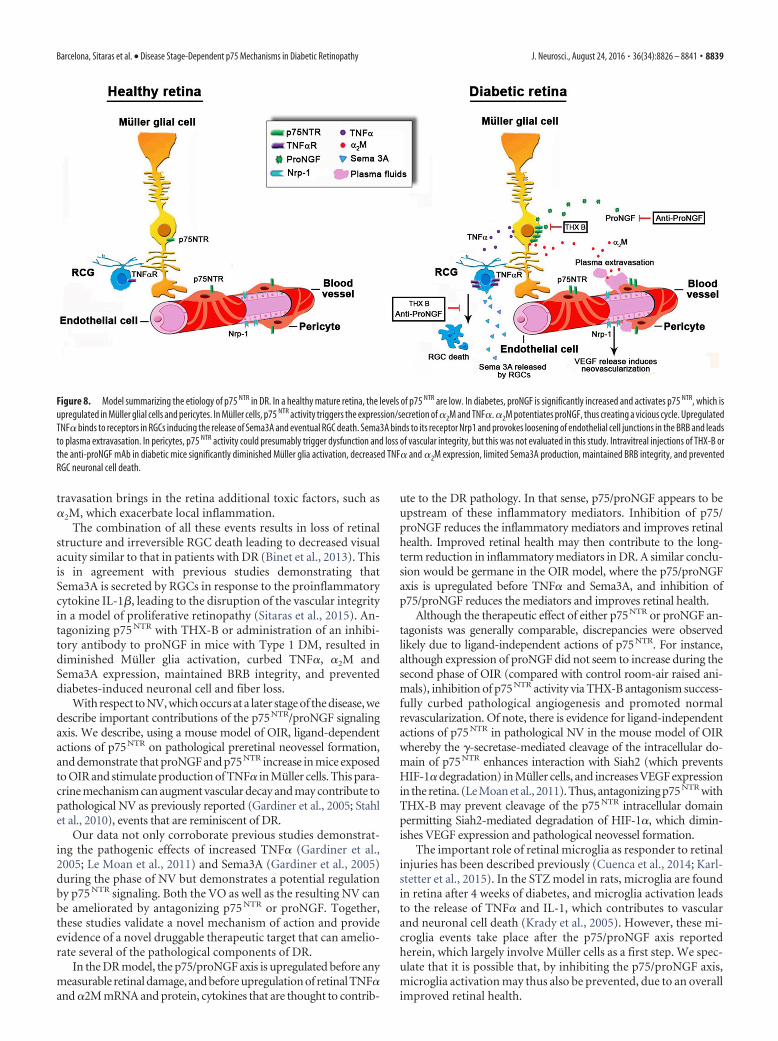
travasation brings in the retina additional toxic factors, such as�2M, which exacerbate local inflammation.
The combination of all these events results in loss of retinalstructure and irreversible RGC death leading to decreased visualacuity similar to that in patients with DR (Binet et al., 2013). Thisis in agreement with previous studies demonstrating thatSema3A is secreted by RGCs in response to the proinflammatorycytokine IL-1�, leading to the disruption of the vascular integrityin a model of proliferative retinopathy (Sitaras et al., 2015). An-tagonizing p75 NTR with THX-B or administration of an inhibi-tory antibody to proNGF in mice with Type 1 DM, resulted indiminished Muller glia activation, curbed TNF�, �2M andSema3A expression, maintained BRB integrity, and preventeddiabetes-induced neuronal cell and fiber loss.
With respect to NV, which occurs at a later stage of the disease, wedescribe important contributions of the p75NTR/proNGF signalingaxis. We describe, using a mouse model of OIR, ligand-dependentactions of p75NTR on pathological preretinal neovessel formation,and demonstrate that proNGF and p75NTR increase in mice exposedto OIR and stimulate production of TNF� in Muller cells. This para-crine mechanism can augment vascular decay and may contribute topathological NV as previously reported (Gardiner et al., 2005; Stahlet al., 2010), events that are reminiscent of DR.
Our data not only corroborate previous studies demonstrat-ing the pathogenic effects of increased TNF� (Gardiner et al.,2005; Le Moan et al., 2011) and Sema3A (Gardiner et al., 2005)during the phase of NV but demonstrates a potential regulationby p75 NTR signaling. Both the VO as well as the resulting NV canbe ameliorated by antagonizing p75 NTR or proNGF. Together,these studies validate a novel mechanism of action and provideevidence of a novel druggable therapeutic target that can amelio-rate several of the pathological components of DR.
In the DR model, the p75/proNGF axis is upregulated before anymeasurable retinal damage, and before upregulation of retinal TNF�and �2M mRNA and protein, cytokines that are thought to contrib-
ute to the DR pathology. In that sense, p75/proNGF appears to beupstream of these inflammatory mediators. Inhibition of p75/proNGF reduces the inflammatory mediators and improves retinalhealth. Improved retinal health may then contribute to the long-term reduction in inflammatory mediators in DR. A similar conclu-sion would be germane in the OIR model, where the p75/proNGFaxis is upregulated before TNF� and Sema3A, and inhibition ofp75/proNGF reduces the mediators and improves retinal health.
Although the therapeutic effect of either p75NTR or proNGF an-tagonists was generally comparable, discrepancies were observedlikely due to ligand-independent actions of p75NTR. For instance,although expression of proNGF did not seem to increase during thesecond phase of OIR (compared with control room-air raised ani-mals), inhibition of p75NTR activity via THX-B antagonism success-fully curbed pathological angiogenesis and promoted normalrevascularization. Of note, there is evidence for ligand-independentactions of p75NTR in pathological NV in the mouse model of OIRwhereby the �-secretase-mediated cleavage of the intracellular do-main of p75NTR enhances interaction with Siah2 (which preventsHIF-1� degradation) in Muller cells, and increases VEGF expressionin the retina. (Le Moan et al., 2011). Thus, antagonizing p75NTR withTHX-B may prevent cleavage of the p75NTR intracellular domainpermitting Siah2-mediated degradation of HIF-1�, which dimin-ishes VEGF expression and pathological neovessel formation.
The important role of retinal microglia as responder to retinalinjuries has been described previously (Cuenca et al., 2014; Karl-stetter et al., 2015). In the STZ model in rats, microglia are foundin retina after 4 weeks of diabetes, and microglia activation leadsto the release of TNF� and IL-1, which contributes to vascularand neuronal cell death (Krady et al., 2005). However, these mi-croglia events take place after the p75/proNGF axis reportedherein, which largely involve Muller cells as a first step. We spec-ulate that it is possible that, by inhibiting the p75/proNGF axis,microglia activation may thus also be prevented, due to an overallimproved retinal health.
Figure 8. Model summarizing the etiology of p75 NTR in DR. In a healthy mature retina, the levels of p75 NTR are low. In diabetes, proNGF is significantly increased and activates p75 NTR, which isupregulated in Muller glial cells and pericytes. In Muller cells, p75 NTR activity triggers the expression/secretion of �2M and TNF�. �2M potentiates proNGF, thus creating a vicious cycle. UpregulatedTNF� binds to receptors in RGCs inducing the release of Sema3A and eventual RGC death. Sema3A binds to its receptor Nrp1 and provokes loosening of endothelial cell junctions in the BRB and leadsto plasma extravasation. In pericytes, p75 NTR activity could presumably trigger dysfunction and loss of vascular integrity, but this was not evaluated in this study. Intravitreal injections of THX-B orthe anti-proNGF mAb in diabetic mice significantly diminished Muller glia activation, decreased TNF� and �2M expression, limited Sema3A production, maintained BRB integrity, and preventedRGC neuronal cell death.
Barcelona, Sitaras et al. • Disease Stage-Dependent p75 Mechanisms in Diabetic Retinopathy J. Neurosci., August 24, 2016 • 36(34):8826 – 8841 • 8839

The data support a model (Fig. 8) explaining the mechanismsby which p75 NTR mediates different phases of the DR. The earlyand persistent upregulation of p75 NTR, mainly in Muller glialcells, plays a pivotal role in disrupting neuro-glia-vascular unit inthe DR. More specifically, p75NTR signaling promotes, in aligand-dependent manner, production of the inflammatory cy-tokines TNF� and �2M and Sema3A, leading to RGC death, BRBbreakdown, and edema. Similarly, p75 NTR activity contributes toretinal VO and NV via increased TNF� and Sema3A production.To illustrate this phenomenon, we propose a hypothetical modelto integrate these observations (Fig. 8). In DR, resident Mullerglia undergo activation and gliosis augmenting the production ofp75 NTR. Activated Muller cells rapidly increase the levels ofproNGF, and autocrine binding of proNGF to p75 NTR promotesthe production and release of proinflammatory cytokines, such asTNF� and �2M. TNF� and �2M act via two different paracrinemechanisms. First, TNF� binds to its receptor on RGCs andprompts the secretion of the vascular cue Sema3A. Excessive ac-cumulation of TNF� and �2M ultimately causes RGC death andthe secretion of Sema3A from RGCs, which influence endothelialcells by binding, most likely, to its receptor Neuropilin-1 (Ceraniet al., 2013). Sema3A causes early BRB breakdown and plasmaextravasation. Ligand-dependent production of inflammatorycytokines, RGC death, BRB breakdown, and edema were signifi-cantly decreased by treatment with anti-proNGF mAb or thep75 NTR antagonist THX-B. Second, TNF� acts directly on endo-thelial cells inducing cell death, which contribute to vessel lossduring the first phase of the vascular component of the disease.Both anti-proNGF mAb and THX-B treatments also reducedpathological NV. In conclusion, p75 NTR and proNGF are keymediators at different stages of the DR. These antagonists may benovel therapeutic targets that offer a wide range spectrum ofaction and an alternative approach to the VEGF-based strategy.
In conclusion, our data identify a novel mechanism implicat-ing neuronal, glial, and vascular interplay in the pathogenesis ofDR. In doing so, we provide evidence for p75 as a promisingtherapeutic target to counter several stages of DR.
ReferencesAbcouwer SF, Gardner TW (2014) Diabetic retinopathy: loss of neuroreti-
nal adaptation to the diabetic metabolic environment. Ann N Y Acad Sci1311:174 –190. CrossRef Medline
Al-Gayyar MM, Matragoon S, Pillai BA, Ali TK, Abdelsaid MA, El-RemessyAB (2011) Epicatechin blocks pro-nerve growth factor (proNGF)-mediated retinal neurodegeneration via inhibition of p75 neurotrophinreceptor expression in a rat model of diabetes [corrected]. Diabetologia54:669 – 680. CrossRef Medline
Ali TK, Matragoon S, Pillai BA, Liou GI, El-Remessy AB (2008) Peroxyni-trite mediates retinal neurodegeneration by inhibiting nerve growth fac-tor survival signaling in experimental and human diabetes. Diabetes 57:889 – 898. CrossRef Medline
Antonetti DA, Klein R, Gardner TW (2012) Diabetic retinopathy. N EnglJ Med 366:1227–1239. CrossRef Medline
Bai Y, Dergham P, Nedev H, Xu J, Galan A, Rivera JC, ZhiHua S, Mehta HM,Woo SB, Sarunic MV, Neet KE, Saragovi HU (2010a) Chronic and acutemodels of retinal neurodegeneration TrkA activity are neuroprotectivewhereas p75NTR activity is neurotoxic through a paracrine mechanism.J Biol Chem 285:39392–39400. CrossRef Medline
Bai Y, Shi Z, Zhuo Y, Liu J, Malakhov A, Ko E, Burgess K, Schaefer H, EstebanPF, Tessarollo L, Saragovi HU (2010b) In glaucoma the up-regulatedtruncated TrkC.T1 receptor isoform in glia causes increased TNF-� pro-duction, leading to retinal ganglion cell death. Invest Ophthalmol Vis Sci51:6639 – 6651. CrossRef Medline
Bai Y, Sivori D, Woo SB, Neet KE, Lerner SF, Saragovi HU (2011) Duringglaucoma, alpha2-macroglobulin accumulates in aqueous humor andbinds to nerve growth factor, neutralizing neuroprotection. Invest Oph-thalmol Vis Sci 52:5260 –5265. CrossRef Medline
Barcelona PF, Saragovi HU (2015) A pro-nerve growth factor (proNGF)and NGF binding protein, alpha2-macroglobulin, differentially regulatesp75 and TrkA receptors and is relevant to neurodegeneration ex vivo andin vivo. Mol Cell Biol 35:3396 –3408. CrossRef Medline
Binet F, Mawambo G, Sitaras N, Tetreault N, Lapalme E, Favret S, Cerani A,Leboeuf D, Tremblay S, Rezende F, Juan AM, Stahl A, Joyal JS, Milot E,Kaufman RJ, Guimond M, Kennedy TE, Sapieha P (2013) Neuronal ERstress impedes myeloid-cell-induced vascular regeneration throughIRE1alpha degradation of netrin-1. Cell Metab 17:353–371. CrossRefMedline
Bringmann A, Pannicke T, Grosche J, Francke M, Wiedemann P, SkatchkovSN, Osborne NN, Reichenbach A (2006) Muller cells in the healthy anddiseased retina. Prog Retin Eye Res 25:397– 424. CrossRef Medline
Cerani A, Tetreault N, Menard C, Lapalme E, Patel C, Sitaras N, Beaudoin F,Leboeuf D, De Guire V, Binet F, Dejda A, Rezende FA, Miloudi K, SapiehaP (2013) Neuron-derived semaphorin 3A is an early inducer of vascularpermeability in diabetic retinopathy via neuropilin-1. Cell Metab 18:505–518. CrossRef Medline
Coorey NJ, Shen W, Chung SH, Zhu L, Gillies MC (2012) The role of glia inretinal vascular disease. Clin Exp Optom 95:266 –281. CrossRef Medline
Cuenca N, Fernandez-Sanchez L, Campello L, Maneu V, De la Villa P, Lax P,Pinilla I (2014) Cellular responses following retinal injuries and thera-peutic approaches for neurodegenerative diseases. Prog Retin Eye Res43:17–75. CrossRef Medline
Dejda A, Mawambo G, Cerani A, Miloudi K, Shao Z, Daudelin JF, Boulet S,Oubaha M, Beaudoin F, Akla N, Henriques S, Menard C, Stahl A, DelisleJS, Rezende FA, Labrecque N, Sapieha P (2014) Neuropilin-1 mediatesmyeloid cell chemoattraction and influences retinal neuroimmune cross-talk. J Clin Invest 124:4807– 4822. CrossRef Medline
Esquiva G, Lax P, Cuenca N (2013) Impairment of intrinsically photosensi-tive retinal ganglion cells associated with late stages of retinal degenera-tion. Invest Ophthalmol Vis Sci 54:4605– 4618. CrossRef Medline
Fletcher EL, Phipps JA, Ward MM, Puthussery T, Wilkinson-Berka JL (2007)Neuronal and glial cell abnormality as predictors of progression of dia-betic retinopathy. Curr Pharm Des 13:2699 –2712. CrossRef Medline
Frade JM, Bovolenta P, Rodríguez-Tebar A (1999) Neurotrophins andother growth factors in the generation of retinal neurons. Microsc ResTech 45:243–251. CrossRef Medline
García-Ayuso D, Salinas-Navarro M, Agudo M, Cuenca N, Pinilla I, Vidal-Sanz M, Villegas-Perez MP (2010) Retinal ganglion cell numbers anddelayed retinal ganglion cell death in the P23H rat retina. Exp Eye Res91:800 – 810. CrossRef Medline
García-Ayuso D, Di Pierdomenico J, Esquiva G, Nadal-Nicolas FM, Pinilla I,Cuenca N, Vidal-Sanz M, Agudo-Barriuso M, Villegas-Perez MP (2015)Inherited photoreceptor degeneration causes the death of melanopsin-positive retinal ganglion cells and increases their coexpression of Brn3a.Invest Ophthalmol Vis Sci 56:4592– 4604. CrossRef Medline
Gardiner TA, Gibson DS, de Gooyer TE, de la Cruz VF, McDonald DM, StittAW (2005) Inhibition of tumor necrosis factor-alpha improves physio-logical angiogenesis and reduces pathological neovascularization in isch-emic retinopathy. Am J Pathol 166:637– 644. CrossRef Medline
Heng LZ, Comyn O, Peto T, Tadros C, Ng E, Sivaprasad S, Hykin PG (2013)Diabetic retinopathy: pathogenesis, clinical grading, management andfuture developments. Diabet Med 30:640 – 650. CrossRef Medline
Jian Y, Wong K, Sarunic MV (2013) Graphics processing unit acceleratedoptical coherence tomography processing at megahertz axial scan rate andhigh resolution video rate volumetric rendering. J Biomed Opt 18:26002.CrossRef Medline
Joyal JS, Sitaras N, Binet F, Rivera JC, Stahl A, Zaniolo K, Shao Z, Polosa A,Zhu T, Hamel D, Djavari M, Kunik D, Honore JC, Picard E, Zabeida A,Varma DR, Hickson G, Mancini J, Klagsbrun M, Costantino S, et al.(2013) Ischemic neurons prevent vascular regeneration of neural tissueby secreting semaphorin 3A. Blood 117:6024 – 6035. CrossRef Medline
Karlstetter M, Scholz R, Rutar M, Wong WT, Provis JM, Langmann T (2015)Retinal microglia: just bystander or target for therapy? Prog Retin Eye Res45:30 –57. CrossRef Medline
Kowluru RA, Chan PS (2007) Oxidative stress and diabetic retinopathy. ExpDiabetes Res 2007:43603. CrossRef Medline
Krady JK, Basu A, Allen CM, Xu Y, LaNoue KF, Gardner TW, Levison SW(2005) Minocycline reduces proinflammatory cytokine expression,microglial activation, and caspase-3 activation in a rodent model of dia-betic retinopathy. Diabetes 54:1559 –1565. CrossRef Medline
8840 • J. Neurosci., August 24, 2016 • 36(34):8826 – 8841 Barcelona, Sitaras et al. • Disease Stage-Dependent p75 Mechanisms in Diabetic Retinopathy

Lebrun-Julien F, Duplan L, Pernet V, Osswald I, Sapieha P, Bourgeois P,Dickson K, Bowie D, Barker PA, Di Polo A (2009a) Excitotoxic death ofretinal neurons in vivo occurs via a non-cell-autonomous mechanism.J Neurosci 29:5536 –5545. CrossRef Medline
Lebrun-Julien F, Morquette B, Douillette A, Saragovi HU, Di Polo A (2009b)Inhibition of p75(NTR) in glia potentiates TrkA-mediated survival ofinjured retinal ganglion cells. Mol Cell Neurosci 40:410 – 420. CrossRefMedline
Lebrun-Julien F, Bertrand MJ, De Backer O, Stellwagen D, Morales CR, DiPolo A, Barker PA (2010) ProNGF induces TNFalpha-dependent deathof retinal ganglion cells through a p75NTR non-cell-autonomous signal-ing pathway. Proc Natl Acad Sci U S A 107:3817–3822. CrossRef Medline
Le Moan N, Houslay DM, Christian F, Houslay MD, Akassoglou K (2011)Oxygen-dependent cleavage of the p75 neurotrophin receptor triggersstabilization of HIF-1alpha. Mol Cell 44:476 – 490. CrossRef Medline
Li J, Bloch P, Xu J, Sarunic MV, Shannon L (2011) Performance and scal-ability of Fourier domain optical coherence tomography acceleration us-ing graphics processing units. Appl Opt 50:1832–1838. CrossRef Medline
Ma N, Hunt NH, Madigan MC, Chan-Ling T (1996) Correlation betweenenhanced vascular permeability, up-regulation of cellular adhesionmolecules and monocyte adhesion to the endothelium in the retina dur-ing the development of fatal murine cerebral malaria. Am J Pathol 149:1745–1762. Medline
Mysona BA, Al-Gayyar MM, Matragoon S, Abdelsaid MA, El-Azab MF, SaragoviHU, El-Remessy AB (2013) Modulation of p75(NTR) prevents diabetes- andproNGF-induced retinal inflammation and blood–retina barrier breakdown inmice and rats. Diabetologia 56:2329–2339. CrossRef Medline
Saragovi HU, Zheng W, Maliartchouk S, DiGugliemo GM, Mawal YR, Ka-men A, Woo SB, Cuello AC, Debeir T, Neet KE (1998) A TrkA-selective,fast internalizing nerve growth factor-antibody complex induces trophicbut not neuritogenic signals. J Biol Chem 273:34933–34940. CrossRefMedline
Sarunic MV, Yazdanpanah A, Gibson E, Xu J, Bai Y, Lee S, Saragovi HU, Beg
MF (2010) Longitudinal study of retinal degeneration in a rat usingspectral domain optical coherence tomography. Opt Express 18:23435–23441. CrossRef Medline
Shi Z, Rudzinski M, Meerovitch K, Lebrun-Julien F, Birman E, Di Polo A,Saragovi HU (2008) Alpha2-macroglobulin is a mediator of retinal gan-glion cell death in glaucoma. J Biol Chem 283:29156 –29165. CrossRefMedline
Siao CJ, Lorentz CU, Kermani P, Marinic T, Carter J, McGrath K, Padow VA,Mark W, Falcone DJ, Cohen-Gould L, Parrish DC, Habecker BA, NykjaerA, Ellenson LH, Tessarollo L, Hempstead BL (2012) ProNGF, a cytokineinduced after myocardial infarction in humans, targets pericytes to pro-mote microvascular damage and activation. J Exp Med 209:2291–2305.CrossRef Medline
Sitaras N, Rivera JC, Noueihed B, Bien-Aime M, Zaniolo K, Omri S, Hamel D,Zhu T, Hardy P, Sapieha P, Joyal JS, Chemtob S (2015) Retinal neuronscurb inflammation and enhance revascularization in ischemic retinopa-thies via proteinase-activated receptor-2. J Pathol 185:581–595. CrossRefMedline
Smith LE, Wesolowski E, McLellan A, Kostyk SK, D’Amato R, Sullivan R,D’Amore PA (1994) Oxygen-induced retinopathy in the mouse. InvestOphthalmol Vis Sci 35:101–111. Medline
Stahl A, Connor KM, Sapieha P, Willett KL, Krah NM, Dennison RJ, Chen J,Guerin KI, Smith LE (2009) Computer-aided quantification of retinalneovascularization. Angiogenesis 12:297–301. CrossRef Medline
Stahl A, Connor KM, Sapieha P, Chen J, Dennison RJ, Krah NM, SeawardMR, Willett KL, Aderman CM, Guerin KI, Hua J, Lofqvist C, Hellstrom A,Smith LE (2010) The mouse retina as an angiogenesis model. InvestOphthalmol Vis Sci 51:2813–2826. CrossRef Medline
Yau JW, Rogers SL, Kawasaki R, Lamoureux EL, Kowalski JW, Bek T, Chen SJ,Dekker JM, Fletcher A, Grauslund J, Haffner S, Hamman RF, Ikram MK,Kayama T, Klein BE, Klein R, Krishnaiah S, Mayurasakorn K, O’Hare JP,Orchard TJ, et al. (2012) Global prevalence and major risk factors ofdiabetic retinopathy. Diabetes Care 35:556 –564. CrossRef Medline
Barcelona, Sitaras et al. • Disease Stage-Dependent p75 Mechanisms in Diabetic Retinopathy J. Neurosci., August 24, 2016 • 36(34):8826 – 8841 • 8841


![[XLS] · Web viewSheet1 P.S. KAPSULI BARAIT MODEL V.M. BHAUNKHAL P.S. TARAM P.S. TARAM NEW H.S. TARAM P.S. GHJEERA P.S BAURARA G.I.C. BHAUNKHAL P.S. AJOLI U.P.S. AJOLI TALLI P.S. AJOLI](https://img.dokumen.tips/doc/110x75/5aad8db97f8b9a8f498e67bf/xls-viewsheet1-ps-kapsuli-barait-model-vm-bhaunkhal-ps-taram-ps-taram.jpg)