Embed Size (px)
Citation preview
General rights Copyright and moral rights for the publications made accessible in the public portal are retained by the authors and/or other copyright owners and it is a condition of accessing publications that users recognise and abide by the legal requirements associated with these rights.
Users may download and print one copy of any publication from the public portal for the purpose of private study or research.
You may not further distribute the material or use it for any profit-making activity or commercial gain
You may freely distribute the URL identifying the publication in the public portal If you believe that this document breaches copyright please contact us providing details, and we will remove access to the work immediately and investigate your claim.
Downloaded from orbit.dtu.dk on: Oct 18, 2021
Novel insights into the dynamics behavior of glucagon-like peptide-1 receptor with itssmall molecule agonists
Girdhar, Khyati; Dehury, Budheswar; Kumar Singh, Mahender; Daniel, Vineeth P.; Choubey, Abhinav;Dogra, Surbhi; Kumar, Sunil; Mondal, Prosenjit
Published in:Journal of Biomolecular Structure and Dynamics
Link to article, DOI:10.1080/07391102.2018.1532818
Publication date:2019
Document VersionPeer reviewed version
Link back to DTU Orbit
Citation (APA):Girdhar, K., Dehury, B., Kumar Singh, M., Daniel, V. P., Choubey, A., Dogra, S., Kumar, S., & Mondal, P. (2019).Novel insights into the dynamics behavior of glucagon-like peptide-1 receptor with its small molecule agonists.Journal of Biomolecular Structure and Dynamics, 37(15), 3976-3986.https://doi.org/10.1080/07391102.2018.1532818
Full Terms & Conditions of access and use can be found athttp://www.tandfonline.com/action/journalInformation?journalCode=tbsd20
Journal of Biomolecular Structure and Dynamics
ISSN: 0739-1102 (Print) 1538-0254 (Online) Journal homepage: http://www.tandfonline.com/loi/tbsd20
Novel insights into the dynamics behavior ofGlucagon-Like Peptide-1 Receptor with its smallmolecule Agonists
Khyati Girdhar, Budheswar Dehury, Mahender Kumar Singh, Vineeth PDaniel, Abhinav Choubey, Surbhi Dogra, Sunil Kumar & Prosenjit Mondal
To cite this article: Khyati Girdhar, Budheswar Dehury, Mahender Kumar Singh, Vineeth P Daniel,Abhinav Choubey, Surbhi Dogra, Sunil Kumar & Prosenjit Mondal (2018): Novel insights into thedynamics behavior of Glucagon-Like Peptide-1 Receptor with its small molecule Agonists, Journalof Biomolecular Structure and Dynamics, DOI: 10.1080/07391102.2018.1532818
To link to this article: https://doi.org/10.1080/07391102.2018.1532818
Accepted author version posted online: 08Oct 2018.
Submit your article to this journal
View Crossmark data
Novel insights into the dynamics behavior of Glucagon-Like Peptide-1
Receptor with its small molecule Agonists
Khyati Girdhar#$
, Budheswar Dehury¥,$,ƪ
, Mahender Kumar Singh£, Vineeth P Daniel
#, Abhinav Choubey
#, Surbhi
Dogra#, Sunil Kumar
¶, Prosenjit Mondal
*#
#School of Basic Sciences, Indian Institute of Technology Mandi, Mandi-175001, H.P, India
¥Biomedical Informatics Centre, ICMR-Regional Medical Research Centre, Chandrasekharpur, Bhubaneswar-
751023, Odisha, India
ƪDepartment of Chemistry, Technical University of Denmark, DTU Chemistry, DK-2800 Kongens Lyngby,
Denmark (Present Address)
£National Brain Research Centre, Manesar, Gurugram, Haryana-122051
¶ICAR-National Bureau of Agriculturally Important Microorganism, Kushmaur, Mau Nath Bhanjan -275103, U.P.,
India
$These authors contributed equally to this work
Corresponding Authors
*E-mail: Prosenjit Mondal [email protected], Phone No.+91-8894296497
Accep
ted
Man
uscr
ipt
Abstract
The glucagon-like peptide-1 receptor (GLP-1R) is a well-known target of therapeutics industries for the treatment of
various metabolic diseases like Type2 Diabetes and Obesity. The structural-functional relationships of small
molecule agonists and GLP-1R are yet to be understood. Therefore, an attempt was made on structurally known
GLP-1R agonists (Compound 1, Compound 2, Compound A, Compound B, and (S)-8) to study their interaction
with the extracellular domain of GLP-1R. In this study, we explored the dynamics, intrinsic stability and binding
mechanisms of these molecules through computational modeling, docking, molecular dynamics (MD) simulations
and MM/PBSA binding free energy estimation. Molecular docking study depicted that hydrophobic interaction (pi-
pi stacking) play a crucial role in maintaining the stability of the complex, which was also supported by inter-
molecular analysis from MD simulation study. Principal component analysis suggested that the terminal ends along
with the turns/loops connecting adjacent helix and strands exhibits comparatively higher movement of main chain
atoms in most of the complexes. MM/PBSA binding free energy study revealed that non-polar solvation (van der
Waals and electrostatic) energy subsidizes significantly to the total binding energy and the polar solvation energy
opposes the binding agonists to GLP-1R. Overall, we provide structural features information about GLP-1R
complexes that would be conducive for the discovery of new GLP-1R agonists in the future for treatment of various
metabolic diseases.
Key words: Agonists, docking, Type2 diabetes, GLP-1R, molecular dynamics
Accep
ted
Man
uscr
ipt
Introduction
International Diabetes Federation (IDF) estimates that there are about approximately 64,200,000 individuals with
Type 2 Diabetes Mellitus (T2DM) in India and by 2030 the number will reach to 100,000,000, as predicted by the
World Health Organization (WHO) (Interantional Diabetes Federation, 2015).
T2DM is characterized by a deficit in “functional” beta-cell mass, resulting in insufficient insulin secretion to
counter metabolic demands of insulin resistance and to maintain normoglycemia (Jensen, Cnop, Hull, Fujimoto, &
Kahn, 2002). To overcome the impaired beta-cells secretory response to glucose in T2DM patients as well as in
subjects vulnerable to develop T2DM, it is desirable to introduce a therapeutic molecule in pancreatic beta-cells
which can modulate insulin production and secretion. Numbers of therapeutics are available in the market for
treatment of T2DM like metformin, sulfonylurea, thiazolidinedione, inhibitors or modulators of sodium-glucose
cotransporter 2 (SGLT2), dipeptidyl peptidase 4 (DPP-4) and GLP-1R agonists (Yang, Zhou, Liu, & Wang, 2015).
Among all therapeutics, GLP-1R based therapy is a well-known promising approach in the treatment of diabetes by
augmenting glucose-dependent insulin secretion with low risk of hypoglycemia. Incretin-based drugs like analogs of
GLP-1 and its more stable analog exendin-4 (Ex-4) are being used to augment glucose-stimulated insulin secretion
(GSIS) from beta-cells for better glycemic control in T2DM patients (Baggio & Drucker, 2007; Drucker & Nauck,
2006; Nielsen, Young, & Parkes, 2004). GLP-1 exerts its effects after binding to its specific GLP-1R which belongs
to B family of G-protein Coupled Receptor (GPCR), consists of the N-terminal extracellular domain and a
transmembrane domain. The N-terminus extracellular domain of GPCR-B family is known to have a major role in
the determination of the ligand binding (De Maturana, Willshaw, Kuntzsch, Rudolph, & Donnelly, 2003). C-
terminus of the GLP-1(endogenous agonist of GLP-1R) binds to the extracellular domain of receptor and help in
stabilizing its binding while N-terminus binds to the transmembrane domain of the receptor (Santiago, Paci, &
Donnelly, 2018) and activates adenylyl cyclase thereby elevate cAMP (Furman, Ong, & Pyne, 2010; Hansotia et al.,
2004; Tengholm, 2012). However, GLP-1 has its own limitation with its administration and its short biological half-
life in blood because of its metabolic stability and rapid degradation by DPP-4. The limitation of breakdown by
DPP-IV is to some extent overcome by a GLP-1R agonist such as Ex-4 (Parkes, Mace, & Trautmann, 2013) and
Liraglutide (Gupta, 2013) (clinically approved GLP-1R peptide agonists for the treatment of diabetes) (Lotte B.
Knudsen et al., 2000). However, the stability of these peptides, its synthesis, cost and its routes of administration is
still a major challenge. Therefore, there is a need to identify and develop a small organic molecule that binds
orthostatically and activate the receptor to resolve this issue. From the last few decades, there are very few small
molecule GLP-1R agonists has been reported in which most potent agonist Boc5 and its analog WB4-24 show both
antidiabetic and anti-obesity effect (Su et al., 2008).
Compound 1, Compound 2 (Lotte Bjerre Knudsen et al., 2007), Compound A, Compound B (Sloop et al., 2010) and
(S)-8 (G. Morris & Huey, 2009), all are GLP-1R allosteric modulators have been determined by high-throughput
screening on the basis of cAMP and intracellular calcium mobilization. These compounds have known to act as
allosteric activators of the receptor (Bueno et al., 2016; Koole et al., 2010; L. C. Morris et al., 2014). Methods that
were used till now for the screening of above GLP-1R agonist are time-consuming and cost ineffective (Lotte Bjerre
Accep
ted
Man
uscr
ipt
Knudsen et al., 2007; L. C. Morris et al., 2014; Sloop et al., 2010; M. W. Wang, Liu, & Zhou, 2010). This might be
one of the reasons that there is very few small molecules GLP-1R agonist reported so far. Therefore, for the
development of potent GLP-1R allosteric agonists, it becomes a prime need to understand the interaction between
the agonists and the receptor. Some of the reports try to mention the interaction of GLP-1R with its agonists (Lin &
Wang, 2009; Santiago et al., 2018) but till now there is no report on the study of an allosteric agonist of GLP-1R
with the extracellular domain. Here in our report, we selected some of the small molecule GLP-1R agonists based on
the Lipinski rule of five and tried to study their interaction with the extracellular domain of GLP-1R. In our study,
AutoDock software was extensively used to determine the binding site of small molecules with GLP-1R followed by
molecular dynamics (MD) simulation to inspect the dynamics and mode of binding of all agonists. Overall, the
computational investigation of the extracellular domain of GLP-1R was carried out to open up a better
understanding of the mechanism of binding GLP-1R agonists which would probably be an useful information for the
development of potent GLP-1R agonist in near future.
Materials and Methods
Preparation of Protein Structure
The three-dimensional structure of the extracellular domain of GLP-1R in complex with GLP-1 peptide was
retrieved from the RSCB Protein Data Bank [PDB Code: 3IOL]. Protein structure was prepared by removing GLP-1
peptide and water molecules associated with the crystal structure. Heteroatoms other than water molecules were also
removed and the file was saved in PDB format and further used for the docking of reported agonists.
Ligand selection and preparation
The chemical structures of small molecules (Fig. 1) were retrieved as reported. From all reported GLP-1R agonist, 5
out of all are selected because of their reported allosteric property along with small molecules structure. Three-
dimensional PDB structures of all the agonists were obtained by using BIOVIA Discovery Studio (DS) Visualizer
tool. The ligand molecules were then subjected to energy minimization through DS. Gasteiger charges were added
to small molecules through AutoDock Tools.
Molecular docking
AutoDock 4.2.6 and AutoDock tools 1.5.6 (G. Morris & Huey, 2009) were used to perform molecular docking, a
computational docking program based on rapid Lamarckian genetic algorithm search method and empirical free-
energy force field (Forli et al., 2016). AutoDock Tools was utilized to prepare the input pdbqt file of GLP-1R [PDB:
3IOL]. Polar hydrogen atoms and Kollman charges were added to the 3D structure of GLP-1R. All torsional degrees
of freedom, atomic partial charges, non-polar hydrogens and rotatable bonds were assigned to each ligand. The
docking grid box of dimension x, y, and z are 102Å, 126Å, and 104Å respectively with the spacing of 0.375Å was
made to cover the entire extracellular GLP-1R domain. The best docking conformation was calculated by using the
Lamarckian Genetic Algorithm (LGA) that is considered one of the best docking methods in AutoDock. The
parameter used for LGA includes an initial population size of 150, number of energy evaluation of 250000, and
Accep
ted
Man
uscr
ipt
maximum generation of 27000 were defined to find one best individual. Each of molecule was docked using 100
LGA runs. Final docked conformations of each molecule were clustered using 2Å root mean square deviation
(RMSD).
Evaluation and visualization of docked conformations
The docked conformations of each ligand were ranked into clusters based on their binding energy and the best
conformations were visually analyzed. Non-bonded interactions including hydrogen bond, Vander wall, pi-pi and
other electrostatic interaction between docked ligand and GLP-1R was analyzed using DS.
Molecular Dynamics Simulation
To explore the stability and conformational flexibility of all the GLP-1R complex systems, MD simulations were
performed as described previously (Dehury, Behera, & Mahapatra, 2017). AnteChamber Python Parser interface
(ACPYPE) was employed to parameterize the required topologies, atomic types, and charges of ligands. In this
study, Amber99SB-ILDN and General Amber force fields were used for protein and ligand respectively in
GROMACSv2018.2. All complexes were solvated with TIP3P water model. For each system, a cubic box-setting
with a minimal distance of 10 Å between the complex and edge of the box was further solvated using periodic
boundary conditions. Each system was neutralized with counter ions of strength 0.15M (Na+/Cl
-) (summarized in
Table S1). Then, electro-neutralized systems were subjected to energy minimization on a convergence threshold of 1
kcal mol-1
Å achieved using a conjugate gradient algorithm. After energy minimization, all systems equilibrated
using NVT and NPT ensemble for 1 nano-second (ns). Finally, a production runs of 100 ns with an integration time
step of 0.2 ps were performed at a constant temperature (V-rescale method) and pressure (Parrinello-Rahman
method) using the Leapfrog algorithm. To constrain all H-bonds during the equilibration LINCS algorithm was
used, while, long-range ionic interactions approximated using the particle-mesh Ewald (PME) algorithm. Trajectory
snapshots were stored at every 0.2 ps during the simulation period and 3D coordinate files harvested for post-
dynamic analysis.
MD Trajectory Analysis
To analyze the resultant trajectories, GROMACS analysis toolkit utilities were used to determine dynamics stability
parameters including backbone RSMD, the radius of gyration (Rg), root mean square fluctuations (RMSF), and
inter-molecular hydrogen bond (H-Bond) distribution for each complex system. The intermolecular H-bonds formed
between specific amino acids residues and ligand atoms were determined gmx hbond utility tool with the donor-
acceptor set at a maximum of 0.35 nm. VMD was employed to assess the evolution of the secondary structural
elements in the proteins during MD simulations. The 2D graphs were plotted using Xmgrace (Grace 5.1.21;
http://plasma-gate.weizmann.ac.il/Grace/) and DS was used for protein-ligand interaction analysis.
2.7 Principal Component Analysis and Clustering
Principal component analysis (PCA), a promising statistical approach, often separates the collective motions from
the local dynamics to a small subset comprised of principal components (PCs) that defines the collective motion. To
observe the collective motion of GLP-1R complexes, PCA was performed using built-in modules of GROMACS
i.e., gmx covar and gmx anaeig. The covariance matrix was diagonalized to extract a set of eigenvectors and
Accep
ted
Man
uscr
ipt
eigenvalues which reflected concert motion of the molecules. A set of eigenvectors of protein main chain atoms
have been calculated, while coordinates of every 500 ps were used for porcupine plots. Cluster analysis of the
resultant trajectories was performed using the method described by Daura and coworkers (Daura et al., 1999) using
the gmx cluster built-in module of Gromacs. The GROMOS, RMSD conformational clustering algorithm with a cut-
off of 0.2 nm was used to extract the maximally occupied clusters by taking into account the protein conformation
with the lowermost RMSD to the centroid.
MM/PBSA Binding Free Energy
The binding energy of each GLP-1R-ligand complex (GLP-1R with its agonists) was determined using the
g_mmpbsa tool which employs the molecular mechanics-based Poisson–Boltzmann Surface Area (MM/PBSA)
approach. A total of 1000 snapshots structures extracted from 100 ns trajectory at equal interval of time for
determining the binding energy ((ΔGbind) using the followings sets of equation:
Gbind = Gcomplex − (GGLP-1R + Gligand) …………… (1)
Gbind = Egas + Gsol − TΔS ……………………..... (2)
Egas = Eint + Evdw + Eele ……………………….... (3)
Gsol = Gpol + GSA ……………………………… (4)
GSA = γSASA + b ……………………………. (5)
Absolute free energies of the GLP-1R-ligand complexes, receptor and ligands were denoted by Gcomplex, GGLP-1R and
Gligand respectively. The ΔGbind was decomposed to various terms; a gas-phase energy (Egas) can be defined as sum of
bonded (Eint) and non-bonded terms (Evdw and Eele); the solvation free energy (Gsol) was decomposed into polar (Gpol)
and nonpolar (GSA) solvation energy components- and an entropy term (TΔS). Polar solvation energies were
determined by solving the Poisson-Boltzmann linear equation while nonpolar solvation through the solvent
accessible surface area with an offset value (b) of 3.84928 kJmol−1
and surface tension proportionality (γ) set at
0.0226778 kJ.mol−1
Å−2
. A detailed description of methods used in the calculation can be found in the previous
report (C. Wang, Greene, Xiao, Qi, & Luo, 2018). The per-residue energy decomposition was also computed to
understand the contribution of individual amino acids to the total binding energy.
Results and Discussions
Molecular Docking
Based on the structural motifs of the GLP-1R agonists, we divided them into three categories which include
substituted quinoxaline, pyrimidine-based ago-allosteric modulators and pyridoindole based modulators (Graaf et
al., 2016). Substituted quinoxaline derivative include Compound 1 and Compound 2; while, Compound A and
Compound B/BETP belongs to the family of pyrimidine-based agonists and (S)-8 belong to tricyclic pyridoindole-
based series of GLP-1R allosteric modulators (as shown in Fig. 1). All the selected agonist of GLP-1R have
different structure motif, but, they all reported as positive agonist modulator of GLP-1R. The mode of binding of
quinoxaline, pyrimidine and tricyclic pyridoindole agonists of GLP-1R is still unknown. Hence, to identify the
binding sites involved in the allosteric interactions in between the extracellular domain of GLP-1R and above-
mentioned agonists, molecular docking was performed.
Accep
ted
Man
uscr
ipt
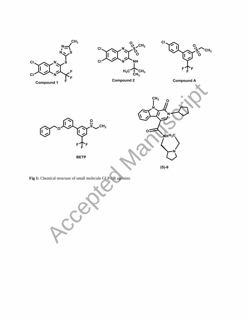
Fig 1: Chemical structure of small molecule GLP-1R agonists
Accep
ted
Man
uscr
ipt
Molecular docking is mostly used for identification of binding affinities of a small molecule with the target protein
(Lee, Jo, Lim, & Im, 2012). In AutoDock tool, docking score is determined in terms of binding affinity in kcal/mol
(Lu et al., 2010). Binding energy is the sum of intermolecular energy (comprised of Hydrogen bond energy,
electrostatic energy, desolvation energy, and van der Wall energy) and torsional energy. A detailed summary of
highest binding affinity, low inhibitory constant, no. of hydrogen bond formed and essential binding sites of all the
above-mentioned compounds is presented in Table 1. The small molecule docking results were evaluated depending
on their binding affinity, binding site and no. of H-Bond formed. The hundred docking conformations for each
agonist were grouped according to their RMSD value of 2Å. After docking, the obtained lower energy cluster
conformations of each molecule were compared with the literature. The binding energy of (S)-8 and BETP was
observed to be higher as compared to Compound 1, Compound 2 and Compound A as mentioned in Table 1. After
performing docking study for each ligand molecules, the best pose (based on its conformation), binding sites and
docking energy were determined. The binding site of the extracellular domain of GLP-1R and the involvement of
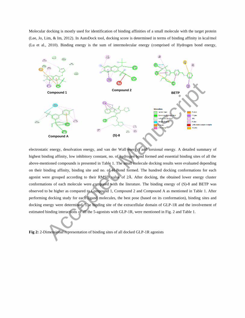
estimated binding interactions of all the 5-agonists with GLP-1R, were mentioned in Fig. 2 and Table 1.
Fig 2: 2-Dimensional representation of binding sites of all docked GLP-1R agonists
Compound 1 Compound 2
BETP
(S)-8
Compound A
Accep
ted
Man
uscr
ipt
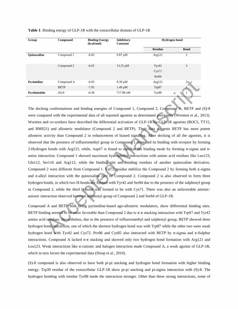
Table 1: Binding energy of GLP-1R with the extracellular domain of GLP-1R
Group Compound Binding Energy
(kcal/mol) Inhibitory
Constant Hydrogen bond
Residue Bond
Quinoxaline Compound 1 -6.83 9.87 µM Arg121
2
Compound 2 -6.65 13.25 µM Tyr42
Cys71
Ser84
3
Pyrimidine Compound A -6.93 8.39 µM Arg121 2
BETP -7.95 1.49 µM Trp87 1
Pyridoindole (S)-8 -8.38 717.98 nM Tyr88 1
The docking conformations and binding energies of Compound 1, Compound 2, Compound A, BETP and (S)-8
were compared with the experimental data of all reported agonists as determined previously (Wootten et al., 2013).
Wootten and co-workers have described the differential activation of GLP-1R-by-GLP-1R agonists (BOC5, TT15,
and BMS21) and allosteric modulator (Compound 2 and BETP). Their data suggests BETP has more potent
allosteric activity than Compound 2 in enhancement of biased signaling. After docking of all the agonists, it is
observed that the presence of trifluoromethyl group in Compound 1 increased its binding with receptor by forming
2-Hydrogen bonds with Arg121, while, Asp67 is found to stabilize the binding mode by forming π-sigma and π-
anion interaction. Compound 1 showed maximum hydrophobic interactions with amino acid residues like Leu123,
Gln112, Ser116 and Arg112, while the binding site and binding residues of another quinoxaline derivative,
Compound 2 were different from Compound 1. Val72 residue stabilize the Compound 2 by forming both π-sigma
and π-alkyl interaction with the quinoxaline ring of Compound 2. Compound 2 is also observed to form three
hydrogen bonds, in which two H-bonds are formed with Tyr42 and Ser84 due to the presence of the sulphonyl group
in Compound 2, while the third H-bond was formed to be with Cys71. There was also an unfavorable anionic-
anionic interaction observed between sulphonyl group of Compound 2 and Ser84 of GLP-1R.
Compound A and BETP both being pyrimidine-based ago-allosteric modulators, show differential binding sites.
BETP binding seemed to be more favorable than Compound 2 due to π-π stacking interaction with Trp87 and Tyr42
amino acid residues. Nevertheless, due to the presence of trifluoromethyl and sulphonyl group, BETP showed three
hydrogen bond interaction, one of which the shortest hydrogen bond was with Trp87 while the other two were weak
hydrogen bond with Tyr42 and Cys72. Pro96 and Cys85 also interacted with BETP by π-sigma and π-Sulphur
interactions. Compound A lacked π-π stacking and showed only two hydrogen bond formation with Arg121 and
Leu123. Weak interactions like π-cationic and halogen interaction made Compound A, a weak agonist of GLP-1R,
which in turn favors the experimental data (Sloop et al., 2010).
(S)-8 compound is also observed to have both pi-pi stacking and hydrogen bond formation with higher binding
energy. Trp39 residue of the extracellular GLP-1R show pi-pi stacking and pi-sigma interaction with (S)-8. The
hydrogen bonding with residue Tyr88 made the interaction stronger. Other than these strong interactions, some of
Accep
ted
Man
uscr
ipt
the weaker π-alkyl and alkyl interactions with Trp91, Tyr69 and Leu123 were also found to present in GLP-1R- (S)-
8 complex (Fig. 2).
The obtained docking results of all the five structurally diverse agonists of GLP-1R give us knowledge about the
binding mode and affinity of the ligand which in results allow us to purpose novel binding residues involved in the
activation of GLP-1R. Compound 2 and BETP has been experimentally reported to be a good agonist of GLP-1R
which totally favors our docking results as Compound 2 shows weak allosteric binding site in our docking results
which make us interpret the binding of Compound 2 to be probably at orthosteric binding site. Compound 2 has
been reported more potent in the enhancement of intracellular cAMP than Compound 1, which may be due to the
formation of a maximum number of the hydrogen bond or due to sharing of the orthosteric binding site by
Compound 2. The experimental study on Compound 1 as an allosteric ligand has not reported so far, our data
suggests high allosteric activity of Compound1 than Compound 2. BETP also have the ability to act via multiple
pathways through GLP-1R e.g., cAMP, pERK1/2, iCa, -Arr-1 and -Arr-2. To some extent, this may be explained
by comparing the binding energy of BETP with other ligands and strongest π-π interaction formed by BETP. From
all the above data, it may be concluded that amino acid residue Tyr42, Cys71 and Val83 is mainly involved in the
activation of the GLP-1R receptor. Other than these residual levels receptor activation, - interaction with some of
the amino acid residue like Tyr42 and Trp39 can also stabilize the receptor ligand binding and help in activation of
different pathways. The results obtained from our study co-relate with the study performed by Wootten and
coworkers’ states Compound 2 to have less allosteric activity in presence of Oxyntomodulin (GLP-1 agonist) than
BETP which can be predicted by high cAMP and Ca2+
concentration. Compound 2 seems to be a weak binder on an
extracellular domain which explains its binding with the orthosteric site than the extracellular regions. All the above
mentioned results suggest the binding behavior of concerned agonists with GLP-1R and the difference in the binding
mode or site may explain the distinct functional profiles of these agonists. This knowledge of the appropriate
binding site on receptor provide us the opportunity to target physiological important responses which can extend in
to drug development strategies.
Analysis of MD trajectory of GLP-1R complexes
MD simulations study was conducted for the five complex systems to understand the structural and dynamic
behavior of the GLP-1R when coupled with agonists. The intrinsic dynamic stability of each complex was
investigated by backbone RMSD, Rg, and Cα RMSF as a function of simulation time. RMSD measures the
difference between the backbones of a protein from its initial structural conformation to its final position. The
stability of the protein/enzymes/receptor-ligand complex relative to its conformation can be assessed by plotting the
deviations produced during the course of the simulation. The smaller the deviations, the more stable the protein
structure. RMSD value for the backbone was calculated for 100 ns simulation in order to check for the stability of all
the systems. Close inspection of all the systems showed a stable trend in RMSD (with an average deviation of ~0.19
nm) profile demonstrating the stable nature of the systems considered in the present study. Although GLP1-R-
Compound A system was found show little higher RMSD up to 80 ns, but, later it attained equilibrium with a stable
RMSD of ~0.23 nm (Fig. 3A). Overall, most of the systems showed least backbone deviation indicating the stable
Accep
ted
Man
uscr
ipt
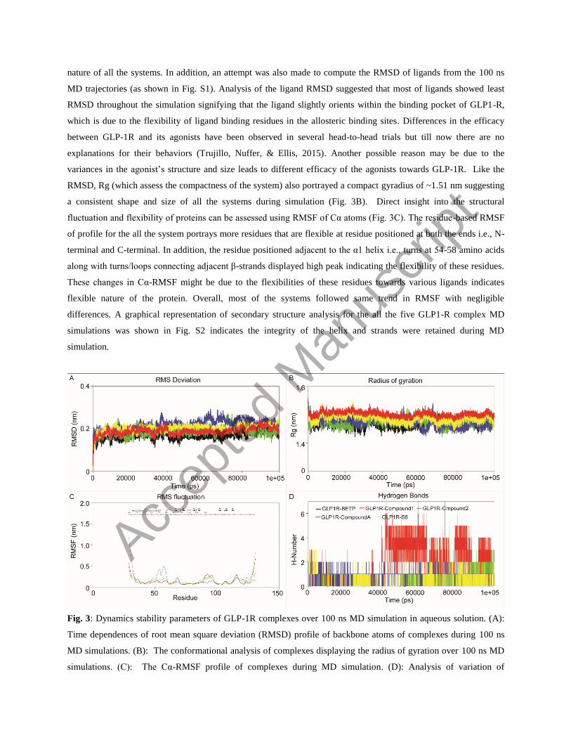
nature of all the systems. In addition, an attempt was also made to compute the RMSD of ligands from the 100 ns
MD trajectories (as shown in Fig. S1). Analysis of the ligand RMSD suggested that most of ligands showed least
RMSD throughout the simulation signifying that the ligand slightly orients within the binding pocket of GLP1-R,
which is due to the flexibility of ligand binding residues in the allosteric binding sites. Differences in the efficacy
between GLP-1R and its agonists have been observed in several head-to-head trials but till now there are no
explanations for their behaviors (Trujillo, Nuffer, & Ellis, 2015). Another possible reason may be due to the
variances in the agonist’s structure and size leads to different efficacy of the agonists towards GLP-1R. Like the
RMSD, Rg (which assess the compactness of the system) also portrayed a compact gyradius of ~1.51 nm suggesting
a consistent shape and size of all the systems during simulation (Fig. 3B). Direct insight into the structural
fluctuation and flexibility of proteins can be assessed using RMSF of Cα atoms (Fig. 3C). The residue-based RMSF
of profile for the all the system portrays more residues that are flexible at residue positioned at both the ends i.e., N-
terminal and C-terminal. In addition, the residue positioned adjacent to the α1 helix i.e., turns at 54-58 amino acids
along with turns/loops connecting adjacent β-strands displayed high peak indicating the flexibility of these residues.
These changes in Cα-RMSF might be due to the flexibilities of these residues towards various ligands indicates
flexible nature of the protein. Overall, most of the systems followed same trend in RMSF with negligible
differences. A graphical representation of secondary structure analysis for the all the five GLP1-R complex MD
simulations was shown in Fig. S2 indicates the integrity of the helix and strands were retained during MD
simulation.
Fig. 3: Dynamics stability parameters of GLP-1R complexes over 100 ns MD simulation in aqueous solution. (A):
Time dependences of root mean square deviation (RMSD) profile of backbone atoms of complexes during 100 ns
MD simulations. (B): The conformational analysis of complexes displaying the radius of gyration over 100 ns MD
simulations. (C): The Cα-RMSF profile of complexes during MD simulation. (D): Analysis of variation of
Accep
ted
Man
uscr
ipt
intermolecular H-bonds of GLP-1R complexes during 100 ns MD simulation (Black: GLP-1R-BETP complex, Red:
GLP-1R-Compound 1 complex, Green: GLP-1R-Compound 2 complex, Blue: GLP-1R-Compound A complex, and
Yellow: GLP-1R-(S)-8 complex).
Intermolecular H-bond and clustering analysis
The stability of a protein and its binding components can be measured by the intermolecular H-bonds. In the GLP-
1R-BETP complex, an average of ~0.98 numbers H-bonds was observed for each frame as a function of simulation
time; whereas, comparatively higher numbers of H-bonds (~3.87) was noticed in the GLP-1R-Compound 1 complex
(Fig. 3D). In contrast, GLP-1R-Compound A complex displayed an average of ~2.08 number of H-bonds for each
frame throughout the simulation period followed by GLP-1R-(S)-8 complex with ~1.87 bonds. Complex with
compound A displayed an average of ~1.59 numbers of H-bonds during 100 ns MD. Comparative analysis portrayed
that in all complexes differential H-bonding pattern was observed, which might be due to the preferential affinity of
GLP-1R towards each ligand within the active pocket and/or due to the slight change in ligand orientation within the
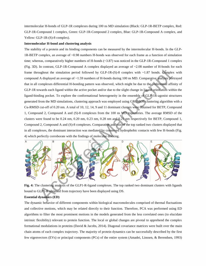
ligand-binding pocket. To explore the conformational heterogeneity in the ensemble of GLP-1R-agonist structures
generated from the MD simulations, clustering approach was employed using GROMOS clustering algorithm with a
Cα-RMSD cut-off of 0.20 nm. A total of 10, 12, 14, 9 and 11 dominant clusters were obtained for BETP, Compound
1, Compound 2, Compound A and (S)-8 complexes from the 100 ns MD trajectories. The average RMSD of the
clusters were found to be 0.24 nm, 0.20 nm, 0.23 nm, 0.28 nm and 0.21 nm respectively for BETP, Compound 1,
Compound 2, Compound A and (S)-8 complexes. Comparative analysis of the top ranked two clusters displayed that
in all complexes, the dominant interaction was mediated by conserved hydrophobic contacts with few H-bonds (Fig.
4) which perfectly corroborate with the findings of molecular docking.
Fig. 4: The clustering analysis of the GLP1-R-ligand complexes. The top ranked two dominant clusters with ligands
bound to GLP1-R obtained from trajectory have been displayed using DS.
Essential dynamics (ED)
The dynamic behavior of different components within biological macromolecules comprised of thermal fluctuations
and collective motions, which may be related directly to their function. Therefore, PCA was performed using ED
algorithms to filter the most prominent motions in the models generated from the less correlated ones (to elucidate
intrinsic flexibility) relevant to protein function. The local or global changes are pivotal to apprehend the complex
formational modulations in proteins (David & Jacobs, 2014). Diagonal covariance matrices were built over the main
chain atoms of each complex trajectory. The majority of protein dynamics can be successfully described by the first
few eigenvectors (EVs) or principal components (PCs) of the entire system (Amadei, Linssen, & Berendsen, 1993)
Accep
ted
Man
uscr
ipt
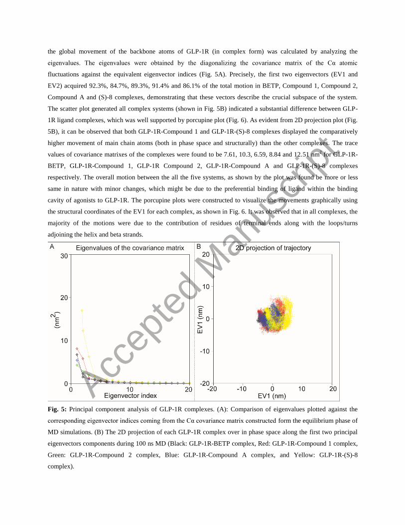
the global movement of the backbone atoms of GLP-1R (in complex form) was calculated by analyzing the
eigenvalues. The eigenvalues were obtained by the diagonalizing the covariance matrix of the Cα atomic
fluctuations against the equivalent eigenvector indices (Fig. 5A). Precisely, the first two eigenvectors (EV1 and
EV2) acquired 92.3%, 84.7%, 89.3%, 91.4% and 86.1% of the total motion in BETP, Compound 1, Compound 2,
Compound A and (S)-8 complexes, demonstrating that these vectors describe the crucial subspace of the system.
The scatter plot generated all complex systems (shown in Fig. 5B) indicated a substantial difference between GLP-
1R ligand complexes, which was well supported by porcupine plot (Fig. 6). As evident from 2D projection plot (Fig.
5B), it can be observed that both GLP-1R-Compound 1 and GLP-1R-(S)-8 complexes displayed the comparatively
higher movement of main chain atoms (both in phase space and structurally) than the other complexes. The trace
values of covariance matrixes of the complexes were found to be 7.61, 10.3, 6.59, 8.84 and 12.51 nm2 for GLP-1R-
BETP, GLP-1R-Compound 1, GLP-1R Compound 2, GLP-1R-Compound A and GLP-1R-(S)-8 complexes
respectively. The overall motion between the all the five systems, as shown by the plot was found be more or less
same in nature with minor changes, which might be due to the preferential binding of ligand within the binding
cavity of agonists to GLP-1R. The porcupine plots were constructed to visualize the movements graphically using
the structural coordinates of the EV1 for each complex, as shown in Fig. 6. It was observed that in all complexes, the
majority of the motions were due to the contribution of residues of terminal ends along with the loops/turns
adjoining the helix and beta strands.
Fig. 5: Principal component analysis of GLP-1R complexes. (A): Comparison of eigenvalues plotted against the
corresponding eigenvector indices coming from the Cα covariance matrix constructed form the equilibrium phase of
MD simulations. (B) The 2D projection of each GLP-1R complex over in phase space along the first two principal
eigenvectors components during 100 ns MD (Black: GLP-1R-BETP complex, Red: GLP-1R-Compound 1 complex,
Green: GLP-1R-Compound 2 complex, Blue: GLP-1R-Compound A complex, and Yellow: GLP-1R-(S)-8
complex).
Accep
ted
Man
uscr
ipt
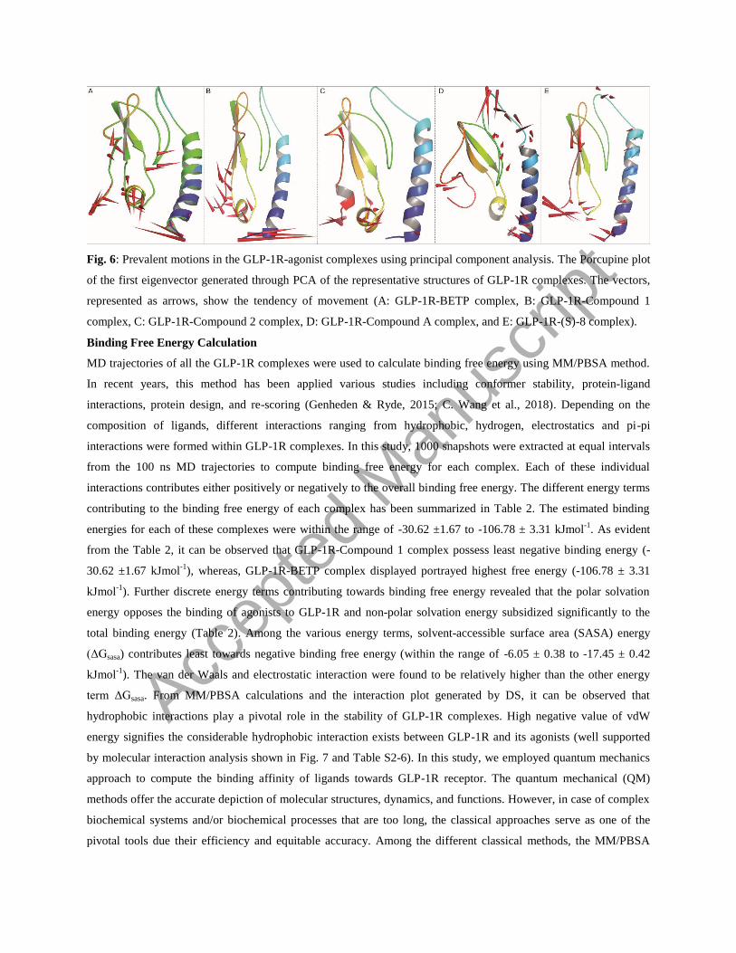
Fig. 6: Prevalent motions in the GLP-1R-agonist complexes using principal component analysis. The Porcupine plot
of the first eigenvector generated through PCA of the representative structures of GLP-1R complexes. The vectors,
represented as arrows, show the tendency of movement (A: GLP-1R-BETP complex, B: GLP-1R-Compound 1
complex, C: GLP-1R-Compound 2 complex, D: GLP-1R-Compound A complex, and E: GLP-1R-(S)-8 complex).
Binding Free Energy Calculation
MD trajectories of all the GLP-1R complexes were used to calculate binding free energy using MM/PBSA method.
In recent years, this method has been applied various studies including conformer stability, protein-ligand
interactions, protein design, and re-scoring (Genheden & Ryde, 2015; C. Wang et al., 2018). Depending on the
composition of ligands, different interactions ranging from hydrophobic, hydrogen, electrostatics and pi-pi
interactions were formed within GLP-1R complexes. In this study, 1000 snapshots were extracted at equal intervals
from the 100 ns MD trajectories to compute binding free energy for each complex. Each of these individual
interactions contributes either positively or negatively to the overall binding free energy. The different energy terms
contributing to the binding free energy of each complex has been summarized in Table 2. The estimated binding
energies for each of these complexes were within the range of -30.62 ±1.67 to -106.78 ± 3.31 kJmol-1
. As evident
from the Table 2, it can be observed that GLP-1R-Compound 1 complex possess least negative binding energy (-
30.62 ±1.67 kJmol-1
), whereas, GLP-1R-BETP complex displayed portrayed highest free energy (-106.78 ± 3.31
kJmol-1
). Further discrete energy terms contributing towards binding free energy revealed that the polar solvation
energy opposes the binding of agonists to GLP-1R and non-polar solvation energy subsidized significantly to the
total binding energy (Table 2). Among the various energy terms, solvent-accessible surface area (SASA) energy
(ΔGsasa) contributes least towards negative binding free energy (within the range of -6.05 ± 0.38 to -17.45 ± 0.42
kJmol-1
). The van der Waals and electrostatic interaction were found to be relatively higher than the other energy
term ΔGsasa. From MM/PBSA calculations and the interaction plot generated by DS, it can be observed that
hydrophobic interactions play a pivotal role in the stability of GLP-1R complexes. High negative value of vdW
energy signifies the considerable hydrophobic interaction exists between GLP-1R and its agonists (well supported
by molecular interaction analysis shown in Fig. 7 and Table S2-6). In this study, we employed quantum mechanics
approach to compute the binding affinity of ligands towards GLP-1R receptor. The quantum mechanical (QM)
methods offer the accurate depiction of molecular structures, dynamics, and functions. However, in case of complex
biochemical systems and/or biochemical processes that are too long, the classical approaches serve as one of the
pivotal tools due their efficiency and equitable accuracy. Among the different classical methods, the MM/PBSA
Accep
ted
Man
uscr
ipt
approach has materialized as an efficient and reliable method to understand crucial molecular recognition processes
(Kollman et al., 2000; C. Wang et al., 2018). Moreover, this method has been often a choice for efficient and reliable
calculation of free energy of protein-ligand and protein-peptide and protein-protein interactions. For simple and
small systems, the free energy perturbation and thermodynamic integration methods (developed much earlier than
the MM/PBSA) shown to be more accurate than the MM/PBSA method. Due to low efficiency and slow
convergence, their applications to typically large and complex molecular recognition processes are relatively
limited. MM/PBSA methods have been used in wide spectrum of applications to estimate the binding free energies
at a reasonable computational cost. The applications of the MMPBSA method have grown considerably in many
different areas of bio-molecular study including protein-ligand binding affinity calculations due to their utility in
drug discovery research. At present due to lack of experimental evidence on the binding energy, we cannot correlate
our computed energy of the ligand towards receptor. However, our study has portrayed the energetic component that
drives the receptor-ligand interaction at molecular scale.
Table 2: MM/PBSA binding free energies calculated using the g_mmpbsa method. Various energy terms
contributing to the total free energy of GLP-1R ligand complexes
Energetic components (kJ/mol)
Ligands
BETP Compound 1
Compound 2 Compound A (S)-8
van der Waal energy (ΔGvwd) -183.03 ± 4.31 -58.21 ± 3.44 -64.12 ± 3.78 -67.00 ± 3.51 -58.44 ± 3.76
Electrostatic energy (ΔGele) -67.31 ± 1.71 -70.39 ± 4.59 -58.95 ± 3.57 -174.99 ± 9.32 -134.79 ± 6.03
Polar solvation energy (ΔGps) 160.68 ± 3.34 104.86 ±7.29 96.27 ± 6.62 179.01 ± 15.53 97.10 ± 7.81
SASA energy (ΔGsasa) -17.45 ± 0.42 -6.83±0.40 -6.59 ± 0.40 -9.04 ± 0.49 -6.05 ± 0.38
Binding energy (ΔGbinding) -106.78 ± 3.31 -30.62 ±1.67 -33.67 ± 1.69 -72.19 ± 2.31 -100.63 ± 3.48
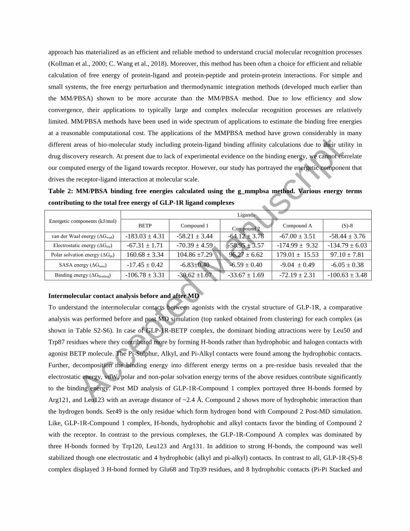
Intermolecular contact analysis before and after MD
To understand the intermolecular contacts between agonists with the crystal structure of GLP-1R, a comparative
analysis was performed before and post MD simulation (top ranked obtained from clustering) for each complex (as
shown in Table S2-S6). In case of GLP-1R-BETP complex, the dominant binding attractions were by Leu50 and
Trp87 residues where they contributed more by forming H-bonds rather than hydrophobic and halogen contacts with
agonist BETP molecule. The Pi-Sulphur, Alkyl, and Pi-Alkyl contacts were found among the hydrophobic contacts.
Further, decomposition the binding energy into different energy terms on a pre-residue basis revealed that the
electrostatic energy, vdW, polar and non-polar solvation energy terms of the above residues contribute significantly
to the binding energy. Post MD analysis of GLP-1R-Compound 1 complex portrayed three H-bonds formed by
Arg121, and Leu123 with an average distance of ~2.4 Å. Compound 2 shows more of hydrophobic interaction than
the hydrogen bonds. Ser49 is the only residue which form hydrogen bond with Compound 2 Post-MD simulation.
Like, GLP-1R-Compound 1 complex, H-bonds, hydrophobic and alkyl contacts favor the binding of Compound 2
with the receptor. In contrast to the previous complexes, the GLP-1R-Compound A complex was dominated by
three H-bonds formed by Trp120, Leu123 and Arg131. In addition to strong H-bonds, the compound was well
stabilized though one electrostatic and 4 hydrophobic (alkyl and pi-alkyl) contacts. In contrast to all, GLP-1R-(S)-8
complex displayed 3 H-bond formed by Glu68 and Trp39 residues, and 8 hydrophobic contacts (Pi-Pi Stacked and
Accep
ted
Man
uscr
ipt
Pi-Alkyl) (Table S6). Furthermore, we have also computed the energetic contribution of each residue towards
negative free energy (as shown Fig. S3). As evident from Fig. S3, it can be observed that most of the ligand binding
residues significantly contribute to the overall negative free energy. Altogether, the results of intermolecular contact
analysis perfectly corroborate with the contribution of different energetic terms i.e., vdW and non-polar solvation
energies favorable for binding.
Fig. 7 Molecular interaction of the agonist’s with GLP1-R receptor obtained from clustering analysis (Agonists
displayed in ball and stick model, interacting residues in stick model along with the receptor colored by secondary
structure elements)
Conclusion
In the present study, we have performed molecular docking of five structurally known diverse GLP-1R agonists.
The docking studies revealed the significance of variable binding sites of these agonists and critical amino acid
residues involved in the recognition of these agonists. MD simulations of the complexes of GLP-1R with all five
different agonists were performed to understand the dynamics behavior of these complexes at the molecular scale.
Our results suggested that all the agonists could exactly bind to the active pocket of GLP-1R to display agonistic
activity. Similar to the reported data of BETP and Compound 2, our data suggests a probable high agonist activity
of (S)-8 in comparison to experimentally validated Compound 2 (Lotte Bjerre Knudsen et al., 2007; L. C. Morris et
al., 2014). However, there is no experimental evidence for (S)-8 in comparison with BETP and Compound 2. Our
results also presented higher agonist activity of the above-mentioned compounds to be driven due to more
hydrophobic interactions of agonists which the receptor protein. In our report, we have tried to describe all the
possible allosteric interactions between the five-small molecule GLP-1R agonists with extracellular domain of GLP-
1R along with its probable explanation for their differential experimental behavior. It is our expectations, mapping
all these possible interacting avenues between GLP-1R and its agonists will significantly broaden our understanding
of regulatory mechanisms within the GLP-1R and its agonist’s axis. In turn, this will provide new strategies to
screen and identify novel insulin secretagogue compounds having a greater efficacy of glucose-dependent
insulinotropic action and restoration of islet glucosensitivity with improved intrinsic protection against
hypoglycemia without compromising the efficacy of treatment. In summary, we have demonstrated the agonist
binding mechanism of GLP-1R, which can be useful to develop other agonist-based receptor models in order to
Accep
ted
Man
uscr
ipt
answer imperative questions associated with structure to function, and to propose agonist-mediated receptor
activation mechanisms in near future.
Conflicts of interest
No potential conflicts of interest relevant to this article were reported.
Acknowledgments
We sincerely thank the BioX Centre of IIT Mandi for the facility and this work was partially supported by Seed
Research grant from IIT Mandi and Khyati thanks the UGC, India for her fellowship.
Author Contributions
KG and BD conducted the majority of the experiments and performed data analyses. MKS performed part of the
molecular dynamic simulation and SK reviewed the manuscript. PM designed study, and supervised the project;
KG, BD, VD, AC, SD, SK and PM wrote the manuscript. PM is the guarantor of this work and, as such, had full
access to all the data in the study and takes responsibility for the integrity of the data and the accuracy of the data
analysis.
References
Amadei, A., Linssen, A. B. M., & Berendsen, H. J. C. (1993). Essential dynamics of proteins. Proteins: Structure,
Function, and Bioinformatics. https://doi.org/10.1002/prot.340170408
Baggio, L. L., & Drucker, D. J. (2007). Biology of Incretins: GLP-1 and GIP. Gastroenterology.
https://doi.org/10.1053/j.gastro.2007.03.054
Bueno, A. B., Showalter, A. D., Wainscott, D. B., Stutsman, C., Marín, A., Ficorilli, J., … Sloop, K. W. (2016).
Positive allosteric modulation of the glucagon-like peptide-1 receptor by diverse electrophiles. Journal of
Biological Chemistry. https://doi.org/10.1074/jbc.M115.696039
Daura, X., Gademann, K., Jaun, B., Seebach, D., van Gunsteren, W. F., & Mark, A. E. (1999). Peptide Folding:
When Simulation Meets Experiment. Angewandte Chemie International Edition.
https://doi.org/10.1002/(SICI)1521-3773(19990115)38:1/2<236::AID-ANIE236>3.3.CO;2-D
David, C. C., & Jacobs, D. J. (2014). Principal component analysis: a method for determining the essential dynamics
of proteins. Methods in Molecular Biology (Clifton, N.J.). https://doi.org/10.1007/978-1-62703-658-0_11
De Maturana, R. L., Willshaw, A., Kuntzsch, A., Rudolph, R., & Donnelly, D. (2003). The isolated N-terminal
domain of the glucagon-like peptide-1 (GLP-1) receptor binds exendin peptides with much higher affinity than
GLP-1. Journal of Biological Chemistry, 278(12), 10195–10200. https://doi.org/10.1074/jbc.M212147200
Dehury, B., Behera, S. K., & Mahapatra, N. (2017). Structural dynamics of Casein Kinase I (CKI) from malarial
parasite Plasmodium falciparum (Isolate 3D7): Insights from theoretical modelling and molecular simulations.
Journal of Molecular Graphics and Modelling. https://doi.org/10.1016/j.jmgm.2016.11.012
Drucker, D. J., & Nauck, M. A. (2006). The incretin system: glucagon-like peptide-1 receptor agonists and
Accep
ted
Man
uscr
ipt
dipeptidyl peptidase-4 inhibitors in type 2 diabetes. Lancet. https://doi.org/10.1016/S0140-6736(06)69705-5
Forli, S., Huey, R., Pique, M. E., Sanner, M. F., Goodsell, D. S., & Olson, A. J. (2016). Computational protein-
ligand docking and virtual drug screening with the AutoDock suite. Nature Protocols.
https://doi.org/10.1038/nprot.2016.051
Furman, B., Ong, W. K., & Pyne, N. J. (2010). Cyclic AMP signaling in pancreatic islets. Advances in Experimental
Medicine and Biology. https://doi.org/10.1007/978-90-481-3271-3_13
Genheden, S., & Ryde, U. (2015). The MM/PBSA and MM/GBSA methods to estimate ligand-binding affinities.
Expert Opinion on Drug Discovery. https://doi.org/10.1517/17460441.2015.1032936
Graaf, C. de, Donnelly, D., Wootten, D., Lau, J., Sexton, P. M., Miller, L. J., … Wang, M.-W. (2016). Glucagon-
Like Peptide-1 and Its Class B G Protein-Coupled Receptors: A Long March to Therapeutic Successes.
Pharmacological Reviews. https://doi.org/10.1124/pr.115.011395
Gupta, V. (2013). Glucagon-like peptide-1 analogues: An overview. Indian Journal of Endocrinology and
Metabolism. https://doi.org/10.4103/2230-8210.111625
Hansotia, T., Baggio, L. L., Delmeire, D., Hinke, S. A., Yamada, Y., Tsukiyama, K., … Drucker, D. J. (2004).
Double Incretin Receptor Knockout (DIRKO) Mice Reveal an Essential Role for the Enteroinsular Axis in
Transducing the Glucoregulatory Actions of DPP-IV Inhibitors. Diabetes.
https://doi.org/10.2337/diabetes.53.5.1326
Interantional Diabetes Federation. (2015). IDF Diabetes Atlas. International Diabetes Federation.
https://doi.org/10.1289/image.ehp.v119.i03
Jensen, C. C., Cnop, M., Hull, R. L., Fujimoto, W. Y., & Kahn, S. E. (2002). beta-cell function is a major
contributor to oral glucose tolerance in high-risk relatives of four ethnic groups in the U.S. Diabetes.
https://doi.org/10.2337/diabetes.51.7.2170
Knudsen, L. B., Kiel, D., Teng, M., Behrens, C., Bhumralkar, D., Kodra, J. T., … Lau, J. (2007). Small-molecule
agonists for the glucagon-like peptide 1 receptor. Proceedings of the National Academy of Sciences of the
United States of America. https://doi.org/10.1073/pnas.0605701104
Knudsen, L. B., Nielsen, P. F., Huusfeldt, P. O., Johansen, N. L., Madsen, K., Pedersen, F. Z., … Agersø, H. (2000).
Potent derivatives of glucagon-like peptide-1 with pharmacokinetic properties suitable for once daily
administration. Journal of Medicinal Chemistry. https://doi.org/10.1021/jm9909645
Kollman, P. A., Massova, I., Reyes, C., Kuhn, B., Huo, S., Chong, L., … Cheatham, T. E. (2000). Calculating
structures and free energies of complex molecules: Combining molecular mechanics and continuum models.
Accounts of Chemical Research. https://doi.org/10.1021/ar000033j
Koole, C., Wootten, D., Simms, J., Valant, C., Sridhar, R., Woodman, O. L., … Sexton, P. M. (2010). Allosteric
Accep
ted
Man
uscr
ipt
Ligands of the Glucagon-Like Peptide 1 Receptor (GLP-1R) Differentially Modulate Endogenous and
Exogenous Peptide Responses in a Pathway-Selective Manner: Implications for Drug Screening. Molecular
Pharmacology. https://doi.org/10.1124/mol.110.065664
Lee, H. S., Jo, S., Lim, H. S., & Im, W. (2012). Application of binding free energy calculations to prediction of
binding modes and affinities of MDM2 and MDMX inhibitors. Journal of Chemical Information and
Modeling. https://doi.org/10.1021/ci3000997
Lin, F., & Wang, R. (2009). Molecular modeling of the three-dimensional structure of GLP-1R and its interactions
with several agonists. Journal of Molecular Modeling. https://doi.org/10.1007/s00894-008-0372-2
Lu, S. Y., Jiang, Y. J., Lv, J., Wu, T. X., Yu, Q. Sen, & Zhu, W. L. (2010). Molecular docking and molecular
dynamics simulation studies of GPR40 receptor-agonist interactions. Journal of Molecular Graphics and
Modelling. https://doi.org/10.1016/j.jmgm.2010.02.001
Morris, G., & Huey, R. (2009). AutoDock4 and AutoDockTools4: Automated docking with selective receptor
flexibility. Journal of …. https://doi.org/10.1002/jcc.21256.AutoDock4
Morris, L. C., Nance, K. D., Gentry, P. R., Days, E. L., Weaver, C. D., Niswender, C. M., … Lindsley, C. W.
(2014). Discovery of (S)-2-cyclopentyl-N-((1-isopropylpyrrolidin2-yl)-9-methyl-1-oxo-2,9-dihydro-1H-
pyrrido[3,4-b]indole-4-carboxamide (VU0453379): a novel, CNS penetrant glucagon-like peptide 1 receptor
(GLP-1R) positive allosteric modulator (PAM). Journal of Medicinal Chemistry.
https://doi.org/10.1021/jm501375c
Nielsen, L. L., Young, A. A., & Parkes, D. G. (2004). Pharmacology of exenatide (synthetic exendin-4): A potential
therapeutic for improved glycemic control of type 2 diabetes. Regulatory Peptides.
https://doi.org/10.1016/j.regpep.2003.10.028
Parkes, D. G., Mace, K. F., & Trautmann, M. E. (2013). Discovery and development of exenatide: the first
antidiabetic agent to leverage the multiple benefits of the incretin hormone, GLP-1. Expert Opinion on Drug
Discovery. https://doi.org/10.1517/17460441.2013.741580
Santiago, C. G., Paci, E., & Donnelly, D. (2018). A mechanism for agonist activation of the glucagon-like Peptide-1
(GLP-1) receptor through modelling & molecular dynamics. Biochemical and Biophysical Research
Communications, 498(2), 359–365. https://doi.org/10.1016/j.bbrc.2018.01.110
Sloop, K. W., Willard, F. S., Brenner, M. B., Ficorilli, J., Valasek, K., Showalter, A. D., … Coghlan, M. J. (2010).
Novel small molecule glucagon-like peptide-1 receptor agonist stimulates insulin secretion in rodents and
from human islets. Diabetes. https://doi.org/10.2337/db10-0689
Su, H., He, M., Li, H., Liu, Q., Wang, J., Wang, Y., … Wang, M. W. (2008). Boc5, a non-peptidic glucagon-like
peptide-1 receptor agonist, invokes sustained glycemic control and weight loss in diabetic mice. PLoS ONE.
https://doi.org/10.1371/journal.pone.0002892
Accep
ted
Man
uscr
ipt
Tengholm, A. (2012). Cyclic AMP dynamics in the pancreatic β-cell. Upsala Journal of Medical Sciences.
https://doi.org/10.3109/03009734.2012.724732
Trujillo, J. M., Nuffer, W., & Ellis, S. L. (2015). GLP-1 receptor agonists: A review of head-to-head clinical studies.
Therapeutic Advances in Endocrinology and Metabolism. https://doi.org/10.1177/2042018814559725
Wang, C., Greene, D., Xiao, L., Qi, R., & Luo, R. (2018). Recent Developments and Applications of the MMPBSA
Method. Frontiers in Molecular Biosciences. https://doi.org/10.3389/fmolb.2017.00087
Wang, M. W., Liu, Q., & Zhou, C. H. (2010). Non-peptidic glucose-like peptide-1 receptor agonists: Aftermath of a
serendipitous discovery. Acta Pharmacologica Sinica. https://doi.org/10.1038/aps.2010.107
Wootten, D., Savage, E. E., Willard, F. S., Bueno, A. B., Sloop, K. W., Christopoulos, A., & Sexton, P. M. (2013).
Differential Activation and Modulation of the Glucagon-Like Peptide-1 Receptor by Small Molecule Ligands.
Molecular Pharmacology. https://doi.org/10.1124/mol.112.084525
Yang, D. H., Zhou, C. H., Liu, Q., & Wang, M. W. (2015). Landmark studies on the glucagon subfamily of GPCRs:
From small molecule modulators to a crystal structure. Acta Pharmacologica Sinica.
https://doi.org/10.1038/aps.2015.78
Amadei, A., Linssen, A. B. M., & Berendsen, H. J. C. (1993). Essential dynamics of proteins. Proteins: Structure,
Function, and Bioinformatics. https://doi.org/10.1002/prot.340170408
Baggio, L. L., & Drucker, D. J. (2007). Biology of Incretins: GLP-1 and GIP. Gastroenterology.
https://doi.org/10.1053/j.gastro.2007.03.054
Bueno, A. B., Showalter, A. D., Wainscott, D. B., Stutsman, C., Marín, A., Ficorilli, J., … Sloop, K. W. (2016).
Positive allosteric modulation of the glucagon-like peptide-1 receptor by diverse electrophiles. Journal of
Biological Chemistry. https://doi.org/10.1074/jbc.M115.696039
Daura, X., Gademann, K., Jaun, B., Seebach, D., van Gunsteren, W. F., & Mark, A. E. (1999). Peptide Folding:
When Simulation Meets Experiment. Angewandte Chemie International Edition.
https://doi.org/10.1002/(SICI)1521-3773(19990115)38:1/2<236::AID-ANIE236>3.3.CO;2-D
David, C. C., & Jacobs, D. J. (2014). Principal component analysis: a method for determining the essential dynamics
of proteins. Methods in Molecular Biology (Clifton, N.J.). https://doi.org/10.1007/978-1-62703-658-0_11
De Maturana, R. L., Willshaw, A., Kuntzsch, A., Rudolph, R., & Donnelly, D. (2003). The isolated N-terminal
domain of the glucagon-like peptide-1 (GLP-1) receptor binds exendin peptides with much higher affinity than
GLP-1. Journal of Biological Chemistry, 278(12), 10195–10200. https://doi.org/10.1074/jbc.M212147200
Dehury, B., Behera, S. K., & Mahapatra, N. (2017). Structural dynamics of Casein Kinase I (CKI) from malarial
Accep
ted
Man
uscr
ipt
parasite Plasmodium falciparum (Isolate 3D7): Insights from theoretical modelling and molecular simulations.
Journal of Molecular Graphics and Modelling. https://doi.org/10.1016/j.jmgm.2016.11.012
Drucker, D. J., & Nauck, M. A. (2006). The incretin system: glucagon-like peptide-1 receptor agonists and
dipeptidyl peptidase-4 inhibitors in type 2 diabetes. Lancet. https://doi.org/10.1016/S0140-6736(06)69705-5
Forli, S., Huey, R., Pique, M. E., Sanner, M. F., Goodsell, D. S., & Olson, A. J. (2016). Computational protein-
ligand docking and virtual drug screening with the AutoDock suite. Nature Protocols.
https://doi.org/10.1038/nprot.2016.051
Furman, B., Ong, W. K., & Pyne, N. J. (2010). Cyclic AMP signaling in pancreatic islets. Advances in Experimental
Medicine and Biology. https://doi.org/10.1007/978-90-481-3271-3_13
Genheden, S., & Ryde, U. (2015). The MM/PBSA and MM/GBSA methods to estimate ligand-binding affinities.
Expert Opinion on Drug Discovery. https://doi.org/10.1517/17460441.2015.1032936
Graaf, C. de, Donnelly, D., Wootten, D., Lau, J., Sexton, P. M., Miller, L. J., … Wang, M.-W. (2016). Glucagon-
Like Peptide-1 and Its Class B G Protein-Coupled Receptors: A Long March to Therapeutic Successes.
Pharmacological Reviews. https://doi.org/10.1124/pr.115.011395
Gupta, V. (2013). Glucagon-like peptide-1 analogues: An overview. Indian Journal of Endocrinology and
Metabolism. https://doi.org/10.4103/2230-8210.111625
Hansotia, T., Baggio, L. L., Delmeire, D., Hinke, S. A., Yamada, Y., Tsukiyama, K., … Drucker, D. J. (2004).
Double Incretin Receptor Knockout (DIRKO) Mice Reveal an Essential Role for the Enteroinsular Axis in
Transducing the Glucoregulatory Actions of DPP-IV Inhibitors. Diabetes.
https://doi.org/10.2337/diabetes.53.5.1326
Interantional Diabetes Federation. (2015). IDF Diabetes Atlas. International Diabetes Federation.
https://doi.org/10.1289/image.ehp.v119.i03
Jensen, C. C., Cnop, M., Hull, R. L., Fujimoto, W. Y., & Kahn, S. E. (2002). beta-cell function is a major
contributor to oral glucose tolerance in high-risk relatives of four ethnic groups in the U.S. Diabetes.
https://doi.org/10.2337/diabetes.51.7.2170
Knudsen, L. B., Kiel, D., Teng, M., Behrens, C., Bhumralkar, D., Kodra, J. T., … Lau, J. (2007). Small-molecule
agonists for the glucagon-like peptide 1 receptor. Proceedings of the National Academy of Sciences of the
United States of America. https://doi.org/10.1073/pnas.0605701104
Knudsen, L. B., Nielsen, P. F., Huusfeldt, P. O., Johansen, N. L., Madsen, K., Pedersen, F. Z., … Agersø, H. (2000).
Potent derivatives of glucagon-like peptide-1 with pharmacokinetic properties suitable for once daily
administration. Journal of Medicinal Chemistry. https://doi.org/10.1021/jm9909645
Kollman, P. A., Massova, I., Reyes, C., Kuhn, B., Huo, S., Chong, L., … Cheatham, T. E. (2000). Calculating
Accep
ted
Man
uscr
ipt
structures and free energies of complex molecules: Combining molecular mechanics and continuum models.
Accounts of Chemical Research. https://doi.org/10.1021/ar000033j
Koole, C., Wootten, D., Simms, J., Valant, C., Sridhar, R., Woodman, O. L., … Sexton, P. M. (2010). Allosteric
Ligands of the Glucagon-Like Peptide 1 Receptor (GLP-1R) Differentially Modulate Endogenous and
Exogenous Peptide Responses in a Pathway-Selective Manner: Implications for Drug Screening. Molecular
Pharmacology. https://doi.org/10.1124/mol.110.065664
Lee, H. S., Jo, S., Lim, H. S., & Im, W. (2012). Application of binding free energy calculations to prediction of
binding modes and affinities of MDM2 and MDMX inhibitors. Journal of Chemical Information and
Modeling. https://doi.org/10.1021/ci3000997
Lin, F., & Wang, R. (2009). Molecular modeling of the three-dimensional structure of GLP-1R and its interactions
with several agonists. Journal of Molecular Modeling. https://doi.org/10.1007/s00894-008-0372-2
Lu, S. Y., Jiang, Y. J., Lv, J., Wu, T. X., Yu, Q. Sen, & Zhu, W. L. (2010). Molecular docking and molecular
dynamics simulation studies of GPR40 receptor-agonist interactions. Journal of Molecular Graphics and
Modelling. https://doi.org/10.1016/j.jmgm.2010.02.001
Morris, G., & Huey, R. (2009). AutoDock4 and AutoDockTools4: Automated docking with selective receptor
flexibility. Journal of …. https://doi.org/10.1002/jcc.21256.AutoDock4
Morris, L. C., Nance, K. D., Gentry, P. R., Days, E. L., Weaver, C. D., Niswender, C. M., … Lindsley, C. W.
(2014). Discovery of (S)-2-cyclopentyl-N-((1-isopropylpyrrolidin2-yl)-9-methyl-1-oxo-2,9-dihydro-1H-
pyrrido[3,4-b]indole-4-carboxamide (VU0453379): a novel, CNS penetrant glucagon-like peptide 1 receptor
(GLP-1R) positive allosteric modulator (PAM). Journal of Medicinal Chemistry.
https://doi.org/10.1021/jm501375c
Nielsen, L. L., Young, A. A., & Parkes, D. G. (2004). Pharmacology of exenatide (synthetic exendin-4): A potential
therapeutic for improved glycemic control of type 2 diabetes. Regulatory Peptides.
https://doi.org/10.1016/j.regpep.2003.10.028
Parkes, D. G., Mace, K. F., & Trautmann, M. E. (2013). Discovery and development of exenatide: the first
antidiabetic agent to leverage the multiple benefits of the incretin hormone, GLP-1. Expert Opinion on Drug
Discovery. https://doi.org/10.1517/17460441.2013.741580
Santiago, C. G., Paci, E., & Donnelly, D. (2018). A mechanism for agonist activation of the glucagon-like Peptide-1
(GLP-1) receptor through modelling & molecular dynamics. Biochemical and Biophysical Research
Communications, 498(2), 359–365. https://doi.org/10.1016/j.bbrc.2018.01.110
Sloop, K. W., Willard, F. S., Brenner, M. B., Ficorilli, J., Valasek, K., Showalter, A. D., … Coghlan, M. J. (2010).
Novel small molecule glucagon-like peptide-1 receptor agonist stimulates insulin secretion in rodents and
from human islets. Diabetes. https://doi.org/10.2337/db10-0689
Accep
ted
Man
uscr
ipt
Su, H., He, M., Li, H., Liu, Q., Wang, J., Wang, Y., … Wang, M. W. (2008). Boc5, a non-peptidic glucagon-like
peptide-1 receptor agonist, invokes sustained glycemic control and weight loss in diabetic mice. PLoS ONE.
https://doi.org/10.1371/journal.pone.0002892
Tengholm, A. (2012). Cyclic AMP dynamics in the pancreatic β-cell. Upsala Journal of Medical Sciences.
https://doi.org/10.3109/03009734.2012.724732
Trujillo, J. M., Nuffer, W., & Ellis, S. L. (2015). GLP-1 receptor agonists: A review of head-to-head clinical studies.
Therapeutic Advances in Endocrinology and Metabolism. https://doi.org/10.1177/2042018814559725
Wang, C., Greene, D., Xiao, L., Qi, R., & Luo, R. (2018). Recent Developments and Applications of the MMPBSA
Method. Frontiers in Molecular Biosciences. https://doi.org/10.3389/fmolb.2017.00087
Wang, M. W., Liu, Q., & Zhou, C. H. (2010). Non-peptidic glucose-like peptide-1 receptor agonists: Aftermath of a
serendipitous discovery. Acta Pharmacologica Sinica. https://doi.org/10.1038/aps.2010.107
Wootten, D., Savage, E. E., Willard, F. S., Bueno, A. B., Sloop, K. W., Christopoulos, A., & Sexton, P. M. (2013).
Differential Activation and Modulation of the Glucagon-Like Peptide-1 Receptor by Small Molecule Ligands.
Molecular Pharmacology. https://doi.org/10.1124/mol.112.084525
Yang, D. H., Zhou, C. H., Liu, Q., & Wang, M. W. (2015). Landmark studies on the glucagon subfamily of GPCRs:
From small molecule modulators to a crystal structure. Acta Pharmacologica Sinica.
https://doi.org/10.1038/aps.2015.78
Accep
ted
Man
uscr
ipt