Embed Size (px)
Citation preview

5 Tall Tree LanePleasantville, NY 10570
www.nevetdermatology.com
UPCOMING MEETINGS
> 2016 8th World Congress of Veterinary Dermatology
> 2017 NAVDF April 26-29 Disney Contemporary Resort
MARK YOUR CALENDAR
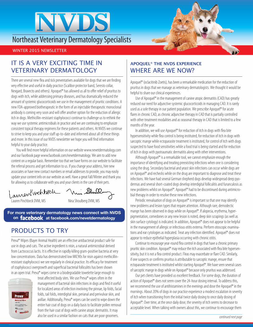
Northeast Veterinary Dermatology Specialists
Northeast Veterinary Dermatology Specialists
IT IS A VERY EXCITING TIME IN VETERINARY DERMATOLOGYThere are several new flea and tick preventatives available for dogs that we are finding very effective and useful in daily practice (Scalibor protector band, Seresto collar, Nexgard, Bravecto and others). Apoquel® has allowed us all to offer relief of pruritus to dogs with itch, while addressing primary diseases, and has dramatically reduced the amount of systemic glucocorticoids we use in the management of pruritic conditions. A new FDA-approved biotherapeutic in the form of an injectable therapeutic monoclonal antibody is coming very soon and will offer another option for the reduction of allergic itch in dogs. Methicillin-resistant staphylococci continue to challenge us to rethink the way we use systemic antimicrobials in practice and we are continuing to emphasize consistent topical therapy regimens for these patients and others. At NVDS we continue to strive to keep you and your staff up-to-date and informed about all of these things and more. In this issue of our NVDS newsletter we hope you will find information helpful to your daily practice.
You will find more helpful information on our website www.nevetdermatology.com and our Facebook page www.facebook.com/nevetdermatology. We aim to add new content on a regular basis. Remember too that we have forms on our website to facilitate the referral process and get information to us. If you change your address, hire new associates or have new contact numbers or email addresses to provide, you may easily update your content info on our website as well. Have a great Fall/Winter and thank you for allowing us to collaborate with you and your clients in the care of their pets.
Lauren Pinchbeck DVM, MS Nina Shoulberg DVM, MS
WINTER 2015 NEWSLETTER
PRODUCTS TO TRYPreva® Wipes (Bayer Animal Health) are an effective antibacterial product safe for use in dogs and cats. The active ingredient is nisin, a natural antimicrobial derived from Lactococcus lactis. It is effective in rapidly killing gram-positive bacteria at very low concentrations. Data has demonstrated low MIC90s for nisin against methicillin- resistant staphylococci we see regularly in clinical practice. Its efficacy for treatment of staphylococci overgrowth and superficial bacterial folliculitis has been shown in an open trial. Preva® wipes come in a biodegradable towelette large enough to
treat affected body sites. We use Preva® wipes often in the management of bacterial skin infections in dogs and find it useful for localized areas of infection involving the pinnae, lip folds, facial folds, tail folds, interdigital skin, perianal and perivulvar skin, and axillae. Additionally, Preva® wipes can be used to wipe down the entire hair coat of dogs on a daily basis to facilitate pollen removal from the hair coat of dogs with canine atopic dermatitis. It may also be used in a similar fashion on cats that are poor groomers.
APOQUEL® THE NVDS EXPERIENCEWHERE ARE WE NOW?Apoquel® (oclacitinib Zoetis), has been a remarkable medication for the reduction of pruritus in dogs that we manage as veterinary dermatologists. We thought it would be helpful to share our clinical experiences.
Use of Apoquel® in the management of canine atopic dermatitis (CAD) has greatly reduced our need for adjunctive systemic glucocorticoids in managing CAD. It is rarely used as a sole therapy in our patient population. We prescribe Apoquel® for acute flares in chronic CAD, as chronic adjunctive therapy in CAD that is partially controlled with other treatment modalities and as seasonal therapy in CAD that is limited to a few months of the year.
In addition, we will use Apoquel® for reduction of itch in dogs with flea bite hypersensitivity while flea control is being instituted, for reduction of itch in dogs with sarcoptic mange while ectoparasite treatment is instituted, for control of itch with dogs suspected to have food sensitivities while a food trial is being started and for reduction of itch in dogs with pyotraumatic dermatitis along with other interventions.
Although Apoquel® is a remarkable tool, we cannot emphasize enough the importance of identifying and treating preexisting infections when one is considering using the drug. Secondary bacterial and yeast skin infections can occur while dogs are on Apoquel® and rechecks while on the drug are important to diagnose and treat these infections. We have had several German shepherd dogs develop widespread deep pyo-dermas and several short-coated dogs develop interdigital folliculitis and furunculosis as new problems whilst on Apoquel®. Apoquel® had to be discontinued during antimicro-bial therapy in order to resolve these new infections.
Periodic reevaluation of dogs on Apoquel® is important so that one may identify new problems and lesion types that require attention. Although rare, demodectic mange has been observed in dogs while on Apoquel®. If alopecia, erythema, hyper-pigmentation, comedones or any new lesion is noted, deep skin scrapings (as well as skin surface cytology) is indicated. In addition, Apoquel® does not appear to be helpful in the management of allergic or infectious otitis externa. Perform otoscopic examina-tions and ear cytologies as indicated. Treat any infection identified. Apoquel® does not appear to reduce epithelial hyperplasia occurring with chronic otitis.
Continue to encourage year-round flea control in dogs that have a chronic primary pruritic skin condition. Apoquel® may reduce the itch associated with flea bite hypersen-sitivity, but it is not a flea control product. Fleas may exacerbate or flare CAD. Similarly, if one suspects or confirms pruritus is attributable to sarcoptic mange, ensure that ectoparasite treatment is instituted whilst starting Apoquel®. We have seen several cases of sarcoptic mange in dogs while on Apoquel® because only pruritus was addressed.
Our pet clients have provided us excellent feedback. For some dogs, the duration of action of Apoquel® does not seem cover the 24-hour dosing interval. To address this, we recommend the use of antihistamines in the evenings and dose the Apoquel® in the mornings. About 20% of dogs in our practice experience a modest escalation in severity of itch when transitioning from the initial twice daily dosing to once daily dosing of Apoquel®. Over time, at the once daily dose, the severity of itch seems to decrease to acceptable level. When talking with owners about this, we continue to encourage them
continued next page
For more veterinary dermatology news connect with NVDS on at facebook.com/nevetdermatology
Northeast Veterinary Dermatology Specialists
Apoquel® from page 1
to work through the differential diagnoses to identify and control the primary diseasecausing pruritus. We are also noting that there seems to be a diminished response to Apoquel® over time. At this time, we do not have an immunologic explanation for this observation. However, knowing that this is potential, we continue to pursue the primary disease causing pruritus.
One of the greatest benefits in veterinary dermatology practice is that we now have an effective medication that can control itch while we are in the process of allergy testing pets. For the most part, serum and intradermal testing may be done while a pet is taking Apoquel®. From time to time, we may discontinue Apoquel® for testing. Previ-ously, we would have to discontinue antipruritic therapies before intradermal testing and pets were uncomfortable during this time. In some cases, we would be unable to allergy test a dog due to the inability to withdraw from glucocorticoids.
FDA approval studies have evaluated the safety of Apoquel®. The product label reviews those results. We do not recommend off-label usage of Apoquel®, and do not recommend use in cats at this time. Side effects to Apoquel® are few. The very rare dog will have diarrhea. Several observations have been made by other veterinary dermatologists using Apoquel®. None have been critically evaluated in the peer-reviewed literature to-date. Studies are ongoing. There have been anecdotal reports of individual cases of demodicosis, viral papillomatosis, weight gain thought to be related to the decrease in calorie expenditure from previously pruritic behaviors, urinary tract infections (UTI), proteinuria that may be associated with occult UTI or other cause, notable leukopenias and ALP elevations. Dogs on Apoquel® have developed malignancies, but care should be taken in attributing their development to use of Apoquel® at the prescribed dosages; use of Apoquel® has not been found to be causal. As per the product label, we do not recommend use of Apoquel® in dogs with a previous history of malignant neoplasia of any type.
How do we monitor pets on Apoquel®? Routine scheduled reevaluations are recommended. Some dogs should have pretreatment lab work and urinalysis. We use our clinical judgment in making those recommendations. Typically, a CBC, blood chemistry and UA are done one-to-three months into treatment with the drug and then every six-to-12 months thereafter while the dog remains on the Apoquel®.
IN THE PIPELINETHERAPEUTIC MONOCLONAL ANTI-BODY TREATMENT FOR PRURITUSBiologic medicine uses principles of what we know about normal immune responses to rebalance, restore or stimulate a patient’s own immune system to fight disease. Biotherapeutics used in biologic medicine include immunostimulating cytokines (interferon), colony stimulating factors (erythropoietin) and therapeutic monoclonal antibodies (mAbs). Therapeutic mAbs can be used to block disease relevant proteins (cytokines, receptors) and target viruses or bacteria and aid in destruction and elimi-nation of pathogens. A new biotherapeutic is in the pipeline and will have a specific application in veterinary dermatology.
Acquired knowledge about disease pathogenesis has resulted in the development of targeted therapies for chronic diseases for which the target antigen is now known. Therapeutic mAbs are designed to selectively target such proteins/antigens. These mAbs are created using recombinant DNA techniques. By use of recombinant DNA techniques one achieves therapeutic mAbs that are tolerated by the target species (humanized, caninzed, felinized) and can be administered directly to the patient.
Therapeutic mAbs are given by injection, have a long half-life and are protected from destruction. Therapeutic mAbs are eliminated by intracellular catabolism within the lysosome where they are broken down into peptides or amino acids. These may be reused for synthesis of new proteins or renally excreted.
Examples of therapeutic mAbs in human medicine include omalizumab (Xolair) for allergic asthma. This mAb binds free IgE in plasma and intercellular fluid. Another, rituximab (Rituxan) targets CD20 on B lymphocytes and is used in the treatment of autoimmune disease and lymphomas. Therapeutic mAbs are well-tolerated in humans; the safety is attributable to specific targeting and no intracellular activity. Early human mAbs had a high proportion of mouse-derived sequences that were recognized as foreign. This resulted in immunogenicity, destruction of the mAb, de-creased efficacy and side effects. Development of humanized and fully human mAbs addressed these problems. During the development of therapeutic mAbs for dogs, generation of fully caninized mAbs has been the goal.
Early examples of therapeutic mAbs for canines have been evaluated. An anti-IgE mAb has been shown to reduce house dust mite IgE hypersensitivity for a duration of five weeks in house dust mite sensitized beagles. This was done by use of mAbs that inhibit production of IgE via its promoting cytokines (IL4 and IL13), their cytokine receptors, or IgE itself. Therapeutic mAbs have also been shown to neutralize the pruritogenic IL-31 cytokine in dogs and reduce the pruritic response for three weeks after injection.
TJ Fleck et. al. (abstract 2015 NAVDF) identified and characterized a therapeutic mAb, ZTS-00103289, that neutralized IL-31 mediated pruritus in beagles. Recall that IL-31 has recently been shown to induce pruritus in multiple species. The increased expression of Il-31 is associated with severity of AD in humans. To evaluate the role of IL-31 in canine atopic dermatitis, a caninized mAb that inhibits IL-31 mediated cell-based signaling was identified and characterized. A subcutaneous dose of the mAb or an injection of placebo was administered at 1 mg/kg to the beagle dogs. Pruritus was assessed over time. Pruritus scores for mAb treated dogs were significantly lower than for placebo on day one and day 28. This study in beagle dogs supported the use of ZTS-00103289 subcutaneously to achieve a one-month duration of efficacy. A next step was to evaluate this in naturally occurring canine atopic dermatitis.
One had to first determine dose and duration of activity. A laboratory dose titration efficacy study with ZTS-00103289 was done by RR Walters et. al. (abstract, NAVDF 2015), wherein they used this caninized anti-IL-31 mAb in a canine beagle model of IL-31 induced pruritus. This study demonstrated the clear relationship between dose and duration of activity. In this study, the pruritogenic and pro-inflammatory cytokine IL-31 was administered to dogs to induce short-term pruritus. A single injection of ZTS-00103289 was then administered at various doses. Dogs then received IL-31 challenges. Results showed a significant reduction in pruritus with use of ZTS-0013289 and determined that free mAb ZTS-00103289 half-life of was 11.4 days. Anti-drug an-tibodies produced by the dog against the mAb were minimal and none persisted. The pharmacodynamic data concluded that a dose 2 mg/kg of ZTS-0013289 would result in a serum mAb concentration that remained above the EC50 for 28 days.
GM Michels et. al. (abstract, NAVDF 2015) reported a proof of concept efficacy and safety study of ZTS-00103289 anti-IL-31 mAb in the treatment of atopic dermatitis in client-owned dogs. ZTS-00103289 was dosed twice subcutaneously at 14-day intervals in dogs with AD. This was a masked placebo 42-day study including 78 dogs. Owner assessment of itch and dermatologist assessment of lesions were evaluated. Results showed that ZTS-00103289 treated dogs had significantly (>50%) greater reduction of pruritus from baseline in owner assessed pruritus scores compared to placebo on days two-42. Lesion scores improved 40% when evaluated at day 14 and day 28. No serious adverse events were reported. Antidrug-antibody levels showed no immunogenicity. The results of this study support the effectiveness and safety of ZTS-001-3289 for the treatment of canine atopic dermatitis.
Thus, with the advancement basic science and new knowledge about the molecules responsible for pruritus in dogs, a new biotherapeutic (ZTS-00103289) has being generated and tested in the laboratory and then in dogs with natural occurring disease. Based upon the studies reported, we anticipate this therapeutic mAb to be another safe and effective therapy we have to use in the management of dogs with canine atopic dermatitis.
HOSPITAL LOCATIONS
We offer patient appointments and services in New York and Connecticut, at the following veterinary hospitals:
County Animal Clinic 1574 Central Park Avenue
Yonkers, NY 10710
Hudson Highlands Veterinary Specialty Group
222 Lime Kiln Road Hopewell Junction, NY 12533
Katonah-Bedford Veterinary Center 546 N. Bedford Road (Rte. 117)
Bedford Hills, NY 10507
Central Hospital for Veterinary Medicine
4 Devine Street North Haven, CT 06473
Newtown Veterinary Specialists 52 Church Hill Road Newtown, CT 06470
Norwalk Veterinary Hospital 726 Connecticut Avenue
South Norwalk, CT 06854
Norwalk Veterinary Referral and Emergency Center 123 West Cedar Street
Norwalk, CT 06854
Shoreline Veterinary Referral and Emergency Center 895 Bridgeport Avenue
Shelton, CT 06484
To schedule a dermatology appointment at any of our locations,
please call us at
914-777-DERM (3376).
Nina Shoulberg, DVM, MS
Lauren Pinchbeck, DVM, MS
www.nevetdermatology.com
COMMON INFECTIOUS DISEASES AFFECTING THE NASAL PLANUM OF DOGSSeveral skin diseases in the dog may affect the nasal planum. Salient features of the common infectious skin diseases affecting the nasal planum of dogs is presented. Visit our website blog for more information on the diseases affecting the nasal planum.
Mucocutaneous pyoderma (MCP) is a common condition that may affect the nasal planum. MCP is a bacterial infection usually caused by Staphylococcus pseudintermedius. German shepherd dogs are the most commonly affected breed. MCP primarily affects the mucocutaneous junctions of the nasal planum and lips, but other mucocutaneous junction sites may be involved. If the nasal planum is affected, there is initial erythema and swelling of the nasal planum, typically at the alar folds, followed by crusts, then fissures and erosions. Beneath elevated crusts there is thick, purulent exudate and an erosive to ulcerative dermatitis. Diagnosis is made via dermatologic examination, cytology and response to treatment. Cytology of exudate reveals bacteria and inflammatory cells. Bacterial culture and susceptibility testing of purulent exudate from under a protected crust is indicated if there is a failed response to appropriate antibiotic therapy. If cytology does not reveal bacteria, a bacterial culture is sterile and there is a failed response to antimicrobial therapy, submission of a tissue biopsy for dermatohistopathology is indicated, as discoid lupus erythematosus is a condition with similar clinical appearance. A fungal culture may also be indicated, as some fungal diseases share clinical features with MCP. Treatment of MCP involves topical therapies and systemic antibiotics. Maintenance topical therapy is recommended for long-term control.
Trichophyton dermatophytosis is one of the most common fungal infections we see affecting the nasal planum in dogs. This condition is caused by infection with the soil-borne dermatophyte Trichophyton mentagrophytes and dogs that hunt or root in soil, such as the Jack Russell terrier, are at an increased risk of developing the disease. Lesions of the skin and nasal planum may include, alopecia, erythema, papules, scaling, crusting, erosions or ulcerations. The clinical presentation may resemble autoimmune diseases such as discoid lupus erythematosus, pemphigus erythematosus or dermatomyositis. Pruritus of lesional sites varies from absent to severe. Diagnosis is via dermatologic examination, skin scrapings, cytology, culture and dermatohistopathology.
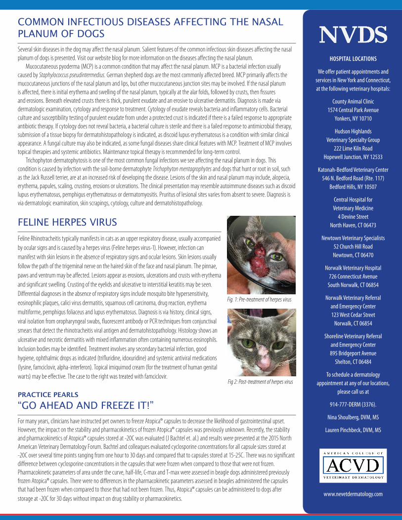
FELINE HERPES VIRUSFeline Rhinotracheitis typically manifests in cats as an upper respiratory disease, usually accompanied by ocular signs and is caused by a herpes virus (Feline herpes virus-1). However, infection can manifest with skin lesions in the absence of respiratory signs and ocular lesions. Skin lesions usually follow the path of the trigeminal nerve on the haired skin of the face and nasal planum. The pinnae, paws and ventrum may be affected. Lesions appear as erosions, ulcerations and crusts with erythema and significant swelling. Crusting of the eyelids and ulcerative to interstitial keratitis may be seen. Differential diagnoses in the absence of respiratory signs include mosquito bite hypersensitivity, eosinophilic plaques, calici virus dermatitis, squamous cell carcinoma, drug reaction, erythema multiforme, pemphigus foliaceus and lupus erythematosus. Diagnosis is via history, clinical signs, viral isolation from oropharyngeal swabs, fluorescent antibody or PCR techniques from conjunctival smears that detect the rhinotracheitis viral antigen and dermatohistopathology. Histology shows an ulcerative and necrotic dermatitis with mixed inflammation often containing numerous eosinophils. Inclusion bodies may be identified. Treatment involves any secondary bacterial infection, good hygiene, ophthalmic drops as indicated (trifluridine, idoxuridine) and systemic antiviral medications (lysine, famciclovir, alpha-interferon). Topical imiquimod cream (for the treatment of human genital warts) may be effective. The case to the right was treated with famciclovir.
PRACTICE PEARLS“GO AHEAD AND FREEZE IT!”For many years, clinicians have instructed pet owners to freeze Atopica® capsules to decrease the likelihood of gastrointestinal upset. However, the impact on the stability and pharmacokinetics of frozen Atopica® capsules was previously unknown. Recently, the stability and pharmacokinetics of Atopica® capsules stored at -20C was evaluated (J Bachtel et. al.) and results were presented at the 2015 North American Veterinary Dermatology Forum. Bachtel and colleagues evaluated cyclosporine concentrations for all capsule sizes stored at -20C over several time points ranging from one hour to 30 days and compared that to capsules stored at 15-25C. There was no significant difference between cyclosporine concentrations in the capsules that were frozen when compared to those that were not frozen. Pharmacokinetic parameters of area under the curve, half-life, C-max and T-max were assessed in beagle dogs administered previously frozen Atopica® capsules. There were no differences in the pharmacokinetic parameters assessed in beagles administered the capsules that had been frozen when compared to those that had not been frozen. Thus, Atopica® capsules can be administered to dogs after storage at -20C for 30 days without impact on drug stability or pharmacokinetics.
Fig 2: Post-treatment of herpes virus
Fig. 1: Pre-treatment of herpes virus