Embed Size (px)
Citation preview
The Journal of Neuroscience, September 1993, 13(g): 38783883
Noradrenaline Hyperpolarizes Identified Rat Mesopontine Cholinergic Neurons in vitro
Julie A. Williams and Peter B. Reiner
Kinsmen Laboratory of Neurological Research, Department of Psychiatry, University of British Columbia, Vancouver, British Columbia V6T 123, Canada
Inhibition of brainstem cholinergic neurons by noradrenergic neurons of the locus ceruleus has long been suggested as a key mechanism of behavioral state control. In particular, the commonly held view is that noradrenaline (NA) plays a permissive role in rapid eye movement (REM) sleep gener- ation by disinhibiting brainstem cholinergic neurons. While this notion has been supported by numerous investigations, the inhibition of cholinergic neurons by NA has never been directly demonstrated. The purpose of this study was to investigate the effects of NA upon identified cholinergic neu- rons in the rat mesopontine tegmentum.
Using whole-cell patch-clamp recordings in slices, 175 cells were studied during bath application of 50 I.IM NA. Cho- linergic neurons were positively identified by intracellular labeling with biocytin and subsequent staining with NADPH- diaphorase, a reliable marker for brainstem cholinergic neu- rons (Vincent et al., 1983). Successful intracellular labeling was obtained in 98 cells. Ninety-two percent (36 of 39) of cholinergic neurons hyperpolarized in response to NA, while noncholinergic cells (n = 57) exhibited mixed responses. Application of NA in a low-Ca2+, high-Mg2+ solution elicited the same hyperpolarizing effect as in normal solution, which indicated that the effect of NA on cholinergic neurons was direct. The noradrenergic hyperpolarization was mimicked by the a,-adrenoceptor agonist UK-1 4,304, and was blocked by the a2-adrenoceptor antagonist idazoxan, which sug- gested an a2-mediated response. Finally, voltage-clamp ex- periments revealed that NA activates the inwardly rectifying potassium current, /,,.
These data unambiguously confirm the hypothesis that NA inhibits brainstem cholinergic neurons, and are fundamental to understanding the role of noradrenergic-cholinergic in- teractions in behavioral state control.
[Key words: REM sleep, locus ceruleus, at-adrenergic re- ceptors, potassium currents, NADPH-diaphorase, behavioral state control]
The most widely accepted model of rapid eye movement (REM) sleep generation is that release of ACh within the medial pontine reticular formation (MPRF) is one, if not the, natural trigger for
Received Nov. 5, 1992; revised Feb. 17, 1993; accepted Mar. 18, 1993. We thank Andy Laycock for technical assistance. This work was supported by
grants from the British Columbia Health Care Research Foundation and MRC. J.A.W. received partial support from the IODE. P.B.R. is an MRC Scholar.
Correspondence should be addressed to Peter B. Reiner, Kinsmen Laboratory of Neurological Research, Department of Psychiatry, University of British Co- lumbia, 2255 Wesbrook Mall, Vancouver, BC V6T 123 Canada.
Copyright 0 1993 Society for Neuroscience 0270-6474/93/133878-06$05.00/O
REM sleep generation. This is based upon the well-established observation that microinjection of cholinergic agonists into the MPRF induces a state indistinguishable from REM sleep (Bax- ter, 1969; Mitler and Dement, 1974; Amatruda et al., 1975). That amines inhibit this cholinergic trigger was suggested by the observation that systemic administration of the anti-AChE agent eserine induces a state indistinguishable from REM sleep, but only after depletion of amines by reserpine (Karczmar et al., 1970). Based upon extracellular recordings of (1) neurons se- lectively active during REM sleep in the gigantocellular teg- mental field (FTG) where AChE-positive neurons are found, and (2) neurons that fall silent during REM sleep in the nor- adrenergic locus ceruleus (LC) (Hobson et al., 1975) and sero- tonergic dorsal raphe (DR) nuclei (McGinty and Harper, 1972) McCarley and Hobson (1975) proposed the reciprocal-inter- action hypothesis: that the sleep cycle is controlled by an in- hibitory aminergic population and a reciprocally excitatory cho- linergic population, in which the silence of aminergic neurons would disinhibit cholinergic neurons and thereby evoke REM sleep by release of ACh in the MPRF.
The striking behavioral neurophysiological profile of norad- renergic and serotonergic neurons has been fully confirmed in numerous studies (McGinty and Harper, 1976; Trulson and Jacobs, 1979; Aston-Jones and Bloom, 198 1; Reiner, 1985). However, two features of the original reciprocal-interaction model were subsequently shown to be invalid. First, the REM selectivity of FTG neuronal activity was found to be an artifact of head restraint (Siegel and McGinty, 1977; Vertes, 1977). Second, the AChE-positive neurons of the FTG were found not to be cholinergic when studied with ChAT immunohistochem- istry (Jones and Beaudet, 1987; Vincent and Reiner, 1987; Shi- romani et al., 1988). However, these same studies unambigu- ously demonstrated that the neurons of the laterodorsal (LDT) and pedunculopontine (PPT) tegmental nuclei represent the pre- dominant group of brainstem cholinergic neurons, and these neurons give rise to a massive innervation of the thalamus (So- froniew et al., 1985; Satoh and Fibiger, 1986; Woolf and Butch- er, 1986; Hallanger and Wainer, 1988; Steriade et al., 1988), as well as direct projections to the MPRF (Mitani et al., 1988; Shiromani et al., 1988; Quattrochi et al., 1989; Jones, 1990; Semba et al., 1990). Unfortunately, the behavioral neurophys- iology of mesopontine cholinergic neurons is still not known with certainty. A small percentage of neurons in this region exhibit REM-selective discharge patterns, but the majority do not (El Mansari et al., 1989; Steriade et al., 1990). Based upon these data, Steriade et al. (1990) have concluded that cholinergic neurons are active during both wake and REM sleep, while other authors have argued otherwise (Sakai, 1988; Kamondi et al.,
The Journal of Neuroscience, September 1993, 13(9) 3879
1992). Barring more direct evidence, the behavior of mesopon- tine cholinergic neurons across states cannot be stated with com- plete confidence. Nonetheless, both the LDT and PPT contain neurons that are at least active during REM, and are thus can- didates for being the cholinergic neurons that trigger the state of REM sleep.
Given these observations, the original reciprocal-interaction hypothesis has been considerably modified by various groups (Hobson et al., 1986; Sakai, 1988; Steriade and McCarley, 1990). One explicit prediction of all these models is that noradrenaline (NA) and 5-HT inhibit mesopontine cholinergic neurons. We have recently shown that 5-HT inhibits identified cholinergic neurons (Luebke et al., 1992). Utilizing whole-cell patch-clamp recordings of LDT neurons in rat brain slices, we have now directly tested the other arm of this hypothesis, that NA hy- perpolarizes mesopontine cholinergic neurons.
Materials and Methods
Methods for whole-cell recording, intracellular labeling, and histochem- ical identification of LDT cholinergic neurons in rat brain slices were similar to those of Kamondi et al. (1992). Briefly, rats 7-l 5 d old were anesthetized with halothane and decapitated, and the brains were rapidly removed and immersed in cold artificial cerebrospinal fluid (ACSF). The brain was trimmed to a block containing the pontomesencephalic tegmentum and cut into 400 pm coronal sections with a Vibratome. Usually, two slices containing the LDT were obtained from each brain. Slices were stored in a holding chamber for at least 1 hr before being transferred to a recording chamber where they were superfused with ACSF at 2 ml/min. ACSF contained (in mM) 126 NaCli25 NaHCO,, 1.2 NaH,PO,. 2.5 KCl. 2.5 CaCl,, 1.2 M&l,. and 11 glucose. DH 7.4, when saturated with 95% 0,, 5% CO,. All>xperiments\ere carried out at room temperature.
Drugs were applied by superfusing the slice in ACSF containing a given concentration of the drug. Except for idazoxan and barium, drugs were stored in frozen aliquots at high concentrations and were thawed and diluted in ACSF prior to the experiment. NA, phenylephrine, and isoproterenol were obtained from Sigma Chemical Co.; idazoxan and UK-14,304, from Research Biochemicals, Inc.; and clonidine, from Boehringer Ingelheim, Ltd.
Patch pipettes were constructed from thin-wall (1.5 mm o.d., 1.1 mm i.d.) borosilicate glass (Sutter Inst. Co.). The electrode solution contained (in mM) 15 NaCl, 10 Na-HEPES, 11 EGTA, 140 KOH, 1 CaCl,, 2 Mg- ATP, 0.3 GTP, and 0.2% biocytin, and was pH balanced with methane- sulfonic acid to 7.4. Electrode resistance ranged from 4 to 6 MQ in the recording chamber, typical electrode seals were 9-12 GQ, and access resistance following establishment of the whole-cell recording configu- ration ranged from 10 to 80 MQ. Both bridge mode and continuous single-electrode voltage-clamp recordings were obtained with an Axo- clamp-2A. For voltage-clamp experiments, gains were from 5 to 10 nA/ mV, and the output filter reduced from 30 to 1 kHz; neither capacitance nor series resistance compensation was utilized, as described elsewhere (Kamondi et al., 1992). Data were collected through an Axolab interface using ~CLAMP computer software, version 5.0 (Axon Instruments), and recorded onto videotape in a digitized format. Data are reported as the mean + SD.
Histochemical identification of recorded neurons. In order to identify biocytin-filled neurons unambiguously, only one cell was obtained from each side of a slice. Slices were nicked on one side during preparation to differentiate one side from the other. After experiments were com- plete, each slice was fixed overnight in 2% paraformaldehyde and 15% picric acid in 0.1 M phosphate-buffered saline (PBS), pH 7.4. The fol- lowing day, slices were removed from the fixative and rinsed three times for 20 min in 0.1 M PBS. Slices were then incubated in PBS containing 20 &ml Texas red-conjugated avidin and 0.3% T&on-X for 2 hr, rinsed in PBS as before, and left in a T&on/PBS solution with 15% sucrose overnight. The next day slices were cut into 40-50 pm sections
37°C for 45-60 min. Afterward, slices were rinsed in PBS and stored in the dark to dry. Slides were coverslipped and examined under a microscope equipped with both bright-field optics and epifluorescence for identification of NADPH-positive and Texas red-positive neurons, respectively.
Results
Identification of cholinergic and noncholinergic neurons This study is based upon whole-cell patch-clamp recordings of 175 cells in the LDT. Successful intracellular labeling with bio- cytin was obtained in 96 cells, and these were all processed for NADPH-diaphorase histochemistry. Forty-one percent (39 of 96) of biocytin-labeled cells were NADPH-diaphorase positive and thus identified as cholinergic. The remaining cells (57 of 96, or 59%) were noncholinergic (Fig. 1). The focus ofthe results reported below is largely upon the identified cholinergic neurons (n = 39).
The salient biophysical properties of the cholinergic neurons of the LDT were identical to those reported previously (Ka- mondi et al., 1992; Luebke et al., 1992). In brief, the majority of cholinergic neurons exhibited both the transient outward K+ current IA and the transient inward Ca*+ current I,. In bridge mode these could be observed on the offset of hyperpolarizing current pulses: there was a delayed return to the resting potential (IA) followed by a depolarizing overshoot (IT) that often evoked a burst of action potentials.
NA hyperpolarizes LDT cholinergic neurons
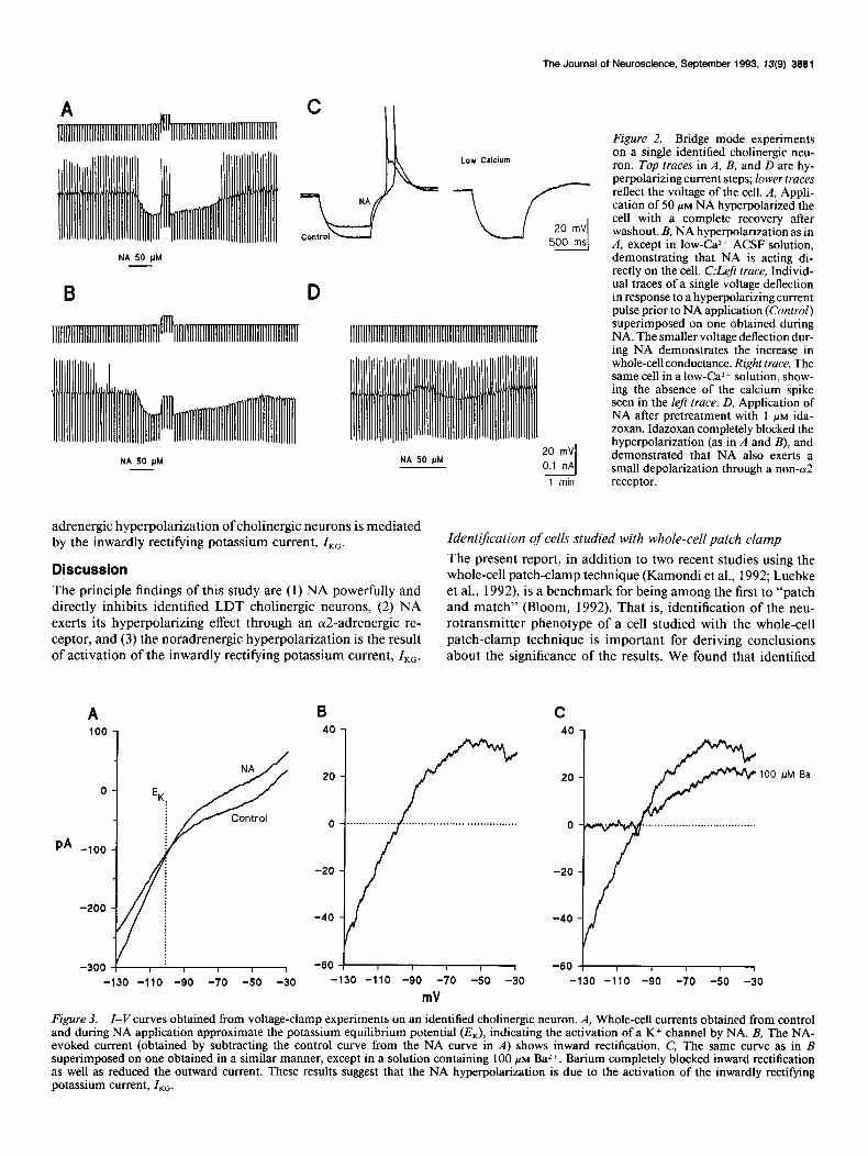
Ninety-two percent (36 of 39) of identified cholinergic neurons hyperpolarized by 10.1 f 4.1 mV in response to 50 PM NA. In contrast, noncholinergic neurons exhibited heterogeneous re- sponses to NA [35% (20 of 57) hyperpolarized, 46% (26 of 57) depolarized, 19% (11 of 57) no response]. The hyperpolarization of cholinergic neurons was characterized by an increase in con- ductance, as illustrated in Figure 2, A and B. When the cell hyperpolarized by NA was returned to the resting potential by injection of depolarizing current, the input resistance was de- creased as evidenced by smaller voltage deflections during the hyperpolarizing pulses as compared to control. Thus, NA hy- perpolarizes cholinergic neurons by increasing an ionic con- ductance.
We next carried out experiments to determine if the norad- renergic effect was direct. To test this, two paradigms were uti- lized. First, in the majority of cases (n = 27), 300 nM TTX was included in the bath solution to block voltage-dependent sodium channels. The noradrenergic hyperpolarization always persisted under such conditions. Second, some slices (n = 4) were bathed in ACSF with low Ca2+ and high Mg*+ concentration (0.5 mM and 10 mM, respectively) in addition to TTX to abolish Caz+- dependent synaptic transmission. As shown in Figure 2B, the low-Ca*+ ACSF abolished the low-threshold Ca2+ spike nor- mally seen after a 500 msec hyperpolarizing pulse, thus ensuring a minimal synaptic Caz+ influx and thereby synaptic transmis- sion. Responses to NA in low Ca*+ were identical to those seen in the normal solution, which indicates that the noradrenergic effect is indeed direct (compare Fig. 2A,C).
Noradrenergic hyperpolarization is mediated by cu2-receptors
Application of 1 PM idazoxan, an cY2-adrenergic antagonist, completely blocked the hyperpolarizing effect of NA on five of eight cholinergic cells and reduced the hyperpolarization in the other three cells by 2.3 f 0.5 mV. Interestingly, in three of the
with a cryostat and mounted onto coated slides. Choline@ neurons were identified using NADPH-diaphorase his-
tochemistry, a reliable marker of LDT cholinergic neurons (Vincent et al., 1983). Slides were immersed in a solution containing 1 mg/ml NADPH and 0.1 mg/ml nitroblue tetrazolium in PBS and incubated at

3880 Williams and Reiner - Noradrenergic Inhibition of Cholinergic Neurons
Figure I. Histochemical identification of LDT nuclei. A and C, Biocytin-filled neurons. B, NADPH-diaphorase histochemistry shows that the labeled neuron in A is NADPH positive and therefore choline&. D, The labeled cell in C did not stain for NADPH and is therefore noncholinergic. Scale bar, 25 w for A-D.
five cells in which the hyperpolarization was abolished by id- azoxan, NA induced a depolarization of 5.0 + 1.6 mV when a2-receptors were blocked (Fig. 20). Thus, NA may have an additional effect on cholinergic neurons mediated by receptors other than the cY2-receptor. However, these experiments were not carried out in low-Ca2+ ACSF, and therefore we do not know if the depolarization is a direct effect of NA. Both the (Y,- agonist phenylephrine (10 PM, n = 2) and the ,&agonist iso- proterenol (5 PM, n = 1) elicited no response from cells that exhibited a large hyperpolarization to NA. UK-14,304, a full ol2-adrenoceptor agonist (Cambridge, 198 l), induced a hyper- polarization of 5.8 f 2.3 mV on cholinergic neurons (10 PM, n = 3). Clonidine, a partial ar2-adrenoceptor agonist (Medgett et al., 1978) had no effect on cholinergic neurons that were hy- perpolarized by NA (l-10 PM, n = 6). Because clonidine has also been reported to act as a competitive cr2-antagonist, we tested its ability to block the noradrenergic hyperpolarization. It did not (n = 3). We therefore concluded that NA exerts its hyperpolarizing effect through an a2-adrenoceptor.
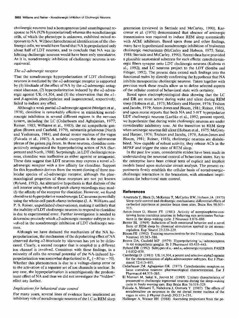
NA activates ZKc As described above, the bridge mode data indicated an increase in conductance during the noradrenergic hyperpolarization of cholinergic neurons. Based upon the calculated equilibrium po- tentials of each of the ions in solution, we hypothesized that the increase in conductance was due to the activation of an outward potassium current. We tested this hypothesis by obtaining cur- rent-voltage (Z-I’) ramps before and during application of NA (n = 5). As shown in Figure 3A, the ramps crossed at a position that approximated the calculated potassium equilibrium poten- tial of - 10 1.5 mV. When the control I-Vramps were subtracted from ones obtained during NA, the resulting curves showed inward rectification (Fig. 3B). This phenomenon is characteristic of the K+ current directly activated by G-proteins, ZKG (Hille, 1992). Addition of 100 PM Ba*+ (a nonspecific K+ channel block- er) to the bath markedly reduced the current in three of three cells, as illustrated in Figure 3C, consistent with the hypothesis that NA is activating a K+ current. We conclude that the nor-
The Journal of Neuroscience, September 1993, 13(9) 3881
Low Calcium
NA 50 UM
NA 50 PM NA 50 PM
1 mln
adrenergic hyperpolarization of cholinergic neurons is mediated by the inwardly rectifying potassium current, ZKG.
Discussion The principle findings of this study are (1) NA powerfully and directly inhibits identified LDT cholinergic neurons, (2) NA exerts its hyperpolarizing effect through an a2-adrenergic re- ceptor, and (3) the noradrenergic hyperpolarization is the result of activation of the inwardly rectifying potassium current, ZKG.
PA
0
-100
-200
-300
Figure 2. Bridge mode experiments on a single identified cholinergic neu- ron. Top traces in A, B, and D are hy- perpolarizing current steps; lower traces reflect the voltage of the cell. A, Appli- cation of 50 PM NA hyperpolarized the cell with a complete recovery after washout. B, NA hyperpolarization as in A, except in low-Caz+ ACSF solution, demonstrating that NA is acting di- rectly on the cell. C:Left trace, Individ- ual traces of a single voltage deflection in response to a hyperpolarizing current pulse prior to NA application (Control) superimposed on one obtained during NA. The smaller voltage deflection dur- ing NA demonstrates the increase in whole-cell conductance. Right trace, The same cell in a low-Ca2+ solution, show- ing the absence of the calcium spike seen in the left trace. D, Application of NA after pretreatment with 1 PM ida- zoxan. Idazoxan completely blocked the hyperpolarization (as in A and B), and demonstrated that NA also exerts a small depolarization through a non-a2 receptor.
Identification of cells studied with whole-cell patch clamp The present report, in addition to two recent studies using the whole-cell patch-clamp technique (Kamondi et al., 1992; Luebke et al., 1992) is a benchmark for being among the first to “patch and match” (Bloom, 1992). That is, identification of the neu- rotransmitter phenotype of a cell studied with the whole-cell patch-clamp technique is important for deriving conclusions about the significance of the results. We found that identified
-60 j-y-----
-130 -110 -90 -70 -50 -30
mV
-60
Figure 3. I-Vcurves obtained from voltage-clamp experiments on an identified cholinergic neuron. A, Whole-cell currents obtained from control and during NA application approximate the potassium equilibrium potential (ZQ, indicating the activation of a K+ channel by NA. B, The NA- evoked current (obtained by subtracting the control curve from the NA curve in A) shows inward rectification. C, The same curve as in B superimposed on one obtained in a similar manner, except in a solution containing 100 PM Ba *+. Barium completely blocked inward rectification as well as reduced the outward current. These results suggest that the NA hyperpolarization is due to the activation of the inwardly rectifying potassium current, Z,,.
3882 Williams and Reiner * Noradrenergic Inhibition of Cholinergic Neurons
cholinergic neurons had a homogeneous (and unambiguous) re- sponse to NA (92% hyperpolarized) whereas the noncholinergic cells, of which the phenotype is unknown, exhibited mixed re- sponses to NA. Without histochemical identification of the cho- linergic cells, we would have found that NA hyperpolarized only about half of LDT neurons, and to conclude that NA was in- hibiting cholinergic neurons would have been only speculative. As it is, noradrenergic inhibition of cholinergic neurons is un- equivocal.
The cu2-adrenergic receptor
That the noradrenergic hyperpolarization of LDT cholinergic neurons is mediated by the a2-adrenergic receptor is supported by (1) blockade of the effect of NA by the a2-adrenergic antag- onist idazoxan, (2) hyperpolarization induced by the a2-adren- ergic agonist UK- 14,304, and (3) the observation that the al- and P-agonists phenylephrine and isoproterenol, respectively, failed to induce any effect.
Although a weak partial cu2-adrenergic agonist (Medgett et al., 1978) clonidine is remarkably effective in mimicking norad- renergic inhibition in several different regions in the nervous system, including the LC (Cedarbaum and Aghajanian, 1977; Reiner, 1985; Williams et al., 1985) the rat sympathetic gan- glion (Brown and Caufield, 1979), substantia gelatinosa (North and Yoshimura, 1984) and dorsal motor nucleus of the vagus (Fukuda et al., 1987). A notable exception is the submucous plexus of the guinea pig ileum. In these neurons, clonidine com- petitively antagonized the hyperpolarizing action of NA (Sur- prenant and North, 1988). Surprisingly, in LDT cholinergic neu- rons, clonidine was ineffective as either agonist or antagonist. These data suggest that LDT neurons may express a novel a2- adrenergic receptor with a low affinity for clonidine. Support for this hypothesis derives from the recent cloning of three mo- lecular species of a2-adrenergic receptor, although the phar- macological properties of these receptors are not yet known (Bylund, 1992). An alternative hypothesis is that dialysis of the cell interior using whole-cell patch-clamp recordings may mod- ify the affinity of the receptor for clonidine. However, we found clonidine to hyperpolarize noradrenergic LC neurons powerfully using the whole-cell patch-clamp technique (J. A. Williams and P. B. Reiner, unpublished observations), making it unlikely that the inability of LDT cholinergic neurons to respond to clonidine is due to experimental error. Further investigation is needed to determine precisely which cY2-adrenergic receptor subtype is in- volved in the noradrenergic inhibition of LDT cholinergic neu- rons.
Although we have deduced the mechanism of the NA hy- perpolarization, the mechanism of the depolarizing effect of NA observed during a2-blockade by idazoxan has yet to be delin- eated. Clearly, a second receptor that is coupled to a different ion channel is involved. Consistent with these findings, in a minority of cells the reversal potential of the NA-induced hy- perpolarization was somewhat depolarized to E, (- 80 to - 70). Whether this phenomenon is due to a voltage-clamp error or to the activation of a separate set of ion channels is unclear. In any case, the hyperpolarization is unambiguously the predom- inant effect of NA and thus we did not investigate the “hidden” effect any further.
Implications for behavioral state control
For many years, several lines of evidence have implicated an inhibitory role of noradrenergic neurons of the LC in REM sleep
generation (reviewed in Steriade and McCarley, 1990). Kar- czmar et al. (1970) demonstrated that absence of aminergic transmission was required to induce REM sleep systemically with AChE inhibitors. Based upon these and other findings, many have hypothesized noradrenergic inhibition of brainstem cholinergic mechanisms (McCarley and Hobson, 1975; Sakai, 1988; Steriade and McCarley, 1990). Recent data have provided a plausible anatomical substrate for such effects: catecholamin- ergic fibers synapse onto LDT cholinergic neurons (Kubota et al., 1992), and LC neurons project to the LDT (Semba and Fibiger, 1992). The present data extend such findings into the functional realm by directly confirming the hypothesis that NA inhibits mesopontine cholinergic neurons. Taken together with previous work these results allow us to define selected aspects of the cellular control of behavioral state with certainty.
Based upon electrophysiological data showing that the LC and DR are most active during wake and silent during REM sleep (Hobson et al., 1975; McGinty and Harper, 1976; Trulson and Jacobs, 1979; Aston-Jones and Bloom, 198 1; Reiner, 1985) and upon recent reports that both NA and 5-HT hyperpolarize LDT cholinergic neurons (Luebke et al., 1992; present report), we hypothesize that during wake cholinergic neurons are under considerable inhibitory tone. At the transition to REM sleep when aminergic neurons fall silent (Hobson et al., 1975; McGinty and Harper, 1976; Trulson and Jacobs, 1979; Aston-Jones and Bloom, 198 1; Reiner, 1985), cholinergic neurons are disinhi- bited. Now capable of robust activity, they release ACh in the MPRF and trigger the state of REM sleep.
In the past few years, considerable strides have been made in understanding the neuronal control of behavioral states. Key to the enterprise have been critical tests of explicit and implicit models based upon molecular circuit analysis. The present ex- periments firmly establish the cellular basis of noradrenergic- cholinergic interaction in the brainstem, with attendant impli- cations for behavioral state control.
References
Amatruda T, Black D, McKenna T, McCarley RW, Hobson JA (1975) Sleep cvcle control and cholineraic mechanisms: differential effects of card&o1 injections at pontinebrain stem sites. Brain Res 98:501- 515.
Aston-Jones G, Bloom FE (198 1) Activity of norepinephrine-con- taining locus coeruleus neurons in behaving rats anticipates fluctua- tions in the sleepwaking cycle. J Neurosci 1876-886.
Baxter BL (1969) Induction of both emotional behavior and a novel form of REM sleep by chemical stimulation applied to cat mesen- cephalon. Exp Neurol 23:220-229.
Bloom FE (1992) Training neuroscientists for the 2 1 st century. Trends Neurosci 15:383-386.
Brown DA, Caufield MP (1979) Hyperpolarizing ‘a,‘-adrenoceptors in rat sympathetic ganglia. Br J Pharmacol 65:435-445.
Bylund DB (1992) Subtypes of Ly, - and a,-adrenergic receptors. FASEB J 6:832-839.
Cambridge D (198 1) UK 14,304, a potent and selective alpha2-agonist for the characterization of alpha-adrenoceptor subtypes. Eur J Phar- macol 72:413-415.
Cedarbaum JM, Aghajanian GK (1977) Catecholamine receptors on locus coeruleus neurons: pharmacological characterization. Eur J Pharmacol44:375-385.
El Mansari M, Sakai K, Jouvet M (1989) Unitary characteristics of presumptive cholinergic tegmental neurons during the sleep-waking cycle in freely moving cats. Exp Brain Res 76:5 19-529.
Fukuda A, Minami T, Nabekura J, Oomura Y (1987) The effects of noradrenaline on neurones in the rat dorsal motor nucleus of the vagus in vitro. J Physiol (Lond) 393:2 13-23 1.
Hallanger A, Wainer BH (1988) Ascending projections from the pe-

The Journal of Neuroscience, September 1993, 73(9) 3883
dunculopontine tegmental nucleus and the adjacent mesopontine teg- mentum in the rat. J Comp Neurol 274:483-5 15.
Hille B (1992) G protein-coupled mechanisms and nervous signalling. Neuron 9: 187-195.
Hobson JA, McCarley RW, Wyzinski PW (1975) Sleep cycle oscil- lation: reciprocal discharge by two brainstem neuronal groups. Science 189:55-58.
Hobson JA, Lydic R, Baghdoyan HA (1986) Evolving concepts of sleep cycle generation: from brain centers to neuronal populations. Behav Brain Sci 9:371-448.
Jones BE (1990) Immunohistochemical study of choline acetyltrans- ferase-immunoreactive processes and cells ~innervating the ponto- medullan, reticular formation in the rat. J Camp Neurol 295:485- 514. -
Jones BE, Beaudet A (1987) Distribution of acetylcholine and cate- cholamine neurons in the cat brain stem: a choline acetyltransferase and tyrosine hydroxylase immunohistochemical study. J Comp Neu- rol 261:15-32.
Kamondi A, Williams JA, Hutcheon B, Reiner PB (1992) Membrane properties of mesopontine cholinergic neurons studied with the whole- cell patch-clamp technique: implications for behavioral state control. J Neurophysiol 68:1359-1371.
Karczmar AC, Longo VG, Scotti de Carolis A (1970) A pharmaco- logical model of paradoxical sleep: the role of cholinergic and mono- amine systems. Physiol Behav 5: 175-l 82.
Kubota Y, Leung E, Vincent SR (1992) Ultrastructure of cholinergic neurons in the laterodorsal tegmental nucleus of the rat: interaction with catecholamine fibers. Brain Res Bull 29:479-49 1.
Luebke JI, Greene RW, Semba K, Kamondi A, McCarley RW, Reiner PB (1992) Serotonin hyperpolarizes cholinergic low-threshold burst neurons in the rat laterodorsal tegmental nucleus in vitro. Proc Nat1 Acad Sci USA 89~743-747.
McCarley RW, Hobson JA (1975) Neuronal excitability modulation over the sleep cycle: a structural and mathematical model. Science 189:58-60.
McGinty DJ, Harper RM (1972) 5-HT containing neurons: unit ac- tivity during sleep. Sleep Res 1:27.
McGintv DJ, Harper RM (1976) Dorsal raphe neurons: depression of firing during sleep in cats. Brain Res 101:569-575.
Medaett IC. McCulloch MW. Rand MJ (1978) Partial agonist action of clonidine on prejunctional and postjunctional cY-adrenoceptors. Naunyn Schmiedebergs Arch Pharmacol 304:2 15-22 1.
Mitani A, Ito K, Hallanger AE, Wainer BH, Kataoka K, McCarley RW (1988) Cholinergic projections from the laterodorsal and peduncu- lopontine tegmental nuclei to the pontine gigantocellular tegmental field in the cat. Brain Res 45 1:397402.
Mitler MM, Dement WC (1974) Cataplectic-like behavior in cats after microinjections of carbachol in pontine reticular formation. Brain Res 68:335-343.
North RA, Yoshimura M (1984) The actions of noradrenaline on neurones of the rat substantia gelatinosa in vitro. J Physiol (Lond) 349:43-55.
Quattrochi JJ, Mamelak AN, Madison RD, Macklis JD, Hobson JA (1989) Mapping neuronal inputs to REM sleep induction sites with carbachol-fluorescent microspheres. Science 245:984-986.
Reiner PB (1985) Clonidine inhibits central noradrenergic neurons in unanesthetized cats. Eur J Pharmacol 115:249-257.
Sakai K (1988) Executive mechanisms of paradoxical sleep. Arch Ital Biol 1261239-257.
Satoh K, Fibiger HC (1986) Cholinergic neurons of the laterodorsal tegmental nucleus: efferent and afferent connections. J Comp Neurol 2531277-302.
Semba K, Fibiger HC (1992) Afferent connections of the laterodorsal and the pedunculopontine tegmental nuclei in the rat: a retro- and antero-grade transport and immunohistochemical study. J Comp Neurol 323:387410.
Semba K, Reiner PB, Fibiger HC (1990) Single cholinergic mesopon- tine tegmental neurons project to both the pontine reticular formation and the thalamus in the rat. Neuroscience 38:643-654.
Shiromani PJ, Armstrong DM, Gillin JC (1988) Cholinergic neurons from the dorsolateral pons project to the medial pons: a WGA-HRP and choline acetyltransferase immunohistochemical study. Neurosci Lett 95: 19-23.
Siegel JM, McGinty DJ (1977) Pontine reticular formation neurons: relationship of discharge to motor activity. Science 196:678-680.
Sofroniew MV, Priestly JV, Consolazione A, Eckenstein F, Cue110 AC (1985) Cholinergic projections from the midbrain and pons to the thalamus in the rat, identified by combined retrograde tracing and choline acetyltransferase immunohistochemistry. Brain Res 329:2 13- 223.
Steriade M, McCarley RW (1990) Brainstem control of wakefulness and sleep. New York: Plenum.
Steriade M, Pare D, Parent A, Smith Y (1988) Projections of cholin- ergic and noncholinergic neurons of the brain stem core to relay and associational thalamic nuclei in the cat and macaque monkey. Neu- roscience 25:47-67.
Steriade M, Datta S, Pare D, Oakson G, Curro Dossi R (1990) Neu- ronal activities in brain-stem cholinergic nuclei related to tonic ac- tivation processes in thalamocortical systems. J Neurosci 10:2541- 2559.
Surprenant A, North RA (1988) Mechanism of synaptic inhibition by noradrenaline acting at oc,-adrenoceptors. Proc R Sot Lond [Biol] 234: 85-l 14.
Trulson ME, Jacobs BL (1979) Raphe unit activity in freely moving cats: correlation with level of behavioral arousal. Brain Res 163: 135- 150.
Vertes RP (1977) Selective firing of rat pontine gigantocellular neurons during movement and REM sleep. Brain Res 128: 146-152.
Vincent SR, Reiner PB (1987) The immunohistochemical localization of choline acetyltransferase in the cat brain. Brain Res Bull 18:371- 415.
Vincent SR, Satoh K, Armstrong DM, Fibiger HC (1983) NADPH- diaphorase: a selective histochemical marker for the cholinergic neu- rons of the pontine reticular formation. Neurosci Lett 43:3 l-36.
Williams JT, Henderson G, North RA (1985) Characterization of (Ye adrenoceptors which increase potassium conductance in rat locus coeruleus neurons. Neuroscience 14:95-102.
Woolf NJ, Butcher LL (1986) Cholinergic systems in the rat brain. III. Projections from the pontomesencephalic tegmentum to the thal- amus, tectum, basal ganglia and basal forebrain. Brain Res Bull 16: 603-637.