Embed Size (px)
Citation preview
Non-operative Management of Proximal Humerus Fractures:
What’s Best for Our Patients?
Brian D. Solberg MD
1. Define our patient(s) a. What’s their baseline?
i. DASH/ Constant scores b. What’s the goal of treatment?
i. ADLs, dominant non-dominant arm c. What is possible?
i. Is a Constant score of 90 possible in an 80 year old? 2. Our previous experience –What can we learn from the past?
a. Operative repair b. Hemi-arthroplasty
3. What does the recent data tell us? Are we repeating past mistakes? a. Does ORIF make our patients better?
i. Statistical vs. clinical difference b. Does ORIF burn reconstruction bridges?
i. What if fixation fails? c. Is ORIF ANY better than non-op?
i. Good evidence? 4. Our patient
a. What’s best for them?
Bibliography
Beks RB, Ochen Y, Frima H, et al. Operative versus nonoperative treatment of proximal humeral fractures: a systematic review, meta-analysis, and comparison of observational studies and randomized controlled trials. J Shoulder Elbow Surg. 2018 Aug;27(8):1526-1534.
Handoll HH, Keding A, Corbacho B, et al. Five-year follow-up results of the PROFHER trial comparing operative and non-operative treatment of adults with a displaced fracture of the proximal humerus. Bone Joint J. 2017 Mar;99-B(3):383-392. Xie L, Ding F, Zhao Z,et al. Operative versus non-operative treatment in complex proximal humeral fractures: a meta-analysis of randomized controlled trials. Springerplus. 2015 Nov 25;4:728.
Rabi S, Evaniew N, Sprague SA, et al. Operative vs non-operative management of displaced proximal humeral fractures in the elderly: A systematic review and meta-analysis of randomized controlled trials. World J Orthop. 2015 Nov 18;6(10):838-46.
Proximal Humerus Fractures-Operative Brian Mullis, MD
1. 2015 Cochrane Review-“insufficient evidence to inform management of these fractures”[1]
a. Following recommendations are based on author’s opinion and review of the literature, which is considered weak
b. Keep in mind elderly are 16 times more likely to have a proximal humerus fracture than young, most studies are predominantly composed of elderly
2. Young patients or highly functional elderly (author’s opinion) a. General recs for isolated tuberosities or surgical neck
i. ORIF for greater tuberosty great then 3-5 mm displaced ii. Closed treatment for lesser tuberosity fracture, consider ORIF for significant
displacement 1cm or greater (little evidence to inform given rare isolated injury) iii. 2-part surgical neck good outcomes with closed treatment, consider ORIF or IM
nail for open fractures and polytrauma[2] b. 3-part or 4-part fractures
i. Equivalent outcomes with ORIF and IM nail techniques[3] ii. Consider fibula allograft for anatomic neck fractures with poor screw purchase
with ORIF iii. Hemiarthroplasty may be better option for head splitting
3. Low demand elderly patients a. Closed treatment appears to be equivalent to any surgical procedure with lower
complication rates, ORIF may be worst choice for all fracture patterns, reverse total shoulder may be best choice for 3 or 4-part fractures if surgery is chosen and may be superior to closed treatment (weak evidence)[4-6]
References
1. Kleinlugtenbelt, Y.V. and M. Bhandari, Cochrane in CORR ((R)): Interventions for Treating Proximal Humeral Fractures in Adults (Review). Clin Orthop Relat Res, 2015. 473(9): p. 2750-6. 2. Handoll, H.H., et al., Five-year follow-up results of the PROFHER trial comparing operative and non-operative treatment of adults with a displaced fracture of the proximal humerus. Bone Joint J, 2017. 99-B(3): p. 383-392. 3. Li, M., et al., Intramedullary nail versus locking plate for treatment of proximal humeral fractures: A meta-analysis based on 1384 individuals. J Int Med Res, 2018. 46(11): p. 4363-4376. 4. Dillon, M.T., et al., The increasing role of reverse total shoulder arthroplasty in the treatment of proximal humerus fractures. Injury, 2019. 5. Beks, R.B., et al., Operative versus nonoperative treatment of proximal humeral fractures: a systematic review, meta-analysis, and comparison of observational studies and randomized controlled trials. J Shoulder Elbow Surg, 2018. 27(8): p. 1526-1534. 6. Du, S., et al., Interventions for Treating 3- or 4-part proximal humeral fractures in elderly patient: A network meta-analysis of randomized controlled trials. Int J Surg, 2017. 48: p. 240-246.
Long vs. Short Cephalomedullary Nail – Why Longer is Better
William Min, MD
1. Introduction
a. Current literature supporting short over long i. EBL
ii. Surgery Time iii. Similar healing outcomes
b. Commonly stated benefit of a longer nail i. Periprosthetic advantage of long over short
2. Other (less recognized) benefits of going longer a. Metaphyseal instability
i. Analogous to using short retrograde femoral IMN b. Thigh pain c. Protection of atypical fractures in patients on bisphosphonates
i. Aren’t all of these patient’s suffering from a “pathologic fracture”? 3. Conclusion
a. Role for both short and long nails i. Short nails in:
1. EBL, surgery time concerns a. Clinically relevant?
2. Stable fracture patterns a. What is truly stable?
ii. Long nails in: 1. Everyone else (default)
Mangled Extremity: Never Amputate on the First Trip to the O.R.
William J. Ertl, MD
The definition of a mangled extremity has been varied and at times complex. Numerous scoring systems have been developed to predict limb salvage versus amputation. However, these have not been able to demonstrate predictability, sensitivity nor specificity for either limb salvage or amputation. (1, 2, 3) Further, functional outcome could also not be predicted utilizing different scoring systems. (4 The American College of Surgeons characterizes the mangled extremity “as a high energy transfer or crush causing a combination of injuries to the artery, bone, tendon, nerve and/or soft tissue.” From this surgeon’s perspective, as viewed as organ system, a mangled extremity is a limb that has the majority of its components injured, requiring surgical intervention. Therefore the limb threatened. The purpose of this short presentation is to provide support and examples of threatened limbs that were thought to require amputation on the day of presentation. Through the combined effort of orthopaedic surgeons, trauma surgeons, vascular surgeons and plastic surgeons, application of temporizing external fixation, vascular shunts and wound containment with negative pressure wound therapy (NPWT), immediate amputation was avoided. Upper Extremity Case example 17 year old male sustained an accidental gunshot wound from a black powder muzzle loading rifle to his left distal humerus. A field tourniquet was applied. No median nerve function, ulnar nerve dysthesias and brachial artery disruption with no distal pulses. Orthopaedic surgery asked to perform an urgent amputation due to large wound and lost pulses. Films reviewed and discussed with trauma surgeon not to amputate but pursue limb salvage. Patient aware of limb threatening nature of the injury. Temporary shunt placed, spanning external fixation placed and forearm fasciotomies performed. Wound contained with NPWT. Vascular reconstruction of segmental arterial loss with reverse saphenous vein graft. Humeral reconstruction was accomplished two days later at a more controlled time and setting, followed by split thickness skin grafting. Sural nerve grafting occurred four months later. Lower Extremity Case example 44 year old female sustained a 3B open left tibia/fibula fracture with segmental bone loss and gross contamination. Orthopaedic trauma consulted for operative stabilization versus amputation. Soft tissue loss was circumferential and a 5 cm segment of bone was missing. Patient understood the limb threatening nature of her injury. Underwent medullary nailing of her tibia. Ultimately required a latissimus flap with STSG.
Suggested overall approach
1) Rapid efficient transport of the patient from the injury scene. 2) Complete overall assessment of the injured limb
a. Size of wounds b. Amount of contamination c. Bone loss? d. Ankle/Brachial Index (ABI) to rule occult vascular injury e. Resuscitation (plug the holes)
3) Quality imaging a. Advanced imaging should not delay the patient to the O.R. for surgical stabilization
4) Emergent surgical intervention a. Temporize (external fixation)? b. Definitive fixation?
Summary Management of the patient with the mangled extremity requires the utilization of multiple surgical specialties. The role of the orthopaedic surgeon is early and has an important impact of the outcome the patient may ultimately have. In some instances, the decision is obvious to perform an immediate amputation when there is significant soft tissue loss, bone loss and/or limited reconstructive choices. However, temporizing the limb allows the surgeon to engage the patient and family in treatment options and the potential futility of limb salvage. An amputation may ultimately be required but it can now be performed on in a controlled, reconstructive manner. References
– 1) J Trauma 34(1): 99, 1993.
– 2) Am Surg 60(1): 50-5, 1994.
– 3) J Am Acad Orthop Surg 4(4): 182-190, 1996.
– 4) JBJS 83A(1): 3-14, 2001.
– 5) Am J Surg, 172(5): 569-73, 1996.
Session 1: Orthopaedic Trauma Hot Topics
Nail or Plate: Distal Tibia-Nail
Daniel N Segina MD
Krettek C, Stephan C, Schandelmaier P, et al. The use of Poller screws as blocking screws in stabilizing tibial fractures treated with small diameter intramedullary nails. J Bone Joint Surg Br. 1999;81-B:963–968 Nork S, Schwartz A, Agel J, et al. Intramedullary nailing of distal metaphyseal tibia fractures. J Bone Joint Surg Am. 2005;87:1213–1221 Fan CY, Chiang CC, et.al. Interlocking Nails for Displaced Metaphyseal Fractures of the Distal Tibia. Injury. 2005 May;36(5);699-74 Im GI, Tae SK. Distal metaphyseal fractures of tibia: a prospective randomized trial of closed reduction and intramedullary nail versus open reduction and plate and screws fixation. J Trauma. 2005;59:1219–1223 Zelle BA, Bhandari M, Espiritu M, et al; Evidence-Based Orthopaedic Trauma Working Group. Treatment of distal tibia fractures without articular involvement: a systematic review of 1125 fractures. J Orthop Trauma. 2006;20:76–79 Guo JJ, Tang N, Yang HL, et al.. A prospective, randomized trial comparing closed intramedullary nailing with percutaneous plating in the treatment of distal metaphyseal fractures of the tibia. J Bone Joint Surg Br. 2010;92:984–988 Vallier HA, Cureton BA, Patterson BM. Randomized, prospective comparison of plate versus intramedullary nail for distal tibial shaft fractures. J Orthop Trauma. 2011;25:736–741 Vallier HA, Cureton BA, Patterson BM. Factors influencing functional outcomes after distal tibia shaft fractures. J Orthop Trauma. 2012;26:178–183 Kwok CS, Crossman PT, Loizou CL. Plate versus nail for distal tibial fractures: a systematic review and meta-analysis. J Orthop Trauma. 2014;28:542–548 Avilucea F, Triantafillou K, Whiting P, et al. Suprapatellar intramedullary nail technique lowers rate of malalignment of distal tibia fractures. J Orthop Trauma. 2016;30:557–560 Costa ML, Achten J, Intramedullary nail fixation versus locking plate fixation for adults with a fracture of the distal tibia: the UK FixDT RCT. Health Technol Assess. 2018 May;22(25): 1-148
1
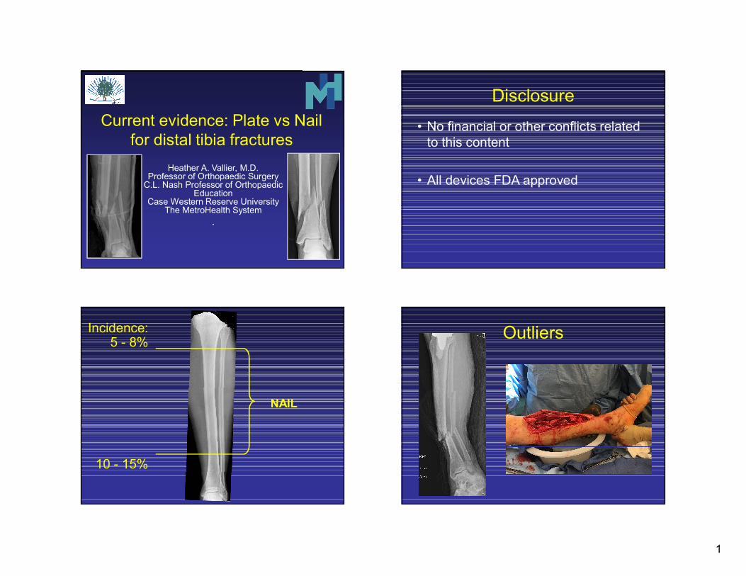
Current evidence: Plate vs Nail for distal tibia fractures
Heather A. Vallier, M.D.Professor of Orthopaedic Surgery
C.L. Nash Professor of Orthopaedic Education
Case Western Reserve UniversityThe MetroHealth System
.
Disclosure
• No financial or other conflicts related to this content
• All devices FDA approved
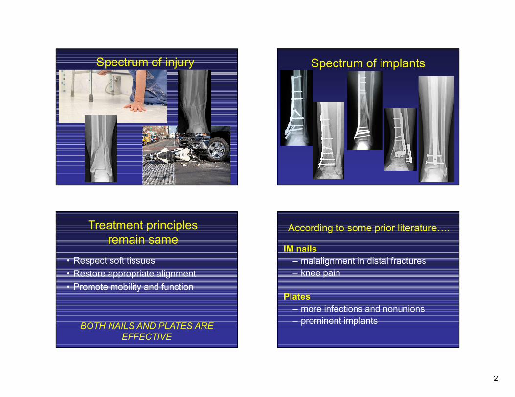
Incidence: 5 - 8%
10 - 15%
NAIL
Outliers
2
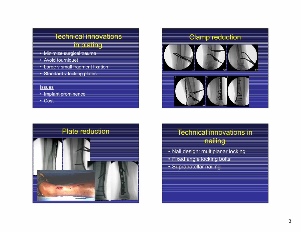
Spectrum of injury Spectrum of implants
Treatment principles remain same
• Respect soft tissues
• Restore appropriate alignment
• Promote mobility and function
BOTH NAILS AND PLATES ARE EFFECTIVE
IM nails– malalignment in distal fractures– knee pain
Plates– more infections and nonunions– prominent implants
According to some prior literature….
3
Technical innovations in plating
• Minimize surgical trauma
• Avoid tourniquet
• Large v small fragment fixation
• Standard v locking plates
Issues
• Implant prominence
• Cost
Clamp reduction
Plate reduction Technical innovations in nailing
• Nail design: multiplanar locking
• Fixed angle locking bolts
• Suprapatellar nailing
4
Review of comparative literature
• 7 RCTs
–3 with locking plates
–2 with anterolateral tibia plates
–2 with conventional medial plates
Summary of existing literature• Alignment: Nails more malalignment than plates
• Union: Locked plates more nonunions than nails; Nails with more nonuions than standard plates
• Outcomes similar: depends on time to WB; nails show better outcomes 6wk-3mo, then no difference
• Costs: Locked plate > Nail >> Standard plate
Review of comparative literature
• Methodological issues
–Small samples
–Selection bias
–Variable implant selection
–Variable technique
–Fibula fixation
Hypotheses
• Nails will be associated with malalignment, nonunion, and knee pain
• Plates will be associated with prominent hardware
5
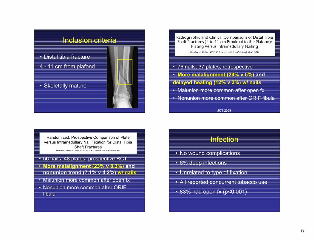
Inclusion criteria
• Distal tibia fracture
4 - 11 cm from plafond
• Skeletally mature
• 76 nails, 37 plates, retrospective
• More malalignment (29% v 5%) and
delayed healing (12% v 3%) w/ nails
• Malunion more common after open fx
• Nonunion more common after ORIF fibula
JOT 2008
• 56 nails, 48 plates, prospective RCT
• More malalignment (23% v 8.3%) and nonunion trend (7.1% v 4.2%) w/ nails
• Malunion more common after open fx
• Nonunion more common after ORIF fibula
Randomized, Prospective Comparison of Plate versus Intramedullary Nail Fixation for Distal Tibia
Shaft FracturesHeather A. Vallier, MD, Beth Ann Cureton, BS, and Brendan M. Patterson, MD
Infection
• No wound complications
• 6% deep infections
• Unrelated to type of fixation
• All reported concurrent tobacco use
• 83% had open fx (p<0.001)
6
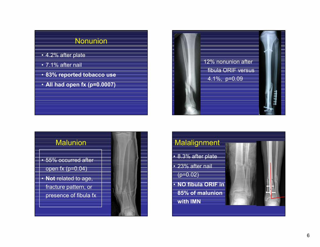
Nonunion
• 4.2% after plate
• 7.1% after nail
• 83% reported tobacco use
• All had open fx (p=0.0007)
12% nonunion after
fibula ORIF versus
4.1%, p=0.09
Malunion
• 55% occurred after
open fx (p=0.04)
• Not related to age,
fracture pattern, or
presence of fibula fx
Malalignment
• 8.3% after plate
• 23% after nail
(p=0.02)
• NO fibula ORIF in
85% of malunion
with IMN
7
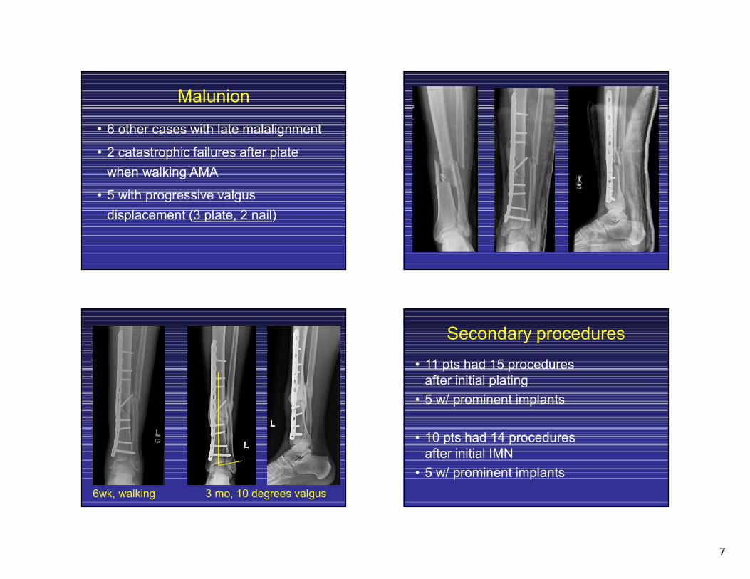
Malunion
• 6 other cases with late malalignment
• 2 catastrophic failures after plate
when walking AMA
• 5 with progressive valgus
displacement (3 plate, 2 nail)
6wk, walking 3 mo, 10 degrees valgus
Secondary procedures
• 11 pts had 15 procedures after initial plating
• 5 w/ prominent implants
• 10 pts had 14 procedures after initial IMN
• 5 w/ prominent implants
8
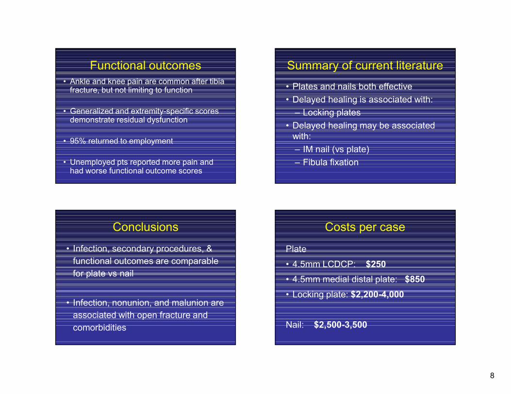
Functional outcomes• Ankle and knee pain are common after tibia
fracture, but not limiting to function
• Generalized and extremity-specific scores demonstrate residual dysfunction
• 95% returned to employment
• Unemployed pts reported more pain and had worse functional outcome scores
Summary of current literature
• Plates and nails both effective
• Delayed healing is associated with:
– Locking plates
• Delayed healing may be associated with:
– IM nail (vs plate)
– Fibula fixation
Conclusions
• Infection, secondary procedures, & functional outcomes are comparable for plate vs nail
• Infection, nonunion, and malunion are associated with open fracture and comorbidities
Costs per case
Plate
• 4.5mm LCDCP: $250
• 4.5mm medial distal plate: $850
• Locking plate: $2,200-4,000
Nail: $2,500-3,500


![Biomechanical Investigation of Locked Plate Fixation with ...duction and internal fixation (ORIF) of displaced proximal humerus fractures is an accepted surgical technique [1]-[6]](https://img.dokumen.tips/doc/110x75/5e32b0b6c428c77b4b67396f/biomechanical-investigation-of-locked-plate-fixation-with-duction-and-internal.jpg)