Embed Size (px)
Citation preview
Non-Infectious Skin Diseases
Dermatology Lecture 7
Dr Tim Scott-Taylor Health and Human Sciences

Topics
Topics covered; nomenclature kinds of lesion scaling lesions blistering lesions dermatitis
Learning Objectives
to be familiar with the description of different kinds of rashes and spots
to be acquainted with the underlying mechanism of pathology in dermatoses
to know particular examples of skin disease

DiagnosisThere is systematic approach involving;
visual interpretation of lesion characteristicsphysical examinationhistory
biopsydiagnostic tests
A special and specific lexicology of terms for diagnostic description
History and Examination
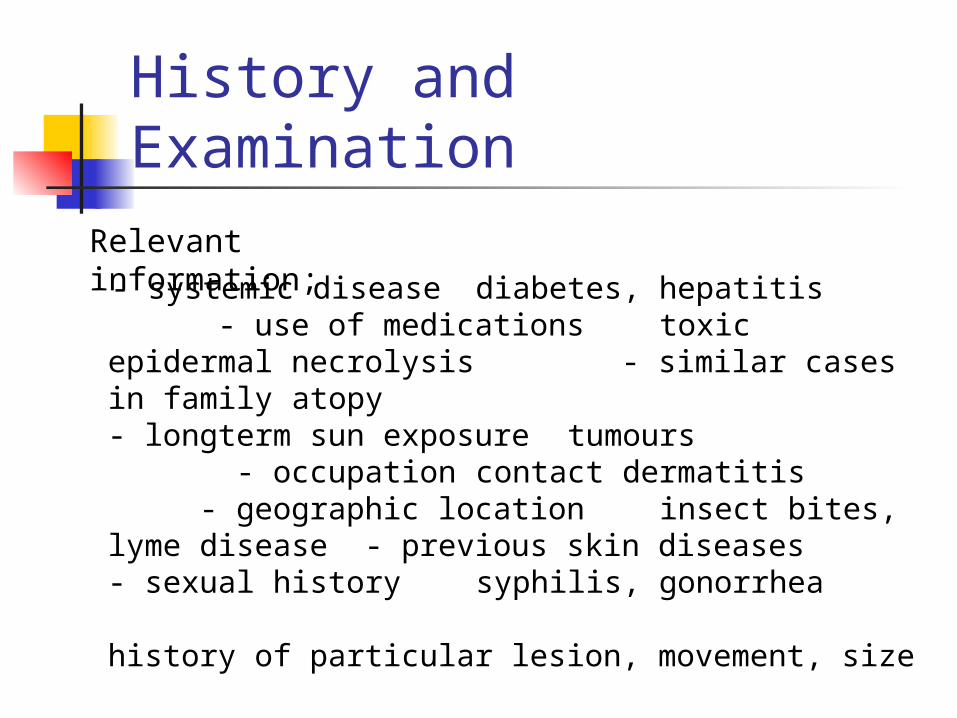
- systemic disease diabetes, hepatitis - use of medications toxic epidermal necrolysis - similar cases in family
atopy- longterm sun exposure tumours - occupation contact dermatitis - geographic locationinsect bites, lyme disease - previous skin diseases- sexual history syphilis, gonorrhea
history of particular lesion, movement, size
Relevant information;

Description
primary: describes morphology or lesion typemacule, papule, plaque, wheal, cyst, nodule, tumor, vesicle, bulla and pustule.
secondary: configuration, texture, scale, crust, lichenification, scar, excoriation, fissure, ulceration, ulcer, erosion and atrophy.
vascular lesions: blood sequellae, pathologypurpura, petechiae, ecchymoses, telangiectasia
shape: annular, serpiginous, flat-topped, domed
A particular terminology has developed to define skin lesions
Primary Characteristics
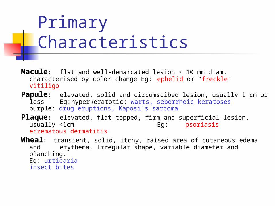
Macule: flat and well-demarcated lesion < 10 mm diam.characterised by color change Eg: ephelid or "freckle"
vitiligo
Papule: elevated, solid and circumscibed lesion, usually 1 cm or less Eg: hyperkeratotic: warts, seborrheic keratoses
purple: drug eruptions, Kaposi's sarcoma
Plaque: elevated, flat-topped, firm and superficial lesion, usually <1cm Eg: psoriasis
eczematous dermatitis
Wheal: transient, solid, itchy, raised area of cutaneous edema and erythema. Irregular shape, variable diameter and blanching.
Eg: urticariainsect bites
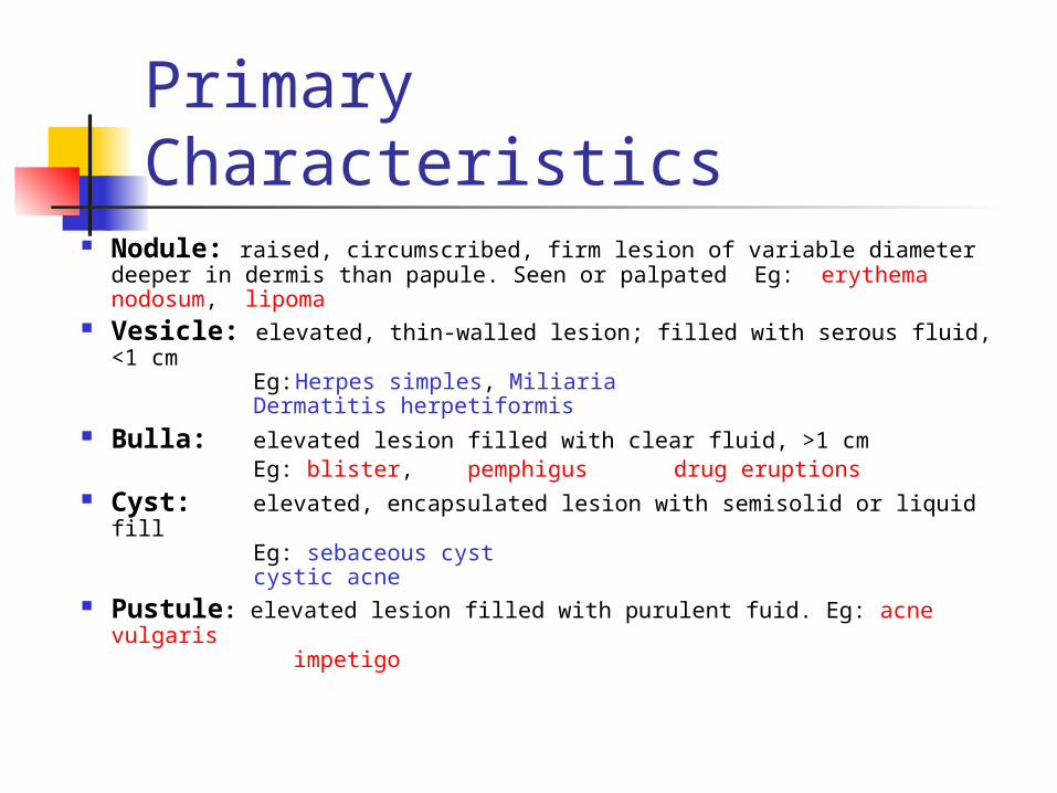
Primary Characteristics Nodule: raised, circumscribed, firm lesion of variable diameter
deeper in dermis than papule. Seen or palpatedEg: erythema nodosum, lipoma
Vesicle: elevated, thin-walled lesion; filled with serous fluid, <1 cmEg: Herpes simples, Miliaria
Dermatitis herpetiformis Bulla: elevated lesion filled with clear fluid, >1 cm
Eg: blister, pemphigus drug eruptions
Cyst: elevated, encapsulated lesion with semisolid or liquid fill Eg: sebaceous cyst
cystic acne Pustule: elevated lesion filled with purulent fuid. Eg: acne vulgaris
impetigo
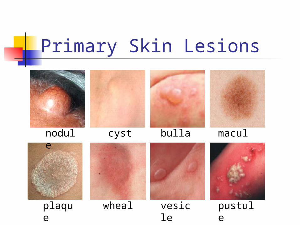
Primary Skin Lesions
cyst
plaque
macule
bullae
vesicle
wheal
nodule
pustule
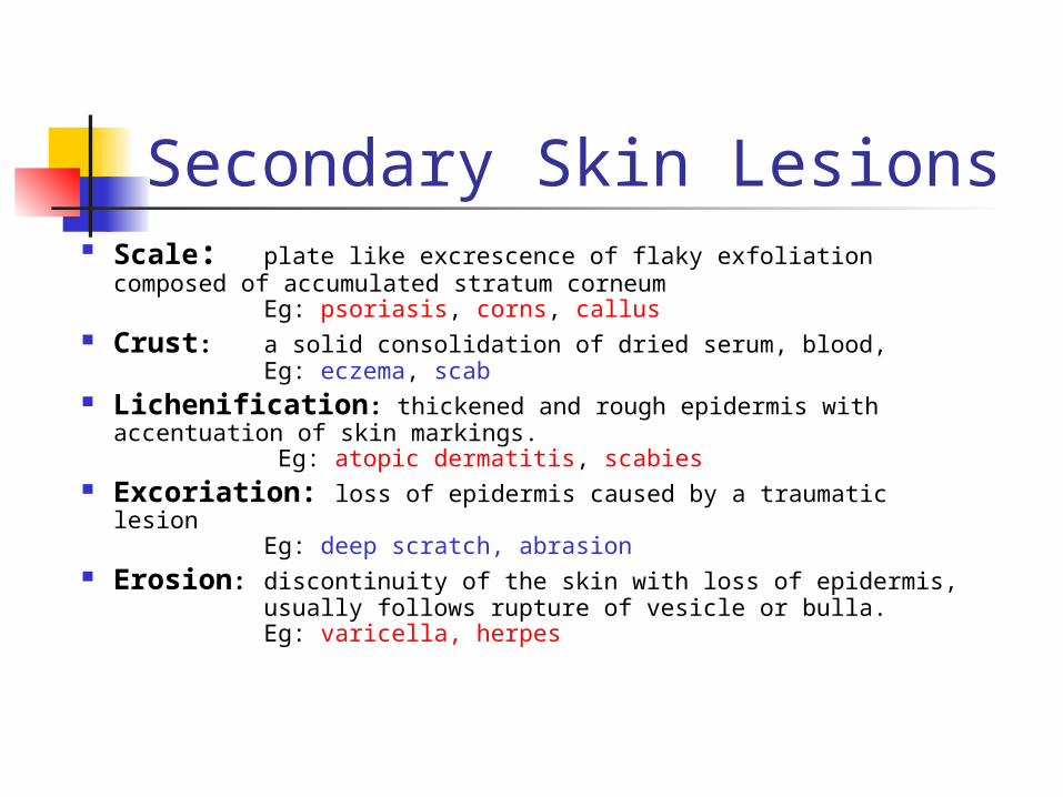
Secondary Skin Lesions Scale: plate like excrescence of flaky exfoliation
composed of accumulated stratum corneumEg: psoriasis, corns, callus
Crust: a solid consolidation of dried serum, blood, Eg: eczema, scab
Lichenification: thickened and rough epidermis with accentuation of skin markings.
Eg: atopic dermatitis, scabies Excoriation: loss of epidermis caused by a traumatic
lesion Eg: deep scratch, abrasion
Erosion: discontinuity of the skin with loss of epidermis, usually follows rupture of vesicle or bulla.Eg: varicella, herpes
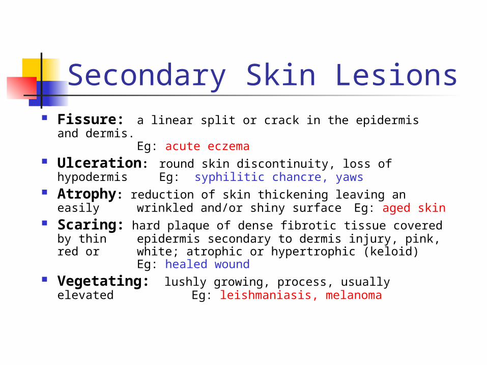
Fissure: a linear split or crack in the epidermis and dermis.
Eg: acute eczema Ulceration: round skin discontinuity, loss of
hypodermis Eg: syphilitic chancre, yaws Atrophy: reduction of skin thickening leaving an easily
wrinkled and/or shiny surfaceEg: aged skin Scaring: hard plaque of dense fibrotic tissue covered
by thin epidermis secondary to dermis injury, pink, red or white; atrophic or hypertrophic (keloid)
Eg: healed wound Vegetating: lushly growing, process, usually elevated
Eg: leishmaniasis, melanoma
Secondary Skin Lesions
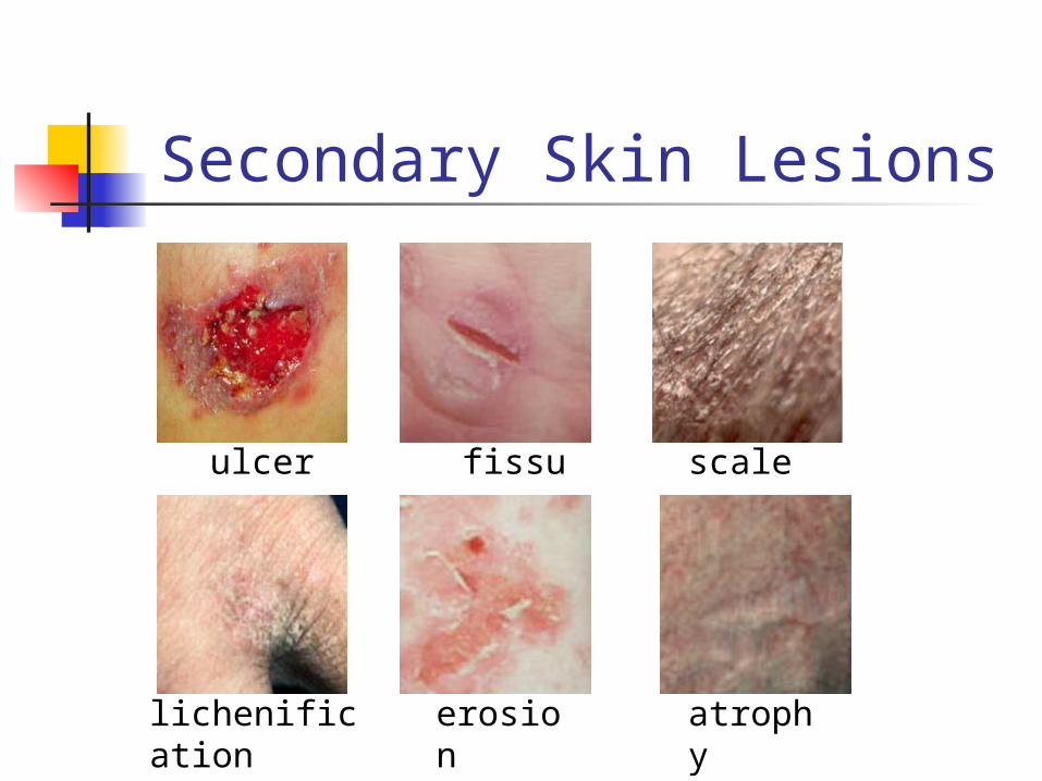
fissure
atrophy
scaleulcer
erosionlichenification
Secondary Skin Lesions
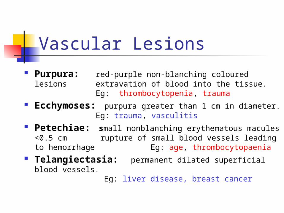
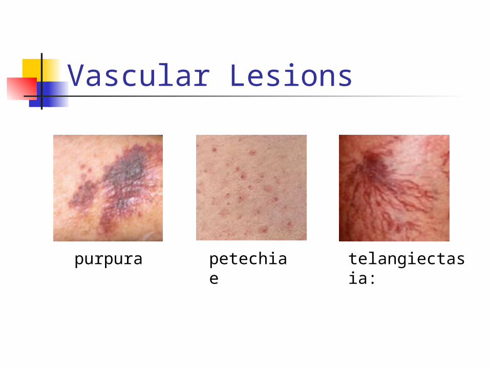
Vascular Lesions Purpura: red-purple non-blanching coloured lesions
extravation of blood into the tissue.Eg: thrombocytopenia, trauma
Ecchymoses: purpura greater than 1 cm in diameter.Eg: trauma, vasculitis
Petechiae: small nonblanching erythematous macules <0.5 cm rupture of small blood vessels leading to hemorrhage Eg: age, thrombocytopaenia
Telangiectasia: permanent dilated superficial blood vessels.
Eg: liver disease, breast cancer
petechiae
telangiectasia: purpura
Vascular Lesions
Diseases
blistering disease: Pemphigus
Bullous Pemphigoid Erythema MultiformesBullous Drug Reactions Dermatitis Herpetiformis
Phytophoto Dermatitis
scaling diseases dermatitis
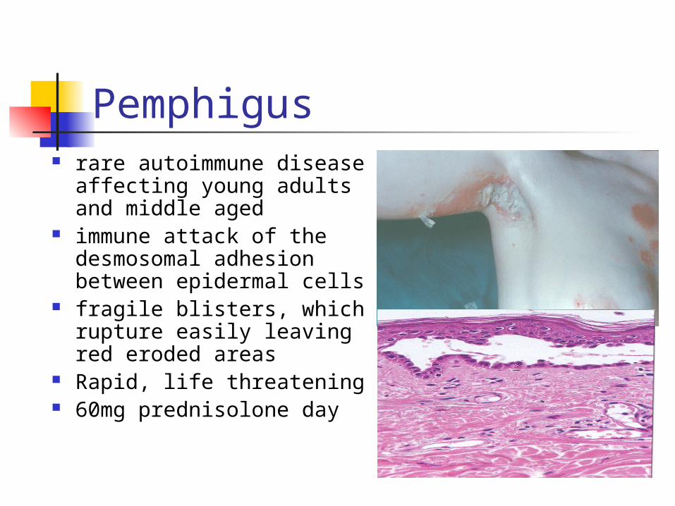
Pemphigus rare autoimmune disease
affecting young adults and middle aged
immune attack of the desmosomal adhesion between epidermal cells
fragile blisters, which rupture easily leaving red eroded areas
Rapid, life threatening 60mg prednisolone day
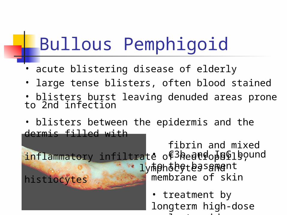
Bullous Pemphigoid• acute blistering disease of elderly• large tense blisters, often blood stained• blisters burst leaving denuded areas prone to 2nd infection
• blisters between the epidermis and the dermis filled with fibrin and mixed inflammatory infiltrate of neutrophils,
lymphocytes and histiocytes
• C3b and IgG bound to the basement membrane of skin
• treatment by longterm high-dose oral steroids
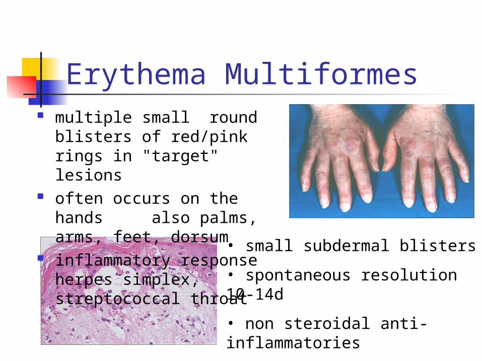
Erythema Multiformes multiple small round
blisters of red/pink rings in "target" lesions
often occurs on the hands also palms, arms, feet, dorsum
inflammatory response herpes simplex, streptococcal throat
• small subdermal blisters
• spontaneous resolution 10-14d
• non steroidal anti-inflammatories asprin, ibuprofen and antibiotics
Bullous Drug Reactions STEVENS-JOHNSON SYNDROME
small multiple subdernal blisters with extensive involvement of the mouth and mucus membranes.
TOXIC EPIDERMAL NECROSIS very severe blisters with extensive
involvement mortality of about 30-50%
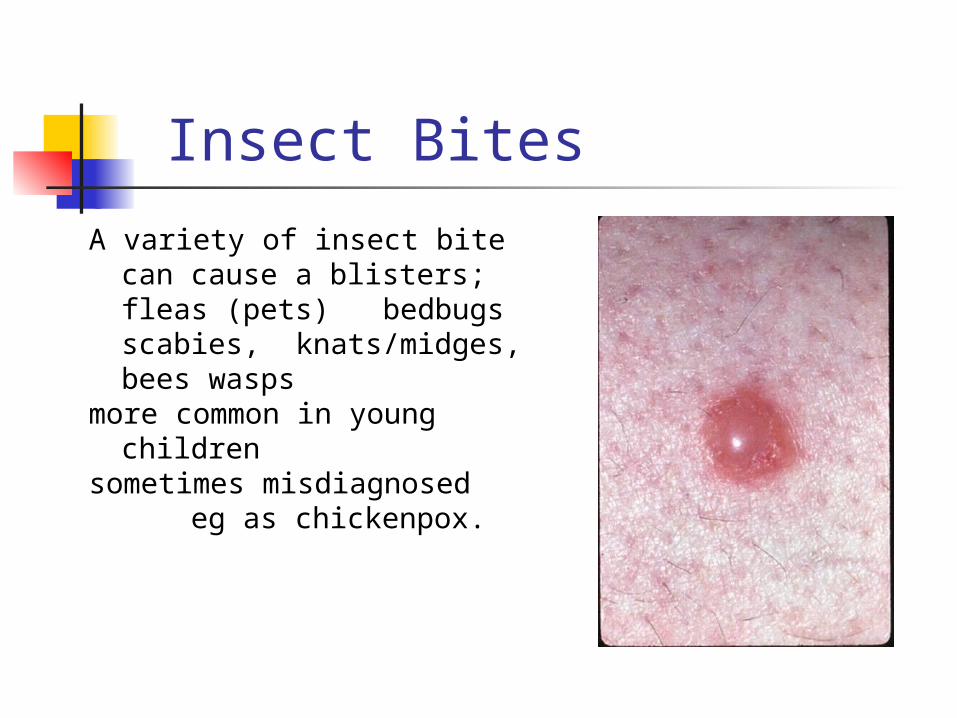
Insect Bites
A variety of insect bite can cause a blisters; fleas (pets)
bedbugsscabies,
knats/midges, bees wasps
more common in young children
sometimes misdiagnosed eg as chickenpox.
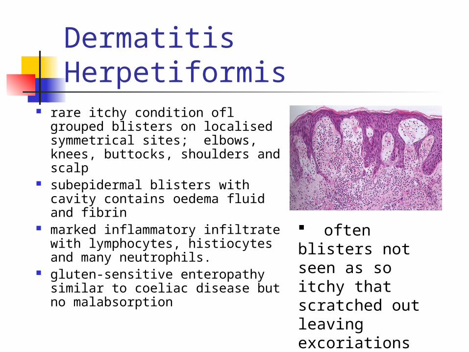
Dermatitis Herpetiformis rare itchy condition ofl grouped
blisters on localised symmetrical sites; elbows, knees, buttocks, shoulders and scalp
subepidermal blisters with cavity contains oedema fluid and fibrin
marked inflammatory infiltrate with lymphocytes, histiocytes and many neutrophils.
gluten-sensitive enteropathy similar to coeliac disease but no malabsorption
often blisters not seen as so itchy that scratched out leaving excoriations
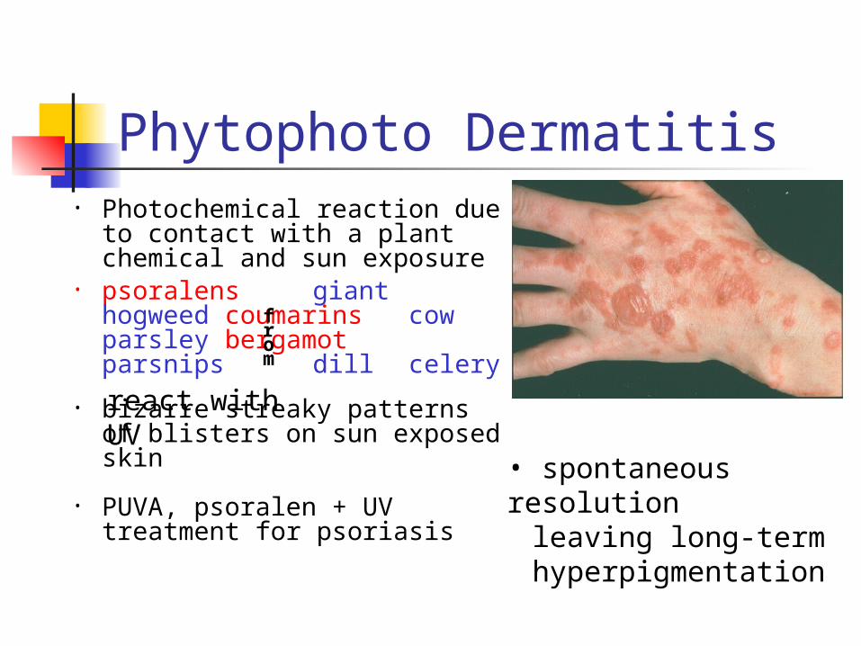
Phytophoto Dermatitis• Photochemical reaction due
to contact with a plant chemical and sun exposure
• psoralens giant hogweed coumarins cow parsley bergamot parsnips dill
celery
• bizarre streaky patterns of blisters on sun exposed skin
• PUVA, psoralen + UV treatment for psoriasis
react with UV
from
• spontaneous resolution leaving long-term
hyperpigmentation

Scaling Diseases
psoriasis
parapsoriasis
pityriasis rosea
lichen planus
dissimilar disorders grouped together by lesions with similar primary characteristics
sharply marginated scaling papules or plaques without wetness, crusts, fissures and excorationsAppearance and distribution of lesions distinguish the diseases
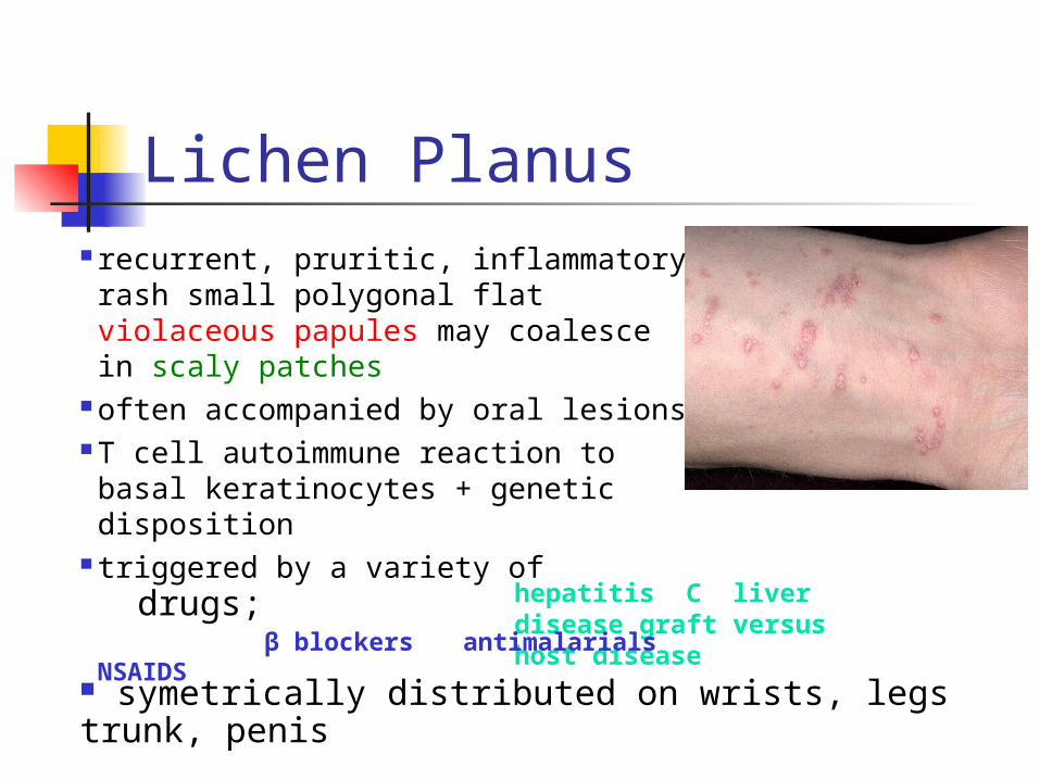
Lichen Planus
hepatitis C liver disease graft versus host disease
recurrent, pruritic, inflammatory rash small polygonal flat violaceous papules may coalesce in scaly patches
often accompanied by oral lesions T cell autoimmune reaction to basal keratinocytes + genetic disposition
triggered by a variety of β blockers antimalarials NSAIDSdrugs;
symetrically distributed on wrists, legs trunk, penis
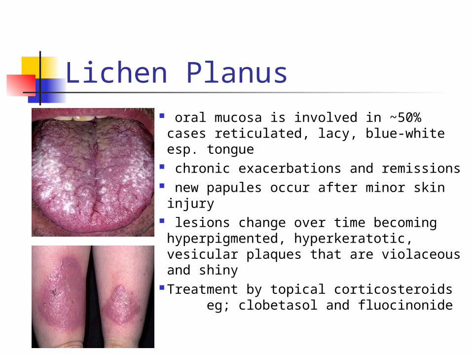
Lichen Planus oral mucosa is involved in ~50% cases reticulated, lacy, blue-white esp. tongue
chronic exacerbations and remissions new papules occur after minor skin injury lesions change over time becoming hyperpigmented, hyperkeratotic, vesicular plaques that are violaceous and shiny
Treatment by topical corticosteroids eg; clobetasol and fluocinonide
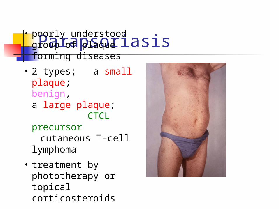
Parapsoriasis • poorly understood group of plaque forming diseases
• 2 types; a small plaque; benign,
a large plaque; CTCL
precursor cutaneous T-cell
lymphoma
• treatment by phototherapy or topical corticosteroids
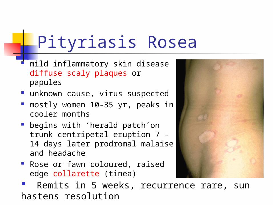
Pityriasis Rosea mild inflammatory skin disease
diffuse scaly plaques or papules unknown cause, virus suspected mostly women 10-35 yr, peaks in
cooler months begins with ‘herald patch’on trunk
centripetal eruption 7 -14 days later prodromal malaise and headache
Rose or fawn coloured, raised edge collarette (tinea)
Remits in 5 weeks, recurrence rare, sun hastens resolution
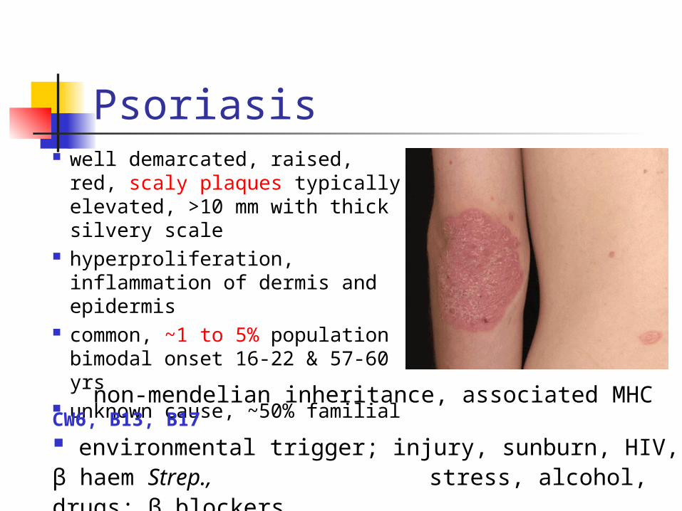
Psoriasis well demarcated, raised, red,
scaly plaques typically elevated, >10 mm with thick silvery scale
hyperproliferation, inflammation of dermis and epidermis
common, ~1 to 5% population bimodal onset 16-22 & 57-60 yrs
unknown cause, ~50% familial
non-mendelian inheritance, associated MHC CW6, B13, B17 environmental trigger; injury, sunburn, HIV, β haem Strep., stress, alcohol, drugs; β blockers chloroquine
Clinical Variants
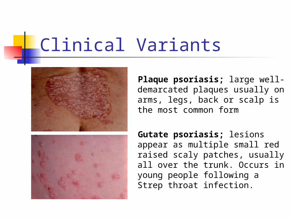
Plaque psoriasis; large well-demarcated plaques usually on arms, legs, back or scalp is the most common form
Gutate psoriasis; lesions appear as multiple small red raised scaly patches, usually all over the trunk. Occurs in young people following a Strep throat infection.
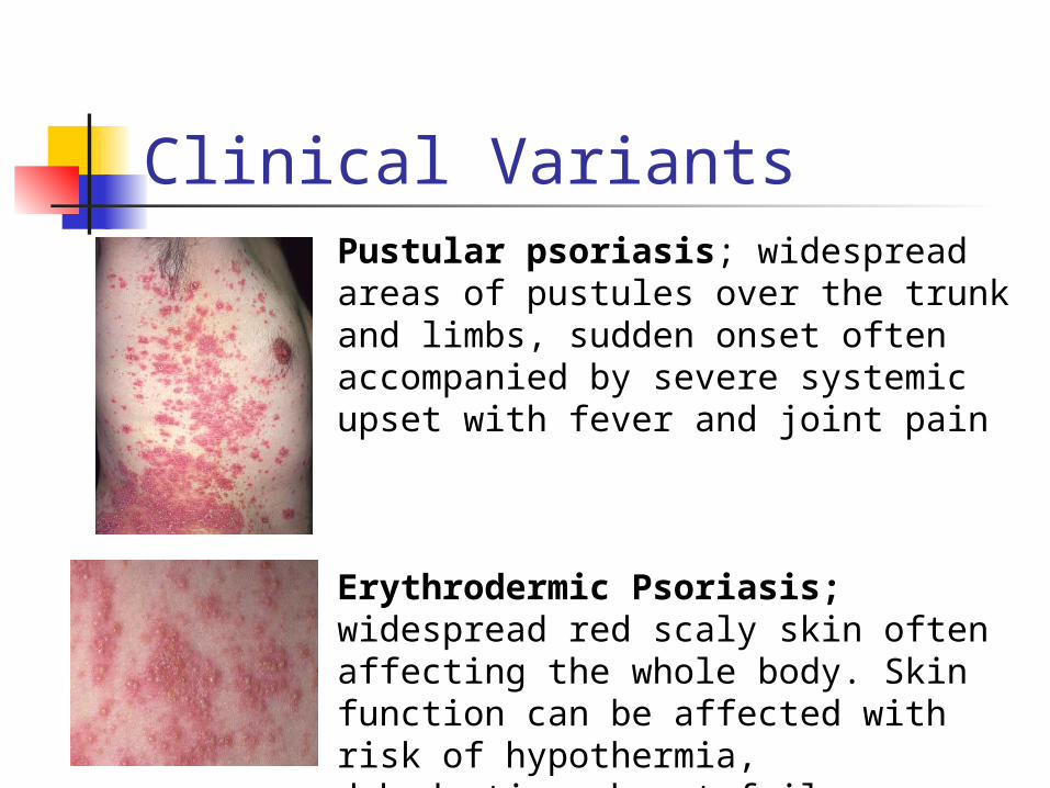
Clinical VariantsPustular psoriasis; widespread areas of pustules over the trunk and limbs, sudden onset often accompanied by severe systemic upset with fever and joint pain
Erythrodermic Psoriasis; widespread red scaly skin often affecting the whole body. Skin function can be affected with risk of hypothermia, dehydration, heart failure.
Treatment of Psoriasis arthritis develops in 5 to 30% of cases, can be disabling rarely life-threatening but severely affects self-image time required to treat extensive skin lesions and
maintain clothing and bedding may adversely affect quality of life
treatments extensive, include emollients, salicylic acid, coal tar, anthralin, corticosteroids, methotrexate
emollient creams, parafin, petrolatum, hydrogenated oils reduce scaling, best applied after bathing
Salicyclic acid is a keratinolytic, softens scales. Coal tar and corticisteroids are anti-inflammatory and
reduce proliferation. Used in combination with UV light (Goekerman regimen)
Dermatitissuperficial inflammation of the skin
characterized by rednessoedema
oozingcrusting
scaling(vesicles)
Eczema used interchangeably with dermatitis
pruritis
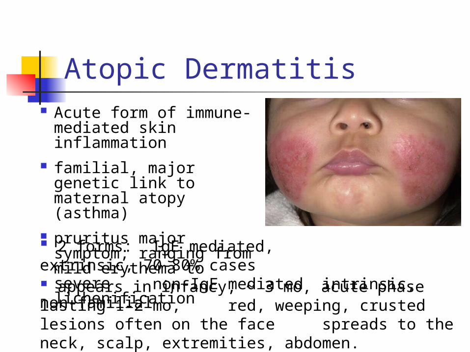
Atopic Dermatitis Acute form of immune-
mediated skin inflammation
familial, major genetic link to maternal atopy (asthma)
pruritus major symptom; ranging from mild erythema to severe lichenification
appears in infancy, ~ 3 mo, acute phase lasting 1-2 mo, red, weeping, crusted lesions often on the face spreads to the neck, scalp, extremities, abdomen.
2 forms: IgE mediated, extrinsic, 70-80% cases
non-IgE mediated intrinsic, non-familial
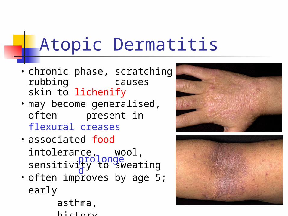
• chronic phase, scratching rubbing causes skin to lichenify
• may become generalised, often present in flexural creases
• associated food intolerance, wool, sensitivity to sweating
• often improves by age 5; early asthma, history
• treatment by emollients, creams moisturising, anti-histamines, corticosteroids
Atopic Dermatitis
prolonged
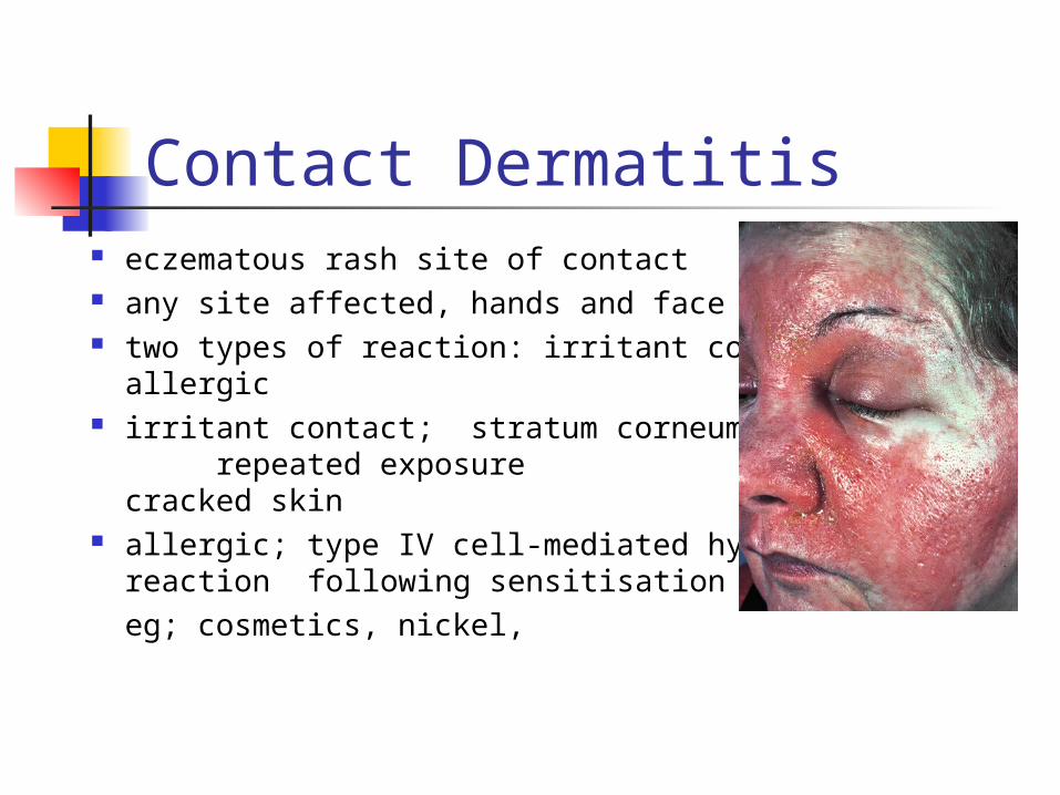
Contact Dermatitis eczematous rash site of contact any site affected, hands and face two types of reaction: irritant contact
allergic irritant contact; stratum corneum
repeated exposure red cracked skin
eg nappy rash allergic; type IV cell-mediated hypersensitivity
reaction following sensitisation eg; cosmetics, nickel,
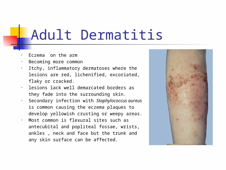
Adult Dermatitis • Eczema on the arm • Becoming more common• Itchy, inflammatory dermatoses where the
lesions are red, lichenified, excoriated, flaky or cracked.
• lesions lack well demarcated borders as they fade into the surrounding skin.
• Secondary infection with Staphylococcus aureus is common causing the eczema plaques to develop yellowish crusting or weepy areas.
• Most common is flexural sites such as antecubital and popliteal fossae, wrists, ankles , neck and face but the trunk and any skin surface can be affected.
Allergic ContactAirborne substances; Ragweed pollen, insecticide spray Chemicals; tanning agents, formaldehyde in press finishesCosmetics; depilatories, nail polish, deodorantDyes; p-Phenylenediamine in hair and textile dyesFragrances, acrylic monomers, epoxy, formaldehydeAntibiotics; bacitracin, neomycin, penicillin, sulfonamides Antihistamines; diphenhydramine, promethazine Antiseptics; thimerosal, hexachloropheneLatex; gloves, condoms, catheters, balloonsMetals; nickel, cobalt, chromates, mercury Plants; poison ivy, oak, sumac, ragweed, primrose
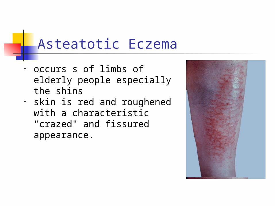
Asteatotic Eczema
• occurs s of limbs of elderly people especially the shins
• skin is red and roughened with a characteristic "crazed" and fissured appearance.
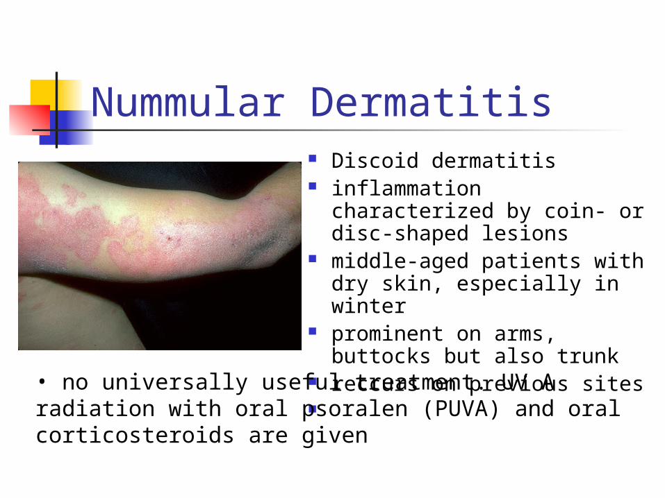
Nummular Dermatitis Discoid dermatitis inflammation characterized
by coin- or disc-shaped lesions
middle-aged patients with dry skin, especially in winter
prominent on arms, buttocks but also trunk
reccurs on previous sites
• no universally useful treatment. UV A radiation with oral psoralen (PUVA) and oral corticosteroids are given
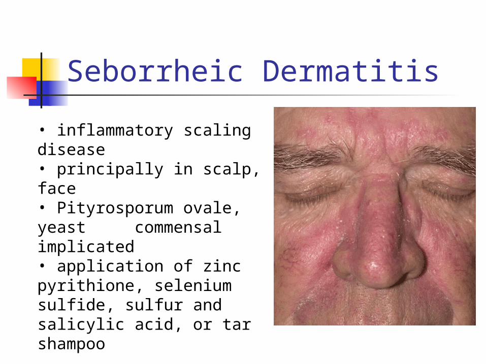
Seborrheic Dermatitis
• inflammatory scaling disease• principally in scalp, face• Pityrosporum ovale, yeast commensal implicated• application of zinc pyrithione, selenium sulfide, sulfur and salicylic acid, or tar shampoo
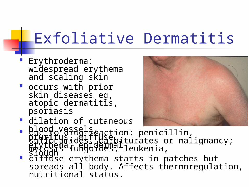
Exfoliative Dermatitis Erythroderma:
widespread erythema and scaling skin
occurs with prior skin diseases eg, atopic dermatitis, psoriasis
dilation of cutaneous blood vessels, pruritus, diffuse erythema, epidermal slough
due to drug reaction; penicillin, sulfonamides, barbiturates or malignancy; mycosis fungoides, leukemia,
diffuse erythema starts in patches but spreads all body. Affects thermoregulation, nutritional status.
Summary Many types of skin lesion, Many with unknown cause Many with element of
autoimmunity Diagnosis requires thorough
knowledge of symptoms, history and terminology