Embed Size (px)
Citation preview
RESEARCH Open Access
Nogo receptor complex expressiondynamics in the inflammatory foci ofcentral nervous system experimentalautoimmune demyelinationPaschalis Theotokis1, Olga Touloumi1, Roza Lagoudaki1, Evangelia Nousiopoulou1, Evangelia Kesidou1,Spyridon Siafis1, Theodoros Tselios2, Athanasios Lourbopoulos1,3, Dimitrios Karacostas1, Nikolaos Grigoriadis1
and Constantina Simeonidou4*
Abstract
Background: Nogo-A and its putative receptor NgR are considered to be among the inhibitors of axonal regenerationin the CNS. However, few studies so far have addressed the issue of local NgR complex multilateral localization withininflammation in an MS mouse model of autoimmune demyelination.
Methods: Chronic experimental autoimmune encephalomyelitis (EAE) was induced in C57BL/6 mice. Analyses wereperformed on acute (days 18–22) and chronic (day 50) time points and compared to controls. The temporal and spatialexpression of the Nogo receptor complex (NgR and coreceptors) was studied at the spinal cord using epifluorescentand confocal microscopy or real-time PCR. Data are expressed as cells/mm2, as mean % ± SEM, or as arbitrary units ofintegrated density.
Results: Animals developed a moderate to severe EAE without mortality, followed by a progressive, chronic clinicalcourse. NgR complex spatial expression varied during the main time points of EAE. NgR with coreceptors LINGO-1 andTROY was increased in the spinal cord in the acute phase whereas LINGO-1 and p75 signal seemed to be dominant inthe chronic phase, respectively. NgR was detected on gray matter NeuN+ neurons of the spinal cord, within the whitematter inflammatory foci (14.2 ± 4.3 % NgR+ inflammatory cells), and found to be colocalized with GAP-43+ axonalgrowth cones while no β-TubIII+, SMI-32+, or APP+ axons were found as NgR+. Among the NgR+ inflammatory cells, 75.6 ± 9.0 % were microglial/macrophages (lectin+), 49.6 ± 14.2 % expressed CD68 (phagocytic ED1+ cells), and no cellswere Mac-3+. Of these macrophages/monocytes, only Arginase-1+/NgR+ but not iNOS+/NgR+ were present in lesionsboth in acute and chronic phases.
Conclusions: Our data describe in detail the expression of the Nogo receptor complex within the autoimmuneinflammatory foci and suggest a possible immune action for NgR apart from the established inhibitory one on axonalgrowth. Its expression by inflammatory macrophages/monocytes could signify a possible role of these cells on axonalguidance and clearance of the lesioned area during inflammatory demyelination.
Keywords: Nogo receptor complex, NgR, LINGO-1, p75, TROY, Experimental autoimmune encephalomyelitis,Neuroinflammation, Macrophages
* Correspondence: [email protected] of Experimental Physiology, Faculty of Medicine, AristotleUniversity of Thessaloniki, 546 36 Thessaloniki, Central Macedonia, GreeceFull list of author information is available at the end of the article
© 2016 The Author(s). Open Access This article is distributed under the terms of the Creative Commons Attribution 4.0International License (http://creativecommons.org/licenses/by/4.0/), which permits unrestricted use, distribution, andreproduction in any medium, provided you give appropriate credit to the original author(s) and the source, provide a link tothe Creative Commons license, and indicate if changes were made. The Creative Commons Public Domain Dedication waiver(http://creativecommons.org/publicdomain/zero/1.0/) applies to the data made available in this article, unless otherwise stated.
Theotokis et al. Journal of Neuroinflammation (2016) 13:265 DOI 10.1186/s12974-016-0730-4

BackgroundNumerous studies indicate the dynamic and high potentialrole of neurite outgrowth inhibitor Nogo-A to inhibit,guide, and modulate the injured and demyelinating tissuein various models of disease. These characteristics aredependent on the presence of Nogo-66 receptor (NgR) [1]as well as the subcellular coreceptor components p75 [2],TROY [3], and adaptor molecule LINGO-1 [4], compris-ing the receptor complex that mediates myelin’s inhibitoryaction. The latest finding as a component of this complexis AMIGO3, substituting LINGO-1 under specific circum-stances [5].NgR is expressed by various glial and neuronal cell bodies
or axons except oligodendrocytes, which express its ligandNogo-A [6, 7]. This specific ligand-receptor localization hasbeen accounted for a neurite outgrowth inhibitory mechan-ism, exerting a growth cone collapse effect in response to atleast two more myelin proteins, namely MAG [8] andOMgp [9]. The 473-amino acid NgR protein is a glycosyl-phosphatidylinositol (GPI)-linked molecule linked to aneight-leucine-rich-repeat (LRR) domain by an LRRcarboxy-terminal (LRRCT) domain. It can be found inthree isoforms: NgR1, NgR2, and NgR3 [10], with NgR1being the most common and best correlated to Nogo-A[11]. The other three molecules of the NgR complex,namely p75, TROY, and LINGO-1, are membrane corecep-tors that regulate the downstream effects of NgR dependingon their interactions [12, 13].The value of the Nogo-NgR pathway for control of
axonal regeneration is verified by the development ofNogo-A and LINGO-1 monoclonal antibodies for clin-ical trials [14, 15]. Despite the fact that NgR is key to thepathway activation, only few studies analyzing knockoutmice in experimental autoimmune encephalomyelitis(EAE) [16, 17] and few small interference RNA (siRNA)experiments have been carried out in demyelination ofoptic nerve tracts [18, 19], rendering its precise functionstill enigmatic. In vitro experiments by Takahashi andcolleagues [20] and later further progressed by Davidand colleagues [21, 22] suggest that the presence of NgRin macrophages—apart from neurons or glia—couldsupport the clearance of debris and could confine theinjury away from the normal-appearing white mattertissue. However, the question of whether the NgR com-plex positively or negatively regulates the inflammationspreading in a central nervous system (CNS)-based auto-immune disease remains largely unknown so far.The purpose of this study was to describe the spatiotem-
poral kinetics of NgR within the inflammatory sites ofexperimental autoimmune demyelination, in order tounderstand its roles in the inflammatory milieu. We fo-cused on both NgR and coreceptor molecules LINGO-1,p75, and TROY, in an attempt to characterize the Nogo-A/NgR pathway in the autoimmune inflammatory foci. Based
on our present and previous published data [23], wepropose that NgR may have an additional role in the dis-ease progression—besides the attributed axonal inhibition-,namely confining the inflammatory reaction and/or thesprouting of axons in EAE lesions.
MethodsAnimal handlingFemale C57BL/6 mice (n = 40), 8–10 weeks old, werepurchased from the Hellenic Pasteur Institute (Athens,Greece) and housed in the pathogen-free animal facilityof the B’ Neurology Department, AHEPA UniversityHospital, Thessaloniki, Greece. Animals were fed a nor-mal diet and given ad libitum water without antibiotics.All experimental procedures were conducted accordingto the institutional guidelines, in compliance with theGreek Regulations and the European CommunitiesCouncil Directive of November 24, 1986 (86/609/EEC).Experimentation received approval from the VeterinaryMedicines Directorate (license number 177867/1510).
EAE induction and clinical evaluationChronic EAE was induced in animals (n = 30), with sub-cutaneous injection of 300 μg of myelin oligodendrocyteglycoprotein 35–55 peptide (MOG35–55) as previouslydescribed [23, 24]. Animals inoculated with completeFreund’s adjuvant (CFA, supplemented with 4 % mycobac-terium tuberculosis) only did not develop lesions in theCNS and served as controls (n = 10). All animals wereexamined daily using a 6-grade scale: 0, asymptomatic; 1,partial loss of tail tonicity; 2, flaccid tail paralysis; 3, diffi-culty to roll over from a supine position; 4, hind limbparalysis; 5, forelimb paresis; and 6, death due to EAE.The following indexes were calculated based on their
daily scores: mean maximal score (MMS), area underthe curve (AUC), day of disease onset (dDO), clinicalscore at day 50 (d50 score), and demyelination extent inthe spinal cord.
Tissue collectionAnimals were humanely euthanized and preparedaccordingly for neuropathology and further processing.Animals for immunohistochemistry were transcardiallyperfused using phosphate buffer saline (PBS), followedby ice-cold 4 % paraformaldehyde in PBS (4 % PFA) for5 min. The brain and spinal cords were removed, post-fixed in the corresponding fixation solution for 16–20 hat 4 °C, and routinely processed for paraffin sectioningat 6 μm. Animals for real-time PCR were euthanatized,and their brains and spinal cords were removed, snapfrozen, and stored at −80 °C until further use.
Theotokis et al. Journal of Neuroinflammation (2016) 13:265 Page 2 of 13
Histological stainings, immunohistochemistry, andimmunofluorescenceSix-micrometer-thick paraffin coronal spinal cord sec-tions were stained with routinely used immunohisto-chemical methods: Luxol fast blue (LFB) counterstainedwith nuclear fast red for demyelination evaluation andBielschowsky silver impregnation counterstained withhematoxylin for pervasive infiltratory burden and axonalloss, as previously described [25–27]. Lectin and DAB-based anti-Iba-1 (rabbit, WAKO) protocols were usedfor microglia evaluation, as previously described [27].Localization and neuropathological study of NgR was
performed with double immunofluorescence (dIF) also on6-μm coronal sections. Briefly, following hydration andincubation of the sections with 10 % fetal bovine serum(FBS) for 30 min, primary antibodies were applied over-night. Neuronal and glial components were evaluated forthe presence of NgR with a combination of primaryantibodies, anti-NgR (rabbit, Santa Cruz) with anti-NeuN(mouse, Millipore) for neurons, anti-GFAP (mouse,DAKO) for astrocytes, anti-Nogo-A (goat, Santa Cruz) formature oligodendrocytes, anti-Mac-3 (rat, BD Biosciences)for detection of peritoneal/tissue macrophages anddendritic cells, and anti-ED1 (mouse, Serotec) for detec-tion of macrosialin (macrophages; human CD68), whileanti-Arginase-1 (goat, Santa Cruz) and anti-iNOS (mouse,Santa Cruz) were used to distinguish M2 microglia fromthe M1 phenotype [28].Additionally, axonal epitopes were designated with the
following: anti-β-TubIII (mouse, BD Biosciences) andnon-phosphorylated neurofilament H anti-SMI-32(mouse, Calbiochem) were used for detection of physio-logical and pathological neuronal axons, and anti-β-amyloid precursor protein (APP) (mouse, Millipore) andanti-GAP-43 (mouse, Sigma) were used for axonal focaldegeneracy and regeneration, respectively. CoreceptorsLINGO-1 (rabbit, Abcam ab23631, 1:300), TROY (goat,Santa Cruz 13711, 1:100), and p75 (mouse, Abcamab8877, 1:400) were also accessed and combined accord-ingly. For specificity and quality control of the corecep-tor antibodies, a preabsorption protocol previouslydescribed [29] and the use of a second antibody recog-nizing a different epitope were applied upon availabilityand whenever applicable (see Additional file 1: FigureS1). Briefly, the peptide for each preabsorption assay wasmixed with the antibody in a 10× molecular ratio andwas incubated under gentle agitation at roomtemperature for 30 min. The mixture was then used inplace of the primary antibody for the rest of the IFprotocol.Depending on the primary antibodies used, the following
secondary fluorescent antibodies were used at the spectrumof green (488, Biotium; Alexa Fluor) and red (555-568,Biotium; Alexa Fluor) for the rest of the markers. Sections
were mounted with 4′,6-diamidino-2-phenylindole (DAPI)(Biotium).
Neuropathology evaluationPathologic examination of demyelination, axonopathy, in-flammation, and microglia/macrophages was performedunder a Zeiss Axioplan 2 light microscope by two, blindedto the experimental groups, investigators. Photos werecaptured with the aid of a CCD camera (Nikon), and ana-lysis of the designated areas was performed with ImageJsoftware.Demyelination was evaluated on approximately 15
Luxol fast blue-stained sections. Demyelinated areas andthe total area of white matter were carefully circum-scribed and expressed as a percentage (demyelinated/total wm area), as previously described [23]. Axonal losswas assessed on approximately ten randomly selectedsilver-stained spinal cord sections within 150 × 150-μmareas from the three major spinal cord white matter col-umns (ventral, lateral, and dorsal), spaced at least 60 μmapart, and graded as 0 = normal/even silver stainthroughout the white matter compared to unimmunizedmice; 1 = small spurious areas in the white matter thatlack silver stain; 2 = small, but frequent, areas in thewhite matter that lack silver stain; and 3 = extensive lossof silver stain throughout the white matter, as previouslydescribed [25]. Typical images of scores 0–3 are pre-sented in Additional file 1: Figure S1.For the evaluation of inflammation, hematoxylin-stained
fields of the same sections were examined for each animal,using a prefrontal microscope grid. Microglia/macro-phagic populations were assessed in respective whitematter areas with ionized calcium-binding adaptormolecule-1 (Iba-1) staining. The number of perivascularinfiltrates and microglia/macrophages were counted andpresented as cells/mm2.
NgR profile assessmentSpatial analysis for NgR protein was performed in the cen-ter of six to eight inflammatory foci per spinal cord peranimal, spaced at least 50 μm apart. Sections were studiedusing confocal microscopy (Nikon C1-Eclipse TE-2000U)under ×60 optical fields. Briefly, images were captured,within a predefined 305 × 305-μm area, and the NgR-positive signal was evaluated for levels of coexpressionwith miscellaneous markers. GAP-43 and APP signal wasmeasured as an integrated density (IntDen) as previouslydescribed [23], using the ImageJ software. Colocalizationpercentages were also estimated, wherever applicable.
NgR coreceptor real-time PCRMolecular analysis of coreceptors LINGO-1, TROY, andp75 was performed with real-time PCR in spinal cordextracts based on a protocol previously described [23].
Theotokis et al. Journal of Neuroinflammation (2016) 13:265 Page 3 of 13
Briefly, total RNAs were extracted from spinal cord tissuesusing Trizol reagent (Invitrogen); RNA was reverse tran-scribed to complementary DNA (cDNA) with iScriptcDNA Synthesis Kit (BioRad), and real-time PCR was per-formed using BioRad IQ5 ICycler Multicolor DetectionSystem. The relative gene expression was normalized to β-actin which served as an internal control. Additionally,GAPDH was used as a second, quality control, house-keeping gene. Primer sequences for SYBR Green probesof target genes are as follows: LINGO-1 (Lingo1): forward5′ CATCAGGTGAGCGAGAGGAT 3′ and reverse 5′CGTCCTGGTTGAGTGTCTTG 3′ giving rise to a 267-bp product; p75 (Ngfr): forward 5′ CTGCTGCTTCTAGGGGTGTC 3′ and reverse 5′ ACACAGGGAGCGGACATACT 3′ giving rise to a 248-bp product; TROY(Tnfrsf19): forward 5′ AGATTGCAGGCAGCAGGA 3′and reverse 5′ TCCGCACATGGCTTACACTT 3′ givingrise to a 186-bp product; β-actin (Actb): forward 5′TTGTAACCAACTGGGACGATATGC 3′ and reverse 5′GATCTTGATCTTCATGATGCTAGG 3′ giving rise to a139-bp product; and GAPDH (Gapdh): forward 5′ GGATGCAGGGATGATGTTCT 3′ and reverse 5′ AAGGGCTCATGACCACAGTC 3′ giving rise to a 116-bpproduct. The results were analyzed using ΔΔCt method.
Statistical analysisAll data are given as mean ± standard error of the mean(SEM). For statistical analysis, the SPSS Statistics 18package was used. Student’s t test and one-way analysisof variance (ANOVA) with Dunnett’s and Bonferronipost hoc tests were used for comparisons of two or moregroups, respectively. Non-parametric data were com-pared using Mann-Whitney U test. Semi-quantitativedata were analyzed using the Pearson χ2 test or Fisher’sexact test, where appropriate, and the ordinal data weredisplayed as bar graphs. Two-tailed values of p < 0.05were considered statistically significant for all tests.
ResultsClinical course and inflammatory pathologyMOG-inoculated animals developed a typical chronicMOG-EAE pattern (Fig. 1A) with MMS = 3.76 ± 0.28,AUC = 85.89 ± 11.73, dDO = 14.50 ± 0.47, and d50score = 2.06 ± 0.44. Percentage of demyelination of thespinal cord was found 22.20 ± 1.89 % and 14.80 ±1.34 % (p < 0.01) during the acute and chronic phases,respectively (Fig. 1B). Control animals (CFA inoculatedanimals) did not develop EAE.Tissue sections from acute and chronic phases were
compared to control animals. Hematoxylin stainingrevealed perivascular infiltrates (cells/mm2) within thewhite matter of the spinal cord in the acute phase(123.90 ± 9.89 cells/mm2) that was reduced during thechronic phase (87.80 ± 6.85 cells/mm2; p < 0.01) (Fig. 1C,
F1–H2). Axonal loss assessed by Bielschowsky stainingwas found semi-quantitatively more extensive in thechronic phase than the acute (p < 0.05) and control group(p < 0.001), respectively (Fig. 1D, F1–H2). Microglial/mac-rophagic populations were increased in the acute phase(101.40 ± 4.63 cells/mm2 from control animal levels 10.44± 2.45 cells/mm2; p < 0.001) and then decreased towardsthe chronic phase (83.67 ± 5.77 cells/mm2; p < 0.05 versusacute and p < 0.001 versus controls) (Fig. 1E, I1–K2).
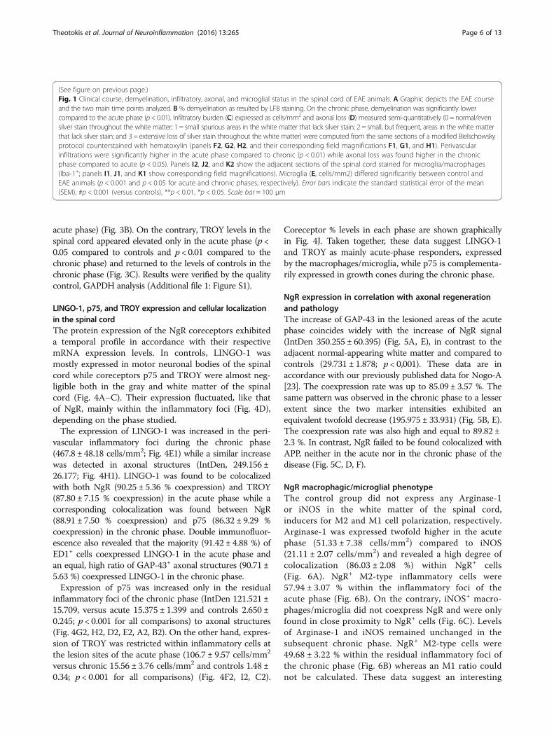
Pattern of NgR expression inside and peripherally to EAElesions of the spinal cordIn the spinal cord of controls, the highest NgR signalwas obtained from neurons of the gray matter (>80 % ofthe total signal) and the rest from axonal elements(DAPI-negative structures).In the acute phase, neuronal NgR signal (Fig. 2A) was de-
creased compared to controls (15.85 ± 9.09 % NeuN+NgR+
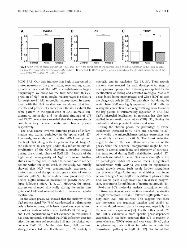
double-positive cells from 82.37 ± 7.81 % of controls; p <0.001), was absent from astrocytes and oligodendrocytes(Fig. 2B, C), and found to be highly expressed in thelesioned white matter (306.7 ± 32.06 cells/mm2, 75.6 ±9.04 % lectin+NgR+ inflammatory cell double positive)(Fig. 2D). NgR was found to be expressed in specific micro-glial/macrophagic cells; 49.6 ± 9.5 % were ED1+ (Fig. 2F),but Mac-3 negative (Fig. 2E), while the remaining per-centage (~10 % the total signal) was found in axonalstructures and specifically only in regenerative growthcones (GAP-43+ structures, Fig. 2I) and nowhere elsethroughout the axon (β-TubIII and SMI-32 negative,Fig. 2G, H). On the contrary, this specific localizationwas not observed in control animals.In the chronic phase, the inflammatory process de-
clines with a diminished appearance of inflammatorycells resulting in a decreased detection of cellular NgRlocalization within residual inflammations (<20 % of thetotal signal) while increasing the axonal localization ingrowth cones (IntDen, 195.975 ± 33.931) alongside asimilar increase, 2.5-fold (39.44 ± 8.61 cells/mm2 versusacute 15.85 ± 9.09 cells/mm2; p < 0.01) in neurons of thegray matter of the spinal cord. NgR localization in eachphase is shown graphically in Fig. 2J.
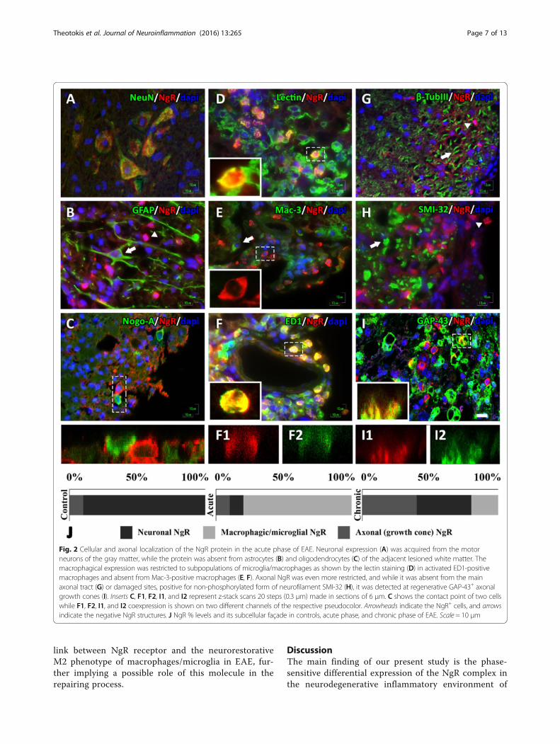
Analysis of LINGO-1, p75, and TROY mRNA expression inthe spinal cord by real-time PCRThe messenger RNA (mRNA) levels of the NgR corecep-tors were found to fluctuate, depending on the stagestudied. Expression of LINGO-1 in the spinal cord wasincreased in the acute phase compared to controls andfollowed a statistically significant increase in the chronicphase (p < 0.001 compared to controls) (Fig. 3A). Thecorresponding normalized expression of p75 showed adecrease during the acute phase but increased signifi-cantly in the chronic phase (p < 0.001 compared to the
Theotokis et al. Journal of Neuroinflammation (2016) 13:265 Page 4 of 13
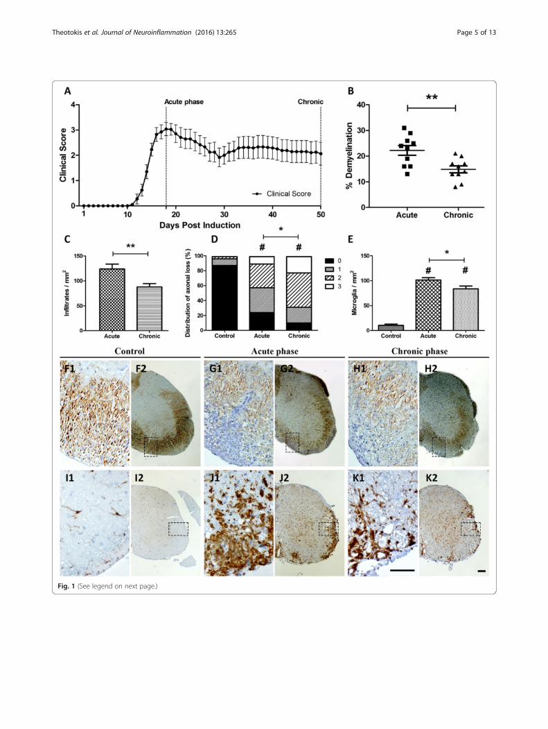
Fig. 1 (See legend on next page.)
Theotokis et al. Journal of Neuroinflammation (2016) 13:265 Page 5 of 13
acute phase) (Fig. 3B). On the contrary, TROY levels in thespinal cord appeared elevated only in the acute phase (p <0.05 compared to controls and p < 0.01 compared to thechronic phase) and returned to the levels of controls in thechronic phase (Fig. 3C). Results were verified by the qualitycontrol, GAPDH analysis (Additional file 1: Figure S1).
LINGO-1, p75, and TROY expression and cellular localizationin the spinal cordThe protein expression of the NgR coreceptors exhibiteda temporal profile in accordance with their respectivemRNA expression levels. In controls, LINGO-1 wasmostly expressed in motor neuronal bodies of the spinalcord while coreceptors p75 and TROY were almost neg-ligible both in the gray and white matter of the spinalcord (Fig. 4A–C). Their expression fluctuated, like thatof NgR, mainly within the inflammatory foci (Fig. 4D),depending on the phase studied.The expression of LINGO-1 was increased in the peri-
vascular inflammatory foci during the chronic phase(467.8 ± 48.18 cells/mm2; Fig. 4E1) while a similar increasewas detected in axonal structures (IntDen, 249.156 ±26.177; Fig. 4H1). LINGO-1 was found to be colocalizedwith both NgR (90.25 ± 5.36 % coexpression) and TROY(87.80 ± 7.15 % coexpression) in the acute phase while acorresponding colocalization was found between NgR(88.91 ± 7.50 % coexpression) and p75 (86.32 ± 9.29 %coexpression) in the chronic phase. Double immunofluor-escence also revealed that the majority (91.42 ± 4.88 %) ofED1+ cells coexpressed LINGO-1 in the acute phase andan equal, high ratio of GAP-43+ axonal structures (90.71 ±5.63 %) coexpressed LINGO-1 in the chronic phase.Expression of p75 was increased only in the residual
inflammatory foci of the chronic phase (IntDen 121.521 ±15.709, versus acute 15.375 ± 1.399 and controls 2.650 ±0.245; p < 0.001 for all comparisons) to axonal structures(Fig. 4G2, H2, D2, E2, A2, B2). On the other hand, expres-sion of TROY was restricted within inflammatory cells atthe lesion sites of the acute phase (106.7 ± 9.57 cells/mm2
versus chronic 15.56 ± 3.76 cells/mm2 and controls 1.48 ±0.34; p < 0.001 for all comparisons) (Fig. 4F2, I2, C2).
Coreceptor % levels in each phase are shown graphicallyin Fig. 4J. Taken together, these data suggest LINGO-1and TROY as mainly acute-phase responders, expressedby the macrophages/microglia, while p75 is complementa-rily expressed in growth cones during the chronic phase.
NgR expression in correlation with axonal regenerationand pathologyThe increase of GAP-43 in the lesioned areas of the acutephase coincides widely with the increase of NgR signal(IntDen 350.255 ± 60.395) (Fig. 5A, E), in contrast to theadjacent normal-appearing white matter and compared tocontrols (29.731 ± 1.878; p < 0,001). These data are inaccordance with our previously published data for Nogo-A[23]. The coexpression rate was up to 85.09 ± 3.57 %. Thesame pattern was observed in the chronic phase to a lesserextent since the two marker intensities exhibited anequivalent twofold decrease (195.975 ± 33.931) (Fig. 5B, E).The coexpression rate was also high and equal to 89.82 ±2.3 %. In contrast, NgR failed to be found colocalized withAPP, neither in the acute nor in the chronic phase of thedisease (Fig. 5C, D, F).
NgR macrophagic/microglial phenotypeThe control group did not express any Arginase-1or iNOS in the white matter of the spinal cord,inducers for M2 and M1 cell polarization, respectively.Arginase-1 was expressed twofold higher in the acutephase (51.33 ± 7.38 cells/mm2) compared to iNOS(21.11 ± 2.07 cells/mm2) and revealed a high degree ofcolocalization (86.03 ± 2.08 %) within NgR+ cells(Fig. 6A). NgR+ M2-type inflammatory cells were57.94 ± 3.07 % within the inflammatory foci of theacute phase (Fig. 6B). On the contrary, iNOS+ macro-phages/microglia did not coexpress NgR and were onlyfound in close proximity to NgR+ cells (Fig. 6C). Levelsof Arginase-1 and iNOS remained unchanged in thesubsequent chronic phase. NgR+ M2-type cells were49.68 ± 3.22 % within the residual inflammatory foci ofthe chronic phase (Fig. 6B) whereas an M1 ratio couldnot be calculated. These data suggest an interesting
(See figure on previous page.)Fig. 1 Clinical course, demyelination, infiltratory, axonal, and microglial status in the spinal cord of EAE animals. A Graphic depicts the EAE courseand the two main time points analyzed. B % demyelination as resulted by LFB staining. On the chronic phase, demyelination was significantly lowercompared to the acute phase (p< 0.01). Infiltratory burden (C) expressed as cells/mm2 and axonal loss (D) measured semi-quantitatively (0 = normal/evensilver stain throughout the white matter; 1 = small spurious areas in the white matter that lack silver stain; 2 = small, but frequent, areas in the white matterthat lack silver stain; and 3 = extensive loss of silver stain throughout the white matter) were computed from the same sections of a modified Bielschowskyprotocol counterstained with hematoxylin (panels F2, G2, H2, and their corresponding field magnifications F1, G1, and H1). Perivascularinfiltrations were significantly higher in the acute phase compared to chronic (p < 0.01) while axonal loss was found higher in the chronicphase compared to acute (p < 0.05). Panels I2, J2, and K2 show the adjacent sections of the spinal cord stained for microglia/macrophages(Iba-1+; panels I1, J1, and K1 show corresponding field magnifications). Microglia (E, cells/mm2) differed significantly between control andEAE animals (p < 0.001 and p < 0.05 for acute and chronic phases, respectively). Error bars indicate the standard statistical error of the mean(SEM), #p < 0.001 (versus controls), **p < 0.01, *p < 0.05. Scale bar = 100 μm
Theotokis et al. Journal of Neuroinflammation (2016) 13:265 Page 6 of 13
link between NgR receptor and the neurorestorativeM2 phenotype of macrophages/microglia in EAE, fur-ther implying a possible role of this molecule in therepairing process.
DiscussionThe main finding of our present study is the phase-sensitive differential expression of the NgR complex inthe neurodegenerative inflammatory environment of
Fig. 2 Cellular and axonal localization of the NgR protein in the acute phase of EAE. Neuronal expression (A) was acquired from the motorneurons of the gray matter, while the protein was absent from astrocytes (B) and oligodendrocytes (C) of the adjacent lesioned white matter. Themacrophagical expression was restricted to subpopulations of microglia/macrophages as shown by the lectin staining (D) in activated ED1-positivemacrophages and absent from Mac-3-positive macrophages (E, F). Axonal NgR was even more restricted, and while it was absent from the mainaxonal tract (G) or damaged sites, positive for non-phosphorylated form of neurofilament SMI-32 (H), it was detected at regenerative GAP-43+ axonalgrowth cones (I). Inserts C, F1, F2, I1, and I2 represent z-stack scans 20 steps (0.3 μm) made in sections of 6 μm. C shows the contact point of two cellswhile F1, F2, I1, and I2 coexpression is shown on two different channels of the respective pseudocolor. Arrowheads indicate the NgR+ cells, and arrowsindicate the negative NgR structures. J NgR % levels and its subcellular façade in controls, acute phase, and chronic phase of EAE. Scale = 10 μm
Theotokis et al. Journal of Neuroinflammation (2016) 13:265 Page 7 of 13
MOG-EAE. Our data indicate that NgR is expressed inmotor neurons of the gray matter, regenerating axonalgrowth cones and the M2 microglial/macrophages.Surprisingly, we show for the first time that this ex-pression of NgR on microglia/macrophages is selectivefor Arginase-1+ M2 microglia/macrophages. In agree-ment with the NgR localization, we showed that bothmRNA and protein of coreceptor LINGO-1 exhibit thesame pattern in the spinal cord of EAE animals. Fur-thermore, molecular and histological findings of p75and TROY coreceptors revealed that their expression iscomplementary between acute and chronic phases,respectively.The EAE course involves different phases of inflam-
mation and axonal pathology in the spinal cord [27].Previously, we established that the mRNA and proteinlevels of NgR along with its putative ligand, Nogo-A,are subjected to changes under this inflammatory de-myelination of the CNS, showing a notable increaseduring the chronic phase of EAE [23]. Because of thehigh local heterogeneity of NgR expression, furtherstudies were required in order to decode more refinedactions within the spinal cord. In this study, we initiallyshowed that NgR was predominately expressed inmotor neurons of the spinal cord gray matter of controlanimals (>80 %). In vitro data have previously con-nected NgR’s neuronal presence to growth cone col-lapse following injury [1, 8, 30]. However, the spatialexpression changed drastically during the main timepoints of EAE and seemed to shift in terms of cellularlocalization.In the acute phase, we showed that the majority of the
NgR protein signal (70–75 %) was detected in inflammatorycells of lesioned areas, while lesser signal was acquired fromaxonal elements of white matter (~10–15 %). Although Band T cell populations were not examined in this study, ithas been previously published that NgR deficiency does notaffect the immune cell repertoire [16] nor the clinical out-come of EAE [17]. On the other hand, NgR has beenstrongly connected to cell adhesion [31, 32], motility of
microglia and its regulation [22, 33, 34]. Thus, specificmarkers were selected for each developmental stage ofmicroglia/macrophages; lectin staining was applied for theidentification of resting and activated microglia, Mac-3 todetect blood-borne macrophages, and CD68 (ED1) to labelthe phagocytic cells [6, 22]. Our data show that during theacute phase, NgR was highly expressed by ED1+ cells, re-vealing the connection of an outgrowth regulator to one ofthe key players of inflammatory regulation in EAE [35].NgR’s microglial localization in microglia has also beenstudied in traumatic brain injury (TBI) [36], linking themolecule to developmental functions and aging.During the chronic phase, the percentage of axonal
localization increased to 40–45 % and neuronal to 30–40 % while the microglial/macrophage expression wasdramatically reduced to <20 %. The latest reductionmight be due to the less inflammatory burden of thephase, while the neuronal reappearance might be con-nected to axonal remodeling and plasticity of corticosp-inal tract found during EAE rehabilitation period [37].Although we failed to detect NgR on normal (β-TubIII)or pathological (SMI-32) axonal tracts, a significantcolocalization with GAP-43 was seen on regenerativeaxonal growth cones. Such result further strengthensour previous Nogo-A findings, establishing that inter-action of Nogo-A and NgR in the different phases of theEAE course plays a significant role in disease progres-sion, accounting for inhibition of neurite outgrowth [23].Real-time PCR molecular analysis in conjunction with
dIF tissue stainings of serial sections revealed the kineticsof NgR coreceptors. LINGO-1 followed NgR’s pattern mo-tility, both level- and cell-wise. This suggests that thesetwo molecules are regulated together and exhibit anactivity-induced neural plasticity response, especially forthe neuronal coexpression [38]. On the other hand, p75and TROY exhibited a more specific phase-dependentexpression. It has been reported that p75 is present inareas where no TROY exists and vice versa [39, 40], thuscomplementing their actions in order to activate thedownstream pathway of NgR [41, 42]. We found that
Fig. 3 mRNA levels of coreceptors LINGO-1, p75, and TROY in the spinal cord of EAE animals. Levels of mRNA expression in the spinal cord byreal-time PCR analysis for the coreceptors (A) LINGO-1, (B) p75, and (C) TROY, respectively. Error bars indicate the standard statistical error of themean (SEM), ***p < 0.001, **p < 0.01, *p < 0.05
Theotokis et al. Journal of Neuroinflammation (2016) 13:265 Page 8 of 13
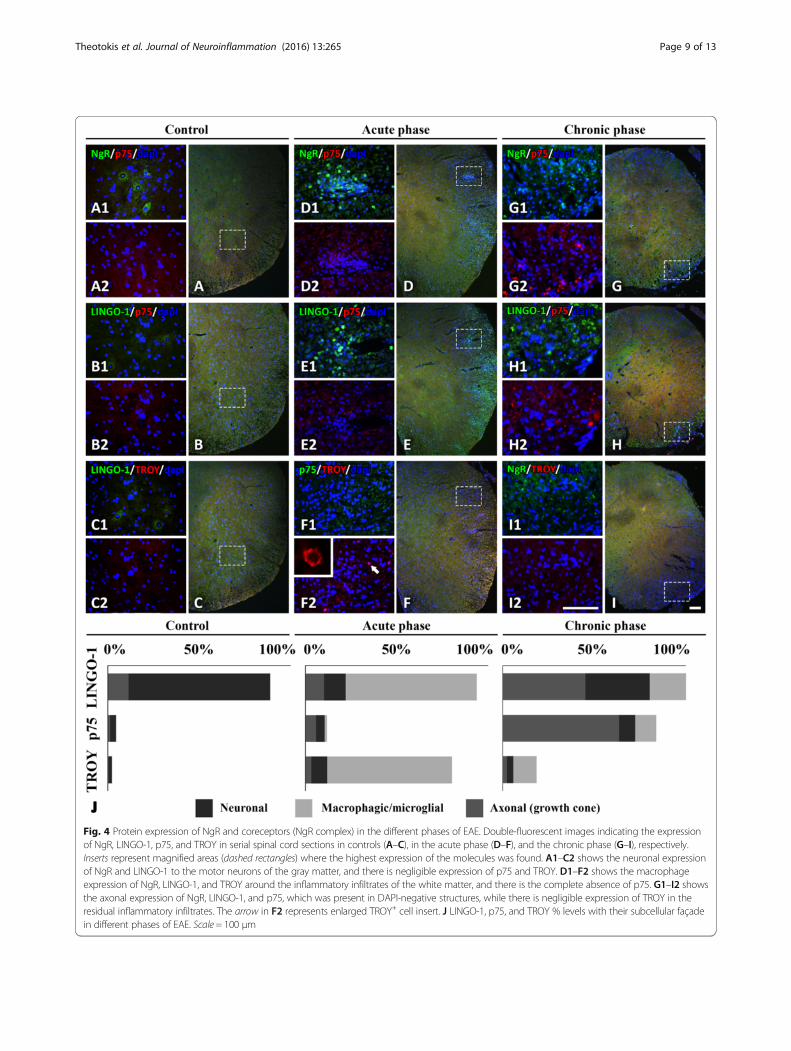
Fig. 4 Protein expression of NgR and coreceptors (NgR complex) in the different phases of EAE. Double-fluorescent images indicating the expressionof NgR, LINGO-1, p75, and TROY in serial spinal cord sections in controls (A–C), in the acute phase (D–F), and the chronic phase (G–I), respectively.Inserts represent magnified areas (dashed rectangles) where the highest expression of the molecules was found. A1–C2 shows the neuronal expressionof NgR and LINGO-1 to the motor neurons of the gray matter, and there is negligible expression of p75 and TROY. D1–F2 shows the macrophageexpression of NgR, LINGO-1, and TROY around the inflammatory infiltrates of the white matter, and there is the complete absence of p75. G1–I2 showsthe axonal expression of NgR, LINGO-1, and p75, which was present in DAPI-negative structures, while there is negligible expression of TROY in theresidual inflammatory infiltrates. The arrow in F2 represents enlarged TROY+ cell insert. J LINGO-1, p75, and TROY % levels with their subcellular façadein different phases of EAE. Scale = 100 μm
Theotokis et al. Journal of Neuroinflammation (2016) 13:265 Page 9 of 13

more than 25 % of macrophages of the acute phase werefound to express TROY in their cytoplasm while no p75was found. TROY protein has been found up-regulated inMS brain lesions [43]. The absence of p75 in TROY+ cellshas been also observed in sciatic nerve (PNS) of rats afterinjury [22]. Conversely, in the chronic phase, the appear-ance of p75 in no DAPI-correlated structures appeared tobe unaffected by the lack of TROY from residual macro-phages. Furthermore, neither of two proteins appeared incontrols, leading us to assume that this spatiotemporalrearrangement of the molecules at a systematic level is
associated with the processes occurring in the variousstages of EAE, seen under certain circumstances [42, 44,45]. The significance of these findings would be increasedby functional experiments (KO mice or blocking peptidesfor LINGO-1, p75, and TROY), which unfortunately arenot feasible for the time being, establishing a weakness inthis study.In order to understand the possible role of NgR in the
inflammatory processes, we correlated its expression withmicroglia/macrophage polarization [28, 46]. Our datashowed that NgR is expressed only by M2 macrophages/
Fig. 5 NgR protein expression in correlation with axonal regeneration and degeneration markers. Double-fluorescent immunostaining of NgRwith molecular markers GAP-43 (A, B) and APP (C, D), respectively. E The data is displayed as integrated density (in arbitrary units) GAP-43, APP,and NgR in controls, acute phase, and chronic phase. F Percentage of GAP-43+ or APP+ axons coexpressing NgR. *** and # denote p < 0.001 forcomparison of the acute and chronic phases, respectively, compared to controls, ‡p < 0.001 for comparisons indicated on the bars. ns non significantcomparison. Scale = 20 μm
Fig. 6 M1–M2 phenotype of NgR+ macrophagic/microglial populations within the inflammatory foci of the spinal cord. A A high expression ofthe Arginase-1 marker was observed in NgR+ cells (juxtapositions of separate color channels in A1–A3) within the acute lesions in the white matter. BLevels did not differ significantly in the chronic phase. C No connection (coexpression) was observed for iNOS+ (arrows) and NgR+ (arrowheads) cells inboth acute and chronic phases. ***p < 0.001, ns non significant comparison. Scale = 20 μm
Theotokis et al. Journal of Neuroinflammation (2016) 13:265 Page 10 of 13
microglia suggesting that it may have an important but un-known role so far for their functions [46, 47]. Previousstudies on rat EAE lesions support a positive role of theinflammatory ED1+ macrophages for the promotion of therepair process and recovery [28]. Additionally, the role ofM2 microglia has been proven beneficial in EAE, by creat-ing an anti-inflammatory environment, accompanied bytissue repair [46]. As a de novo synthesis of NgR in macro-phages is possible, as seen after sciatic nerve crush in rats[21, 22], it may further prevent the spread of inflammationin the adjacent normal-appearing white matter [22]. Takentogether, along with the high ratio of ED1+ macrophagespresent within the inflammatory foci, we propose that thosephagocytic Arginase-1+NgR+ cells contribute to inflamma-tory regulation facilitating the repair process in the tissue.In conclusion, we provide descriptive evidence for a pos-
sible action of NgR within inflammatory lesions of EAEacute and chronic phases. We show that NgR, LINGO-1,and TROY are expressed by macrophages of the acutephase and that NgR is also expressed on GAP-43+ axonalgrowth cones. Interestingly, the majority of NgR+ macro-phages present in the inflammatory foci acquire the anti-inflammatory M2 phenotype which might ultimately leadto area clearance. As the kinetics of NgR, LINGO-1, p75,and TROY are tightly regulated and interchange bothcellularly and time-wise, we propose that this system mightbe involved in the regulation, resolution, and repairinglocal processes after the inflammatory axonal injury in thespinal cord of EAE animals.
ConclusionsThis study demonstrates the expression kinetics of theNogo receptor complex in autoimmune demyelinatinglesions of EAE. Our data supports a phase-driven differen-tial expression of all the molecules of the complex with adistinct temporal profile pattern, thereby defined by theEAE course. We further provide a possible underlyingmechanism based on the selective expression milieu ofNgR in GAP-43+ axonal growth cones and its coexpres-sion in Arginase-1+, M2 phenotype alternatively activatedmacrophages.
Additional file
Additional file 1: Figure S1. Bielschowsky staining, immunofluorescence,and real-time PCR quality controls. (A1–4) Typical images of scores 0–3 inBielschowsky silver staining. (B, C) Preabsorption assay for LINGO-1 and TROYin serial sections of chronic and acute phases, respectively (the phasewhere signal is most abundant). (B1) LINGO-1 antibody specifications:rabbit polyclonal, Abcam ab23631, LOT N/A, dilution 1:300. (B2) LINGO-1peptide specifications: rabbit, Abcam ab25890, LOT #GR41007-1, incubationwith ab in 10× molecular ratio. (C1) TROY antibody specifications: goatpolyclonal, Santa Cruz sc-13711 (E-19), LOT #H0707, epitope mapping nearthe C-terminus of TROY of mouse origin, dilution 1:100. (C2) TROY peptidespecifications: goat, Santa Cruz sc-13711 P, LOT #B0402, incubation with abin 10× molecular ratio. (D, E) Antibody specificity test for p75 and NgR with
the use of another antibody (different company) recognizing a differentepitope in serial sections of chronic and acute phases, respectively (thephase where signal is most abundant). (D1) p75 antibody #1 specifications:mouse monoclonal, Abcam ab8877, LOT GR136825-1, ME20.4, dilution1:400. (D2) p75 antibody #2 specifications: mouse monoclonal, Santa Cruzp75 (B-1) sc-271708, LOT #J0611, epitope mapping between amino acids393–427 at the C-terminus of NGFR p75 of human origin, 1:100. (E1) NgRantibody #1 specifications: rabbit polyclonal, Santa Cruz sc-25659 (H-120),LOT E1209, epitope corresponding to amino acids 31–150 mapping nearthe N-terminus of Nogo-R of human origin, dilution 1:100. (E2) NgRantibody #2 specifications: rabbit polyclonal, Abcam ab26291, LOT N/A,epitope from within residues 150–250 of rat Nogo receptor, dilution 1:100.(F) β-actin real-time PCR quality control showing the specific amplificationproducts on agarose gel and the melting curves of their respective genes;curve identifier: light green TROY, orange p75, dark green LINGO-1. (G)mRNA levels of coreceptors LINGO-1, p75, and TROY in the spinal cord ofEAE animals by real-time PCR analysis using GAPDH as a second, qualitycontrol, house-keeping gene. The levels of mRNA expression for thecoreceptors (G1) LINGO-1, (G2) p75, and (G3) TROY, followed the samepattern with those that underwent β-actin analysis. Error bars indicate thestandard statistical error of the mean (SEM), ***p < 0.001, **p < 0.01. Blackscale bar = 20 μm.
AbbreviationsANOVA: Analysis of variance; APP: β-Amyloid precursor protein;CFA: Complete Freund’s adjuvant; CNS: Central nervous system; DAPI: 4′,6-Diamidino-2-phenylindole; EAE: Experimental autoimmuneencephalomyelitis; GFAP: Glial fibrillary acidic protein;GPI: Glycosylphosphatidylinositol; Iba-1: Ionized calcium-binding adaptormolecule-1; iNOS: Inducible nitric oxide synthase; LFB: Luxol fast blue;LRR: Leucine-rich-repeat; MOG: Myelin oligodendrocyte glycoprotein;MS: Multiple sclerosis; NeuN: Neuronal nuclei; SEM: Standard error of themean
AcknowledgementsThe authors would like to thank Evangelia Kofidou for her technicalassistance in the EAE experiments.
FundingThis work was supported by the State Scholarships Foundation (IKY).
Availability of data and materialsThe datasets supporting the conclusions of this article are included withinthe article.
Authors’ contributionsPT performed the experiments, collected and analyzed the data, and wrotethe manuscript. OT analyzed part of the histopathology and edited themanuscript. RL performed and analyzed the real-time PCR data and editedthe manuscript. EN performed the histopathology. EK and SS assisted withthe data collection. TT provided the MOG peptide. AL performed the EAEexperiments, interpreted the data, and revised the manuscript. DK, NG, andCS designed the study, interpreted the data, and revised the manuscript. Allauthors read and approved the final manuscript.
Competing interestsThe authors declare that they have no competing interests.
Consent for publicationNot applicable
Ethics approvalAll experimental procedures were conducted according to the institutionalguidelines, in compliance with the Greek Regulations and the EuropeanCommunities Council Directive of November 24, 1986 (86/609/EEC).Experimentation received approval from the Veterinary Medicines Directorate(license number 177867/1510).
Theotokis et al. Journal of Neuroinflammation (2016) 13:265 Page 11 of 13
Author details1B’ Department of Neurology, Laboratory of Experimental Neurology andNeuroimmunology, AHEPA University Hospital, Aristotle University ofThessaloniki, Stilponos Kiriakides str. 1, 546 36 Thessaloniki, CentralMacedonia, Greece. 2Department of Chemistry, University of Patras, Rion, 26504 Patras, Greece. 3Institute for Stroke and Dementia Research (ISD),Feodor-Lynen-Strasse 17, 81377 Munich, Germany. 4Department ofExperimental Physiology, Faculty of Medicine, Aristotle University ofThessaloniki, 546 36 Thessaloniki, Central Macedonia, Greece.
Received: 30 May 2016 Accepted: 22 September 2016
References1. Fournier AE, GrandPre T, Strittmatter SM. Identification of a receptor mediating
Nogo-66 inhibition of axonal regeneration. Nature. 2001;409:341–6.2. Wang KC, Kim JA, Sivasankaran R, Segal R, He Z. P75 interacts with the
Nogo receptor as a co-receptor for Nogo, MAG and OMgp. Nature. 2002;420:74–8.
3. Park JB, Yiu G, Kaneko S, Wang J, Chang J, He XL, Garcia KC, He Z. A TNFreceptor family member, TROY, is a coreceptor with Nogo receptor inmediating the inhibitory activity of myelin inhibitors. Neuron. 2005;45:345–51.
4. Mi S, Lee X, Shao Z, Thill G, Ji B, Relton J, Levesque M, Allaire N, Perrin S,Sands B, et al. LINGO-1 is a component of the Nogo-66 receptor/p75signaling complex. Nat Neurosci. 2004;7:221–8.
5. Ahmed Z, Douglas MR, John G, Berry M, Logan A. AMIGO3 is an NgR1/p75co-receptor signalling axon growth inhibition in the acute phase of adultcentral nervous system injury. PLoS One. 2013;8:e61878.
6. Satoh J, Onoue H, Arima K, Yamamura T. Nogo-A and nogo receptorexpression in demyelinating lesions of multiple sclerosis. J NeuropatholExp Neurol. 2005;64:129–38.
7. Wang X, Chun SJ, Treloar H, Vartanian T, Greer CA, Strittmatter SM. Localizationof Nogo-A and Nogo-66 receptor proteins at sites of axon-myelin and synapticcontact. J Neurosci. 2002;22:5505–15.
8. Domeniconi M, Cao Z, Spencer T, Sivasankaran R, Wang K, Nikulina E,Kimura N, Cai H, Deng K, Gao Y, et al. Myelin-associated glycoproteininteracts with the Nogo66 receptor to inhibit neurite outgrowth. Neuron.2002;35:283–90.
9. Wang KC, Koprivica V, Kim JA, Sivasankaran R, Guo Y, Neve RL, He Z.Oligodendrocyte-myelin glycoprotein is a Nogo receptor ligand that inhibitsneurite outgrowth. Nature. 2002;417:941–4.
10. Barton WA, Liu BP, Tzvetkova D, Jeffrey PD, Fournier AE, Sah D, Cate R,Strittmatter SM, Nikolov DB. Structure and axon outgrowth inhibitor bindingof the Nogo-66 receptor and related proteins. EMBO J. 2003;22:3291–302.
11. Cafferty WB, Strittmatter SM. The Nogo-Nogo receptor pathway limits aspectrum of adult CNS axonal growth. J Neurosci. 2006;26:12242–50.
12. Shao Z, Browning JL, Lee X, Scott ML, Shulga-Morskaya S, Allaire N, Thill G,Levesque M, Sah D, McCoy JM, et al. TAJ/TROY, an orphan TNF receptorfamily member, binds Nogo-66 receptor 1 and regulates axonalregeneration. Neuron. 2005;45:353–9.
13. Mosyak L, Wood A, Dwyer B, Buddha M, Johnson M, Aulabaugh A, Zhong X,Presman E, Benard S, Kelleher K, et al. The structure of the Lingo-1ectodomain, a module implicated in central nervous system repairinhibition. J Biol Chem. 2006;281:36378–90.
14. Meininger V, Pradat PF, Corse A, Al-Sarraj S, Rix Brooks B, Caress JB, CudkowiczM, Kolb SJ, Lange D, Leigh PN, et al. Safety, pharmacokinetic, and functionaleffects of the Nogo-A monoclonal antibody in amyotrophic lateral sclerosis: arandomized, first-in-human clinical trial. PLoS One. 2014;9:e97803.
15. Tran JQ, Rana J, Barkhof F, Melamed I, Gevorkyan H, Wattjes MP, de Jong R,Brosofsky K, Ray S, Xu L, et al. Randomized phase I trials of the safety/tolerability of anti-LINGO-1 monoclonal antibody BIIB033. NeurolNeuroimmunol Neuroinflamm. 2014;1:e18.
16. Litwak SA, Payne NL, Campanale N, Ozturk E, Lee JY, Petratos S, Siatskas C,Bakhuraysah M, Bernard CC. Nogo-receptor 1 deficiency has no influenceon immune cell repertoire or function during experimental autoimmuneencephalomyelitis. PLoS One. 2013;8:e82101.
17. Steinbach K, McDonald CL, Reindl M, Schweigreiter R, Bandtlow C, Martin R.Nogo-receptors NgR1 and NgR2 do not mediate regulation of CD4 T helperresponses and CNS repair in experimental autoimmune encephalomyelitis.PLoS One. 2011;6:e26341.
18. Pourabdolhossein F, Mozafari S, Morvan-Dubois G, Mirnajafi-Zadeh J, Lopez-Juarez A, Pierre-Simons J, Demeneix BA, Javan M. Nogo receptor inhibitionenhances functional recovery following lysolecithin-induced demyelinationin mouse optic chiasm. PLoS One. 2014;9:e106378.
19. Cui Z, Kang J, Hu D, Zhou J, Wang Y. Oncomodulin/truncated protamine-mediated Nogo-66 receptor small interference RNA delivery promotes axonregeneration in retinal ganglion cells. Mol Cells. 2014;37:613–9.
20. Takahashi K, Rochford CD, Neumann H. Clearance of apoptotic neuronswithout inflammation by microglial triggering receptor expressed onmyeloid cells-2. J Exp Med. 2005;201:647–57.
21. David S, Fry EJ, Lopez-Vales R. Novel roles for Nogo receptor ininflammation and disease. Trends Neurosci. 2008;31:221–6.
22. Fry EJ, Ho C, David S. A role for Nogo receptor in macrophage clearancefrom injured peripheral nerve. Neuron. 2007;53:649–62.
23. Theotokis P, Lourbopoulos A, Touloumi O, Lagoudaki R, Kofidou E,Nousiopoulou E, Poulatsidou KN, Kesidou E, Tascos N, Spandou E,Grigoriadis N. Time course and spatial profile of Nogo-A expression inexperimental autoimmune encephalomyelitis in C57BL/6 mice. JNeuropathol Exp Neurol. 2012;71:907–20.
24. Theotokis P, Kleopa KA, Touloumi O, Lagoudaki R, Lourbopoulos A,Nousiopoulou E, Kesidou E, Poulatsidou KN, Dardiotis E, Hadjigeorgiou G, etal. Connexin43 and connexin47 alterations after neural precursor cellstransplantation in experimental autoimmune encephalomyelitis. Glia. 2015;63:1772–83.
25. de Luca LE, Pikor NB, O’Leary J, Galicia-Rosas G, Ward LA, Defreitas D, FinlayTM, Ousman SS, Osborne LR, Gommerman JL. Substrain differences revealnovel disease-modifying gene candidates that alter the clinical course of arodent model of multiple sclerosis. J Immunol. 2010;184:3174–85.
26. Gur-Wahnon D, Mizrachi T, Maaravi-Pinto FY, Lourbopoulos A, Grigoriadis N,Higazi AA, Brenner T. The plasminogen activator system: involvement incentral nervous system inflammation and a potential site for therapeuticintervention. J Neuroinflammation. 2013;10:124.
27. Lourbopoulos A, Grigoriadis N, Lagoudaki R, Touloumi O, Polyzoidou E,Mavromatis I, Tascos N, Breuer A, Ovadia H, Karussis D, et al. Administrationof 2-arachidonoylglycerol ameliorates both acute and chronic experimentalautoimmune encephalomyelitis. Brain Res. 2011;1390:126–41.
28. Ahn M, Yang W, Kim H, Jin JK, Moon C, Shin T. Immunohistochemical studyof arginase-1 in the spinal cords of Lewis rats with experimentalautoimmune encephalomyelitis. Brain Res. 2012;1453:77–86.
29. Lourbopoulos A, Mourouzis I, Karapanayiotides T, Nousiopoulou E,Chatzigeorgiou S, Mavridis T, Kokkinakis I, Touloumi O, Irinopoulou T,Chouliaras K, et al. Changes in thyroid hormone receptors after permanentcerebral ischemia in male rats. J Mol Neurosci. 2014.
30. Kim JE, Liu BP, Park JH, Strittmatter SM. Nogo-66 receptor preventsraphespinal and rubrospinal axon regeneration and limits functionalrecovery from spinal cord injury. Neuron. 2004;44:439–51.
31. McDonald CL, Steinbach K, Kern F, Schweigreiter R, Martin R, Bandtlow CE,Reindl M. Nogo receptor is involved in the adhesion of dendritic cells tomyelin. J Neuroinflammation. 2011;8:113.
32. Pool M, Niino M, Rambaldi I, Robson K, Bar-Or A, Fournier AE. Myelinregulates immune cell adhesion and motility. Exp Neurol. 2009;217:371–7.
33. Yan J, Zhou X, Guo JJ, Mao L, Wang YJ, Sun J, Sun LX, Zhang LY, Zhou XF,Liao H. Nogo-66 inhibits adhesion and migration of microglia via GTPaseRho pathway in vitro. J Neurochem. 2012;120:721–31.
34. Fang Y, Yan J, Li C, Zhou X, Yao L, Pang T, Yan M, Zhang L, Mao L, Liao H.The Nogo/Nogo receptor (NgR) signal is involved in neuroinflammationthrough the regulation of microglial inflammatory activation. J Biol Chem.2015;290:28901–14.
35. Shin T, Kang B, Tanuma N, Matsumoto Y, Wie M, Ahn M, Kang J. Intrathecaladministration of endothelin-1 receptor antagonist amelioratesautoimmune encephalomyelitis in Lewis rats. Neuroreport. 2001;12:1465–8.
36. Liu G, Ni J, Mao L, Yan M, Pang T, Liao H. Expression of Nogo receptor 1in microglia during development and following traumatic brain injury.Brain Res. 2015.
37. Kerschensteiner M, Bareyre FM, Buddeberg BS, Merkler D, Stadelmann C,Bruck W, Misgeld T, Schwab ME. Remodeling of axonal connectionscontributes to recovery in an animal model of multiple sclerosis. J Exp Med.2004;200:1027–38.
38. Trifunovski A, Josephson A, Ringman A, Brene S, Spenger C, Olson L.Neuronal activity-induced regulation of Lingo-1. Neuroreport. 2004;15:2397–400.
Theotokis et al. Journal of Neuroinflammation (2016) 13:265 Page 12 of 13
39. Mandemakers WJ, Barres BA. Axon regeneration: it’s getting crowded at thegates of TROY. Curr Biol. 2005;15:R302–5.
40. McDonald CL, Bandtlow C, Reindl M. Targeting the Nogo receptor complexin diseases of the central nervous system. Curr Med Chem. 2011;18:234–44.
41. Borrie SC, Baeumer BE, Bandtlow CE. The Nogo-66 receptor family in theintact and diseased CNS. Cell Tissue Res. 2012.
42. Kraemer BR, Yoon SO, Carter BD. The biological functions and signalingmechanisms of the p75 neurotrophin receptor. Handb Exp Pharmacol.2014;220:121–64.
43. Satoh J, Tabunoki H, Yamamura T, Arima K, Konno H. TROY and LINGO-1expression in astrocytes and macrophages/microglia in multiple sclerosislesions. Neuropathol Appl Neurobiol. 2007;33:99–107.
44. Meeker R, Williams K. Dynamic nature of the p75 neurotrophin receptor inresponse to injury and disease. J Neuroimmune Pharmacol. 2014;9:615–28.
45. Zhou XF, Li HY. Roles of glial p75NTR in axonal regeneration. J Neurosci Res.2007;85:1601–5.
46. Jiang Z, Jiang JX, Zhang GX. Macrophages: a double-edged sword inexperimental autoimmune encephalomyelitis. Immunol Lett. 2014;160:17–22.
47. Berard JL, Kerr BJ, Johnson HM, David S. Differential expression of SOCS1 inmacrophages in relapsing-remitting and chronic EAE and its role in diseaseseverity. Glia. 2010;58:1816–26.
• We accept pre-submission inquiries
• Our selector tool helps you to find the most relevant journal
• We provide round the clock customer support
• Convenient online submission
• Thorough peer review
• Inclusion in PubMed and all major indexing services
• Maximum visibility for your research
Submit your manuscript atwww.biomedcentral.com/submit
Submit your next manuscript to BioMed Central and we will help you at every step:
Theotokis et al. Journal of Neuroinflammation (2016) 13:265 Page 13 of 13