Embed Size (px)
Citation preview
Proc. Natl. Acad. Sci. USAVol. 88, pp. 6368-6371, July 1991Neurobiology
Nitric oxide mediates glutamate neurotoxicity in primarycortical cultures
(endothelium-derived relaxing factor/N-methyl-n-aspartate)
VALINA L. DAWSON*, TED M. DAWSONt, EDYTHE D. LONDON*, DAVID S. BREDTt,AND SOLOMON H. SNYDERtt*Neuropharmacology Laboratory, National Institute on Drug Abuse Addiction Research Center, P.O. Box 5180, Baltimore, MD 21224; and tDepartments ofNeuroscience, Pharmacology and Molecular Sciences, and of Psychiatry, The Johns Hopkins University School of Medicine, 725 North Wolfe Street,Baltimore, MD 21205
Contributed by Solomon H. Snyder, April 22, 1991
ABSTRACT Nitric oxide (NO) mediates several biologicalactions, including relaxation of blood vessels, cytotoxicity ofactivated macrophages, and formation of cGMP by activationofglutamate receptors in cerebellar slices. Nitric oxide synthase(EC 1.14.23.-) immunoreactivit is colocalized with nicotina-mide adenine di-nucleotide phosphate diaphorase in neuronsthat are uniquely resistant to toxic insults. We show that thenitric oxide synthase inhibitors, N0-nitro-L-arginine (EC50 = 20INM) andNw-nmonomethyl-L-arginine (EC,5 = 170 jtM), preventneurotoxicity elicited by N-methyl-D-aspartate and related ex-citatory amino acids. This effect is competitively reversed byL-arginine. Depletion of the culture medium of argnine byargnase or arginine-free growth medium completely attenuatesN-methyl-D-aspartate toxicity. Sodium nitroprusside, whichspontaneously releases NO, produces dose-dependent cell deaththat parallels cGMP formation. Hemoglobin, which complexesNO, prevents neurotoxic effects of both N-methyl-D-aspartateand sodium nitroprusside. These data establish that NO medi-ates the neurotoxicity of glutamate.
Nitric oxide (NO), first identified as endothelium-derivedrelaxing factor (1-3), is also an important neuronal messengermolecule (4, 5). A physiological role for NO in the nervoussystem has been established by demonstrations that argininederivatives, which are potent and selective inhibitors of nitricoxide synthase (NOS; EC 1.14.23.-), block neuronally me-diated relaxation of the intestine (6-8) and stimulation ofcGMP formation by glutamate in the cerebellum (9, 10). NOis formed from arginine in brain and endothelial cells by NOS,which has been purified to homogeneity (11) and molecularlycloned (12) from rat brain. Macrophages and other blood cellsalso produce NO, which mediates their bactericidal andtumoricidal effects (13). However, the NOS of macrophagesis a distinct protein from NOS in brain and endothelialtissues, differing in cofactor requirements. Immunocyto-chemical studies have localized NOS to select neuronalpopulations in the brain, as well as to neurons in the retina,adrenal medulla, and intestine, and to nerve fibers in theposterior pituitary (14, 15).
Glutamate may mediate the neurotoxicity observed inhypoxic-ischemic brain injury, as selective antagonists of theN-methyl-D-aspartate (NMDA) subtype of glutamate recep-tor prevent neuronal cell death in animal models of hypoxic-ischemic brain injury (16). Glutamate neurotoxicity has alsobeen implicated in neurodegenerative disorders such as Alz-heimer and Huntington diseases (16, 17). Glutamate neuro-toxicity mediated by NMDA receptors involves calciumentry into cells via ligand-gated ion channels (17). Interest-ingly, the enhancement of NOS activity by NMDA stimula-
tion of cerebellar slices also derives from channel-associatedentry of calcium, which binds to calmodulin associated withNOS (9-11). To ascertain the relationship ofNO to glutamateneurotoxicity, we have employed rat primary cortical cul-tures and demonstrate that glutamate neurotoxicity is pre-vented selectively by inhibitors of NOS.
MATERIALS AND METHODSCell Culture. Primary dissociated cell cultures were pre-
pared from fetal rats (13- to 14-day gestation for cortex andcaudate-putamen cultures and 19- to 20-day gestation forhippocampal cultures). The tissue of interest was dissected,incubated for 15 min in 0.027% trypsin in Brooks-Logansolution (5% phosphate-buffered saline/0.04 M sucrose/10mM Hepes/0.03 M glucose, pH 7.4), and then transferred tomodified Eagle's medium (MEM)/10%o horse serum/10%ofetal bovine serum/2 mM glutamine for trituration. Dissoci-ated cells were plated at a density of 3-4 x 10⁵ cells per wellin polyornithine-coated 15-mm multiwell plates. After 4 daysthe cells were treated with 10 ,g of 5-fluoro-2'-deoxyuridineto prevent proliferation of nonneuronal cells. Cells weremaintained in MEM/5% horse serum/2 mM glutamine in 8%C02/humidified atmosphere at 37°C. The medium waschanged twice weekly. In the present study, mature neurons(3-4 weeks) were used.
Cytoxicity. Cells were exposed to excitatory amino acidsaccording to the method of Koh and Choi (18). Beforeexposure, the cells were washed three times with Tris-buffered control salt solution (CSS) (18), containing 120 mMNaCl/5.4 mM KCl/1.8 mM CaCI2/25 mM Tris hydrochlo-ride, pH 7.4 at room temperature/15 mM glucose). Briefexposures to glutamate, NMDA (plus 10 ,uM glycine), quis-qualate, and sodium nitroprusside (SNP) were performed for5 min in CSS. The exposure solution was then washed awayand replaced by MEM with 21 mM glucose; then the cellswere placed in an incubator for 20-24 hr. Long exposures tokainate were performed in MEM/21 mM glucose for 20-24 hrin the incubator. After exposure to the excitatory aminoacids, the medium was replaced by CSS/0.4% trypan blue,which stains nonviable cells. Two to four photographs (10-20x) were made of each well, and viable versus nonviablecells were counted. The cytotoxicity data represent 6-24separate wells assayed per data point, with -500-1500 cellscounted per well. In some experiments, overall neuronal cellinjury was also assessed by the measurement of lactate
Abbreviations: CSS, control salt solution; Hb, hemoglobin; L-Arg,L-arginine; D-Arg, D-arginine; MEM, modified Eagle's medium;NADPH, nicotinamide adenine dinucleotide phosphate; NO, nitricoxide; NOS, nitric oxide synthase; N-Arg, N"-nitro-L-arginine;NMDA, N-methyl-D-aspartate; Me-Arg, N"-monomethyl-L-arginine; SNP, sodium nitroprusside.*To whom reprint requests should be addressed.
6368
The publication costs of this article were defrayed in part by page chargepayment. This article must therefore be hereby marked "advertisement"in accordance with 18 U.S.C. §1734 solely to indicate this fact.
Proc. Natl. Acad. Sci. USA 88 (1991) 6369
dehydrogenase released by damaged or destroyed cells intothe extracellular fluid after drug exposure (18).
Biochemical Assays. For determination of cGMP the cellswere washed three times with CSS. After a 1-min exposureto drug solutions the cells were inactivated with 15% tri-chloroacetic acid. After ether extraction, cGMP levels weredetermined by RIA. [3H]MK-801 binding to the NMDAreceptor/channel complex was assayed, as described (19).NO synthase activity was assayed by the conversion of[3H]arginine to [3H]citrulline (9, 11). For immunoblots, 200-400 gg of crude tissue protein was separated on a 7.5%SDS/polyacrylamide gel and transferred to nitrocellulose.Lanes were incubated with affinity-purified antibody (1:1000)(14).
Electrophysiology. Cortical neurons in sister cultures werevoltage clamped by using the whole-cell version of thepatch-clamp technique (20).
Materials. [3H]Arginine (53 Ci/mmol; 1 Ci = 37 GBq) wasobtained from DuPont/NEN. cGMP RIA kits were obtainedfrom Amersham. Nw-monomethyl-L-arginine (Me-Arg) wasobtained from Calbiochem. All other chemicals were pur-chased from Sigma.
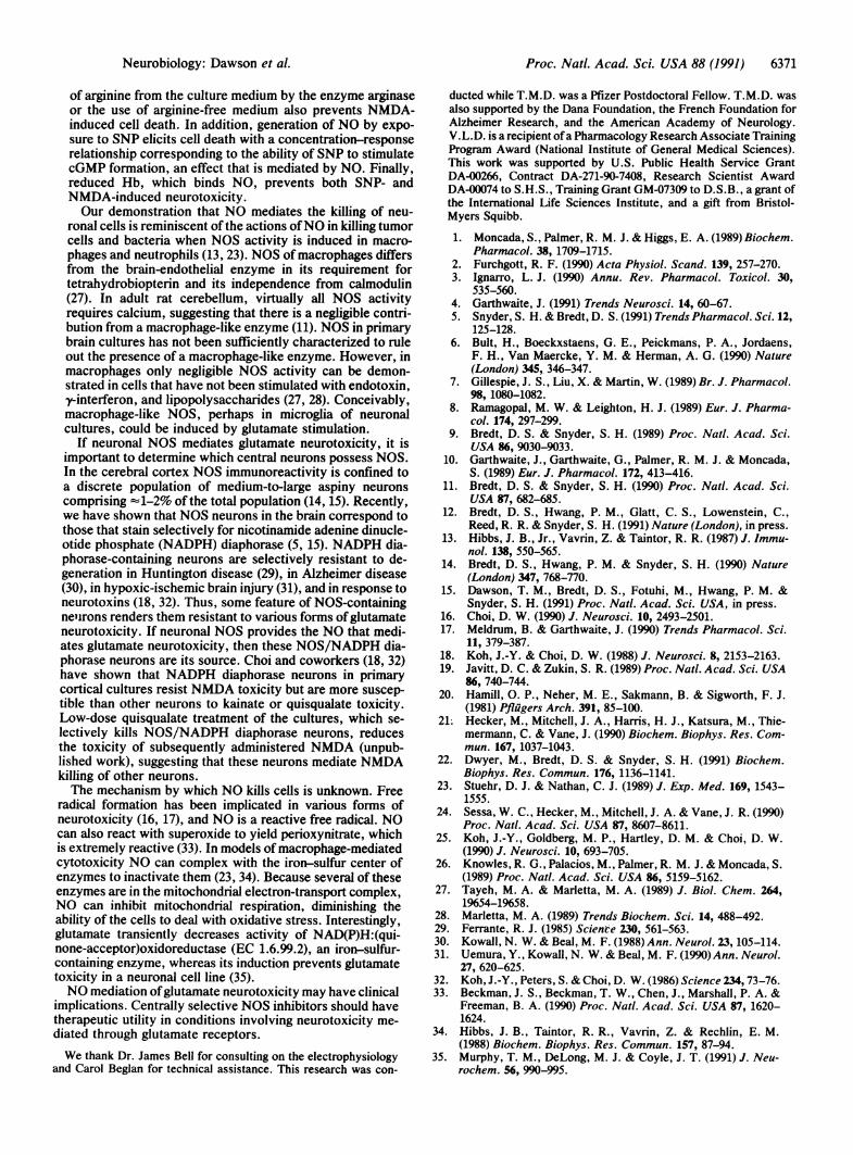
RESULTSBefore initiating cytotoxicity experiments with NOS inhibi-tors, we evaluated the potency of NMDA applied for 5 minin inducing cell death by examining exclusion of0.4% trypanblue by viable cells or measuring lactate dehydrogenasereleased by damaged or destroyed cells into the extracellularfluid. As reported earlier (18), NMDA displays a toxic LD50of 280 ,uM (data not shown), and 300 ,tM NMDA consistentlyyields 60%o cell death (Fig. 1). Simultaneous application of
e-.Muc._* x
-NumA' .8t
Lrj.e Med
FIG. 1. Bright-field photomicrographs of cortical cell cultures 24hr after treatment and after 0.4% trypan blue dye exclusion to stainnonviable cells. Dead cells appear as black dots, and live cells appearas raised cells against the gray background. (A) Control (Cont). (B)NMDA (300 AM plus 10 ,uM glycine). (C) NMDA (300 .M plus 10AM glycine) plus 100 N-Arg demonstrates inhibition of NMDAcytotoxicity. (D) L-Arg (1 mM) reverses the inhibition by 100 ,uMN-Arg of 300 AM NMDA (plus 10 ,uM glycine) cytotoxicity. (E)Treatment of cell cultures for 24 hr in L-Arg-free medium abolishesNMDA toxicity. (F) Reduced Hb plus 300 ,uM NMDA (with 10 AMglycine) reverses cell death. Photomicrographs were taken randomlyfrom culture wells and are representative of 6-24 determinations.
100 AtM NW-nitro-L-arginine (N-Arg), a potent NOS inhibitor(21, 22), with NMDA reduces cell death by 70% (Fig. 1).Addition of 1 mM L-arginine (L-Arg) to the exposure solutioncompletely reverses the effect of N-Arg (Fig. 1). To furtherascertain whetherNO is involved in NMDA neurotoxicity weadded 500AM ofreduced hemoglobin (Hb), which binds NO,simultaneously with 300 ,uM NMDA (Fig. 1). Hb completelyprevents NMDA-induced cell death at 500 ,uM, similar to theconcentration required for reduced myoglobin to preventmacrophage-mediated cell death (23).The cortical cultures possess substantial NOS catalytic
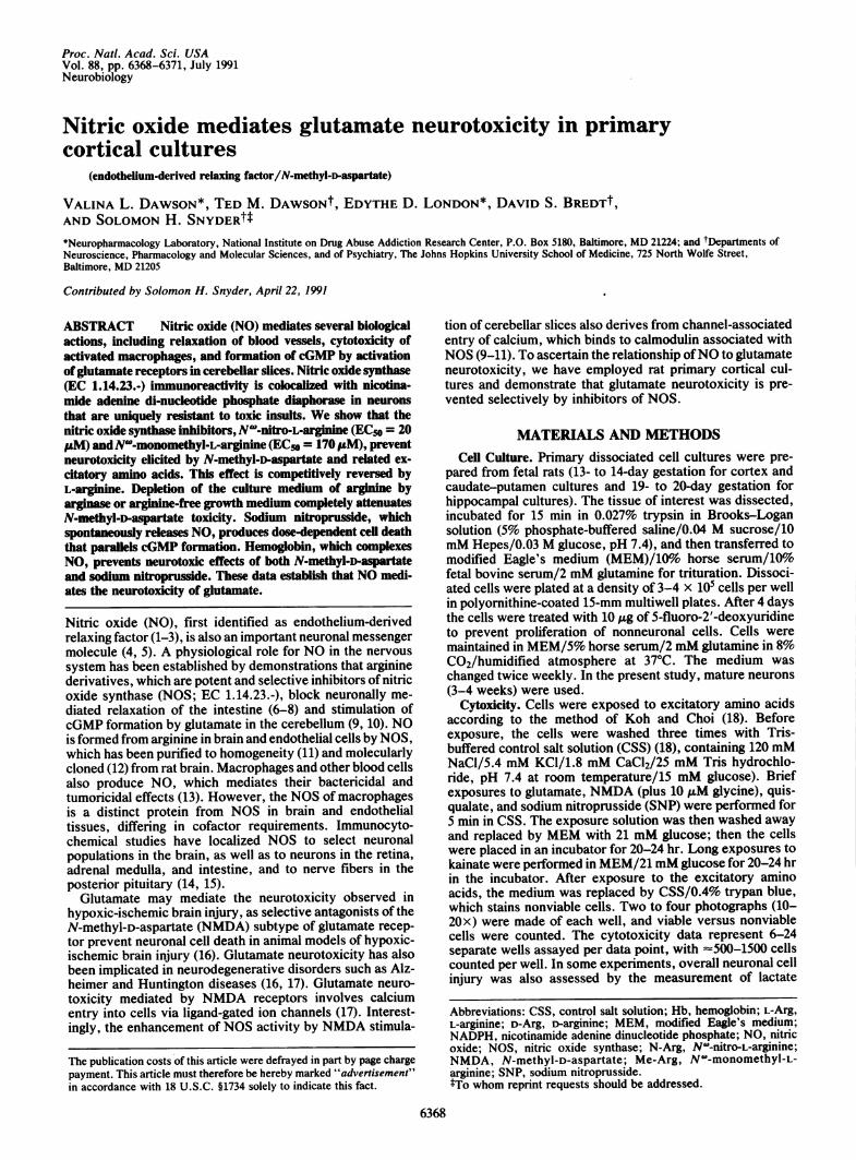
activity and protein, -10% of cerebellar levels (data notshown). Furthermore, NMDA can stimulate the formation ofNO in cortical cultures. Formation of cGMP can be used asan indirect measure ofNO formation (9, 10), as NO binds tothe heme moiety of guanylate cyclase and subsequentlyactivates the enzyme. As has been shown in cerebellar slices,NMDA stimulation ofcGMP levels in cultures is antagonizedby inhibiting NOS with N-Arg or complexing NO with Hb(Fig. 2). N-Arg inhibition of cGMP formation by NMDA iscompetitively reversible by L-Arg. Superoxide dismutase,which removes superoxide that would degrade NO, poten-tiates the formation of cGMP by NMDA (Fig. 2). Thus, as incerebellar slices, in cortical cell cultures NMDA stimulationof cGMP formation involves NO.N-Arg (100 ,M and 500 ,uM) has no effect on [3H]MK-801
binding to NMDA receptor channels in rat cerebral corticalmembranes (data not shown). Similarly, 100 ,uM N-Arg hasno effect on NMDA-induced currents measured in the cor-tical cultures by whole-cell patch-clamp analysis (data notshown). Therefore, N-Arg does not directly act on NMDAreceptors.To ascertain the potency ofN-Arg in preventing cell death,
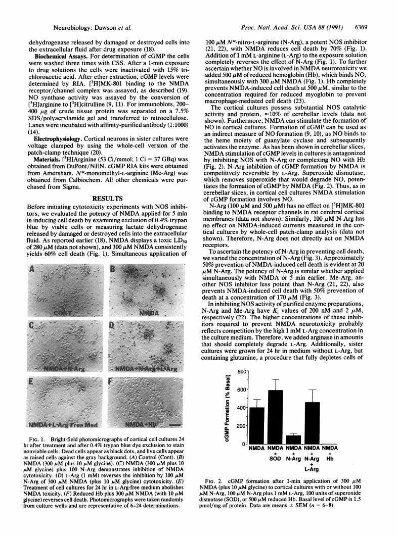
we varied the concentration ofN-Arg (Fig. 3). Approximately50% prevention ofNMDA-induced cell death is evident at 20,uM N-Arg. The potency of N-Arg is similar whether appliedsimultaneously with NMDA or 5 min earlier. Me-Arg, an-other NOS inhibitor less potent than N-Arg (21, 22), alsoprevents NMDA-induced cell death with 50% prevention ofdeath at a concentration of 170 ,uM (Fig. 3).
In inhibiting NOS activity of purified enzyme preparations,N-Arg and Me-Arg have Ki values of 200 nM and 2 ,uM,respectively (22). The higher concentrations of these inhib-itors required to prevent NMDA neurotoxicity probablyreflects competition by the high 1 mM L-Arg concentration inthe culture medium. Therefore, we added arginase in amountsthat should completely degrade L-Arg. Additionally, sistercultures were grown for 24 hr in medium without L-Arg, butcontaining glutamine, a procedure that fully depletes cells of
800-
to
m 600-
0
° 400-E _
0l
U. 200-
-
TT
NMDA NMDA NMDA NMDA
SOD N-Arg N-Arg Hb
L-Arg
FIG. 2. cGMP formation after 1-min application of 300 ,uMNMDA (plus 10 ,uM glycine) to cortical cultures with or without 100,uM N-Arg, 100 ,uM N-Arg plus 1 mM L-Arg, 100 units of superoxidedismutase (SOD), or 500 iM reduced Hb. Basal level ofcGMP is 1.5pmol/mg of protein. Data are means + SEM (n = 6-8).
Neurobiology: Dawson et al.
iL-
'AN.40
qp-MOA
6370 Neurobiology: Dawson et al.
M- ~~~~~~EC50
100 M A N-Arg=20tl
co 80 2AgM80-O 60-
-40-
s 20-z
0 1 lb 5o01o 30050061000NOS Inhibitors (jiM)
FIG. 3. Concentration-response relationship of NOS inhibitorsin inhibiting NMDA neurotoxicity. Data are means + SEM (n = 6).
L-Arg (24). In both arginase-treated preparations and in ex-periments with L-Arg-free medium, 300 pLM NMDA no longercauses cell death (Fig. 1), and the 50% lethal concentration ofNMDA is increased >20-fold to 7.5 mM (data not shown).Adding graded concentrations of L-Arg to L-Arg-free mediumreveals a requirement for 100 ,IM L-Arg to obtain maximalNMDA effects (data not shown).Glutamate neurotoxicity in primary cortical cultures can be
elicited by NMDA, quisqualate, and kainate, although theNMDA receptor presumably accounts for most neurotoxicityassociated with synaptically released glutamate in variouspathologic conditions (16, 17). Quisqualate and kainate actmore slowly and less potently thanNMDA and via somewhatdifferent mechanisms (25). To examine the role of NO inother forms of glutamate neurotoxicity, we compared theeffects of N-Arg on cytotoxicity induced by quisqualate,kainate, andNMDA (Table 1). A portion of cell death elicitedby quisqualate is prevented by N-Arg with reversal by L-Arg,but higher concentrations of N-Arg are required than withNMDA toxicity. N-Arg provides no protection against kain-ate-induced cell death. The slight protection afforded byN-Arg with quisqualate neurotoxicity may relate to theportion of quisqualate cell death occurring via NMDA re-ceptor activation (25). The relative effects of the glutamateanalogs and N-Arg are similar in cultures from caudate-putamen, hippocampus, and cerebral cortex (data notshown).To ascertain the specificity of L-Arg in reversing N-Arg
effects by arginine, we compared L-Arg with homoarginine
Table 1. Inhibition of glutamate neurotoxicity by N-Arg andreversal by L-Arg in cortical culture
Cell death, % ± SEM
500 ,uM glutamate 48.4 ± 4.4+ 100 jAM N-Arg 28.9 ± 6.0*+ 100 LM N-Arg + 1 mM L-Arg 48.2 ± 3.2
300 ILM NMDA + 10 juM glycine 57.8 ± 2.6+ 100 .&M N-Arg 17.7 ± 3.0**+ 100 jAM N-Arg + 1 mM L-Arg 63.4 ± 5.5
500,M quisqualate 64.2 ± 3.7+ 500 AM N-Arg 51.1 ± 2.4***+ 500,uM N-Arg + 5 mM L-Arg 66.6 ± 5.3
100 AM kainate 81.4 ± 7.1+ 500,uM N-Arg 87.0 ± 3.2
Data are means ± SEM (n = 6-24). Cell death was determined by0.4% trypan blue exclusion by viable cells (see text). Significantoverall F values were obtained by using a one-way, between-groupsanalysis of variance. Specific comparisons on all possible pairwisecombinations were made with the Student's t test for independentmeans, P < 0.05. *F, 2, 16, 29.3, P < 0.001; **F, 2, 33, 59.7, P <0.001; ***F, 2, 46, 6.1, P < 0.005.
a)on 4un -
) -)
o 20
L-Arg.....
.-.*.......Fomoarg
.4....A
-D-Arg----K~...
NMDA
NMDA + N-Arg
Concentration (LtM)
FIG. 4. Selectivity for reversal of N-Arg protection from NMDAcytotoxicity by L-Arg, homoarginine (Homoarg), and D-Arg. Solidhorizontal lines represent the percent of cell death after exposure to300 gM NMDA (plus 10 uM glycine) or 300 ,uM NMDA (with 10 uMglycine) plus 100 j.M N-Arg. Shading represents the mean and SEMfor NMDA or NMDA plus N-Arg. Data are means ± SEM (n = 6).
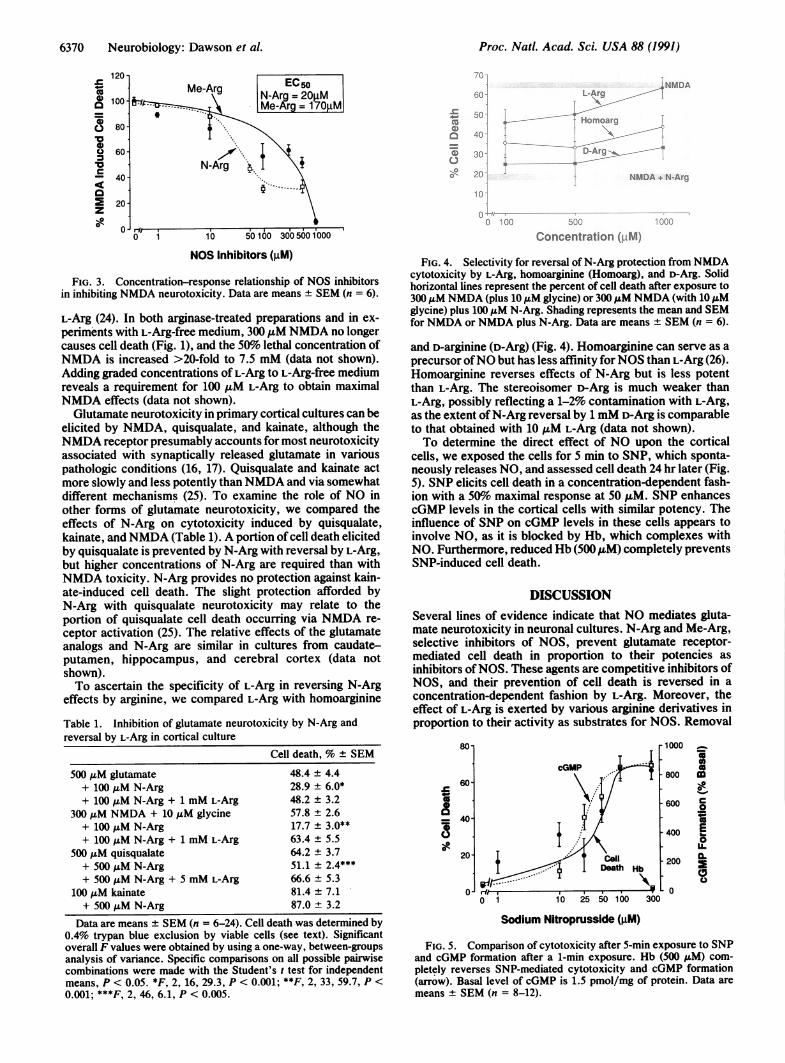
and D-arginine (D-Arg) (Fig. 4). Homoarginine can serve as aprecursor ofNO but has less affinity forNOS than L-Arg (26).Homoarginine reverses effects of N-Arg but is less potentthan L-Arg. The stereoisomer D-Arg is much weaker thanL-Arg, possibly reflecting a 1-2% contamination with L-Arg,as the extent ofN-Arg reversal by 1 mM D-Arg is comparableto that obtained with 10 ,uM L-Arg (data not shown).To determine the direct effect of NO upon the cortical
cells, we exposed the cells for 5 min to SNP, which sponta-neously releases NO, and assessed cell death 24 hr later (Fig.5). SNP elicits cell death in a concentration-dependent fash-ion with a 50%o maximal response at 50 ,uM. SNP enhancescGMP levels in the cortical cells with similar potency. Theinfluence of SNP on cGMP levels in these cells appears toinvolve NO, as it is blocked by Hb, which complexes withNO. Furthermore, reduced Hb (500 ,uM) completely preventsSNP-induced cell death.
DISCUSSIONSeveral lines of evidence indicate that NO mediates gluta-mate neurotoxicity in neuronal cultures. N-Arg and Me-Arg,selective inhibitors of NOS, prevent glutamate receptor-mediated cell death in proportion to their potencies asinhibitors ofNOS. These agents are competitive inhibitors ofNOS, and their prevention of cell death is reversed in aconcentration-dependent fashion by L-Arg. Moreover, theeffect of L-Arg is exerted by various arginine derivatives inproportion to their activity as substrates for NOS. Removal
80-
60
- 40
820
0
1000
800
600
400
200
-0
5'aaa0m0A
U
0-E0
U.
Sodium Nitroprusside (gM)
FIG. 5. Comparison of cytotoxicity after 5-min exposure to SNPand cGMP formation after a 1-min exposure. Hb (500 ,uM) com-pletely reverses SNP-mediated cytotoxicity and cGMP formation(arrow). Basal level of cGMP is 1.5 pmol/mg of protein. Data aremeans + SEM (n = 8-12).
Proc. Natl. Acad. Sci. USA 88 (1991)
Proc. Natl. Acad. Sci. USA 88 (1991) 6371
of arginine from the culture medium by the enzyme arginaseor the use of arginine-free medium also prevents NMDA-induced cell death. In addition, generation of NO by expo-sure to SNP elicits cell death with a concentration-responserelationship corresponding to the ability of SNP to stimulatecGMP formation, an effect that is mediated by NO. Finally,reduced Hb, which binds NO, prevents both SNP- andNMDA-induced neurotoxicity.Our demonstration that NO mediates the killing of neu-
ronal cells is reminiscent of the actions ofNO in killing tumorcells and bacteria when NOS activity is induced in macro-phages and neutrophils (13, 23). NOS of macrophages differsfrom the brain-endothelial enzyme in its requirement fortetrahydrobiopterin and its independence from calmodulin(27). In adult rat cerebellum, virtually all NOS activityrequires calcium, suggesting that there is a negligible contri-bution from a macrophage-like enzyme (11). NOS in primarybrain cultures has not been sufficiently characterized to ruleout the presence of a macrophage-like enzyme. However, inmacrophages only negligible NOS activity can be demon-strated in cells that have not been stimulated with endotoxin,y-interferon, and lipopolysaccharides (27, 28). Conceivably,macrophage-like NOS, perhaps in microglia of neuronalcultures, could be induced by glutamate stimulation.
If neuronal NOS mediates glutamate neurotoxicity, it isimportant to determine which central neurons possess NOS.In the cerebral cortex NOS immunoreactivity is confined toa discrete population of medium-to-large aspiny neuronscomprising -1-2% of the total population (14, 15). Recently,we have shown that NOS neurons in the brain correspond tothose that stain selectively for nicotinamide adenine dinucle-otide phosphate (NADPH) diaphorase (5, 15). NADPH dia-phorase-containing neurons are selectively resistant to de-generation in Huntingtori disease (29), in Alzheimer disease(30), in hypoxic-ischemic brain injury (31), and in response toneurotoxins (18, 32). Thus, some feature of NOS-containingneurons renders them resistant to various forms ofglutamateneurotoxicity. If neuronal NOS provides the NO that medi-ates glutamate neurotoxicity, then these NOS/NADPH dia-phorase neurons are its source. Choi and coworkers (18, 32)have shown that NADPH diaphorase neurons in primarycortical cultures resist NMDA toxicity but are more suscep-tible than other neurons to kainate or quisqualate toxicity.Low-dose quisqualate treatment of the cultures, which se-lectively kills NOS/NADPH diaphorase neurons, reducesthe toxicity of subsequently administered NMDA (unpub-lished work), suggesting that these neurons mediate NMDAkilling of other neurons.The mechanism by which NO kills cells is unknown. Free
radical formation has been implicated in various forms ofneurotoxicity (16, 17), and NO is a reactive free radical. NOcan also react with superoxide to yield perioxynitrate, whichis extremely reactive (33). In models of macrophage-mediatedcytotoxicity NO can complex with the iron-sulfur center ofenzymes to inactivate them (23, 34). Because several of theseenzymes are in the mitochondrial electron-transport complex,NO can inhibit mitochondrial respiration, diminishing theability of the cells to deal with oxidative stress. Interestingly,glutamate transiently decreases activity of NAD(P)H:(qui-none-acceptor)oxidoreductase (EC 1.6.99.2), an iron-sulfur-containing enzyme, whereas its induction prevents glutamatetoxicity in a neuronal cell line (35).NO mediation ofglutamate neurotoxicity may have clinical
implications. Centrally selective NOS inhibitors should havetherapeutic utility in conditions involving neurotoxicity me-diated through glutamate receptors.We thank Dr. James Bell for consulting on the electrophysiology
and Carol Beglan for technical assistance. This research was con-
ducted while T.M.D. was a Pfizer Postdoctoral Fellow. T.M.D. wasalso supported by the Dana Foundation, the French Foundation forAlzheimer Research, and the American Academy of Neurology.V.L.D. is a recipient of a Pharmacology Research Associate TrainingProgram Award (National Institute of General Medical Sciences).This work was supported by U.S. Public Health Service GrantDA-00266, Contract DA-271-90-7408, Research Scientist AwardDA-00074 to S.H.S., Training Grant GM-07309 to D.S.B., a grant ofthe International Life Sciences Institute, and a gift from Bristol-Myers Squibb.
1. Moncada, S., Palmer, R. M. J. & Higgs, E. A. (1989) Biochem.Pharmacol. 38, 1709-1715.
2. Furchgott, R. F. (1990) Acta Physiol. Scand. 139, 257-270.3. Ignarro, L. J. (1990) Annu. Rev. Pharmacol. Toxicol. 30,
535-560.4. Garthwaite, J. (1991) Trends Neurosci. 14, 60-67.5. Snyder, S. H. & Bredt, D. S. (1991) Trends Pharmacol. Sci. 12,
125-128.6. Bult, H., Boeckxstaens, G. E., Peickmans, P. A., Jordaens,
F. H., Van Maercke, Y. M. & Herman, A. G. (1990) Nature(London) 345,. 346-347.
7. Gillespie, J. S., Liu, X. & Martin, W. (1989) Br. J. Pharmacol.98, 1080-1082.
8. Ramagopal, M. W. & Leighton, H. J. (1989) Eur. J. Pharma-col. 174, 297-299.
9. Bredt, D. S. & Snyder, S. H. (1989) Proc. Nati. Acad. Sci.USA 86, 9030-9033.
10. Garthwaite, J., Garthwaite, G., Palmer, R. M. J. & Moncada,S. (1989) Eur. J. Pharmacol. 172, 413-416.
11. Bredt, D. S. & Snyder, S. H. (1990) Proc. Nadl. Acad. Sci.USA 87, 682-685.
12. Bredt, D. S., Hwang, P. M., Glatt, C. S., Lowenstein, C.,Reed, R. R. & Snyder, S. H. (1991) Nature (London), in press.
13. Hibbs, J. B., Jr., Vavrin, Z. & Taintor, R. R. (1987) J. Immu-nol. 138, 550-565.
14. Bredt, D. S., Hwang, P. M. & Snyder, S. H. (1990) Nature(London) 347, 768-770.
15. Dawson, T. M., Bredt, D. S., Fotuhi, M., Hwang, P. M. &Snyder, S. H. (1991) Proc. Nadl. Acad. Sci. USA, in press.
16. Choi, D. W. (1990) J. Neurosci. 10, 2493-2501.17. Meldrum, B. & Garthwaite, J. (1990) Trends Pharmacol. Sci.
11, 379-387.18. Koh, J.-Y. & Choi, D. W. (1988) J. Neurosci. 8, 2153-2163.19. Javitt, D. C. & Zukin, S. R. (1989) Proc. Nati. Acad. Sci. USA
86, 740-744.20. Hamill, 0. P., Neher, M. E., Sakmann, B. & Sigworth, F. J.
(1981) Pflugers Arch. 391, 85-100.21; Hecker, M., Mitchell, J. A., Harris, H. J., Katsura, M., Thie-
mermann, C. & Vane, J. (1990) Biochem. Biophys. Res. Com-mun. 167, 1037-1043.
22. Dwyer, M., Bredt, D. S. & Snyder, S. H. (1991) Biochem.Biophys. Res. Commun. 176, 1136-1141.
23. Stuehr, D. J. & Nathan, C. J. (1989) J. Exp. Med. 169, 1543-1555.
24. Sessa, W. C., Hecker, M., Mitchell, J. A. & Vane, J. R. (1990)Proc. Nati. Acad. Sci. USA 87, 8607-8611.
25. Koh, J.-Y., Goldberg, M. P., Hartley, D. M. & Choi, D. W.(1990) J. Neurosci. 10, 693-705.
26. Knowles, R. G., Palacios, M., Palmer, R. M. J. & Moncada, S.(1989) Proc. Nadl. Acad. Sci. USA 86, 5159-5162.
27. Tayeh, M. A. & Marletta, M. A. (1989) J. Biol. Chem. 264,1%54-1%58.
28. Marletta, M. A. (1989) Trends Biochem. Sci. 14, 488-492.29. Ferrante, R. J. (1985) Scienee 230, 561-563.30. Kowall, N. W. & Beal, M. F. (1988) Ann. Neurol. 23, 105-114.31. Uemura, Y., Kowall, N. W. & Beal, M. F. (1990) Ann. Neurol.
27, 620-625.32. Koh, J.-Y., Peters, S. & Choi, D. W. (1986) Science 234, 73-76.33. Beckman, J. S., Beckman, T. W., Chen, J., Marshall, P. A. &
Freeman, B. A. (1990) Proc. Nati. Acad. Sci. USA 87, 1620-1624.
34. Hibbs, J. B., Taintor, R. R., Vavrin, Z. & Rechlin, E. M.(1988) Biochem. Biophys. Res. Commun. 157, 87-94.
35. Murphy, T. M., DeLong, M. J. & Coyle, J. T. (1991) J. Neu-rochem. 56, 990-995.
Neurobiology: Dawson et al.