Embed Size (px)
Citation preview

KAPOSI’S SARCOMA–ASSOCIATED HERPESVIRUSIMMUNOEVASION AND TUMORIGENESIS: TWO SIDES OF THESAME COIN?
Patrick S. Moore and Yuan ChangMolecular Virology Program, Hillman Cancer Research Center, University of Pittsburgh,Pittsburgh, Pennsylvania 15213-1863Patrick S. Moore: [email protected]; Yuan Chang: [email protected]
AbstractKaposi’s sarcoma–associated herpesvirus (KSHV) [or human herpesvirus 8 (HHV-8)] is the mostfrequent cause of malignancy among AIDS patients. KSHV and related herpesviruses haveextensively pirated cellular cDNAs from the host genome, providing a unique opportunity toexamine the range of viral mechanisms for controlling cell proliferation. Many of the viralregulatory homologs encode proteins that directly inhibit host adaptive and innate immunity.Other viral proteins target retinoblastoma protein and p53 control of tumor suppressor pathways,which also play key effector roles in intracellular immune responses. The immune evasionstrategies employed by KSHV, by targeting tumor suppressor pathways activated during immunesystem signaling, may lead to inadvertent cell proliferation and tumorigenesis in susceptible hosts.
KeywordsKSHV; HHV-8; antiviral immunity; tumor virus; viral oncogenes
INTRODUCTIONThe virus causing Kaposi’s sarcoma (KS) has generated considerable scientific interest as anew human (h) tumor virus. Kaposi’s sarcoma–associated herpesvirus (KSHV) [or humanherpesvirus 8 (HHV-8)] is unique because of its extensive molecular piracy of critical cellregulatory genes. Functions for these genes can be deduced directly from their sequence, butin general the viral (v) proteins are modified to escape normal cellular regulation and hencebehave differently from their cellular counterparts. Whereas KSHV initially appears to beunique among human tumor viruses, the opposite is actually the case because the regulatorycircuits and control points targeted by KSHV are the same as those targeted by other tumorviruses. Examining the mechanisms that KSHV uses to induce cell proliferation can lead tounexpected insights into unrelated viruses and highlights important relationships betweenviral immune evasion and tumorigenesis.
BIOLOGY AND PATHOGENESISDisease Associations
KAPOSI’S SARCOMA—Chang et al. (20) discovered KSHV in 1993 usingrepresentational difference analysis during a molecular search for an infectious etiology ofKS. KS consists of proliferating spindle cells that form irregular microvascular channels.These tumors are most commonly found in the dermis but also occur in viscera includinglungs, liver, and intestines (Figure 1). The large majority of spindle cells are infected with
NIH Public AccessAuthor ManuscriptAnnu Rev Microbiol. Author manuscript; available in PMC 2013 August 03.
Published in final edited form as:Annu Rev Microbiol. 2003 ; 57: 609–639. doi:10.1146/annurev.micro.57.030502.090824.
NIH
-PA Author Manuscript
NIH
-PA Author Manuscript
NIH
-PA Author Manuscript
virus, but because of the infiltrative nature of these lesions, uninfected tissues not harboringviral genome are intertwined with tumor cells (41).
All clinical forms of KS are infected with KSHV, a necessary but not sufficient cofactor forKS pathogenesis. The high rate of KS among AIDS patients, particularly gay men, appearsto be due to both immunosuppression as well as shared sexual risk factors for infectionbetween HIV and KSHV. To date, there is no compelling evidence to support a direct rolefor HIV in KS pathogenesis other than its part in causing immunosuppression. KSHVinfection is uncommon overall (approximately 1%–5% of persons are infected in NorthAmerica and Northern Europe), but higher infection rates occur among gay men (~40%) andMediterranean populations (5%–20%). Sub-Saharan Africa has the highest rates ofinfection, with many countries experiencing seroprevalence rates exceeding 60% [fordetailed descriptions of clinical and epidemiologic aspects KSHV infection, see (116)]. Ascould be expected from the explosion of AIDS in Africa, KS is now the most commonlyreported neoplasm in many African countries and represents a largely unseen andunchallenged public health problem (4, 153).
PRIMARY EFFUSION LYMPHOMA—In addition to KS, several hematologic,predominantly B cell, disorders are associated with KSHV. The two most well characterizedare primary effusion lymphomas (PELs) and a subset of multicentric Castleman’s disease(MCD). Other unusual or rare entities associated with KSHV infection have also beendescribed including posttransplantation plasmacytic proliferations (109), posttransplantationbone marrow failure (105), MCD-associated plasmablastic lymphomas (40), andgerminotropic lymphoproliferations (39). Multiple myeloma has been linked to KSHVinfection in the past (137), but careful analyses show that the virus is unlikely to play asignificant role in this disease (129).
PELs are monoclonal, non-Hodgkin’s B cell lymphomas that frequently lack B cell–specificsurface markers (13, 19) and are commonly coinfected with Epstein-Barr virus (EBV) (84).They display immunoblastic/centroblastic morphology and postgerminal centerimmunophenotype (50). Cell lines established from PEL, unlike KS tumor explants, stablymaintain viral episomes at high copy number (50– 150 copies per cell) and are the source ofvirus for most virologic and serologic studies.
MULTICENTRIC CASTLEMAN’S DISEASE—Multicentric Castleman’s disease(MCD), a B cell lymphoproliferative disorder, has a heterogeneous pathogenesis with onlyabout 50% of Castleman’s disease tumors being infected with KSHV in HIV-negative,healthy persons. The cause of the remaining KSHV-negative MCD patients remains obscurebut almost certainly involves dysregulation of endogenous interleukin (IL)-6 secretion.Nearly all MCD occurring among AIDS patients, however, is KSHV positive. MCD is apolyclonal tumor in which the bulk of the cell mass is composed of uninfected lymphocytesrecruited to the site of infection by cytokines elaborated from a small proportion of B cellsinfected with KSHV (41, 76, 132), and displays an unusual immunoglobulin (Ig)M lightchain restriction (40). There is also a high rate of secondary non-Hodgkin’s lymphomaamong MCD patients, which suggests that this tumor can either predispose or evolve into aneoplastic disorder (127).
Genome Structure and OrganizationThe gammaherpesvirus subfamily is composed of two genera: the Lymphocryptovirus(gamma-1 herpesviruses), which includes EBV (or human herpesvirus 4), and theRhadinovirus (gamma-2 herpesviruses), which includes KSHV and the new world monkeyvirus, herpesvirus saimiri (saimiirine herpesvirus 2, HVS).
Moore and Chang Page 2
Annu Rev Microbiol. Author manuscript; available in PMC 2013 August 03.
NIH
-PA Author Manuscript
NIH
-PA Author Manuscript
NIH
-PA Author Manuscript

CONSERVED AND UNIQUE GENE BLOCKS—Like other herpesviruses, KSHV is alarge double-stranded DNA virus that replicates in the nucleus as a closed circular episomeduring latency but linearizes during virion packaging and replication. All identified KSHVtranscripts are encoded on a continuous 145-kb long unique region. The long unique regionis flanked by the 20–35 kb terminal repeat region composed of 801-bp high G+C contentterminal repeat units (140).
Over 80 genes have been identified in the long unique region, and new open reading frames(ORFs) are continuously described as small gene products, alternative reading frames, andalternative splicing patterns are being investigated (Figure 2).
KSHV shares structural and biological features with the human tumor virus EBV butpossesses none of its latency genes involved in cell immortalization and transformation(140). Although KSHV latency genes have no evolutionary homology to EBV latencygenes, there is a clear genetic correspondence between the two viruses (116). In general,cellular genes that are induced by EBV proteins have been captured and modified by KSHVduring its evolution. This is not unexpected because both viruses use B lymphocytes asreservoirs during latency and face similar biological challenges in establishing persistentinfections in a B cell environment. KSHV also infects endothelial cells, as occurs in KStumors, and possibly monocytes and epithelial cells (at least in transit during initialinfection).
The origin of the pirated cDNAs is unknown. No clear mechanism has been shown whichincorporates cellular cDNAs into the viral genomes of not only KSHV and relatedrhadinoviruses, but also other viruses including poxviruses, which also have extensivelycaptured cellular cDNAs.
KSHV GENE EXPRESSION—KSHV gene expression depends on a variety of factorsincluding whether the virus is latent or lytic, the type of host cell infected, and the host cellenvironment. When induced into lytic replication, the virus genome replicates through arolling circle mechanism with individual viral genomes being cleaved in the terminal repeatregion and packaged as linear molecules into viral capsids.
A salient feature of the genome reveals itself when the functions and expression patterns ofthe genes are examined: Structural genes and highly conserved genes involved in lyticreplication tend to cluster in islands separated by novel genes, including many of the cDNAhomologs of cellular regulatory genes (144). When KSHV enters lytic replication, theclusters of lytic replication genes are induced in an orderly cascade. As shown by Sun et al.and Zhu et al. (161, 179), the progression of lytic gene activation follows an ordered patternsimilar to that of other herpesviruses. Unique gene clusters have more complicatedexpression patterns, and many genes are expressed at low levels during latency but areinduced during lytic replication, a pattern referred to as class II transcription, distinguishingit from constitutive (class I) or lytic (class III) expression (144).
Differentiating gene expression during lytic and latent replication has been useful forclassifying KSHV genes, but it is evident that mutually exclusive latent and lytic genecategories are too simplistic to adequately describe the biology of KSHV. For example, ORFK10.5 [latency-associated nuclear antigen (LANA2)] is only constitutive in hematopoieticcells but not in KS tumors (138), and even the constitutive genes encoding vFLIP (FLICE-inhibitory protein), vCYC (cyclin), and LANA1 at the major latency locus are expressed in aG1/S cell cycle–dependent pattern (147). vIL-6 is induced during lytic replication (114) butis also activated by interferon (IFN) signaling independent of replication cycle (22). Phorbolester treatment may directly activate some genes, such as ORF K5 [modulators of immune
Moore and Chang Page 3
Annu Rev Microbiol. Author manuscript; available in PMC 2013 August 03.
NIH
-PA Author Manuscript
NIH
-PA Author Manuscript
NIH
-PA Author Manuscript

response (MIR2)] (128), further complicating whether these viral genes are solely activatedduring lytic replication. Two genes, ORF K12 (kaposin) and ORF K7 (PAN, polyadenylatednuclear RNA), which are highly expressed and commonly used as markers for latent andlytic virus replication, respectively, are induced during lytic replication in PEL cells (141,144). This complexity is not unexpected because KSHV is a large virus with the capacity torespond in complex ways to its cellular environment.
One rationale for classifying viral genes into latent and lytic replication categories is thatlatency is generally assumed to be the state leading to cell proliferation because lyticreplication results in cell death and is therefore antitumorigenic. Even this assumption,however, is increasingly being called into question and does not apply to all types of virus-induced proliferation. The viralGprotein–coupled receptor (ORF74), for example, hastranscriptional kinetics resembling that of an early lytic gene (27, 83), but evidence suggeststhat it may play an important paracrine role in KS pathogenesis (2, 177). In contrast, latentKSHV gene expression together with host cell mutations leads to PEL cell transformationand monoclonal proliferation.
Examining viral gene expression directly in tumors is necessary to determine genes likely tobe involved in disease pathogenesis. This has been undertaken for a number of differentviral proteins using antibodies and in situ hybridization (27, 75, 76, 131, 132, 138, 157, 160,174). These studies show that the three major proliferative syndromes associated withKSHV have different virus expression programs that contribute to the tumor phenotype.
To better understand the roles that pirated genes play in the biology of KSHV, they arediscussed under the broad categories of immune evasion, apoptosis inhibition, andregulation of the cell cycle. These are artificial divisions and some KSHV proteins, such asvIL-6, have roles in all three biologic functions. The overlap between these areas reflects notonly the incomplete information on viral protein functions, but also the interconnectednature of these broad areas of cell biology.
FUNCTIONS OF KSHV HOMOLOG GENESImmune Evasion by KSHV Immunomodulatory Proteins
Active viral infections rely on highly efficient replication to outpace the development of aneffective adaptive immune response. Viruses causing chronic infections must instead evadeinnate immunity as well as adaptive immune responses so infection can be immediatelyestablished to maintain long-term infection. Latency itself, in which nonessential viralprotein expression is repressed to limit foreign antigen presentation, is a fundamental viralimmune evasion strategy. KSHV employs a broad repertoire of immune evasion proteinsduring both latency and lytic replication—perhaps more than any other virus described todate (Table 1) (Figure 3). Although individual mechanisms may be unique to KSHV andrelated viruses, a wide range of viruses employ similar strategies.
ADAPTIVE IMMUNE EVASION STRATEGIES: THE MIR PROTEINS, vFLIP, ANDVIRAL CHEMOKINES—Viral antigen processing and presentation through majorhistocom-patibility complex (MHC) I is a critical step in initiating an effective antiviral cell-mediated immune response. Burgert et al. (16) first identified viral inhibition of MHC Ifunction by adenovirus E3 protein, and Ploegh and colleagues have elegantly describedmechanisms to prevent antigen presentation for herpes simplex virus, cytomegalovirus(CMV), and other viruses [for review see (166)]. Building on this work, Coscoy & Ganem(30) systematically tested each unique KSHV K protein for the ability to downregulateMHC I cell surface expression. Two transmembrane proteins, MIR1 and MIR2, encoded byORFs K3 and K5, respectively, efficiently inhibit MHC I surface expression through a novel
Moore and Chang Page 4
Annu Rev Microbiol. Author manuscript; available in PMC 2013 August 03.
NIH
-PA Author Manuscript
NIH
-PA Author Manuscript
NIH
-PA Author Manuscript

mechanism not found in other viruses. These results were quickly confirmed and extendedby others (72, 158).
MIR1 and MIR2 remove MHC I from the plasma membrane through enhanced endocytosis,which results in lysosomal targeting and degradation of MHC molecules (30). Theendocytic, lysosomal trafficking and protein targeting functions of MIR1 and MIR2 aregenetically separable and have been examined through domain swapping studies (110, 142).MIR1 and MIR2 possess N-terminal C4HC3 zinc-finger domains, called plant homeodomainmotifs, that are characteristic for a class of E3 ubiquitin ligases (32), and ubiquitinate plasmamembrane-bound MHC I resulting in its endocytosis. While protein ubiquitination generallyresults in proteosomal degradation, it also serves to signal for protein trafficking. The K3homolog of the mouse gammaherpesvirus MHV68 possesses a similar MHC ubiquitinationfunction but prevents egress of MHC from the endoplasmic reticulum (103). MIR2ubiquitinates and downregulates the accessory immune receptors ICAM-1 and B7.2 inaddition to MHC I (31, 71). A newly recognized family of cellular and viral MIR1/2-likeproteins exist, whose functions are beginning to be explored. For example, a poxvirushomolog of K3 has been described to ubiquitinate and downregulate surface expression ofCD4 (49, 107).
Downregulation of MHC I and its accessory immune receptors poses the risk of initiating anatural killer (NK) cell response (122). NK cells interrogate cells for MHC I expression andinitiate receptor-activated apoptosis through Fas (CD95/Apo-1) in cells lacking appropriateMHC I expression. Human and murine CMVs avoid this by presenting virus-encoded MHC-like molecules to thwart NK cell recognition (166). KSHV and related gammaherpesvirusesinhibit NK-mediated killing through expression of v-FLICE-inhibitory proteins (vFLIPs)(165).
KSHV vFLIP encoded by ORF K13 (ORF71) possesses two death effector domains and actsas a dominant-negative inhibitor of receptor-activated apoptosis by binding to Fas-associated death domain protein and caspase 8 (FLICE) (6). This prevents activated caspaserecruitment into the death-inducing signaling complex [reviewed in detail elsewhere (87)].The discovery of vFLIPs in gammaherpesviruses and poxviruses by database searches fordeath effector domain containing proteins (9, 68, 165) led to the subsequent discovery ofcellular FLIPs existing in multiple splice forms (cFLIPL and cFLIPs) that have similarfunctions (87).
Because of its constitutive expression and role in inhibiting receptor-mediated apoptosis,vFLIP is a candidate contributor to KSHV-induced cell transformation and tumorigenesis.Transduction studies using vFLIP show that it enhances tumorigenicity of mouse Blymphoma cells in immunocompetent mice strains (38). KSHV vFLIP shares with cFLIPsthe ability to activate NF-κB through IκB kinase activation, which may also contribute to Bcell proliferation (23, 77, 102). NF-κB is a pro-proliferative transcription factor for most Bcell tumors, and use of specific NF-κB inhibitors induces apoptosis in PEL cell lines (80).vFLIP is expressed constitutively on two latent transcripts (LT1 and LT2) as a bi- ortricistronic message translated from an internal ribosomal entry site (IRES) located in the 3′region of the vCYC gene ORF72 (11, 60, 104). IRES-regulated translation of cellular anti-apoptotic transcripts maintains cap-independent translation during cellular stress (63, 67).
Recruitment of helper T cell subtype 2 (Th2) rather than Th1 CD4+ lymphocytes to the siteof infection polarizes immune responses toward an antibody-predominant Th2 immunereaction (119). KSHV inhibits effective cell-mediated immune responses through secretionof virus-encoded chemoattractant cytokines (chemokines) to enhance Th2 polarization.Patients with active KS can have antibody titers against LANA1 exceeding 1:100,000,
Moore and Chang Page 5
Annu Rev Microbiol. Author manuscript; available in PMC 2013 August 03.
NIH
-PA Author Manuscript
NIH
-PA Author Manuscript
NIH
-PA Author Manuscript
indicating that specific antibody responses are robust but ineffectual in clearing infection(53, 153). KSHV encodes three secreted chemokines, vCCL1 (ORF K6), vCCL2 (ORF K4),and vCCL3 (ORF K4.1), formerly known as vMIP-I/MIP-1a, vMIP-II/MIP-1b, and vMIP-III/BCK, respectively, that act on receptors involved in Th2 chemotactic immune responses.
All three chemokines have dicysteine motifs but have different receptor specificities. vCCL2initiates a strong chemotactic response through CCR3 activation (12), while vCCL1 andvCCL3 activate CCR8 (35, 43) and CCR4 receptors (159), respectively. Unlike cellularchemokines, the viral chemokines have broad antagonistic activities for CC and CXCreceptors to inhibit chemotaxis of Th1 and NK lymphocytes (12, 24). Weber et al. (173)demonstrated that vCCL2 effectively blocks RANTES-mediated chemotaxis of Th1-likelymphocytes and initiates firm arrest of cells on human microvascular endothelium underflow conditions (173). The immune-inhibiting properties of vCCL2 were also demonstratedby experiments in which mismatched cardiac allograft survival was increased, and CTLinfiltration diminished, by viral chemokine expression in rodents (36). Furthermore,administration of vCCL2 inhibits fractalkine-mediated inflammatory glomerulonephritis in amouse model (24). While vCCL1 and vCCL2 expression is generally limited to lyticreplication, vCCL3 is found in KS tumors and may contribute to its pathogenesis (159). TheKSHV chemokines are pro-proliferative for some cell types and induce neoangiogenesis, inpart through induction of vascular endothelial growth factor (12, 101, 159). Chemokines andcytokines encoded by EBV, CMV, murine CMV, and poxviruses also have Th2 activity,which suggests that this is a common viral strategy for blunting adaptive immune responses(119).
KSHV encodes a neural cell adhesion molecule (NCAM)-like adhesin (ORF K14, vOX-2/vAdh) homologous to CD200 (OX-2) that promotes Th2 polarization and/or foreign antigenpresentation (58, 66). Initial studies suggest, however, that it activates rather than inhibitsinflammatory macrophage responses (29). Homologs of this protein are found amongpoxviruses as well.
INNATE IMMUNE EVASION STRATEGIES: KCP, KIS, vIL-6, ORF45 PROTEIN,AND vIRF1—In addition to inhibiting adaptive immune responses through the MIRproteins, vFLIP, and the viral chemokines, KSHV possesses means to block innate immuneresponses as well. These mechanisms include complement binding, downregulation of the Bcell receptor (BCR), and inhibition of IFN initiation and signaling.
Complement control and the B cell receptor: The KSHV ORF4 encodes a proteindesignated KSHV complement control protein (KCP), which has homology to humancomplement regulators (140). In addition to an unspliced, full-length mRNA of 1679 bp, atleast two alternatively, internally spliced transcripts have also been described that aretranslated into 175-, 82-, and 62-kDa proteins detectable only in induced KSHV-infectedPEL cell cultures. These three proteins retain C-terminal transmembrane domains and fourN-terminal complement control protein SUSHI domains required for membrane attachmentand complement regulation, respectively. Stable expression in cells exposed to human serumshows that all three KCP isoforms regulate complement activation by inhibiting C3deposition on the cell surface (156). Some viruses including poxviruses and other membersof herpesvirus family (HVS and MHV-68) have developed similar strategies to circumventthe complement component of the host immune response, which plays an important role inlimiting virus infection (74). Other viruses including HCMV, HIV, and human T celllymphotropic virus (HTLV)-1 upregulate host cell surface expression of complementregulators or acquire these proteins in their envelopes on egress from the cell.
Moore and Chang Page 6
Annu Rev Microbiol. Author manuscript; available in PMC 2013 August 03.
NIH
-PA Author Manuscript
NIH
-PA Author Manuscript
NIH
-PA Author Manuscript

The BCR, interacting with the complement-binding proteins CD19 and CD21, regulates Bcell development and response to antigen. ORF K1 encodes a small transmembrane,immunoglobulin-like, glycoprotein called KIS (K ITAM-signaling), which possesses acytoplasmic immunoreceptor tyrosine activation motif (ITAM) similar to that of the BCR(57). The KIS protein ITAM is tyrosine-phosphorylated in situ, allowing recruitment of SH2domain proteins, Syk pathway activation, and intracellular calcium mobilization (88, 92).Jung and colleagues found that overexpression of KIS causes transformation of Rat-1fibroblasts and that when KIS is substituted for the STP oncogene in recombinant HVSvirus, it can immortalize primary T lymphocytes and induce lymphomas in marmosetmonkeys (93). KIS protein resembles the BCR signal transduction subunits Igα and Igβ inits ability to induce signaling and to interact with mu chains of the BCR. However, unlikeIgα and Igβ, which interact with mu chains to direct BCR complexes to the cell surface, KISinteracts with mu chains to block the intracellular transport of BCR complexes to the cellsurface (90).
Viral hijacking of BCR signaling results in cell transformation, but it is unclear how thispromotes virus survival. The specific downregulation of BCR by KIS implies an importancein controlling B cell viral infections. The BCR interacts with complement receptors CD19and CD21; it remains to be determined what, if any, relationship KIS and ORF4 proteinshave to each other in the infected cell.
Interferon inhibition: Interferons (IFNs) are central to antiviral innate immunity,particularly in limiting initial viral replication prior to development of a specific immuneresponse (70). IFN activation, one of the first immune responses to virus infection, caneffectively limit establishment of infection so that subsequent humoral and cellularimmunities are not overwhelmed by viral load. The importance of the IFN response toKSHV is shown by the multiple mechanisms it employs to subvert IFN signaling. Thisinvolves inhibition of IFN-α signaling, antagonism of IFN-initiated gene transcription, andblockade of interferon regulatory factors (IRF) 3 and 7.
ANTAGONISM OF INTERFERON RECEPTOR SIGNALING BY vIL-6KSHV has evolved a complex mechanism for IFN-α receptor inhibition involving autocrinesignaling through the virus-encoded IL-6 cytokine. IL-6 is mitogenic for B cells and isinvolved in orchestrating Th2-type inflammatory responses (168, 169). vIL-6 is 26%identical to cellular IL-6, and extensive analyses demonstrate that it has nearly identicalsignaling patterns (15, 64, 114, 124, 130). Both cytokines activate STAT1 and STAT3phosphorylation, as well as additional IL-6 response pathways involving MAP-kinase andother serine/threonine kinases (130). vIL-6 substitutes for hIL-6 in maintaining cellproliferation of mouse and human B cell lines dependent on IL-6 signaling (15, 114, 124).
vIL-6 expression is limited to a subpopulation of cells in KSHV-associated hematolymphoiddisorders (Figure 1D). In MCD, vIL-6 is primarily localized to scattered B cells in themantle zone, and the bulk of each tumor is caused by vIL-6-induced proliferation ofuninfected B cells. vIL-6 expression is also common in PEL tumors and PEL cell lines (14,131), where it is expressed in a portion of cells through two alternative transcripts that aredifferentially regulated (37). Studies of PEL cell lines demonstrate that they are autocrinedependent (xenocrine-dependence) on vIL-6, together with hIL-10, but not on hIL-6 (73).
Despite similarities, hIL-6 and vIL-6 differ in their receptor engagement and utilization.hIL-6 signals at the plasma membrane by first binding gp80 (IL-6Rα), which complexes togp130, the molecule responsible for signal transduction across the membrane, at its cytokine—homology–binding region composed of domains 1 and 2 (D1D2). Subsequent recruitment
Moore and Chang Page 7
Annu Rev Microbiol. Author manuscript; available in PMC 2013 August 03.
NIH
-PA Author Manuscript
NIH
-PA Author Manuscript
NIH
-PA Author Manuscript

of gp130 D3 domain fully activates gp130 dimerization and transmembrane signaling.Crystallographic studies reveal that vIL-6 directly interacts with D1D2D3 epitopes throughhydrophobic interactions to activate gp130 in the absence of the IL-6Rα receptor (28). Thisprovides structural evidence supporting the experimental finding (112) that vIL-6 directlyactivates gp130 independent of gp80.
Autocrine-dependence of PEL cells on vIL-6 but not hIL-6 led Chatterjee et al. to examinethe effects of vIL-6 on IFN-α signaling because IFN-α induces cell arrest through inductionof the p21CIP cyclin-dependent kinase inhibitor (22) (Figure 4). Cell cycle arrest by IFN isan effective and reversible means to establish an antiviral state by limiting nucleic acidprecursors available to the virus. vIL-6 inhibits Tyk2 and STAT2 phosphorylation by IFN-αR, effectively blocking downstream signaling (M. Chatterjee, P.S. Moore & Y. Chang,unpublished observation) and IFN induction of p21CIP. The vIL-6 promoter itself possessesIFN-stimulated response element sequences inducible by IFN-α, providing a novel negativefeedback mechanism in which the virus senses and regulates IFN signaling. hIL-6 cannotreproduce this effect because IFN-α downregulates surface expression of IL-6Rα receptor,preventing gp130 signal transduction. Because vIL-6 interacts directly with gp130, itbypasses this regulatory mechanism, making PEL cell lines autocrine dependent on thisvirus-derived cytokine. This strategy blocks IFN from inducing an antiviral cellular state butat the price of increased cell proliferation for infected and nearby uninfected B cells.
INHIBITION OF INTERFERON ACTIVATION AND TRANSCRIPTIONKSHV also interferes with initiation and continued transcription of the IFN response. Yeasttwo-hybrid screening of ORF45 protein, a protein with an immediate-early expressionpattern (179), revealed binding to cellular IRF7 and blocking of its phosphorylation aftervirus infection (180). This prevents translocation of this transcription factor to the nucleus,an effect previously seen with the poxvirus E3L protein (154). The significance of IRF7inhibition is that IRF7, together with IRF3, initiates an IFN response against viral infectionby activation of IFN gene promoters (3). Once IFN secretion begins, it further activates itsown promoter through induction of the positive IFN transcription factor IRF1 [for review,see (163)].
KSHV vIRF1, encoded by ORF K9 with homology to the IRF family of proteins, performs afunction similar to that of the ORF45 protein but does so through direct inhibition of IFN-induced transcription. vIRF1 lacks the complete cellular IRF DNA-binding motif and doesnot directly bind IFN-stimulated response element; instead, it interacts with cellular IRF3,which provides promoter specificity for this repressor protein (99). vIRF1 prevents IRF3recruitment of p300 and CBP (CREB-binding protein) histone acetyltransferase coactivatorsinto the IFN transcriptional complex (94, 172). Sequestration of p300/CBP by vIRF1 is thepresumed mechanism for its broad inhibition of both class I and class II IFN transcription(46, 52, 95, 181), which is similar to the IFN inhibition reported for the adenovirus E1Aprotein (10). The effect of vIRF1 on histone acetylation is profound, producing globalchromatin condensation that can be measured by differential staining with DNAintercalating dyes (94). vIRF1 acts as a transactivator (139), as well as a repressor protein,and has been shown by Li et al. (95) to induce the vIL-6 gene. When vIRF1 is expressed inNIH3T3 fibroblasts, it causes full cell transformation similar to that of the IRF2 repressorprotein (52). Two related proteins, vIRF2 (ORF K11.1) and LANA2 (ORF K10.5), areencoded by spliced genes that probably arose from gene duplication of vIRF1 and have lowhomology to the cellular IRFs. vIRF2 is expressed at low levels constitutively in PEL celllines and activates genes possessing multimeric NF-κB elements (94). Preliminary evidencesuggests that it binds and inactivates dsRNA-activated protein kinase (PKR), therebypreventing initial activation of IFN through this pathway (17).
Moore and Chang Page 8
Annu Rev Microbiol. Author manuscript; available in PMC 2013 August 03.
NIH
-PA Author Manuscript
NIH
-PA Author Manuscript
NIH
-PA Author Manuscript
The repertoire of KSHV immune inhibitory proteins is at first glance bewildering. But theproteins are expressed in different viral stages and cell types and hence may havespecialized functions. vIRF1, for example, is primarily expressed in KS tumor, but not inPELs. Other proteins, such as vFLIP and vIL-6, may act constitutively in tumors and arelikely to play a critical role in tumorigenesis. More importantly, these viral defenses againstthe host immune system illustrate the overlapping nature of immune evasion and celltransformation (Figure 5).
The Antiapoptotic Proteins: vBCL-2, vIAP, LANA1, LANA2, and vIRF1It is now widely accepted that a cellular response limiting viral infections includes apoptosis(8, 111, 115), although programmed cell death is not universal for all viruses and someviruses may capitalize on cell dissolution during apoptosis to enhance replication (164). Theimportance of apoptosis regulation to the virus can be seen by the variety of KSHV factorsinhibiting this process (Table 2). KSHV encodes proteins targeting downstream apoptoticsignaling at the mitochondria, such as vBCL-2 and viral inhibitor of apoptosis protein(vIAP), as well as upstream p53 apoptotic signaling through proteins such as vIRF, LANA1,and LANA2. Inhibition of receptor-mediated apoptosis by vFLIP has already been describedand future studies may more clearly link the remaining antiapoptotic proteins to immuneevasion functions.
INHIBITION OF MITOCHONDRIAL APOPTOTIC SIGNALING—KSHV like manyother DNA viruses (34) encodes a homolog to the cellular BCL-2 antiapoptotic protein(146). BCL-2 family proteins have both pro- and antiapoptotic functions determined in partby their ability to heterodimerize and regulate release of mitochondrial apoptoticcomponents. vBCL-2 encoded by ORF16 was the first KSHV protein to be investigated forits apoptosis-inhibitory properties (25, 146) and possesses BCL-2 homology (BH)1 and BH2domains characteristic for this family of proteins. Although no sequence similarity to BH3and BH4 domains involved in heterodimerization and antiapoptotic functions are present invBCL-2, solution structure studies reveal strong structural similarities to these domains (69),allowing the viral protein to tightly bind proapoptotic Bak and Bax peptides, as suggestedthrough two-hybrid heterodimerization studies (146). Whereas cellular BCL-2 can becleaved by caspase proteolysis and converted to a proapoptotic version, KSHV vBCL-2lacks this cleavage site and escapes cellular regulation (7).
vBCL-2 in PEL cell culture is primarily expressed as an early gene during lytic replication(146, 161) and allows optimal virion production (34, 117, 145). Immunostaining of KStissues shows vBCL-2 production in a minority of infected spindle cells of advanced nodularlesions consistent with a role primarily in delaying lytic apoptosis (174). Functionally,however, it can inhibit apoptosis owing to latent vCYC-CDK6 complex overexpression,whereas cellular BCL-2 is phosphorylated and inactivated by vCYC-CDK6 (126). Despiteits presumed lytic expression pattern, studies from a related murine gammaherpesvirussuggest that vBCL-2 plays a critical role in maintaining chronic viral infection (51).
Another recently described KSHV antiapoptotic factor acting at the mitochondrialmembrane is the vIAP encoded by ORF K7, which has structural similarity to cellularsurvivin protein (45, 171). vIAP, like vBCL-2, is a glycoprotein that localizes tomitochondria and possesses a BH2 domain (171). Mitochondrial membranepermeabilization, Ca+2 depolarization, and release of cytochrome C are early events in theactivation of apoptotic caspase cascades. vIAP appears to stabilize Ca+2 mitochondrial fluxduring cell stress, in part by binding to the calcium-modulating cyclophilin ligand, and caninhibit apoptosis caused by a variety of agents, including Fas, thapsigargin, andstaurosporine (45).
Moore and Chang Page 9
Annu Rev Microbiol. Author manuscript; available in PMC 2013 August 03.
NIH
-PA Author Manuscript
NIH
-PA Author Manuscript
NIH
-PA Author Manuscript

INHIBITION OF p53-MEDIATED APOPTOSIS—p53, a transcriptional regulator of cellcycle arrest and apoptosis, is a second major target of antiapoptotic KSHV proteins.LANA1, the large multifunctional protein encoded by ORF73 (78, 136) first identified as aserologic antigen expressed constitutively in all infected cells (118), was subsequentlyshown by Friborg et al. (48) to bind p53. LANA1 efficiently inhibits p53 activation of apromoter containing the multimerized p53 element from the p21CIP promoter and preventsapoptosis owing to p53 overexpression, whereas a truncated mutant possessing only the first440 amino acids has no activity. Transcribed at low abundance together with vCYC andvFLIP mRNAs, LANA1 protein is highly stable and is easy to identify on immunostainingby its characteristic speckled, nuclear pattern (53, 79). In interphase nuclei, LANA1associates with heterochromatin through an amino-terminal region (98) and directly interactswith chromatin and methylated DNA-binding proteins, including histone H1 and MeCP1(33, 85).
The mechanism and consequences of p53-LANA1 interaction have not been fully examined.It is an attractive candidate oncoprotein because it may antagonize vCYC-induced apoptosisduring viral latency. LANA1 has broad transcriptional repressor activity (54, 86, 149) andacts on cell cycle regulation as well as on p53. A second latency-expressed nuclear antigen,LANA2, is expressed as a spliced transcript from ORF K10.5 (44, 138). Less is knownabout the properties of LANA2, although it has sequence similarity to cellular IRF4 and isexpressed only in infected B cells. Like LANA1, LANA2 inhibits p53-mediatedtranscription and apoptosis, but direct binding to p53 has not been found.
The IRF homolog vIRF1, in addition to its IFN-inhibiting properties, binds and inactivatesp53 (120, 151). This effect may in part be mediated by sequestration of p300, which is a p53transcription coactivator and a p53 acetylator (61, 97, 150, 155). vIRF1 effectively blocksapoptosis from DNA-damaging agents and Fas and fully transforms NIH3T3 cells to causetumors in severe combined immunodeficiency (SCID) mice. Although the IFN-inhibitionfunction of vIRF1 has also been reported to inhibit the transcription of FasL (82), it isunclear what significance this has for the virus because it does not generally infect immuneeffector T cells. Other KSHV proteins, such as the highly spliced transmembrane K15protein (55), may also regulate apoptotic responses but are in early stages of investigation(152).
Regulation of the Cell Cycle: vCYC, LANA1, and K-bZIP/RAPA third cellular function targeted by KSHV regulatory proteins is control of the cell cycle(Table 3) (Figure 6). The retinoblastoma protein pRB acts at the G1/S cell cycle checkpointto prevent unscheduled entry into S phase. Acting as a transcriptional repressor, pRBinactivates E2F transcription factors and complexes with E2F to bind E2F-responsivepromoter elements in promoters for genes involved in DNA synthesis and chromosomalreplication (167). Histone deacety lase and methyl-transferase recruitment by pRB shuts offtranscription of E2F-responsive genes. pRB itself is regulated and phosphorylated by cyclinsthat complex with CDKs.
Small DNA tumor viruses possess proteins that directly inhibit pRB by binding to the pRBHDAC-interaction domain through LXCXE-containing motifs (91). The KSHV vCYCprotein achieves a similar effect, but does so by mimicking the action of cellular D-typecyclins, which act at the G1/S transition to phosphorylate and inactivate pRB by partneringwith CDK4 and 6 (21, 56, 96). The cellular cyclin–CDK complex itself is activated by acyclin-activating kinase and inhibited by cyclin-dependent kinase inhibitors(CDKIs),including p21CIP and p27KIP. vCYC complexes with only CDK6 and is resistant toinactivation by CDKIs (162) either through structural features to prevent CDKI binding(148) or, in the case of p27KIP, by phosphorylating and inactivating p27KIP (42, 106).
Moore and Chang Page 10
Annu Rev Microbiol. Author manuscript; available in PMC 2013 August 03.
NIH
-PA Author Manuscript
NIH
-PA Author Manuscript
NIH
-PA Author Manuscript
Although vCYC functions as a D cyclin that escapes cell regulatory control, it has a broaderrange of substrates than D cyclins do. Similar to cyclin A or E-CDK2 complexes, vCYC andCDK6 phosphorylate histone H1 and ORC1, triggering DNA synthesis in isolated late G1-phase nuclei (89).
Although vCYC dysregulates the pRB G1/S tumor suppressor checkpoint, direct evidence ofvCYC playing a role in cell transformation has been scant until recently. This is in part dueto apoptosis resulting from vCYC overexpression in cell lines (125, 126). Activation of E2Fafter pRB inhibition results in induction of the p14ARF tumor suppressor (p19ARF in themouse), which in turn increases p53 stability through inhibition of its E3 ubiquitin ligase,MDM2 (5). This feedback control to induce apoptosis serves as a check to prevent tumorformation in the event of isolated loss of pRB function. PEL cell lines are frequently null forp16INK4a, suggesting a possible role in resistance to KSHV-induced apoptosis (133).
Using primary cell lines, Verschuren et al. (170) unexpectedly found that vCYC inducesp53-dependent apoptosis that is independent of E2F or p14ARF. Instead, apoptosis occursfrom unscheduled DNA synthesis without cytokinesis. When transgenic mice expressingvCYC under control of a B cell promoter were crossed with p53-null mice, all animalsdeveloped B cell lymphomas. In addition to vCYC, LANA1 also binds pRB in the pocketregion and together with H-Ras can transform rodent embryonic fibroblasts (135). Thus, likeSV40 T antigen, LANA1 targets both pRB and p53 checkpoints and serves as a viralepisome maintenance protein.
KSHV affects cell cycle regulation through mitogenic pathways as well. Viral G protein–coupled receptor encoded by ORF74 is a constitutively active CXC receptor expressedduring lytic replication (27, 83). It activates MAPK, p38, Akt, and NF-κB pathways,resulting in expression of angiogenic factors, such as VEGF, and in cell transformation (2,18, 108, 113, 134). Another interesting mechanism for KSHV-induced mitogenesis occursduring virus binding to cells. The gB virion glycoprotein expressed on the KSHV envelopebinds α 3β1 integrin as the KSHV receptor through an RGD-containing peptide (1). Viruscross-linking of α 3β1 results in activation of focal adhesion kinase and of the MEK-ERKpathway through PI3-kinase and protein kinase C activation (121). It is postulated thatmitogenic signaling by KSHV generates a suitable intracellular environment for maintainingvirus infection.
One KSHV cell cycle regulator has activity opposing that mentioned for the previous KSHVproteins. The ORF K8 gene product, called K-bZIP or RAP (replication associated protein),is homologous to the EBV transactivator BZLF1 protein (59, 100). Unlike its EBVcounterpart, K-bZIP/RAP is not the primary activator of lytic virus replication but insteadacts to inhibit cell cycle progression by inducing p21CIP through CCAAT/enhancer-bindingprotein(C/EBP)-α (176), a finding which has been replicated for the EBV BZLF1 protein aswell (175). This may activate the G2/M checkpoint during lytic replication to preventmitosis, thereby “fixing” the cell at a point in the cell cycle to maximize viral DNAsynthesis.
KSHV AS A MODEL TUMOR VIRUS: MOLECULAR PIRACY OF CELLULARREGULATORY GENES AND IMMUNE EVASION
Ease in identifying and characterizing potential KSHV oncoproteins has made the rapiddevelopment of KSHV as a tumor virus model possible. Studies examining KSHV-inducedcell proliferation have already generated insights into novel mechanisms applicable to othertumor viruses. This is particularly true for mechanisms inhibiting both innate immune andtumor suppressor signaling pathways (115).
Moore and Chang Page 11
Annu Rev Microbiol. Author manuscript; available in PMC 2013 August 03.
NIH
-PA Author Manuscript
NIH
-PA Author Manuscript
NIH
-PA Author Manuscript

Much of what is currently known about viral carcinogenesis is derived from studies of smallDNA viruses and acutely transforming retroviruses. In general, tumor viruses do not causetumors as part of their natural life cycles: Viral carcinogenesis occurs in nonnative orimmunocompromised hosts, or through adventitious host and viral mutagenesis.Nonetheless, viral oncogenes are highly conserved through convergent evolution, indicatingtheir important roles in enhancing viral replication fitness. The presumed function of tumorvirus oncogenes based on small tumor virus models is to inhibit G1/S checkpoints duringlytic replication. This allows the virus to enhance available replication resources throughillicit S phase entry to generate large amounts of viral DNA during lytic replication (123).There is ample evidence for this, including data from KSHV.
An alternative explanation expands on the limited-resource hypothesis to include cellularresponses to virus infection and is illustrated by the wealth of immune evasion genesencoded by KSHV (115). The latent proteins LANA1, vFLIP, and vCYC inhibit tumorsuppressor pathways and appear to contribute to the transformed PEL phenotype (21, 48,135, 165, 170). But there is little obvious need to overcome the G1/S checkpoint or inhibitapoptosis during latency because the viral episome replicates in tandem with the cell. If thecell senses latent viral infection and activates G1/S checkpoint arrest and apoptoticprograms, then there would be a clear benefit to the virus in inhibiting these innate immunemechanisms.
This assumes that there is activation of tumor suppressor checkpoints during innateimmunity, which increasing evidence suggests is the case. Unicellular yeast possessprogrammed death routines, presumably to limit colony viral infections (47), providingevidence for a primordial innate immune system. Similarly, IFN causes cell cyclecheckpoint activation and arrest in a wide variety of cell types (26, 62, 65, 143). The overlapbetween KSHV proteins having immune evasion functions (e.g., KIS, vFLIP, vIRF1, andvIL-6) and the ability to transform cells or induce cell proliferation gives strong support forthis notion. Other pro-proliferative proteins including vCYC, LANA1, vIAP, and vBCL-2may also play a role in protecting the virus from host immune signaling.
Combining these ideas, one can speculate that cells sense lytic virus replication and respondto it by initiating tumor suppressor checkpoint activation. Lytic viral proteins inhibit thisresponse by targeting tumor suppressor checkpoints and allowing virion replication.Obviously, this does not lead to cancers because the virus only postpones cellular apoptosisduring lytic replication, helping to explain the presence of proto-oncogenes in nontumorviruses as diverse as baculovirus and CMV. What kind of cellular sensor can perform thisfunction? An interesting possibility is the DNA-damage response, which activates p53-mediated apoptosis and cell cycle arrest. Linear viral chromosome replication occurringduring lytic replication should trigger DNA-damage responses. In line with this, DNAdamage activates phosphorylation of IRF3 (81, 178), an initial signaling event for the IFNcascade. Similar mechanisms can be proposed for a latent episome-sensing function.Investigations of how cells initially sense and respond to viral infection will ultimatelydetermine to what degree these hypothetical mechanisms are valid.
CONCLUSIONSKSHV illustrates that viral immune evasion is intimately intertwined with viral oncogenesis.A large fraction of the nonstructural regulatory homologs encoded by KSHV induce cellproliferation but also target pathways leading to development of innate and adaptiveimmunity. Immune system and tumor suppressor signaling are only partially overlapping,and other viruses capable of persistent infection without tumorigenesis may havesuccessfully evolved means of inhibiting immunity without abrogating tumor suppressor
Moore and Chang Page 12
Annu Rev Microbiol. Author manuscript; available in PMC 2013 August 03.
NIH
-PA Author Manuscript
NIH
-PA Author Manuscript
NIH
-PA Author Manuscript
checkpoints. While the methods used by KSHV and related rhadinoviruses to target cellregulatory pathways are unique, the lessons learned from these viruses can be applied tounderstanding unrelated viruses, which face the challenge of infecting the hostileenvironment of the eukaryotic cell.
AcknowledgmentsWe would like to thank Rusung Tan for comments and review of the manuscript. This effort was in part supportedby NIH NCI grants CA83485, CA67391, and CA87661.
LITERATURE CITED1. Akula SM, Pramod NP, Wang FZ, Chandran B. Integrin alpha3beta1 (CD 49c/29) is a cellular
receptor for Kaposi’s sarcoma-associated herpesvirus (KSHV/ HHV-8) entry into the target cells.Cell. 2002; 108:407–419. [PubMed: 11853674]
2. Bais C, Santomasso B, Coso O, Arvanitakis L, Raaka EG, et al. G-protein-coupled receptor ofKaposi’s sarcoma-associated herpesvirus is a viral oncogene and angiogenesis activator. Nature.1998; 391:86–89. [PubMed: 9422510]
3. Barnes B, Lubyova B, Pitha PM. Review: on the role of IRF in host defense. J. Interferon CytokineRes. 2002; 22:59–71. [PubMed: 11846976]
4. Bassett MT, Chokunonga E, Mauchaza B, Levy L, Ferlay J, Parkin DM. Cancer in the Africanpopulation of Harare, Zimbabwe, 1990–1992. Int. J. Cancer. 1995; 63:29–36. [PubMed: 7558448]
5. Bates S, Phillips AC, Clark PA, Stott F, Peters G, et al. p14ARF links the tumour suppressors RBand p53. Nature. 1998; 395:124–25. [PubMed: 9744267]
6. Belanger C, Gravel A, Tomoiu A, Janelle ME, Gosselin J, et al. Human herpesvirus 8 viral FLICE-inhibitory protein inhibits Fas-mediated apoptosis through binding and prevention of procaspase-8maturation. J. Hum. Virol. 2001; 4:62–73. [PubMed: 11437316]
7. Bellows DS, Chau BN, Lee P, Lazebnik Y, Burns WH, Hardwick JM. Antiapoptotic herpesvirusbcl-2 homologs escape caspase-mediated conversion to proapoptotic proteins. J. Virol. 2000;74:5024–5031. [PubMed: 10799576]
8. Benedict CA, Norris PS, Ware CF. To kill or be killed: viral evasion of apoptosis. Nat. Immunol.2002; 3:1013–1018. [PubMed: 12407409]
9. Bertin J, Armstrong RC, Ottilie S, Martin DA, Wang Y, et al. Death effector domain-containingherpesvirus and poxvirus proteins inhibit both Fas- and TNFR1-induced apoptosis. Proc. Natl.Acad. Sci. USA. 1997; 94:1172–1176. [PubMed: 9037025]
10. Bhattacharya S, Eckner R, Grossman S, Oldread E, Arany Z, et al. Cooperation of Stat2 and p300/CBP in signalling induced by interferon-alpha. Nature. 1996; 383:344–347. [PubMed: 8848048]
11. Bieleski L, Talbot SJ. Kaposi’s sarcoma-associated herpesvirus vCyclin open reading framecontains an internal ribosome entry site. J. Virol. 2001; 75:1864–1869. [PubMed: 11160685]
12. Boshoff C, Endo Y, Collins PD, Takeuchi Y, Reeves JD, et al. Angiogenic and HIV inhibitoryfunctions of KSHV-encoded chemokines. Science. 1997; 278:290–294. [PubMed: 9323208]
13. Boshoff C, Gao SJ, Healy LE, Matthews S, Thomas AJ, et al. Establishing a KSHV+ cell line(BCP-1) from peripheral blood and characterizing its growth in Nod/SCID mice. Blood. 1998;91:1671–1679. [PubMed: 9473233]
14. Brousset P, Cesarman E, Meggetto F, Lamant L, Delsol G. Colocalization of the viral interleukin-6with latent nuclear antigen-1 of human herpesvirus-8 in endothelial spindle cells of Kaposi’ssarcoma and lymphoid cells of multicentric Castleman’s disease. Hum. Pathol. 2001; 32:95–100.[PubMed: 11172301]
15. Burger R, Neipel F, Fleckenstein B, Savino R, Ciliberto G, et al. Human herpesvirus type 8interleukin-6 homologue is functionally active on human myeloma cells. Blood. 1998; 91:1858–1863. [PubMed: 9490667]
16. Burgert HG, Maryanski JL, Kvist S. “E3/19K” protein of adenovirus type 2 inhibits lysis ofcytolytic T lymphocytes by blocking cell-surface expression of histocompatibility class I antigens.Proc. Natl. Acad. Sci. USA. 1987; 84:1356–1360. [PubMed: 2950523]
Moore and Chang Page 13
Annu Rev Microbiol. Author manuscript; available in PMC 2013 August 03.
NIH
-PA Author Manuscript
NIH
-PA Author Manuscript
NIH
-PA Author Manuscript

17. Burysek L, Pitha PM. Latently expressed human herpesvirus 8-encoded interferon regulatoryfactor 2 inhibits double-stranded RNA-activated protein kinase. J. Virol. 2001; 75:2345–2352.[PubMed: 11160738]
18. Cannon M, Philpott NJ, Cesarman E. The Kaposi’s sarcoma-associated herpesvirus G protein-coupled receptor has broad signaling effects in primary effusion lymphoma cells. J. Virol. 2003;77:57–67. [PubMed: 12477810]
19. Cesarman E, Moore PS, Rao PH, Inghirami G, Knowles DM, Chang Y. In vitro establishment andcharacterization of two acquired immunodeficiency syndrome-related lymphoma cell lines (BC-1and BC-2) containing Kaposi’s sarcoma-associated herpesvirus-like (KSHV) DNA sequences.Blood. 1995; 86:2708–2714. [PubMed: 7670109]
20. Chang Y, Cesarman E, Pessin MS, Lee F, Culpepper J, et al. Identification of herpesvirus-likeDNA sequences in AIDS-associated Kaposi’s sarcoma. Science. 1994; 265:1865–1869. [PubMed:7997879]
21. Chang Y, Moore PS, Talbot SJ, Boshoff CH, Zarkowska T, et al. Cyclin encoded by KSherpesvirus. Nature. 1996; 382:410. [PubMed: 8684480]
22. Chatterjee M, Osborne J, Bestetti G, Chang Y, Moore PS. Viral IL-6-induced cell proliferation andimmune evasion of interferon activity. Science. 2002; 298:1432–1435. [PubMed: 12434062]
23. Chaudhary PM, Jasmin A, Eby MT, Hood L. Modulation of the NF-kappa B pathway by virallyencoded death effector domains-containing proteins. Oncogene. 1999; 18:5738–5746. [PubMed:10523854]
24. Chen S, Bacon KB, Li L, Garcia GE, Xia Y, et al. In vivo inhibition of CC and CX3C chemokine-induced leukocyte infiltration and attenuation of glomerulonephritis in Wistar-Kyoto (WKY) ratsby vMIP-II. J. Exp. Med. 1998; 188:193–198. [PubMed: 9653095]
25. Cheng EH, Nicholas J, Bellows DS, Hayward GS, Guo HG, et al. A Bcl-2 homolog encoded byKaposi sarcoma-associated virus, human herpesvirus 8, inhibits apoptosis but does notheterodimerize with Bax or Bak. Proc. Natl. Acad. Sci. USA. 1997; 94:690–694. [PubMed:9012846]
26. Chin YE, Kitagawa M, Su WC, You ZH, Iwamoto Y, Fu XY. Cell growth arrest and induction ofcyclin-dependent kinase inhibitor p21 WAF1/CIP1 mediated by STAT1. Science. 1996; 272:719–722. [PubMed: 8614832]
27. Chiou CJ, Poole LJ, Kim PS, Ciufo DM, Cannon JS, et al. Patterns of gene expression and atransactivation function exhibited by the vGCR (ORF74) chemokine receptor protein of Kaposi’ssarcoma-associated herpesvirus. J. Virol. 2002; 76:3421–3439. [PubMed: 11884567]
28. Chow, D-c; He, X-l; Snow, AL.; Rose-John, S.; Garcia, KC. Structure of an extracellular gp130cytokine receptor signaling complex. Science. 2001; 291:2150–2155. [PubMed: 11251120]
29. Chung YH, Means RE, Choi JK, Lee BS, Jung JU. Kaposi’s sarcoma-associated herpesvirus OX2glycoprotein activates myeloid-lineage cells to induce inflammatory cytokine production. J. Virol.2002; 76:4688–4698. [PubMed: 11967286]
30. Coscoy L, Ganem D. Kaposi’s sarcoma-associated herpesvirus encodes two proteins that block cellsurface display of MHC class I chains by enhancing their endocytosis. Proc. Natl. Acad. Sci. USA.2000; 97:8051–8056. [PubMed: 10859362]
31. Coscoy L, Ganem D. Aviral protein that selectively downregulates ICAM-1 and B7-2 andmodulates T cell costimulation. J. Clin. Invest. 2001; 107:1599–1606. [PubMed: 11413168]
32. Coscoy L, Sanchez DJ, Ganem D. A novel class of herpesvirus-encoded membrane-bound E3ubiquitin ligases regulates endocytosis of proteins involved in immune recognition. J. Cell Biol.2001; 155:1265–1273. [PubMed: 11756476]
33. Cotter MA 2nd, Robertson ES. The latency-associated nuclear antigen tethers the Kaposi’ssarcoma-associated herpesvirus genome to host chromosomes in body cavity-based lymphomacells. Virology. 1999; 264:254–264. [PubMed: 10562490]
34. Cuconati A, White E. Viral homologs of BCL-2: role of apoptosis in the regulation of virusinfection. Genes Dev. 2002; 16:2465–2478. [PubMed: 12368257]
35. Dairaghi DJ, Fan RA, McMaster BE, Hanley MR, Schall TJ. HHV8-encoded vMIP-I selectivelyengages chemokine receptor CCR8. Agonist and antagonist profiles of viral chemokines. J. Biol.Chem. 1999; 274:21569–21574. [PubMed: 10419462]
Moore and Chang Page 14
Annu Rev Microbiol. Author manuscript; available in PMC 2013 August 03.
NIH
-PA Author Manuscript
NIH
-PA Author Manuscript
NIH
-PA Author Manuscript
36. DeBruyne LA, Li K, Bishop DK, Bromberg JS. Gene transfer of virally encoded chemokineantagonists vMIP-II and MC148 prolongs cardiac allograft survival and inhibits donor-specificimmunity. Gene Ther. 2000; 7:575–582. [PubMed: 10819572]
37. Deng H, Song MJ, Chu JT, Sun R. Transcriptional regulation of the interleukin-6 gene of humanherpesvirus 8 (Kaposi’s sarcoma-associated herpesvirus). J. Virol. 2002; 76:8252–8264. [PubMed:12134031]
38. Djerbi M, Screpanti V, Catrina AI, Bogen B, Biberfeld P, Grandien A. The inhibitor of deathreceptor signaling, FLICE-inhibitory protein defines a new class of tumor progression factors. J.Exp. Med. 1999; 190:1025–1032. [PubMed: 10510092]
39. Du MQ, Diss TC, Liu H, Ye H, Hamoudi RA, et al. KSHV- and EBV-associated germinotropiclymphoproliferative disorder. Blood. 2002; 100:3415–3418. [PubMed: 12384445]
40. Du MQ, Liu H, Diss TC, Ye H, Hamoudi RA, et al. Kaposi sarcoma-associated herpesvirus infectsmonotypic (IgMlambda) but polyclonal naive B cells in Castleman disease and associated lymphoproliferative disorders. Blood. 2001; 97:2130–2136. [PubMed: 11264181]
41. Dupin N, Fisher C, Kellam P, Ariad S, Tulliez M, et al. Distribution of human herpesvirus-8latently infected cells in Kaposi’s sarcoma, multicentric Castleman’s disease, and primary effusionlymphoma. Proc. Natl. Acad. Sci. USA. 1999; 96:4546–4551. [PubMed: 10200299]
42. Ellis M, Chew YP, Fallis L, Freddersdorf S, Boshoff C, et al. Degradation of p27(Kip) cdkinhibitor triggered by Kaposi’s sarcoma virus cyclin-cdk6 complex. EMBO J. 1999; 18:644–653.[PubMed: 9927424]
43. Endres MJ, Garlisi CG, Xiao H, Shan L, Hedrick JA. The Kaposi’s sarcoma-related herpesvirus(KSHV)-encoded chemokine vMIP-I is a specific agonist for the CC chemokine receptor (CCR)8.J. Exp. Med. 1999; 189:1993–1998. [PubMed: 10377196]
44. Fakhari FD, Dittmer DP. Charting latency transcripts in Kaposi’s sarcoma-associated herpesvirusby whole-genome real-time quantitative PCR. J. Virol. 2002; 76:6213–6223. [PubMed: 12021355]
45. Feng P, Park J, Lee BS, Lee SH, Bram RJ, Jung JU. Kaposi’s sarcoma-associated herpesvirusmitochondrial K7 protein targets a cellular calcium-modulating cyclophilin ligand to modulateintracellular calcium concentration and inhibit apoptosis. J. Virol. 2002; 76:11491–11504.[PubMed: 12388711]
46. Flowers C, Flowers S, Nabel G. Kaposi’s sarcoma-associated herpesvirus viral interferonregulatory factor confers resistance to the antiproliferative effect of interferon-alpha. Mol. Med.1998; 4:402–412. [PubMed: 10780883]
47. Fraser A, James C. Fermenting debate: Do yeast undergo apoptosis? Trends Cell Biol. 1998;8:219–221. [PubMed: 9695845]
48. Friborg J Jr, Kong W, Hottiger MO, Nabel GJ. p53 inhibition by the LANA protein of KSHVprotects against cell death. Nature. 1999; 402:889–894. [PubMed: 10622254]
49. Fruh K, Bartee E, Gouveia K, Mansouri M. Immune evasion by a novel family of viral PHD/LAP-finger proteins of gamma-2 herpesviruses and poxviruses. Virus Res. 2002; 88:55–69. [PubMed:12297327]
50. Gaidano G, Capello D, Cilia AM, Gloghini A, Perin T, et al. Genetic characterization of HHV-8/KSHV-positive primary effusion lymphoma reveals frequent mutations of BCL6: implications fordisease pathogenesis and histogenesis. Genes Chromosomes Cancer. 1999; 24:16–23. [PubMed:9892104]
51. Gangappa S, van Dyk LF, Jewett TJ, Speck SH, Virgin HW. Identification of the in vivo role of aviral bcl-2. J. Exp. Med. 2002; 195:931–940. [PubMed: 11927636]
52. Gao SJ, Boshoff C, Jayachandra S, Weiss RA, Chang Y, Moore PS. KSHV ORF K9 (vIRF) is anoncogene that inhibits the interferon signaling pathway. Oncogene. 1997; 15:1979–1986.[PubMed: 9365244]
53. Gao SJ, Kingsley L, Li M, Zheng W, Parravicini C, et al. KSHV antibodies among Americans,Italians and Ugandans with and without Kaposi’s sarcoma. Nat. Med. 1996; 2:925–928. [PubMed:8705864]
54. Garber AC, Shu MA, Hu J, Renne R. DNA binding and modulation of gene expression by thelatency-associated nuclear antigen of Kaposi’s sarcoma-associated herpesvirus. J. Virol. 2001;75:7882–7892. [PubMed: 11483733]
Moore and Chang Page 15
Annu Rev Microbiol. Author manuscript; available in PMC 2013 August 03.
NIH
-PA Author Manuscript
NIH
-PA Author Manuscript
NIH
-PA Author Manuscript

55. Glenn M, Rainbow L, Aurad F, Davison A, Schulz TF. Identification of a spliced gene fromKaposi’s sarcoma-associated herpesvirus encoding a protein with similarities to latent membraneproteins 1 and 2A of Epstein-Barr virus. J. Virol. 1999; 73:6953–6963. [PubMed: 10400794]
56. Godden-Kent D, Talbot SJ, Boshoff C, Chang Y, Moore P, et al. The cyclin encoded by Kaposi’ssarcoma-associated herpesvirus stimulates cdk6 to phosphorylate the retinoblastoma protein andhistone H1. J. Virol. 1997; 71:4193–4198. [PubMed: 9151805]
57. Gold MR. To make antibodies or not: signaling by the B-cell antigen receptor. Trends Pharmacol.Sci. 2002; 23:316–324. [PubMed: 12119152]
58. Gorczynski RM, Yu K, Clark D. Receptor engagement on cells expressing a ligand for thetolerance-inducing molecule OX2 induces an immunoregulatory population that inhibitsalloreactivity in vitro and in vivo. J. Immunol. 2000; 165:4854–4860. [PubMed: 11046009]
59. Gruffat H, Portes-Sentis S, Sergeant A, Manet E. Kaposi’s sarcoma-associated herpesvirus (humanherpesvirus-8) encodes a homologue of the Epstein-Barr virus bZip protein EB1. J. Gen. Virol.1999; 80:557–561. [PubMed: 10091993]
60. Grundhoff A, Ganem D. Mechanisms governing expression of the v-FLIP gene of Kaposi’ssarcoma-associated herpesvirus. J. Virol. 2001; 75:1857–1863. [PubMed: 11160684]
61. Gu W, Roeder RG. Activation of p53 sequence-specific DNA binding by acetylation of the p53 C-terminal domain. Cell. 1997; 90:595–606. [PubMed: 9288740]
62. Harvat BL, Jetten AM. Gamma-interferon induces an irreversible growth arrest in mid-G1 inmammary epithelial cells which correlates with a block in hyperphosphorylation ofretinoblastoma. Cell Growth Differ. 1996; 7:289–300. [PubMed: 8838859]
63. Henis-Korenblit S, Shani G, Sines T, Marash L, Shohat G, Kimchi A. The caspase-cleaved DAP5protein supports internal ribosome entry site-mediated translation of death proteins. Proc. Natl.Acad. Sci. USA. 2002; 99:5400–5405. [PubMed: 11943866]
64. Hideshima T, Chauhan D, Teoh G, Raje N, Treon SP, et al. Characterization of signaling cascadestriggered by human interleukin-6 versus Kaposi’s sarcoma-associated herpes virus-encoded viralinterleukin 6. Clin. Cancer Res. 2000; 6:1180–1189. [PubMed: 10741750]
65. Hobeika AC, Subramaniam PS, Johnson HM. IFNα induces the expression of the cyclin-dependent kinase inhibitor p21 in human prostate cancer cells. Oncogene. 1997; 14:1165–1170.[PubMed: 9121765]
66. Hoek RM, Ruuls SR, Murphy CA, Wright GJ, Goddard R, et al. Downregulation of themacrophage lineage through interaction with OX2 (CD200). Science. 2000; 290:1768–1771.[PubMed: 11099416]
67. Holcik M, Sonenberg N, Korneluk RG. Internal ribosome initiation of translation and the controlof cell death. Trends Genet. 2000; 16:469–473. [PubMed: 11050335]
68. Hu S, Vincenz C, Buller M, Dixit VM. A novel family of viral death effector domain-containingmolecules that inhibit both CD-95- and tumor necrosis factor receptor-1-induced apoptosis. J.Biol. Chem. 1997; 272:9621–9624. [PubMed: 9092488]
69. Huang Q, Petros AM, Virgin HW, Fesik SW, Olejniczak ET. Solution structure of a Bcl-2homolog from Kaposi sarcoma virus. Proc. Natl. Acad. Sci. USA. 2002; 99:3428–3433. [PubMed:11904405]
70. Hwang SY, Hertzog PJ, Holland KA, Sumarsono SH, Tymms MJ, et al. A null mutation in thegene encoding a type I interferon receptor component eliminates antiproliferative and antiviralresponses to interferons alpha and beta and alters macrophage responses. Proc. Natl. Acad. Sci.USA. 1995; 92:11284–11288. [PubMed: 7479980]
71. Ishido S, Choi JK, Lee BS, Wang C, DeMaria M, et al. Inhibition of natural killer cell-mediatedcytotoxicity by Kaposi’s sarcoma-associated herpesvirus K5 protein. Immunity. 2000; 13:365–374. [PubMed: 11021534]
72. Ishido S, Wang C, Lee BS, Cohen GB, Jung JU. Downregulation of major histocompatibilitycomplex class I molecules by Kaposi’s sarcoma-associated herpesvirus K3 and K5 proteins. J.Virol. 2000; 74:5300–5309. [PubMed: 10799607]
73. Jones KD, Aoki Y, Chang Y, Moore PS, Yarchoan R, Tosato G. Involvement of interleukin-10(IL-10) and viral IL-6 in the spontaneous growth of Kaposi’s sarcoma herpesvirus-associatedinfected primary effusion lymphoma cells. Blood. 1999; 94:2871–2879. [PubMed: 10515891]
Moore and Chang Page 16
Annu Rev Microbiol. Author manuscript; available in PMC 2013 August 03.
NIH
-PA Author Manuscript
NIH
-PA Author Manuscript
NIH
-PA Author Manuscript

74. Kapadia SB, Levine B, Speck SH, Virgin HW. Critical role of complement and viral evasion ofcomplement in acute, persistent, and latent gamma-herpesvirus infection. Immunity. 2002;17:143–155. [PubMed: 12196286]
75. Katano H, Sato Y, Itoh H, Sata T. Expression of human herpesvirus 8 (HHV-8)-encodedimmediate early protein, open reading frame 50, in HHV-8-associated diseases. J. Hum. Virol.2001; 4:96–102. [PubMed: 11437319]
76. Katano H, Sato Y, Kurata T, Mori S, Sata T. Expression and localization of human herpesvirus 8-encoded proteins in primary effusion lymphoma, Kaposi’s sarcoma, and multicentric Castleman’sdisease. Virology. 2000; 269:335–344. [PubMed: 10753712]
77. Kataoka T, Budd RC, Holler N, Thome M, Martinon F, et al. The caspase-8 inhibitor FLIPpromotes activation of NF-kappaB and Erk signaling pathways. Curr. Biol. 2000; 10:640–648.[PubMed: 10837247]
78. Kedes DH, Lagunoff M, Renne R, Ganem D. Identification of the gene encoding the majorlatency-associated nuclear antigen of the Kaposi’s sarcoma-associated herpesvirus. J. Clin. Invest.1997; 100:2606–2610. [PubMed: 9366576]
79. Kedes DH, Operskalski E, Busch M, Kohn R, Flood J, Ganem D. The seroepi-demiology of humanherpesvirus 8 (Kaposi’s sarcoma-associated herpesvirus): distribution of infection in KS riskgroups and evidence for sexual transmission. Nat. Med. 1996; 2:918–924. [PubMed: 8705863]
80. Keller SA, Schattner EJ, Cesarman E. Inhibition of NF-kappaB induces apoptosis of KSHV-infected primary effusion lymphoma cells. Blood. 2000; 96:2537–2542. [PubMed: 11001908]
81. Kim T, Kim TY, Song YH, Min IM, Yim J, Kim TK. Activation of interferon regulatory factor 3in response to DNA-damaging agents. J. Biol. Chem. 1999; 274:30686–30689. [PubMed:10521456]
82. Kirchhoff S, Sebens T, Baumann S, Krueger A, Zawatzky R, et al. Viral IFN-regulatory factorsinhibit activation-induced cell death via two positive regulatory IFN-regulatory factor 1-dependentdomains in the CD95 ligand promoter. J. Immunol. 2002; 168:1226–1234. [PubMed: 11801659]
83. Kirshner JR, Staskus K, Haase A, Lagunoff M, Ganem D. Expression of the open reading frame 74(G-protein-coupled receptor) gene of Kaposi’s sarcoma (KS)-associated herpesvirus: implicationsfor KS pathogenesis. J. Virol. 1999; 73:6006–6014. [PubMed: 10364352]
83a. Knipe, DM.; Howley, PM.; Griffin, D.; Lamb, R.; Martin, M.; Straus, S., editors. Fields Virology.Philadelphia: Lippincott, Williams & Wilkins; 2001.
84. Komanduri KV, Luce JA, McGrath MS, Herndier BG, Ng VL. The natural history and molecularheterogeneity of HIV-associated primary malignant lym-phomatous effusions. J. Acquir. Immun.Defic. Syndr. Hum. Retrovirol. 1996; 13:215–226.
85. Krithivas A, Fujimuro M, Weidner M, Young DB, Hayward SD. Protein interactions targeting thelatency-associated nuclear antigen of Kaposi’s sarcoma-associated herpesvirus to cellchromosomes. J. Virol. 2002; 76:11596–11604. [PubMed: 12388720]
86. Krithivas A, Young DB, Liao G, Greene D, Hayward SD. Human herpesvirus 8 LANA interactswith proteins of the mSin3 corepressor complex and negatively regulates Epstein-Barr virus geneexpression in dually infected PEL cells. J. Virol. 2000; 74:9637–9645. [PubMed: 11000236]
87. Krueger A, Baumann S, Krammer PH, Kirchhoff S. FLICE-inhibitory proteins: regulators of deathreceptor-mediated apoptosis. Mol. Biol. Cell. 2001; 21:8247–8254.
88. Lagunoff M, Majeti R, Weiss A, Ganem D. Deregulated signal transduction by the K1 geneproduct of Kaposi’s sarcoma-associated herpesvirus. Proc. Natl. Acad. Sci. USA. 1999; 96:5704–5709. [PubMed: 10318948]
89. Laman H, Coverley D, Krude T, Laskey R, Jones N. Viral cyclin-cyclin-dependent kinase 6complexes initiate nuclear DNA replication. Mol. Cell Biol. 2001; 21:624–635. [PubMed:11134348]
90. Lee BS, Alvarez X, Ishido S, Lackner AA, Jung JU. Inhibition of intracellular transport of B cellantigen receptor complexes by Kaposi’s sarcoma-associated herpesvirus K1. J. Exp. Med. 2000;192:11–21. [PubMed: 10880522]
91. Lee C, Cho Y. Interactions of SV40 large T antigen and other viral proteins with retinoblastomatumour suppressor. Rev. Med. Virol. 2002; 12:81–92. [PubMed: 11921304]
Moore and Chang Page 17
Annu Rev Microbiol. Author manuscript; available in PMC 2013 August 03.
NIH
-PA Author Manuscript
NIH
-PA Author Manuscript
NIH
-PA Author Manuscript

92. Lee H, Guo J, Li M, Choi JK, DeMaria M, et al. Identification of an immunoreceptor tyrosine-based activation motif of K1 transforming protein of Kaposi’s sarcoma-associated herpesvirus.Mol. Cell Biol. 1998; 18:5219–5228. [PubMed: 9710606]
93. Lee H, Veazey R, Williams K, Li M, Guo J, et al. Deregulation of cell growth by the K1 gene ofKaposi’s sarcoma-associated herpesvirus. Nat. Med. 1998; 4:435–440. [PubMed: 9546789]
94. Li M, Damania B, Alvarez X, Ogryzko V, Ozato K, Jung JU. Inhibition of p300 histoneacetyltransferase by viral interferon regulatory factor. Mol. Cell Biol. 2000; 20:8254–8263.[PubMed: 11027294]
95. Li M, Lee H, Guo J, Neipel F, Fleckenstein B, et al. Kaposi’s sarcoma-associated herpesvirus viralinterferon regulatory factor. J. Virol. 1998; 72:5433–5440. [PubMed: 9620998]
96. Li M, Lee H, Yoon DW, Albrecht JC, Fleckenstein B, et al. Kaposi’s sarcoma-associatedherpesvirus encodes a functional cyclin. J. Virol. 1997; 71:1984–1991. [PubMed: 9032330]
97. Lill NL, Grossman SR, Ginsberg D, De-Caprio J, Livingston DM. Binding and modulation of p53by p300/CBP coactivators. Nature. 1997; 387:823–827. [PubMed: 9194565]
98. Lim C, Sohn H, Lee D, Gwack Y, Choe J. Functional dissection of latency-associated nuclearantigen 1 of Kaposi’s sarcoma-associated herpesvirus involved in latent DNA replication andtranscription of terminal repeats of the viral genome. J. Virol. 2002; 76:10320–10331. [PubMed:12239308]
99. Lin R, Genin P, Mamane Y, Sgarbanti M, Battistini A, et al. HHV-8 encoded vIRF-1 represses theinterferon antiviral response by blocking IRF-3 recruitment of the CBP/p300 coactivators.Oncogene. 2001; 20:800–811. [PubMed: 11314014]
100. Lin SF, Robinson DR, Miller G, Kung HJ. Kaposi’s sarcoma-associated herpesvirus encodes abZIP protein with homology to BZLF1 of Epstein-Barr virus. J. Virol. 1999; 73:1909–1917.[PubMed: 9971770]
101. Liu C, Okruzhnov Y, Li H, Nicholas J. Human herpesvirus 8 (HHV-8)-encoded cytokines induceexpression of and autocrine signaling by vascular endothelial growth factor (VEGF) in HHV-8-infected primary-effusion lymphoma cell lines and mediate VEGF-independent antiapoptoticeffects. J. Virol. 2001; 75:10933–10940. [PubMed: 11602733]
102. Liu L, Eby MT, Rathore N, Sinha SK, Kumar A, Chaudhary PM. The human herpes virus 8-encoded viral FLICE inhibitory protein physically associates with and persistently activates theIkappa B kinase complex. J. Biol. Chem. 2002; 277:13745–13751. [PubMed: 11830587]
103. Lorenzo ME, Jung JU, Ploegh HL. Kaposi’s sarcoma-associated herpesvirus K3 utilizes theubiquitin-proteasome system in routing class major histocompatibility complexes to lateendocytic compartments. J. Virol. 2002; 76:5522–5531. [PubMed: 11991980]
104. Low W, Harries M, Ye H, Du MQ, Boshoff C, Collins M. Internal ribosome entry site regulatestranslation of Kaposi’s sarcoma-associated herpesvirus FLICE inhibitory protein. J. Virol. 2001;75:2938–2945. [PubMed: 11222719]
105. Luppi M, Barozzi P, Schulz TF, Setti G, Staskus K, et al. Bone marrow failure associated withhuman herpesvirus 8 infection after transplantation. N. Engl. J. Med. 2000; 343:1378–1385.[PubMed: 11070102]
106. Mann DJ, Child ES, Swanton C, Laman H, Jones N. Modulation of p27(Kip1) levels by thecyclin encoded by Kaposi’s sarcoma-associated herpesvirus. EMBO J. 1999; 18:654–663.[PubMed: 9927425]
107. Mansouri M, Bartee E, Gouveia K, Hovey Nerenberg BT, Barrett J, et al. The PHD/LAP-domainprotein M153R of myxomavirus is a ubiquitin ligase that induces the rapid internalization andlysosomal destruction of CD4. J. Virol. 2003; 77:1427–1440. [PubMed: 12502858]
108. Masood R, Cesarman E, Smith DL, Gill PS, Flore O. Human herpesvirus-8-transformedendothelial cells have functionally activated vascular endothelial growth factor/vascularendothelial growth factor receptor. Am. J. Pathol. 2002; 160:23–29. [PubMed: 11786394]
109. Matsushima AY, Strauchen JA, Lee G, Scigliano E, Hale EE, et al. Posttransplantationplasmacytic proliferations related to Kaposi’s sarcoma-associated herpesvirus. Am. J. Surg.Pathol. 1999; 23:1393–1400. [PubMed: 10555008]
Moore and Chang Page 18
Annu Rev Microbiol. Author manuscript; available in PMC 2013 August 03.
NIH
-PA Author Manuscript
NIH
-PA Author Manuscript
NIH
-PA Author Manuscript

110. Means RE, Ishido S, Alvarez X, Jung JU. Multiple endocytic trafficking pathways of MHC classI molecules induced by a herpesvirus protein. EMBO J. 2002; 21:1638–1649. [PubMed:11927548]
111. Meinl E, Fickenscher H, Thome M, Tschopp J, Fleckenstein B. Anti-apoptotic strategies oflymphotropic viruses. Immunol. Today. 1998; 19:474–479. [PubMed: 9785672]
112. Molden J, Chang Y, You Y, Moore PS, Goldsmith MA. A Kaposi’s sarcoma-associatedherpesvirus-encoded cytokine homolog (vIL-6) activates signaling through the shared gp130receptor subunit. J. Biol. Chem. 1997; 272:19625–19631. [PubMed: 9235971]
113. Montaner S, Sodhi A, Pece S, Mesri EA, Gutkind JS. The Kaposi’s sarcoma-associatedherpesvirus G protein-coupled receptor promotes endothelial cell survival through the activationof Akt/protein kinase B. Cancer Res. 2001; 61:2641–2648. [PubMed: 11289142]
114. Moore PS, Boshoff C, Weiss RA, Chang Y. Molecular mimicry of human cytokine and cytokineresponse pathway genes by KSHV. Science. 1996; 274:1739–1744. [PubMed: 8939871]
115. Moore PS, Chang Y. Antiviral activity of tumor-suppressor pathways: clues from molecularpiracy by KSHV. Trends Genet. 1998; 14:144–150. [PubMed: 9594662]
116. Moore PS, Chang Y. Kaposi’s sarcoma-associated herpesvirus. 2001:2803–2833. See Ref. 83a.
117. Moore PS, Chang Y. Molecular virology of Kaposi’s sarcoma-associated herpesvirus. Philos.Trans. R. Soc. London B Biol. Sci. 2001; 356:499–516. [PubMed: 11313008]
118. Moore PS, Gao SJ, Dominguez G, Cesarman E, Lungu O, et al. Primary characterization of aherpesvirus agent associated with Kaposi’s sarcoma. J. Virol. 1996; 70:549–558. [PubMed:8523568]
119. Murphy PM. Viral exploitation and subversion of the immune system through chemokinemimicry. Nat. Immunol. 2001; 2:116–122. [PubMed: 11175803]
120. Nakamura H, Li M, Zarycki J, Jung JU. Inhibition of p53 tumor suppressor by viral interferonregulatory factor. J. Virol. 2001; 75:7572–7582. [PubMed: 11462029]
121. Naranatt PP, Akula SM, Zien CA, Krishnan HH, Chandran B. Kaposi’s sarcoma-associatedherpesvirus induces the phosphatidylinositol 3-kinase-PKC-{zeta}-MEK-ERK signaling pathwayin target cells early during infection: implications for infectivity. J. Virol. 2002; 77:1524–1539.[PubMed: 12502866]
122. Natarajan K, Dimasi N, Wang J, Mariuzza RA, Margulies DH. Structure and function of naturalkiller cell receptors: multiple molecular solutions to self, non-self discrimination. Annu. Rev.Immunol. 2002; 20:853–885. [PubMed: 11861620]
123. Nevins J. Cell transformation by viruses. 2001:245–275. See Ref. 83a.
124. Nicholas J, Ruvolo VR, Burns WH, Sand-ford G, Wan X, et al. Kaposi’s sarcoma-associatedhuman herpesvirus-8 encodes homologues of macrophage inflammatory protein-1 andinterleukin-6. Nat. Med. 1997; 3:287–292. [PubMed: 9055855]
125. Ojala PM, Tiainen M, Salven P, Veikkola T, Castanos-Velez E, et al. Kaposi’s sarcoma-associated herpesvirus-encoded v-cyclin triggers apoptosis in cells with high levels of cyclin-dependent kinase 6. Cancer Res. 1999; 59:4984–4989. [PubMed: 10519412]
126. Ojala PM, Yamamoto K, Castanos-Velez E, Biberfeld P, Korsmeyer SJ, Makela TP. Theapoptotic v-cyclin-CDK6 complex phosphorylates and inactivates Bcl-2. Nat. Cell Biol. 2000;2:819–925. [PubMed: 11056537]
127. Oksenhendler E, Boulanger E, Galicier L, Du MQ, Dupin N, et al. High incidence of Kaposisarcoma-associated herpesvirus-related non-Hodgkin lymphoma in patients with HIV infectionand multicentric Castleman disease. Blood. 2002; 99:2331–2336. [PubMed: 11895764]
128. Okuno T, Jiang YB, Ueda K, Nishimura K, Tamura T, Yamanishi K. Activation of humanherpesvirus 8 open reading frame K5 independent of ORF50 expression. Virus Res. 2002; 90:77–89. [PubMed: 12457964]
129. Olsen SJ, Tarte K, Sherman W, Hale EE, Weisse MT, et al. Evidence against KSHV infection inthe pathogenesis of multiple myeloma. Virus Res. 1998; 57:197–202. [PubMed: 9870587]
130. Osborne J, Moore PS, Chang Y. KSHV-encoded viral IL-6 activates multiple human IL-6signaling pathways. Hum. Immunol. 1999; 60:921–927. [PubMed: 10566591]
131. Parravicini C, Chandran B, Corbellino M, Berti E, Paulli M, et al. Differential viral proteinexpression in Kaposi’s sarcoma-associated herpesvirus-infected diseases: Kaposi’s sarcoma,
Moore and Chang Page 19
Annu Rev Microbiol. Author manuscript; available in PMC 2013 August 03.
NIH
-PA Author Manuscript
NIH
-PA Author Manuscript
NIH
-PA Author Manuscript

primary effusion lymphoma, and multicentric Castleman’s disease. Am. J. Pathol. 2000;156:743–749. [PubMed: 10702388]
132. Parravicini C, Corbellino M, Paulli M, Magrini U, Lazzarino M, et al. Expression of a virus-derived cytokine, KSHVvIL-6,in HIV-seronegative Castleman’s disease. Am. J. Pathol. 1997;151:1517–1522. [PubMed: 9403701]
133. Platt G, Carbone A, Mittnacht S. p16INK4a loss and sensitivity in KSHV associated primaryeffusion lymphoma. Oncogene. 2002; 21:1823–1831. [PubMed: 11896614]
134. Polson AG, Wang D, DeRisi J, Ganem D. Modulation of host gene expression by theconstitutively active G protein-coupled receptor of Kaposi’s sarcoma-associated herpesvirus.Cancer Res. 2002; 62:4525–4530. [PubMed: 12154065]
135. Radkov SA, Kellam P, Boshoff C. The latent nuclear antigen of Kaposi sarcoma-associatedherpesvirus targets the retinoblastoma-E2F pathway and with the oncogene hras transformsprimary rat cells. Nat. Med. 2000; 6:1121–1127. [PubMed: 11017143]
136. Rainbow L, Platt GM, Simpson GR, Sarid R, Gao SJ, et al. The 222-to 234-kilodalton latentnuclear protein (LNA) of Kaposi’s sarcoma-associated herpesvirus (human herpesvirus 8) isencoded by orf73 and is a component of the latency-associated nuclear antigen. J. Virol. 1997;71:5915–5921. [PubMed: 9223481]
137. Rettig MB, Ma HJ, Vescio RA, Pold M, Schiller G, et al. Kaposi’s sarcoma-associatedherpesvirus infection of bone marrow dendritic cells from multiple myeloma patients. Science.1997; 276:1851–1854. [PubMed: 9188529]
138. Rivas C, Thlick AE, Parravicini C, Moore PS, Chang Y. Kaposi’s sarcoma-associated herpesvirusLANA2 is a B-cell-specific latent viral protein that inhibits p53. J. Virol. 2001; 75:429–438.[PubMed: 11119611]
139. Roan F, Zimring JC, Goodbourn S, Offermann MK. Transcriptional activation by the humanherpesvirus-8-encoded interferon regulatory factor. J. Gen. Virol. 1999; 80:2205–2209.[PubMed: 10466820]
140. Russo JJ, Bohenzky RA, Chien MC, Chen J, Yan M, et al. Nucleotide sequence of the Kaposisarcoma-associated herpesvirus (HHV8). Proc. Natl. Acad. Sci. USA. 1996; 93:14862–14867.[PubMed: 8962146]
141. Sadler R, Wu L, Forghani B, Renne R, Zhong W, et al. A complex translational programgenerates multiple novel proteins from the latently expressed kaposin (K12) locus of Kaposi’ssarcoma-associated herpesvirus. J. Virol. 1999; 73:5722–5730. [PubMed: 10364323]
142. Sanchez DJ, Coscoy L, Ganem D. Functional organization of MIR2, a novel viral regulator ofselective endocytosis. J. Biol. Chem. 2002; 277:6124–6130. [PubMed: 11751860]
143. Sangfelt O, Erickson S, Einhorn S, Grander D. Induction of Cip/Kip and Ink4 cyclin dependentkinase in-ibitors by interferon-alpha in hematopoietic cell lines. Oncogene. 1997; 14:415–423.[PubMed: 9053838]
144. Sarid R, Flore O, Bohenzky RA, Chang Y, Moore PS. Transcription mapping of the Kaposi’ssarcoma-associated herpesvirus (human herpesvirus 8) genome in a body cavity-based lymphomacell line (BC-1). J. Virol. 1998; 72:1005–1012. [PubMed: 9444993]
145. Sarid R, Olsen SJ, Moore PS. Kaposi’s sarcoma-associated herpesvirus: epidemiology, virologyand molecular biology. Adv. Virus Res. 1999; 52:139–232. [PubMed: 10384236]
146. Sarid R, Sato T, Bohenzky RA, Russo JJ, Chang Y. Kaposi’s sarcoma-associated herpesvirusencodes a functional bcl-2 homologue. Nat. Med. 1997; 3:293–298. [PubMed: 9055856]
147. Sarid R, Wiezorek JS, Moore PS, Chang Y. Characterization and cell cycle regulation of themajor Kaposi’s sarcoma-associated herpesvirus (human herpesvirus 8) latent genes and theirpromoter. J. Virol. 1999; 73:1438–1446. [PubMed: 9882349]
148. Schulze-Gahmen U, Jung JU, Kim SH. Crystal structure of a viral cyclin, a positive regulator ofcyclin-dependent kinase 6. Structure. 1999; 7:245–254. [PubMed: 10368294]
149. Schwam DR, Luciano RL, Mahajan SS, Wong L, Wilson AC. Carboxy terminus of humanherpesvirus 8 latency-associated nuclear antigen mediates dimerization, transcriptionalrepression, and targeting to nuclear bodies. J. Virol. 2000; 74:8532–8540. [PubMed: 10954554]
Moore and Chang Page 20
Annu Rev Microbiol. Author manuscript; available in PMC 2013 August 03.
NIH
-PA Author Manuscript
NIH
-PA Author Manuscript
NIH
-PA Author Manuscript

150. Scolnick DM, Chehab NH, Stavridi ES, Lien MC, Caruso L, et al. CREB-binding protein andp300/CBP-associated factor are transcriptional coactivators of the p53 tumor suppressor protein.Cancer Res. 1997; 57:3693–3696. [PubMed: 9288775]
151. Seo T, Park J, Lee D, Hwang SG, Choe J. Viral interferon regulatory factor 1 of Kaposi’ssarcoma-associated herpesvirus binds to p53 and represses p53-dependent transcription andapoptosis. J. Virol. 2001; 75:6193–6198. [PubMed: 11390621]
152. Sharp TV, Wang HW, Koumi A, Hollyman D, Endo Y, et al. K15 protein of Kaposi’s sarcoma-associated herpesvirus is latently expressed and binds to HAX-1, a protein with antiapoptoticfunction. J. Virol. 2002; 76:802–816. [PubMed: 11752170]
153. Sitas F, Carrara H, Beral V, Newton R, Reeves G, et al. Antibodies against human herpesvirus 8in black South African patients with cancer. N. Engl. J. Med. 1999; 340:1863–1871. [PubMed:10369849]
154. Smith EJ, Marie I, Prakash A, Garcia-Sastre A, Levy DE. IRF3 and IRF7 phosphorylation invirus-infected cells does not require double-stranded RNA-dependent protein kinase R or IkappaB kinase but is blocked by Vaccinia virus E3L protein. J. Biol. Chem. 2001; 276:8951–8957.[PubMed: 11124948]
155. Somasundaram K, El-Deiry WS. Inhibition of p53-mediated transactivation and cell cycle arrestby E1A through its p300/CBP-interacting region. Oncogene. 1997; 14:1047–1057. [PubMed:9070653]
156. Spiller OB, Robinson M, O’Donnell E, Milligan S, Morgan BP, et al. Complement regulation byKaposi’s sarcoma-associated herpesvirus ORF4 protein. J. Virol. 2003; 77:592–599. [PubMed:12477863]
157. Staskus KA, Zhong W, Gebhard K, Herndier B, Wang H, et al. Kaposi’s sarcoma-associatedherpesvirus gene expression in endothelial (spindle) tumor cells. J. Virol. 1997; 71:715–719.[PubMed: 8985403]
158. Stevenson PG, Efstathiou S, Doherty PC, Lehner PJ. Inhibition of MHC class I-restricted antigenpresentation by gamma2-herpesviruses. Proc. Natl. Acad. Sci. USA. 2000; 97:8455–8460.[PubMed: 10890918]
159. Stine JT, Wood C, Hill M, Epp A, Raport CJ, et al. KSHV-encoded CC chemokine vMIP-III is aCCR4 agonist, stimulates angiogenesis, and selectively chemoattracts TH2 cells. Blood. 2000;95:1151–1157. [PubMed: 10666184]
160. Sturzl M, Wunderlich A, Ascherl G, Hohenadl C, Monini P, et al. Human herpesvirus-8 (HHV-8)gene expression in Kaposi’s sarcoma (KS) primary lesions: an in situ hybridization study.Leukemia. 1999; 13(Suppl. 1):S110–S112. [PubMed: 10232382]
161. Sun R, Lin SF, Staskus K, Gradoville L, Grogan E, et al. Kinetics of Kaposi’s sarcoma-associatedherpesvirus gene expression. J. Virol. 1999; 73:2232–2242. [PubMed: 9971806]
162. Swanton C, Mann DJ, Fleckenstein B, Neipel F, Peters G, Jones N. Herpes viral cyclin/Cdk6complexes evade inhibition by CDK inhibitor proteins. Nature. 1997; 390:184–187. [PubMed:9367157]
163. Taniguchi T, Ogasawara K, Takaoka A, Tanaka N. IRF family of transcription factors asregulators of host defense. Annu. Rev. Immunol. 2001; 19:623–655. [PubMed: 11244049]
164. Teodoro JG, Branton PE. Regulation of apoptosis by viral gene products. J. Virol. 1997;71:1739–1746. [PubMed: 9032302]
165. Thome M, Schneider P, Hofman K, Fickenscher H, Meinl E, et al. Viral FLICE-inhibitoryproteins (FLIPs) prevent apoptosis induced by death receptors. Nature. 1997; 386:517–521.[PubMed: 9087414]
166. Tortorella D, Gewurz BE, Furman MH, Schust DJ, Ploegh HL. Viral subversion of the immunesystem. Annu. Rev. Immunol. 2000; 18:861–926. [PubMed: 10837078]
167. Trimarchi JM, Lees JA. Sibling rivalry in the E2F family. Nat. Rev. Mol. Cell Biol. 2002; 3:11–20. [PubMed: 11823794]
168. Urashima M, Ogata A, Chauhan D, Vidriales MB, Teoh G, et al. Interleukin-6 promotes multiplemyeloma cell growth via phosphorylation of retinoblastoma protein. Blood. 1996; 88:2219–2227.[PubMed: 8822942]
Moore and Chang Page 21
Annu Rev Microbiol. Author manuscript; available in PMC 2013 August 03.
NIH
-PA Author Manuscript
NIH
-PA Author Manuscript
NIH
-PA Author Manuscript

169. Urashima M, Teoh G, Chauhan D, Hoshi Y, Ogata A, et al. Interleukin-6 overcomes p21WAF1upregulation and G1 growth arrest induced by dexamethasone and interferon-gamma in multiplemyeloma cells. Blood. 1997; 90:279–289. [PubMed: 9207463]
170. Verschuren EW, Klefstrom J, Evan GI, Jones N. The oncogenic potential of Kaposi’s sarcoma-associated herpesvirus cyclin is exposed by p53 loss in vitro and in vivo. Cancer Cell. 2002;2:229–241. [PubMed: 12242155]
171. Wang HW, Sharp TV, Koumi A, Koentges G, Boshoff C. Characterization of an anti-apoptoticglycoprotein encoded by Kaposi’s sarcoma-associated herpesvirus which resembles a splicedvariant of human survivin. EMBO J. 2002; 21:2602–2615. [PubMed: 12032073]
172. Weaver BK, Kumar KP, Reich NC. Interferon regulatory factor 3 and CREB-binding protein/p300 are subunits of double-stranded RNA-activated transcription factor DRAF1. Mol. Cell Biol.1998; 18:1359–1368. [PubMed: 9488451]
173. Weber KS, Grone HJ, Rocken M, Klier C, Gu S, et al. Selective recruitment of Th2-type cells andevasion from a cytotoxic immune response mediated by viral macrophage inhibitory protein-II.Eur J. Immunol. 2001; 31:2458–2466. [PubMed: 11500830]
174. Widmer I, Wernli M, Bachmann F, Gudat F, Cathomas G, Erb P. Differential expression of viralBcl-2 encoded by Kaposi’s sarcoma-associated herpesvirus and human Bcl-2 in primary effusionlymphoma cells and Kaposi’s sarcoma lesions. J. Virol. 2002; 76:2551–2556. [PubMed:11836434]
175. Wu FY, Chen H, Wang SE, apRhys CMJ, Liao G, et al. CCAAT/enhancer binding protein{alpha} interacts with ZTA and mediates ZTA-induced p21CIP-1 accumulation and G1 cellcycle arrest during the Epstein-Barr virus lytic cycle. J. Virol. 2002; 77:1481–1500. [PubMed:12502863]
176. Wu FY, Tang QQ, Chen H, apRhys C, Farrell C, et al. Lytic replication-associated protein (RAP)encoded by Kaposi sarcoma-associated herpesvirus causes p21CIP-1-mediated G1 cell cyclearrest through CCAAT/enhancer-binding protein-alpha. Proc. Natl. Acad. Sci. USA. 2002;99:10683–10688. [PubMed: 12145325]
177. Yang BT, Chen SC, Leach MW, Manfra D, Homey B, et al. Transgenic expression of thechemokine receptor encoded by human herpesvirus 8 induces an angio-proliferative diseaseresembling Kaposi’s sarcoma. J. Exp. Med. 2000; 191:445–454. [PubMed: 10662790]
178. Yoneyama M, Suhara W, Fukuhara Y, Fukuda M, Nishida E, Fujita T. Direct triggering of thetype I interferon system by virus infection: activation of a transcription factor complexcontaining IRF-3 and CBP/p300. EMBO J. 1998; 17:1087–1095. [PubMed: 9463386]
179. Zhu FX, Cusano T, Yuan Y. Identification of the immediate-early transcripts of Kaposi’ssarcoma-associated herpesvirus. J. Virol. 1999; 73:5556–5567. [PubMed: 10364304]
180. Zhu FX, King SM, Smith EJ, Levy DE, Yuan Y. A Kaposi’s sarcoma-associated herpesviralprotein inhibits virus-mediated induction of type I interferon by blocking IRF-7 phosphorylationand nuclear accumulation. Proc. Natl. Acad. Sci. USA. 2002; 99:5573–5578. [PubMed:11943871]
181. Zimring JC, Goodbourn S, Offermann MK. Human herpesvirus 8 encodes an interferonregulatory factor (IRF) homolog that represses IRF-1-mediated transcription. J. Virol. 1998;72:701–707. [PubMed: 9420276]
Moore and Chang Page 22
Annu Rev Microbiol. Author manuscript; available in PMC 2013 August 03.
NIH
-PA Author Manuscript
NIH
-PA Author Manuscript
NIH
-PA Author Manuscript

Figure 1.Proliferative disorders associated with KSHV infection range from monoclonal neoplasia inprimary effusion lymphoma (PEL) to multiclonal hyperplasia in multicentric Castleman’sdisease (MCD). Kaposi’s sarcoma (KS) has both hyperplastic and neoplastic features.LANA1 immunostaining of (A) KS, (B) MCD, and (C ) PEL shows typical nuclear stainingof virus-infected cells. vIL-6 expressed from (D) infected B cells in the germinal centermantle zone of MCD drives proliferation of uninfected cells.
Moore and Chang Page 23
Annu Rev Microbiol. Author manuscript; available in PMC 2013 August 03.
NIH
-PA Author Manuscript
NIH
-PA Author Manuscript
NIH
-PA Author Manuscript

Figure 2.The ~140-kb KSHV genome composed of a single long unique region with over 80 openreading frames (splice patterns not shown) flanked by G/C-rich terminal repeat units. Geneblocks containing well-conserved herpesviral genes (white) are separated by block of genesunique to KSHV and other rhadinoviruses (black). A full description of the KSHV genes isfound in Reference 116. Gene names correspond to positional and sequence homologs foundin HVS, which is the prototype rhadinovirus. Genes are numbered from left to right, fromORF4 to ORF75, with the first three homologs to HVS genes being absent or displaced fromtheir corresponding positions in HVS. Genes unique to KSHV, including most of the cellregulatory homologs, are given a K prefix, e.g., ORF K1. Because several HVS genes areabsent from the KSHV genome (e.g., ORF1), gene names are not necessarily consecutive.
Moore and Chang Page 24
Annu Rev Microbiol. Author manuscript; available in PMC 2013 August 03.
NIH
-PA Author Manuscript
NIH
-PA Author Manuscript
NIH
-PA Author Manuscript
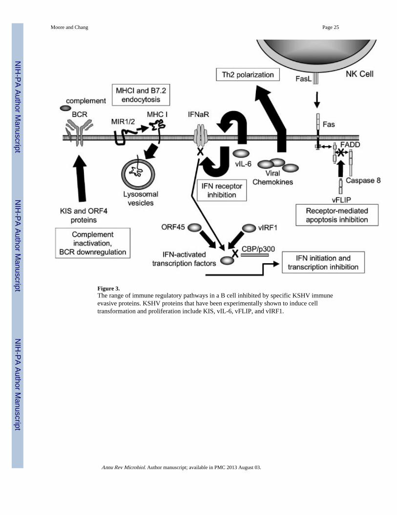
Figure 3.The range of immune regulatory pathways in a B cell inhibited by specific KSHV immuneevasive proteins. KSHV proteins that have been experimentally shown to induce celltransformation and proliferation include KIS, vIL-6, vFLIP, and vIRF1.
Moore and Chang Page 25
Annu Rev Microbiol. Author manuscript; available in PMC 2013 August 03.
NIH
-PA Author Manuscript
NIH
-PA Author Manuscript
NIH
-PA Author Manuscript
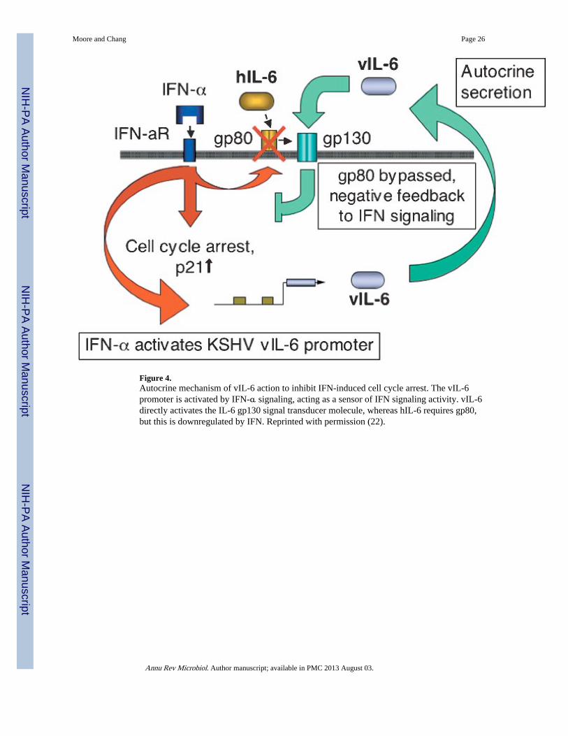
Figure 4.Autocrine mechanism of vIL-6 action to inhibit IFN-induced cell cycle arrest. The vIL-6promoter is activated by IFN-α signaling, acting as a sensor of IFN signaling activity. vIL-6directly activates the IL-6 gp130 signal transducer molecule, whereas hIL-6 requires gp80,but this is downregulated by IFN. Reprinted with permission (22).
Moore and Chang Page 26
Annu Rev Microbiol. Author manuscript; available in PMC 2013 August 03.
NIH
-PA Author Manuscript
NIH
-PA Author Manuscript
NIH
-PA Author Manuscript

Figure 5.Examples of immune activation of tumor suppressor checkpoints. Both cell cycle arrest andapoptosis can be initiated by immune recognition of a virus-infected cell. KSHV vIL-6,vIRF1, and vFLIP, which are putative oncogenes, inhibit these responses.
Moore and Chang Page 27
Annu Rev Microbiol. Author manuscript; available in PMC 2013 August 03.
NIH
-PA Author Manuscript
NIH
-PA Author Manuscript
NIH
-PA Author Manuscript
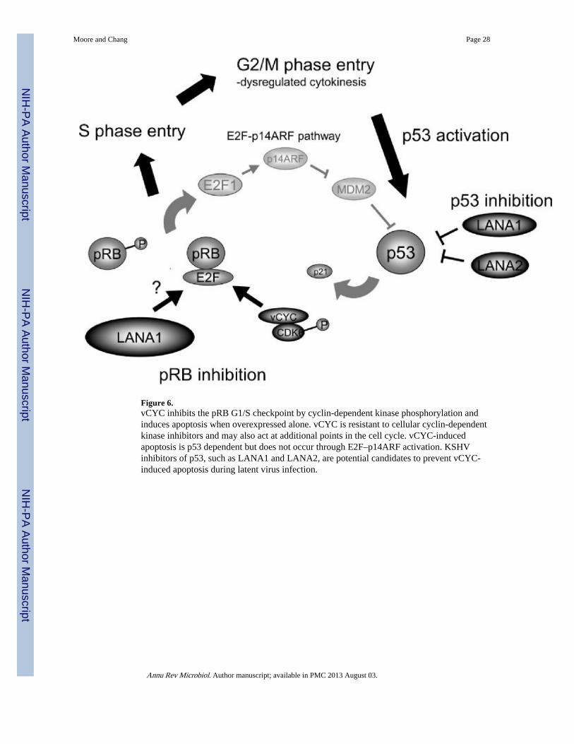
Figure 6.vCYC inhibits the pRB G1/S checkpoint by cyclin-dependent kinase phosphorylation andinduces apoptosis when overexpressed alone. vCYC is resistant to cellular cyclin-dependentkinase inhibitors and may also act at additional points in the cell cycle. vCYC-inducedapoptosis is p53 dependent but does not occur through E2F–p14ARF activation. KSHVinhibitors of p53, such as LANA1 and LANA2, are potential candidates to prevent vCYC-induced apoptosis during latent virus infection.
Moore and Chang Page 28
Annu Rev Microbiol. Author manuscript; available in PMC 2013 August 03.
NIH
-PA Author Manuscript
NIH
-PA Author Manuscript
NIH
-PA Author Manuscript

NIH
-PA Author Manuscript
NIH
-PA Author Manuscript
NIH
-PA Author Manuscript
Moore and Chang Page 29
TABLE 1
KSHV genes with immune modulatory functions
KSHVprotein
KSHVgene Features and functions
Related proteins fromother viruses
vKCP ORF4 Homolog of cellular complement control regulators; inhibits C3 deposition on cell surface
HVS-ORF4, HSVl-gC, Vaccinia-vCP/C21L,Cowpox-IMP, Vaccinia-SPICE
KIS ORFK1 Constitutively active membrane protein containing ITAM motif; interacts with mu chains of B cell receptor complex to block surface transport; transforms rodent fibroblasts and primary T cells; activates Syk signaling pathway; variable ectodomain used for KSHV strain analysis
HVS-STP
MIR1/MIR2 ORFK3/K5 Enhanced endocytosis of surface MHO via ubiquitination; MIR2 also ubiquitinates and downregulates ICAM-1 and B7.2
MHV 68-K3, CMV-US2
vCCL-2(vMIP-II)
ORFK4 Binds to CCR3 and induces angiogenesis in ovo; induces eosinophil and Th2 chemotaxis; CCR8 and vGPCR antagonist.
Molluscum-MC148
vCCL-3(vMIP-III)
ORFK4.1 Agonist for CCR4 and induces Th2 chemotaxis; induces angiogenesis in ovo; protein expressed in KS lesions (by Western blot)
vCCL-1(vMIP-I)
ORFK6 Bids CCR5 and induces angiogenesis in ovo; CCR8 agonist; induces Th2 chemotaxis
Molluscum-MC148
vIRF-1 ORFK9 Inhibits IFN signaling; binds CBP/p300; transforms rodent fibroblasts; protein expressed in MCD and PEL cell lines, not detectable in KS or PEL
EBV-EBNA2, Adeno-EIA, EBV-EBNA2,Adeno-EIA, SV40-LT, HTLV-Tax
vFLIP ORFK13 Homolog of cellular apoptosis inhibitor (FLIP); transcribed on major latency transcripts (LT)1 and LT2; posttranscriptional regulation of protein expression in vivo
HSV-ORF71
ORF45 ORF45 Binds to and inhibits IRF7 nuclear translocation Vaccinia-E3L
Annu Rev Microbiol. Author manuscript; available in PMC 2013 August 03.

NIH
-PA Author Manuscript
NIH
-PA Author Manuscript
NIH
-PA Author Manuscript
Moore and Chang Page 30
TABLE 2
KSHV genes with antiapoptotic functions
KSHVprotein
KSHVgene Features and functions
Functionalhomologsof other viruses
vFLIP ORF K13 Homolog of cellular apoptosis inhibitor (FLIP); transcribed on major latency transcripts (LT)1 and LT2; posttranscriptional regulation of protein expression in vivo
HVS ORF71
vBCL-2 ORF16 Inhibits bax-mediated apoptosis EBV BHRF-1
vIL-6 ORF K2 B cell proliferation factor; prevents apoptosis in IL-6-dependent plasmacytoma cells; binds to gp130 subunit of the IL-6 receptor, does not require IL6Ra subunit; induces angiogenesis and VEGF in rodent fibroblasts; protein expressed only in subpopulation of infected hematopoietic cells
EBV gp350/220 and LMP1 induces cellular IL-6
LANA2 ORF10.5 Inhibits p53-mediated transcription in hematopoietic cells
Mechanism of inhibition unknown
vIAP ORF K7 Homolog of survivin-DeltaEx3; glycoprotein which can inhibit apoptosis; binds to Bcl2 via BH2 domain; inhibits caspase-3 activity via BIR motif
vIRF-1 ORF K9 Inhibits p53; binds CBP/p300; protein expressed in MCD and PEL cell lines
Adeno E1A, Adeno E1A, SV40 LT, EBV EBNA2, HTLV Tax
Annu Rev Microbiol. Author manuscript; available in PMC 2013 August 03.

NIH
-PA Author Manuscript
NIH
-PA Author Manuscript
NIH
-PA Author Manuscript
Moore and Chang Page 31
TABLE 3
KSHV genes with mitogenic or cell cycle regulatory functions
KSHVprotein
KSHVgene Features and functions
Related proteinsfrom other viruses
LANA1 ORF73 Inhibits p53; binds to ori-P and maintains viral episome; protein expressed in all KSHV-infected cells; major latency associated nuclear antigen used in serologic assays
SV40-LT, HPV-E6, Adeno-E1B, EBV-LMP1; SV40-LT, EBV-EBNA-1
vCYC ORF72 Associates with cdk6 to phosphorylate RB and histone H1; resistant to inhibition by p16, p21, and p27; phosphorylates and downregulates p27; latent mRNA expressed in cell cycle–dependent manner
SV40 LT, HPV-E7, Adeno-E1A
vGPCR ORF74 Chemokine receptor homolog; induces transformation, angiogenesis, and VEGF in rodent fibroblast; signals through heterotrimeric G proteins, JNK/Sapk, p38/HOG; signaling inhibited by IFN γ –inducible protein-10 (IP-10) and vMIP-II
HVS-ECRF3; EBV-EBNA-2 activates Burkitt lymphoma receptor-2 (BRL-2/EBI1), a cellular GPCR
vIL-6 ORF K2 Prevents IFN induction of p21waf1 EBV gp350/220 and LMP1 induces cellular IL-6
vIRF-1 ORF K9 Inhibits IFN signaling; binds CBP/p300; transforms rodent fibroblasts; protein expressed in MCD and PEL cell lines, not detectable in KS or PEL
Adeno-E1A; Adeno-E1A, SV40 LT, EBV-EBNA2
K-bZIP/ RAP
ORF K8 Inhibits G2/M transition EBV-BZLF1
Annu Rev Microbiol. Author manuscript; available in PMC 2013 August 03.


