Embed Size (px)
Citation preview
University of Bath
PHD
Nicotinic acetylcholine receptors from the parasitic nematode ascaris suum
Williamson, Sally
Award date:2008
Awarding institution:University of Bath
Link to publication
General rightsCopyright and moral rights for the publications made accessible in the public portal are retained by the authors and/or other copyright ownersand it is a condition of accessing publications that users recognise and abide by the legal requirements associated with these rights.
• Users may download and print one copy of any publication from the public portal for the purpose of private study or research. • You may not further distribute the material or use it for any profit-making activity or commercial gain • You may freely distribute the URL identifying the publication in the public portal ?
Take down policyIf you believe that this document breaches copyright please contact us providing details, and we will remove access to the work immediatelyand investigate your claim.
Download date: 08. Jun. 2019
NICOTINIC ACETYLCHOLINE RECEPTORS
FROM THE PARASITIC NEMATODE ASCARIS SUUM
Sally Miranda Williamson A thesis submitted for the degree of Doctor of Philosophy
University of Bath Department of Biology and Biochemistry
November 2008
COPYRIGHT
Attention is drawn to the fact that copyright of this thesis rest with its author.
This copy of the thesis has been supplied on condition that anyone who consults
it is understood to recognise that its copyright rests with its author and that no
quotation from the thesis and no information derived from it may be published
without the prior written consent of the author.
This thesis may be made available for consultation within the University
Library and may be photocopied or lent to other libraries for the purposes of
consultation.
…………………………………………….
i
Summary
Nematodes of the genus Ascaris are large gastrointestinal parasites. Ascaris
lumbricoides infects ~1 billion people globally; causing malnutrition and
general morbidity, and can block the gut or bile duct causing fatal
complications. Ascaris suum is a parasite of pigs; in addition to its veterinary
significance, it can occasionally be zoonotic, and is a good model of the human
parasite. One of the main classes of drugs used to treat parasitic nematode
infections are the cholinergic anthelmintics, such as levamisole and pyrantel,
which act as agonists of nicotinic acetylcholine receptors at the nematode
neuromuscular junction. The pharmacology of the native neuromuscular
nAChRs from Ascaris suum has previously been well described, but most
detailed studies of the molecular biology of nematode nAChRs have previously
been confined to the model organism C. elegans. The aim of the work presented
here was therefore to attempt to interpret the pharmacology of the nAChRs of
Ascaris suum in terms of the underlying molecular biology.
The genome of Brugia malayi, a parasite within the same phylogenetic clade as
Ascaris suum, was searched for putative nAChR subunit sequences.The results
of this search showed that parasites from this clade appear to have far fewer
nAChR subunit genes than C. elegans, though three subunits known to be
involved in levamisole sensitivity in C. elegans, unc-38, unc-29 and unc-63,
were present and well conserved. Full-length sequences of unc-38 and unc-29
were then amplified from Ascaris suum, though only a partial sequence of unc-
63 was obtained. An additional, novel nAChR subunit with no C. elegans
homologue was also amplified from Ascaris suum.
Antisera were raised against peptides from Ascaris suum UNC-38, UNC-29 and
UNC-63, and were used to perform indirect immunofluorescent labelling of the
nAChR subunits. All three subunits were shown to be present on the membrane
of muscle cells from Ascaris suum, and UNC-38 and UNC-29 were shown to
clearly co-localise.
Expression of UNC-38 and UNC-29 in Xenopus oocytes yielded functional
receptors which were characterised pharmacologically using two-electrode
voltage-clamp electrophysiology. Both UNC-38 and UNC-29 were necessary to
generate a functional receptor, which was sensitive to the agonists levamisole,
ii
acetylcholine and nicotine, and the antagonist mecamylamine. Further
experiments demonstrated that the pharmacology of the receptors could be
changed by altering the stoichiometry, using different ratios of unc-38:unc-29
RNA. Addition of more unc-38 produced a receptor more sensitive to nicotine
and oxantel, but less sensitive to levamisole and insensitive to pyrantel.
Addition of more unc-29 produced a receptor more sensitive to levamisole and
pyrantel, but less sensitive to nicotine and insensitive to oxantel. The
pharmacology of the receptors produced in this study by heterologous
expression in oocytes resembled two distinct pharmacological subtypes of
nAChR previously described from native Ascaris suum muscle cells.
iii
Acknowledgements I would like to thank my supervisor Dr. Adrian Wolstenholme for his support
and advice throughout this project. I would also like to thank everyone from lab
0.47, especially Dr. Sam McCavera, and everyone from Professor David
Sattelle’s lab at the MRC Functional Genomics Unit, Oxford, for their
assistance with the electrophysiology.
I am also grateful to Professor Mike Doenhoff and Professor Mike Lehane,
formerly of the University of Wales, Bangor, for nurturing my interest in
parasites; and to Dr. Simon Webster who first taught me about the pathology
associated with ectopic tapeworm infections. Thanks also to my fiancé Kieren
Tennant for his endless patience and enthusiasm, and to my best friend Adam
Bamforth, my daughter Rhiannon Williamson and my mother Julie Edwards for
keeping life interesting.
This research was funded by a BBSRC-CASE award with Pfizer Animal
Health.
iv
Table of Contents
Summary ...........................................................................................................i
Acknowledgements.........................................................................................iv
Chapter 1: Introduction.................................................................. 1 1.1 Nematodes.................................................................................................2
1.1.1 The diversity of nematodes.................................................................2
1.1.2 Parasitic nematodes.............................................................................5
1.1.3 Ascaris.................................................................................................6
1.2 Anthelmintics.............................................................................................9
1.2.1 Anthelmintics ......................................................................................9
1.2.2 Resistance .........................................................................................10
1.3 Cys-loop ligand gated ion channels .........................................................12
1.3.1 Diversity and general structure of cys-loop LGICs ..........................12
1.3.2 Nicotinic acetylcholine receptors......................................................13
1.3.3 nAChR subunit composition and stoichiometry ...............................15
1.3.4 The ligand binding domain of nAChRs ............................................16
1.3.5 The nAChR ion channel....................................................................19
1.4 Nematode nAChRs ..................................................................................21
1.4.1 Pharmacology of nematode neuromuscular nAChRs .......................21
1.4.2 Molecular biology of nematode nAChRs .........................................24
1.5 Aims and Objectives ................................................................................27
Chapter 2: Methods and Materials.............................................. 28 2.1 Materials ..................................................................................................29
2.1.1 Nematode tissue ................................................................................29
2.1.2 Xenopus laevis oocytes. ....................................................................29
2.1.3 Plasmids ............................................................................................30
2.1.4 Enzymes............................................................................................30
2.1.5 Chemicals and reagants.....................................................................32
2.2 Bioinformatics..........................................................................................32
2.2.1 Reciprocal BLAST searches .............................................................32
2.2.2 Sequence alignments and tree construction ......................................32
v
2.3 PCR and Cloning Methods ......................................................................33
2.3.1 Primer Design ...................................................................................33
2.3.2 RNA extraction .................................................................................33
2.3.3 Reverse Transcription .......................................................................34
2.3.4 PCR...................................................................................................35
2.2.5 5’ and 3’ RACE ...............................................................................36
2.2.6 Agarose gel electrophoresis ..............................................................36
2.3.7 Gel extraction....................................................................................37
2.3.8 Ligation reactions..............................................................................37
2.3.9 Preparation of competent cells..........................................................37
2.3.10 Transformation of competent cells .................................................38
2.3.11 Plasmid extraction and digest .........................................................38
2.3.12 Proof -reading and sequence deposition .........................................39
2.3.13 Sub-cloning into expression vector pT7TS.....................................40
2.3.14 Real-time PCR ................................................................................41
2.4 Antibody Methods ...................................................................................42
2.4.1 Production of antisera .......................................................................43
2.4.2 Affinity purification of antisera ........................................................43
2.4.3 ELISA ...............................................................................................44
2.4.4 Preparation of muscle cells for immunofluorescence.......................44
2.4.5 Immunofluorescence.........................................................................45
2.5 Xenopus oocyte expression and electrophysiology..................................46
2.5.1 Linearisation of plasmid DNA..........................................................46
2.5.2 In vitro transcription .........................................................................46
2.5.5 Microinjection of oocytes .................................................................47
2.5.6 Two-electrode voltage clamp............................................................48
Chapter 3: Bioinformatics ............................................................ 50 3.1 Introduction..............................................................................................51
3.2 The nAChR gene families of Brugia malayi and Trichinella spiralis.....53
3.3 The use of consensus nAChR sequences for primer design ....................57
3.4 Discussion ................................................................................................60
Chapter 4: Amplification of nAChR subunits from Ascaris suum......................................................................................................... 63
vi
4.1 Introduction..............................................................................................64
4.2 Potential components of the levamisole-sensitive nAChR from Ascaris
suum ...............................................................................................................65
4.3 A novel nAChR subunit from Ascaris suum............................................71
4.4 Real-time PCR .........................................................................................74
4.5 Discussion ................................................................................................77
Chapter 5: Immunohistochemical localisation of Ascaris suum UNC-38, UNC-29 and UNC-63..................................................... 79
5.1 Introduction: Ascaris muscle cells and the nematode neuromuscular
junction ..........................................................................................................80
5.2 Choice of peptides for antisera production and ELISA results................82
5.3 Indirect Immunofluorescence ..................................................................87
5.4 Discussion ................................................................................................93
Chapter 6: Electrophysiology....................................................... 95 6.1 Introduction..............................................................................................96
6.2: Initial experiments to co-express Ascaris unc-38 and unc-29 ................98
6.3 Experiments using oocytes injected with 1:5 and 5:1 ratios of Ascaris
unc-38 and unc29 cRNA..............................................................................103
6.4 Further pharmacological studies on oocytes injected with 1:5 and 5:1
ratios of Ascaris unc-38 and unc29 cRNA ..................................................109
6.5 Discussion ..............................................................................................117
Chapter 7: Conclusions............................................................... 120
References..................................................................................... 124
Appendix i: Primers .................................................................... 138
Appendix ii: Abbreviations......................................................... 141
vii
Figures Figure 1.1: The phylogeny of nematodes (Mitreva et al. 2005) ..........................4 Figure 1.2: The lifecycle of Ascaris spp. .............................................................8 Figure 1.3: Structure of the nicotinic acetylcholine receptor (Brown et al. 2006)............................................................................................................................14 Figure 1.4: The ligand binding region at the αδ interface of the vertebrate muscle nAChR (Changeux & Taly 2008)..........................................................18 Figure 1.5: The ion channel of the nAChR (Corringer et al. 2000)...................20 Figure 1.6: The nAChR subtypes present on Ascaris muscle cells (Qian et al. 2006) ..................................................................................................................23 Figure 1.7: The nAChR gene family of C. elegans (Brown et al. 2006) ...........26 Figure 3.1: Identification of putative homologues of C. elegans nAChR genes in parasite genomes............................................................................................52 Figure 3.2: The nAChR gene families of Brugia malayi and Trichinella spiralis............................................................................................................................56 Figure 3.3: ClustalW alignments of the nAChR subunit sequences UNC-29 and UNC-63 from C. elegans and B. malayi. ...........................................................58 Figure 4.1: Reverse transcription, amplification, and 5' and 3' RACE..............67 Figure 4.2: The sequences of Ascaris suum UNC-38, UNC-29 and UNC-63...68 Figure 4.3: Alignment of the novel nAChR subunit ACR-26 from Ascaris suum with the human neuronal α7 subunit..................................................................73 Figure 4.4: Amplification plots showing the relative abundance of Ascaris suum unc-38, unc-29 and unc-63 in tissue taken from a)head and b)body wall. ........75 Figure 5.1: The structure of the muscle cell and neuromuscular junction from Ascaris suum (from Rosenbluth 1965) .............................................................81 Figure 5.2: ProteAn analysis of the sequences of Ascaris suum UNC-38, UNC-29 and UNC-63 ..................................................................................................83 Figure 5.3: ELISA results for antibodies against Ascaris suum UNC-38, UNC-29 and UNC-63 ..................................................................................................85 Figure 5.4: Confocol microscope images of negative controls for indirect immunofluorescent labelling of Ascaris suum muscle cells ..............................88 Figure 5.5: Indirect immunofluorescent labelling of Ascaris suum UNC-38....89 Figure 5.6: Co-localisation of Ascaris suum UNC-38 and UNC-29 on muscle cell membranes ..................................................................................................91 Figure 5.7: Indirect immunofluorescent labelling of Ascaris suum UNC-63....92 Figure 6.1: Both Ascaris unc-38 and unc-29 are required to form a functional nAChR ...............................................................................................................99 Figure 6.2: The nAChRs composed of UNC-38 and UNC-29 are sensitive to the agonists ACh, levamisole and nicotine, and can be reversibly blocked by the antagonist mecamylamine................................................................................100 Figure 6.3: Dose-response relationships for the agonists levamisole, ACh and nicotine applied to oocytes injected with equimolar concentrations of Ascaris unc-38 and unc-29 cRNA ................................................................................102 Figure 6.4: Dose-response relationships for levamisole and nicotine for oocytes injected with a 1:5 ratio of unc-38:unc-29 cRNA............................................105 Figure 6.5: Dose-response relationships for levamisole and nicotine for oocytes injected with a 5:1 ratio of unc-38:unc-29 cRNA............................................107
viii
Figure 6.6: Dose-response relationships for pyrantel and oxantel applied to oocytes injected with a 1:5 ratio of Ascaris unc-38:unc-29 cRNA. ................111 Figure 6.7: Dose-response relationship for oxantel and pyrantel applied to oocytes injected with a 5:1 ratio of Ascaris unc-38:unc-29 cRNA. ................113 Figure 6.8: Sigmoidal curves fitted to separate agonist and antagonist components of dose-response data for pyrantel and oxantel ...........................115
Tables
Table 2.1: Composition of Ascaris ringer solution...........................................29 Table 2.2: Plasmids............................................................................................30 Table 2.3: Enzymes............................................................................................30 Table 2.4: Immunohistochemistry buffers and solutions...................................42 Table 2.5: Solutions used for oocyte incubation and electrophysiology ...........49 Table 3.1: Summary of nAChR subunit sequences from in Brugia malayi and Trichinella spiralis: a comparison with C. elegans ...........................................55 Table 4.1: Similarity between Ascaris suum UNC-38 and the 5 most similar nAChR subunits returned by a search of the GenBank database using the TBLASTN algorithm.........................................................................................70 Table 4.2: Similarity between Ascaris suum UNC-29 and the 5 most similar nAChR subunits returned by a search of the GenBank database using the TBLASTN algorithm. ........................................................................................70 Table 4.3: Similarity between Ascaris suum UNC-63 and the 5 most similar nAChR subunits returned by a search of the GenBank database using the TBLASTN algorithm. ........................................................................................71 Table 4.4: Similarity between Ascaris suum ACR-26 and the 5 most similar nAChR subunits returned by a search of the GenBank database using the TBLASTN algorithm.........................................................................................72 Table 4.5: Critical threshold (Ct) values for real-time PCR amplification of reference gene β-tubulin and nAChR subunits unc-38, unc-29 and unc-63 from Ascaris suum head and body wall tissues ..........................................................76 Table 6.1: EC50 values and Hill slope values for oocytes injected with equimolar concentrations of Ascaris unc-38 and unc-29 cRNA ......................102 Table 6.2: EC50 and Hill slope values for ooctes injectedwith 1:5 and 5:1 ratios of Ascaris unc-38:unc-29 RNA .......................................................................108 Table 6.3: EC50, IC50 and Hill slope values for oxantel and pyrantel on oocytes injected with a 1:5 and 5:1 ratio of Ascaris unc-38:unc-29 RNA....................116
ix

x

Chapter 1: Introduction
1
1.1 Nematodes
1.1.1 The diversity of nematodes The phylum nematoda is incredibly diverse, with estimates of number of
species ranging from tens of thousands to several million (Lambshead 1993).
Nematodes inhabit a diverse array of trophic niches, ranging from free-living
bacteriovores found in soil and marine sediments, to parasites of plants,
invertebrates and vertebrate animals (Baldwin et al., 2004; Blaxter et al., 1998;
Lambshead, 1993). Despite superficial morphological similarities and a lack of
fossil evidence, molecular analysis has greatly increased our understanding of
nematode phylogeny (Blaxter et al., 1998; Conway-Morris, 1981). Analysis of
small subunit ribosomal RNA sequences has produced a robust phylogenetic
framework for the nematodes, resolved into five monophyletic clades (Blaxter
et al., 1998; Dorris et al., 1999). A modified version of this accepted phylogeny
is shown in Figure 1.1. Interestingly, the SSU rRNA phylogeny supports
multiple origins of parasitism in nematodes: plant parasitism evolved 3 times,
and animal parasitism arose independantly at least 6 times during the course of
nematode evolution (Blaxter et al., 1998; Dorris et al., 1999).
Clade I contains nematodes occupying a range of ecological niches: free-living,
plant parasitic, the insect parasitic Mermithida, and the vertebrate parasite
genera Trichinella and Trichuris (Blaxter et al., 1998; Dorris et al., 1999).
Clade II is less well studied, but is known to contain both bacteriovores and
plant parasites (Blaxter et al., 1998). Clade III is composed almost exclusively
of animal parasites, of both invertebrates and vertebrates (Nadler et al., 2007).
Basal members of this clade are often parasites of the arthropod gut, suggesting
that perhaps this pre-adapted clade III nematodes to later become parasites of
vertebrates (Dorris et al., 1999). Clade VI contains notable plant parasites such
as Meloidogyne and Globodera; also represented in this clade are the animal-
parasitic Strongyloides (Blaxter et al., 1998; Dorris et al., 1999; Mitreva et al.,
2005). Clade V contains soil and sediment-dwelling groups alongside several
vertebrate-parasitic taxa of medical and veterinary importance (Blaxter et al.,
2
1998). The most notable member of clade V is perhaps the free-living model
organism Caenorhabditis elegans, the first metazoan animal for which a
complete genome sequence was published (Blaxter, 1998;
C.elegans_Sequencing_Consortium, 1998).
Since the completion of the C. elegans genome sequencing project, knowledge
of the molecular biology of a diverse assemblage of nematodes has expanded:
genome sequencing projects have been undertaken for other Caenorhabditis
species, the plant parasite Meloidogyne, and the animal parasites Haemonchus
contortus, Brugia malayi and Trichinella spiralis (Blaxter, 2003; Ghedin et al.,
2007; Mitreva et al., 2005; Mitreva & Jasmer, 2006). For numerous other
nematodes, EST datasets are being generated (Parkinson et al., 2004; Wylie et
al., 2004).
In summary, the complexity of the phylum Nematoda is revealed both by
disparate ecology and genetic diversity, and recent advances in gene sequencing
and analysis have begun to reveal the similarities, and differences, between
members of this diverse phylum.
3
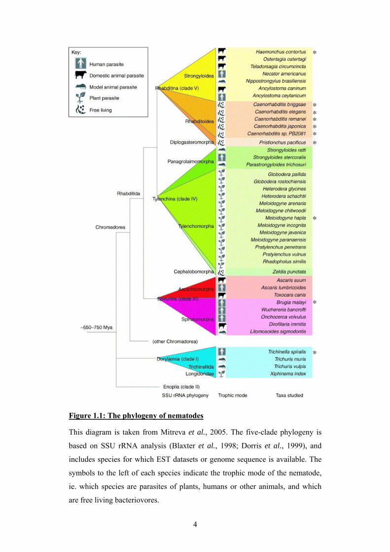
Figure 1.1: The phylogeny of nematodes This diagram is taken from Mitreva et al., 2005. The five-clade phylogeny is
based on SSU rRNA analysis (Blaxter et al., 1998; Dorris et al., 1999), and
includes species for which EST datasets or genome sequence is available. The
symbols to the left of each species indicate the trophic mode of the nematode,
ie. which species are parasites of plants, humans or other animals, and which
are free living bacteriovores.
4
1.1.2 Parasitic nematodes Many nematode parasites of vertebrates are of medical and veterinary
importance as they adversely affect human health and animal welfare. There is
palaentological evidence that the association between vertebrates and their
nematode parasites is ancient; coprolites have revealed that dinosaurs in the
Cretaceous period carried ascarid parasites whose eggs are very similar to those
produced by extant Ascaris species (Poinar & Boucot, 2006). The nematode
parasites of humans also have a long-established history, with pinworm eggs
(Enterobius vermicularis) being found in human faecal material dating from
10,000 years ago (Fry & Moore, 1969). The entire range of nematode parasites
are too numerous to describe, but summarised here are notable examples of the
main types of parasitic nematodes which have an impact on human health.
Soil transmitted helminths are gastrointestinal parasites prevalent in many
tropical countries with poor sanitation; infection rates are estimated at
approximately a billion people worldwide (Bethony et al., 2006; de Silva et al.,
2003; WHO, 2005). This diverse collection of parasites includes the large
roundworm Ascaris lumbricoides, the whipworm Trichuris trichiura, and the
hookworms Ancylostoma duodenale and Necator americanus; many individuals
in developing countries may be chronically infected with all three types
(Bethony et al., 2006). Although complications from such infections may
directly cause mortality, the more widespread and insidious impact of these
parasites is from long-term morbidity (Bethony et al., 2006; WHO, 2005).
Anaemia, malnutrition, and impaired growth and cognitive development are
common sequelae of heavy helminth infections of children living in poverty
(Bethony et al., 2006; WHO, 2005). In addition to this, diseases such as
HIV/AIDS and malaria are exacerbated by co-infection with nematode parasites
(Fincham et al., 2003; Le Hesran et al., 2004).
More obvious pathologies are produced by infection with a sub-group of clade
III nematodes, the filariae. These parasites are transmitted as microfilarial
larvae by intermediate arthropod hosts, usually biting mosquitoes or blackflies,
and develop within the tissues of their human or animal host. Lymphatic
5
filariasis, caused mainly by Brugia malayi and Wuchereria bancrofti, occurs in
~80 countries and infects ~120 million people (Wynd et al., 2007).
Complications of this disease cause blocked and swollen lymphatic ducts,
leading to fluid accumulation and grotesque swelling of the lower limbs or male
genitalia (Partono, 1987). Infection with Onchocerca volvulus, the causative
agent of "river blindness", causes visual impairment in approximately half a
million people in sub-Saharan Africa (WHO, 1995). This symptom is caused by
the inflammatory immune response to microfilarial larvae entering the
conjunctiva and cornea of the eye (Hall & Pearlman, 1999). The microfilariae
also migrate through the skin, causing chronic and disfiguring dermatitis (Kale,
1998).
Infection with parasitic nematodes overall causes a human disease burden
second only to malaria in terms of disability-adjusted life years (Mathers et al.,
2007). In terms of the number of years through which people must live with the
debilitating effects of chronic nematode infections, and indeed the number of
years of life lost through acute infections, diseases caused by parasitic
nematodes rival malaria and schistosomiasis in terms of their impact on human
health in tropical countries (Mathers et al., 2007).
1.1.3 Ascaris Nematodes of the genus Ascaris are large gastrointestinal parasites of pigs (A.
suum) and humans (A. lumbricoides). Related ascarid parasites also occur in
other mammals, birds and reptiles, and fossilised parasite eggs indicate that
dinosaurs were also infected with an ascarid parasite, Ascarites (Poinar &
Boucot, 2006).
Ascaris lumbricoides is transmitted to humans from soil contaminated with
infected faecal material, so has a high prevalence in areas with poor sanitation.
It is estimated that approximately 1000 million people globally are infected
with Ascaris (Bethony et al., 2006). After ingestion of embryonated eggs, the
larvae hatch in the gut, penetrate the intestinal mucosa and migrate via the liver
to the lungs. The pulmonary stage of Ascaris infection may give rise to
associated symptoms such as pulmonary eosinophilia (Chitkara & Krishna,
2006). The larvae ascend the respiratory tract and are swallowed, then
6
subsequently develop to adults within the intestines. The lifecycle of Ascaris is
summarised in Figure 1.2.
Considerable morbidity is associated with Ascaris infection, particularly in
children; malnutrition leading to impaired development is a common
consequence (Crompton, 1986; Hlaing, 1993). Pathologies arising from Ascaris
infection include blockage of the pancreatic and biliary duct, and where a heavy
worm burden is present intestinal occlusion may occur; such complications
result in hospitalisation and may cause fatalities (Bahu, 2001; de Silva, 1997).
The pig parasite Ascaris suum is considered a good experimental model of
human ascariasis (Boes & Helwigh, 2000). There is also evidence of humans
infected with Ascaris suum; microsatellite markers suggest that Ascaris
populations have switched between porcine and human hosts several times
during their recent evolution (Criscione et al., 2007). Ascariasis may primarily
be a zoonosis in communities with no endemic Ascaris lumbricoides and which
have close contact with swine harbouring Ascaris suum (Criscione et al., 2007;
Nejsum et al., 2005). In addition to its role in human disease, Ascaris suum is
studied as a helminth of veterinary significance. Infection of farmed pigs lowers
productivity and has considerable economic consequences; losses of $155
million per year due to Ascaris suum infections in US swine herds have been
reported (Stewart & Hale, 1988).
7
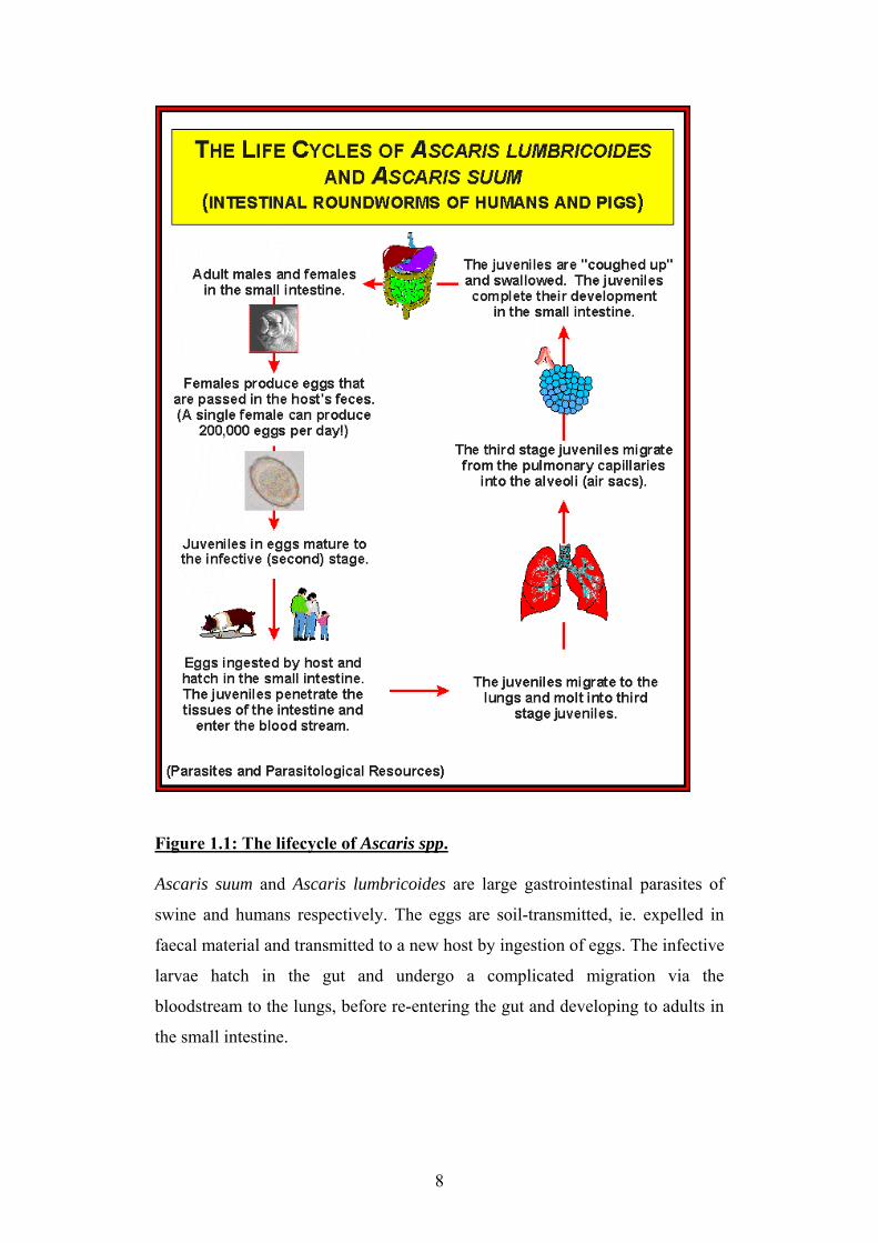
Figure 1.1: The lifecycle of Ascaris spp. Ascaris suum and Ascaris lumbricoides are large gastrointestinal parasites of
swine and humans respectively. The eggs are soil-transmitted, ie. expelled in
faecal material and transmitted to a new host by ingestion of eggs. The infective
larvae hatch in the gut and undergo a complicated migration via the
bloodstream to the lungs, before re-entering the gut and developing to adults in
the small intestine.
8
1.2 Anthelmintics
1.2.1 Anthelmintics Since the 1970s, 3 classes of broad-spectrum anthelmintics have been used to
treat nematode infestations of veterinary and medical significance: the
benzimidazoles, the macrocyclic lactones (eg. avermectins), and the
imidazothiazoles (eg. levamisole) (Burg et al., 1979; Thienpont et al., 1966).
Benzimidazoles were originally used as antifungal agents; their mode of action
is to prevent polymerisation of β-tubulin in the pathogenic organism (Allen &
Gottlieb, 1970; Davidse & Flach, 1978). However, despite the continuing use of
these compounds, resistance rapidly emerged in both fungi and nematodes,
necessitating the development of alternative anthelmintics (Hastie &
Georgopoulos, 1971; Theodorides et al., 1970).
The macrocyclic lactones and the imidathiazoles both target cys-loop ligand-
gated ion channels of nematodes; they cause paralysis of the worm, which in
the case of gastrointestinal nematodes leads to their expulsion from the gut. The
macrocyclic lactones also have the advantage of acting on ion channels of
arthropods, so are also effective against ectoparasites and insect disease vectors
(Goudie et al., 1993; Mellin et al., 1983). The targets of the avermectins are
post-synaptic ligand-gated chloride channels; initially this was thought to be
mainly GABA receptors, but later studies discovered that the glutamate-gated
chloride channels are the main target in nematodes (Holden-Dye & Walker,
1990; Wolstenholme & Rogers, 2005; Yates et al., 2003; Yates &
Wolstenholme, 2004). The avermectins bind with high affinity to nematode
chloride channels and open them irreversibly, leading to an effective inhibition
of muscle contraction and therefore a flaccid paralysis of the parasite
(Wolstenholme & Rogers, 2005).
The imidazothiazoles target excitatory nAChRs at the nematode neuromuscular
junction, causing prolonged muscle contraction and a spastic paralysis in the
nematode (Aceves et al., 1970). The effects of levamisole on nematode
neuromuscular nAChRs are discussed further in section 1.4.1.
More recently, two new classes of anthelmintic drugs have been developed,
with the advantage of being effective against nematodes resistant to the other
classes of anthelmintic: these are the cyclo-octodepsipeptide emodepside, and
9
the aminoacetonitrile derivatives (AADs) (Harder et al., 2003; Kaminsky et al.,
2008). Emodepside has been shown to bind to latrophilin and inhibit pharyngeal
muscle in C. elegans, but its main mode of action is thought to be via calcium-
activated potassium channels, where it acts to cause paralysis (Guest et al.,
2007; Harder et al., 2005; Harder et al., 2003). The AADs, like the
imidazothiazoles, target nematode nAChRs, but in the case of AADs, a
different subtype of nAChR is targeted. This nAChR contains the subunit ACR-
23, belonging to a poorly-characterised but nematode-specific group of
neuronal nAChR subunits (Kaminsky et al., 2008; Mongan et al., 1998; Yassin
et al., 2001). The AAD compounds have been shown to be effective against
levamisole-resistant nematodes, confirming that they target a different nAChR
subtype (Kaminsky, Ducray et al. 2008).
1.2.2 Resistance Drug resistance in nematodes is defined as occuring when a greater frequency
of individuals in the parasite population are no longer affected by the usual
efficacious dose of anthelmintic due to an inherited characteristic (Prichard et
al., 1980). The main mechanisms by which resistance develops are changes in
the drug target site or upregulation of mechanisms which detoxify or remove
the drug (Wolstenholme et al., 2004). In a veterinary context, widespread and
indiscriminate use of anthelmintics has led to selection for resistance alleles in
the nematode parasites of a range of hosts. Drug resistance is a widespread
problem in sheep and other small ruminants, and also occurs in cattle and
horses (Jackson & Coop, 2000; Kuzmina & Kharchenko, 2008; Loveridge et
al., 2003). Resistance has developed to all 3 of the most widely used
anthelmintics, and in some cases, parasite populations may be resistant to all 3
drug types, presenting serious problems for parasite control and animal welfare
(Bartley et al., 2004; Coles et al., 1996). In a medical context, there have been
reports of potential drug resistance in the causative agent of river blindness,
Onchocerca volvulus (Awadzi et al., 2004). With the advent of mass
chemotherapy programmes to treat parasitic diseases of humans, it is likely that
more cases of drug resistance will emerge.
10

Benzimidazole resistance is probably the best understood in terms of the
underlying mechanisms; its history as an antifungal agent, to which fungi could
become resistant, set a precedent for this (Allen & Gottlieb, 1970; Theodorides
et al., 1970). Mutations in β-tubulin which prevent the drug binding have been
found in benzimidazole resistant Haemonchus contortus, Cooperia onchophera,
and Teladorsagia circumcincta; this is therefore likely to be a major resistance
mechanism (Kwa et al., 1994; Silvestre & Cabaret, 2002). However, it is worth
noting that additional non-specific mechanisms such as upregulation of P-
glycoprotein efflux pumps could also contribute to benzimidazole resistance;
this is also likely to be a factor in certain cases of avermectin resistance
(Blackhall et al., 1998; Blackhall et al., 2008; Kerboeuf et al., 2003).
Aside from the involvement of P-glycoproteins, no widespread mechanism of
resistance to the avermectins has been clearly demonstrated. Mutations in
glutamate-gated chloride channel genes confer reduced ivermectin sensitivity to
C. elegans and to parasite glutamate-gated chloride channel genes expressed in
the laboratory, but evidence of such mutations in field populations of parasites
remains scarce (Dent et al., 2000; McCavera et al., 2007).
Levamisole resistance in parasites remains equally hard to define on a
molecular level, although numerous studies have shown that mutating certain
nAChR subunit genes in C. elegans affect levamisole sensitivity (Culetto et al.,
2004; Fleming et al., 1997; Lewis et al., 1980). It has been shown that loss of a
levamisole-sensitive nAChR subtype occurs in levamisole-resistant
Oesophagostomum dentatum, but the genetic changes behind this remain
unknown (Martin & Robertson, 2000; Robertson et al., 1999). A paucity of
information regarding the molecular biology of levamisole-receptors in
parasites has hindered the search for any potential target site mutations
associated with resistance.
11
1.3 Cys-loop ligand gated ion channels
1.3.1 Diversity and general structure of cys-loop LGICs The cys-loop ligand gated ion channels are pentameric proteins; each subunit is
composed of an N-terminal ligand binding domain, and 4 transmembrane
regions which form the ion channel (Sine & Engel, 2006). The term "cys-loop"
refers to a characteristic closed loop motif formed by a disulphide bond
between 2 cysteine residues in the N-terminal part of the protein. The native
ligand is usually a neurotransmitter, and ligand binding causes a conformational
change which induces the channel to open (Lester et al., 2004). The second
transmembrane regions line the ion channel pore, and charged residues in this
region confer cation or anion selectivity (Galzi et al., 1992). The structure of
the ligand binding domain has been well described, due to a resolved crystal
structure of the acetylcholine binding protein from Lymnaea stagnalis (Brejc et
al., 2002). This protein is soluble and lacks transmembrane domains, but has
the pentameric conformation and ligand binding loops characteristic of the cys-
loop LGICs (Brejc et al., 2002).
The cys-loop LGIC superfamily is notable for its diversity, and for the
remarkable degree of conservation of the key motifs among different
organisms. Identification of LGICs from prokaryotes suggest these are the
evolutionary precursors of eukaryotic cys-loop LGICs; the original function of
these channels is thought to be chemotaxis (Tasneem et al., 2005). A molecular
clock approach (ie. analysis of mutation accumulation rates) dates the origin of
the LGICs at approximately 2500 million years ago (Ortells & Lunt, 1995).
Certain members of the cys-loop LGIC superfamily are conserved in both
vertebrates and invertebrates, which implies they were present in the last
bilaterian ancestor (Dent, 2006; Ortells & Lunt, 1995). These include the
acetylcholine and serotonin gated cation channels, and the GABA and glycine
gated anion channels (Ortells & Lunt, 1995). Subsequent to the evolutionary
divergence of the different animal phyla, the cys-loop LGIC families underwent
lineage-specific expansion events and losses resulting in the diversity of
12
channel genes present in extant taxa (Dent, 2006). Perhaps surprisingly, the
vertebrates retain only a relatively small cys-loop LGIC family (Dent, 2006). In
contrast, the nematode C. elegans possesses the largest described cys-loop
LGIC gene family, with 102 members; a large sub-set of these genes form an
"orphan group" for which no ligand or function has yet been identified (Brown
et al., 2006; Jones & Sattelle, 2008). Invertebrates also possess acetylcholine
and serotonin gated anion channels, glutamate-gated cys-loop ligand-gated
anion channels (distinct from the vertebrate tetrameric glutamate receptors) and
also excitatory GABA-gated cation channels (Beg & Jorgensen, 2003; Putrenko
et al., 2005; Ranganathan et al., 2000; Yates & Wolstenholme, 2004). It has
been proposed that animals with a very simple nervous system (such as the 302
neurons of C. elegans) may utilize a large cys-loop LGIC family to enhance
their neurotransmission repertoire; more complex neural circuits may reduce
the need for such molecular diversity in signalling proteins (Littleton &
Ganetzky, 2000).
1.3.2 Nicotinic acetylcholine receptors
Nicotinic acetylcholine receptors are the most well characterised of the cys-loop
ligand-gated ion channels. Like all members of the LGICs, the nAChRs are
pentameric, composed of five subunits arranged around the ion channel pore.
Evidence for the pentameric nature of nAChRs was first provided by studies of
material from the electric ray Torpedo marmorata, the electric organs of which
are a rich source of nAChRs (Brisson & Unwin, 1985). Tubular crystals of
nAChRs from Torpedo have provided the basis for many structural studies of
nAChRs (Unwin, 1998; Unwin, 2005). Each subunit of the nAChR is composed
of 3 distinct domains. The N-terminal region of the protein is extracellular and
protrudes into the synaptic cleft; this is the ligand-binding domain, and also
bears the cys-loop characteristic of this LGIC family (Arias, 1997). The
transmembrane domain forms the ion channel and is composed of 4 membrane-
spanning components, TM1-TM4, believed to be hydrophobic alpha-helices.
The ligand-binding and ion-channel domains and their role in nAChR function
are described in more detail in sections 1.3.3 and 1.3.4. The cytoplasmic region,
13
particularly the long cytoplasmic loop between TM3 and TM4, bears numerous
phosphorylation sites which play a role in modulating receptor sensitivity
(Huganir & Greengard, 1990). The cytoplasmic region also interacts with
proteins involved in nAChR clustering (Phillips et al., 1991).
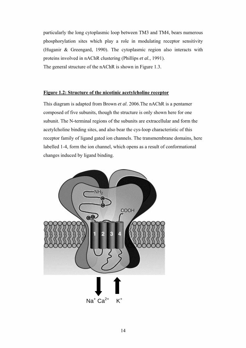
The general structure of the nAChR is shown in Figure 1.3.
Figure 1.2: Structure of the nicotinic acetylcholine receptor This diagram is adapted from Brown et al. 2006.The nAChR is a pentamer
composed of five subunits, though the structure is only shown here for one
subunit. The N-terminal regions of the subunits are extracellular and form the
acetylcholine binding sites, and also bear the cys-loop characteristic of this
receptor family of ligand gated ion channels. The transmembrane domains, here
labelled 1-4, form the ion channel, which opens as a result of conformational
changes induced by ligand binding.
Na+ Ca2+ K+
14
1.3.3 nAChR subunit composition and stoichiometry
Vertebrates possess 17 nAChR subunits, including α1-α10 (though α8 appears
to be specific to birds, and α5 lacks the loop C tyrosine residue characteristic of
α subunits described below, and in section 1.3.4), β1-β4, γ, δ, and ε (Millar &
Gotti, 2008). The distinction between α and non-α subunits was initially based
on the presence of two adjacent cysteine residues involved in ligand binding
first described from the Torpedo α1 subunit cDNA (Noda et al., 1982). It
should be noted that the presence of these two residues alone is not necessarily
an indication that a subunit can form the principal components of a ligand
binding site, and later work suggests that the definition of an α subunit should
also include an important tyrosine residue, ie. the defining motif would be Y-X-
CC (Abramson & Taylor, 1990). The role of these and other amino acids in
nAChR ligand binding is described further in section 1.3.3.
Nicotinic acetylcholine receptors may be homomeric, composed of only one
type of subunit, or heteromeric, composed of 2-4 different subunit types .The
nAChR of the Torpedo electric organ has the same subunit composition as most
vertebrate muscle nAChRs, being composed of two α1 subunits, and also a γ, δ
and β1 subunit (Einarson et al., 1982; Raftery et al., 1976), though in
mammalian muscle an ε subunit replaces γ soon after birth (Witzemann et al.,
1989). Homomeric receptors include human neuronal α7, and the avian subunit
α8 (Couturier et al., 1990; Gerzanich et al., 1994; Peng et al., 1994). Most other
receptors assemble from two or three different subunit types, and variations in
subunit composition and stoichiometry can produce a diverse array of receptor
subtypes with different calcium permeabilities and pharmacological properties
(Millar & Gotti, 2008). An example of how changes to receptor stoichiometry
can generate diversity in the pharmacology and channel properties of nAChRs
is provided by α4β2 receptors, which are abundant in the mammalian brain.
Receptors with the composition (α4)2(β2)3 have different agonist affinity than
receptors with the (α4)3(β2)2 stoichiometry, and addition of a third type of
subunit to give (for example) (α4)2(β2)2α5 further modifies receptor properties
(Kuryatov et al., 2008; Moroni & Bermudez, 2006; Moroni et al., 2006).
15
1.3.4 The ligand binding domain of nAChRs Evidence that the extracellular N-terminal domain bears the nAChR ligand
binding sites has been provided by photoaffinity labelling or radioisotope
labelling of the binding sites using appropriately modified agonist or
competitive antagonist compounds (Changeux et al., 1967; Chiara & Cohen,
1997). The contribution of the labelled sites to ligand binding has also been
confirmed by mutation of these candidate amino acids and electrophysiological
characterisation of the resulting recombinant receptors (Galzi et al., 1990;
Mishina et al., 1985; Sine et al., 1994). Though it is beyond the scope of this
introduction to describe in detail the extensive body of work contributing to our
knowledge of nAChR ligand binding, this has been comprehensively reviewed
by Arias (Arias, 1997; Arias, 2000). Presented here is a summary of the nAChR
components believed to make important contributions to agonist and non-
competitive antagonist binding; this is also shown in Figure 1.3.2.
Initial photoaffinity labelling experiments showed that the α subunits were the
primary contributor to ligand binding (Changeux et al., 1967). It has since been
confirmed that several amino acids characteristic of α subunits play a key role
in ligand binding: in the α1 subunit, these are Y190, C192, C193 and Y198,
which comprise ligand binding loop C (Kao & Karlin, 1986; Mishina et al.,
1985; Sine et al., 1994). Two additional loops from the α subunits have also
been found to contribute residues involved in ligand binding: loop A provides
Y93 and loop B provides W149 in the Torpedo α1 subunit; these residues are
also conserved in α7, occuring at positions 92 and 148 (Galzi et al., 1991a;
Galzi et al., 1990; Galzi et al., 1991b). The aromatic groups provided by the
ligand binding residues has been shown to be of particular importance when
binding ligands with quaternary ammonium groups (Sine et al., 1994).
Although the principal components of ligand binding are contributed by α
subunits, additional components involved in ligand binding are provided by the
adjacent subunit, so that the ligand binding site is actually located at the
interface between subunits; in the absence of non-alpha subunits, the muscle α1
subunit does not bind agonist or d-tubocurarine (Blount & Merlie, 1989;
Kurosaki et al., 1987). In homomeric receptors these additional binding motifs
are also present on the α subunit, but in heteromeric receptors they are provided
16
by non-α subunits. These ligand binding regions are termed loops D, E and F;
loop D includes a tryptophan residue at position 55 of the vertebrate muscle γ
subunit or position 57 of the δ subunit which is also conserved in the α7
receptor at position 54, and has been labelled with both d-tubocurarine and
nicotine (Chiara & Cohen, 1997; Chiara et al., 1998; Corringer et al., 1995).
While conserved residues on the complementary binding loops may confer
similarities in agonist and antagonist affinities, variations between subunits in
the amino acids contributing to ligand binding can also lead to pharmacological
differences. In the vertebrate muscle nAChR, the two binding sites in the same
receptor have different properties, with the αγ binding site showing a low
affinity for ACh but a high affinity for d-tubocurarine, and the αδ binding site
having a higher affinity for ACh but a lower affinity for d-tubocurarine (Blount
& Merlie, 1989; Pedersen & Cohen, 1990). An additional example of the
pharmacological properties which may be confered on a receptor by the
complementary binding components is the dependence of neonicotinoid
insecticide sensitivity on residues present in loop D and loop F (Shimomura et
al., 2003).
17
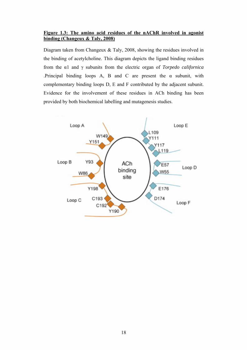
Figure 1.3: The amino acid residues of the nAChR involved in agonist binding (Changeux & Taly, 2008) Diagram taken from Changeux & Taly, 2008, showing the residues involved in
the binding of acetylcholine. This diagram depicts the ligand binding residues
from the α1 and γ subunits from the electric organ of Torpedo californica
.Principal binding loops A, B and C are present the α subunit, with
complementary binding loops D, E and F contributed by the adjacent subunit.
Evidence for the involvement of these residues in ACh binding has been
provided by both biochemical labelling and mutagenesis studies.
18
1.3.5 The nAChR ion channel On binding of ligands to the extracellular domain of the nAChR, the protein
undergoes a conformational change which causes a rotational movement and
consequently opens the ion channel (Sansom, 1995). This has been inferred
from comparison of the structure of the Torpedo nAChR in the open and closed
states, and the conformational change responsible for channel opening has also
been modelled (Taly et al., 2005; Unwin, 1993; Unwin, 1995).
The ion channel domain is formed from the transmembrane regions of the
nAChR, and the second transmembrane region of each subunit forms the pore
of the ion channel (Imoto et al., 1986). There is also evidence that residues at
the N-terminal region of the first transmembrane domain may also contribute to
formation of the channel pore (Zhang & Karlin, 1997). Most of the evidence
supporting the involvement of these regions in ion channel function has been
provided by selective mutation of the amino acids in these regions, then
heterologous expression of the resulting subunits (Akabas et al., 1994; Mishina
et al., 1985). The ion channel of the nAChR is permeable to cations, and this
specificity is conferred by three rings of negatively charged amino acids present
in all the constituent subunits (Corringer et al., 2000; Imoto et al., 1988). These
rings are positioned at both ends of the second membrane-spanning region, and
also at an intermediate position which is within the ion channel pore but below
the hydrophobic alpha helix of the transmembrane region (Imoto et al., 1988).
Figure 1.3.3 shows the positions of the rings of negatively charged residues in
the homomeric α7 nAChR. It has also been demonstrated that these few amino
acids are not only necessary but also sufficient to confer cation specificity to an
ion channel; mutations at just these three charged amino acids in each subunit
can convert a cation channel to an anion channel (Galzi et al., 1992). However,
the channel conductance and relative calcium permeability of an nAChR are
dependant on the properties of additional amino acids in the channel pore
region (Fucile, 2004; Villarroel & Sakmann, 1992).
19
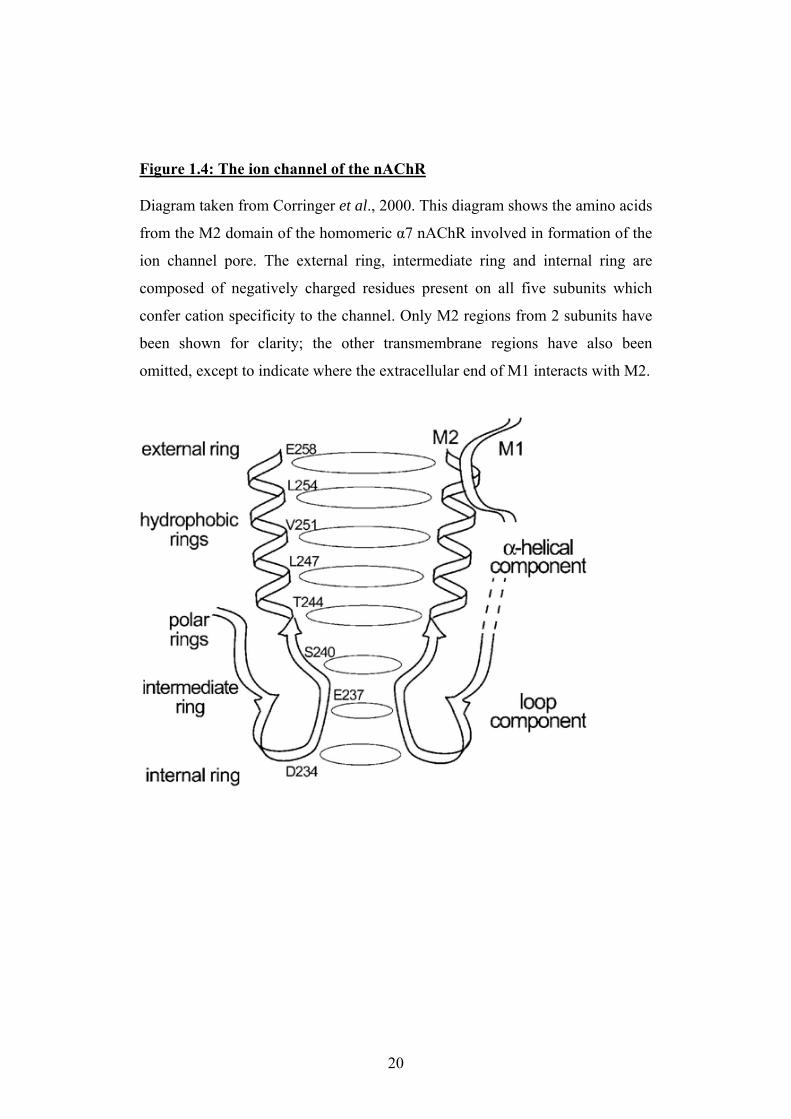
Figure 1.4: The ion channel of the nAChR Diagram taken from Corringer et al., 2000. This diagram shows the amino acids
from the M2 domain of the homomeric α7 nAChR involved in formation of the
ion channel pore. The external ring, intermediate ring and internal ring are
composed of negatively charged residues present on all five subunits which
confer cation specificity to the channel. Only M2 regions from 2 subunits have
been shown for clarity; the other transmembrane regions have also been
omitted, except to indicate where the extracellular end of M1 interacts with M2.
20
1.4 Nematode nAChRs
1.4.1 Pharmacology of nematode neuromuscular nAChRs Much of the research investigating nematode nAChR pharmacology has been
carried out using muscle tissue from Ascaris suum; the large size of the worm
allows muscle strips and individual muscle cells to be removed with relative
ease. The earliest studies were based primarily on muscle contraction assays in
response to application of agonist and antagonist drugs (Baldwin & Moyle,
1949). Ascaris muscle was shown to contract in response to both acetylcholine
and nicotine, and inhibition of ACh-induced contractions could be
accomplished by application of tubocurarine and mecamylamine; this provided
direct evidence for a nAChR involved in Ascaris muscle contractions (Baldwin
& Moyle, 1949; Natoff, 1969; Rozhkova et al., 1980).
Electrophysiological techniques have also been widely used to study the
pharmacology of native Ascaris nAChRs; these recordings utilize extrasynaptic
receptors present on the muscle bag cell (Colquhoun et al., 1991; Martin, 1993).
Such studies confirmed the primarily nicotinic nature of the ACh response:
ACh and nicotine produced dose-dependant depolarisation of the muscle cell,
while muscarinic agonists showed little effect (Colquhoun et al., 1991).
Mecamylamine was again shown to be a potent antagonist at <1µM
concentration, whereas atropine was 100-fold less effective at reducing the ACh
response (Colquhoun et al., 1991). Attempts to further characterise the Ascaris
nAChRs using antagonists selective for different vertebrate nAChR subtypes
proved difficult; for example curare and pancuronium had similar efficacy as
antagonists of Ascaris nAChRs, though these compounds have very different
potencies as antagonists of vertebrate neuromuscular nAChRs (Buckett, 1968;
Colquhoun et al., 1991).
The use of cholinergic anthelmintic compounds such as morantel, oxantel,
pyrantel and levamisole have been perhaps more useful in elucidating the
pharmacology of nematode nAChRs. These compounds are potent agonists of
nematode neuromuscular nAChRs; depolarisation of muscle cells causing
21
muscle contraction and spastic paralysis underlies their efficacy as anthelmintic
agents (Aceves et al., 1970). In addition to this, their selectivity for nematode
rather than vertebrate nAChRs produces minimal toxicity when used as
therapeutic drugs for the treatment of livestock and humans (Aubry et al., 1970;
Eyre, 1970). Electrophysiological recordings of Ascaris muscle cell responses
to the cholinergic anthelminics were first performed by Harrow and Gration
(1985). This demonstrated dose-dependant increases in cation conductance in
response to levamisole, with the EC50 for levamisole being 5-10 fold lower than
for ACh (dependant on method of drug application). Pyrantel and morantel
were shown to be even more potent agonists at low concentrations (10nM-
10µM) but gave a bell-shaped dose response curve, indicating either
desensitization or open channel block at high concentrations (Harrow &
Gration, 1985).
Patch-clamp techniques and single-channel recordings have confirmed the
mode of action of the cholinergic anthelmintics, and allowed different nAChR
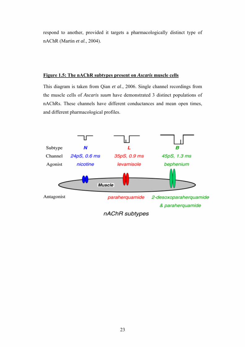
subtypes present on Ascaris muscle to be characterised; 3 nAChR subtypes with
different conductances and mean open times have been described (Qian et al.,
2006; Robertson & Martin, 1993; Robertson et al., 1994). These can be
distinguished pharmacologically by their relative sensitivities to selective
agonists and antagonists, and their properties are summarised in Figure 1.4. The
large conductance channel is the B-subtype, most sensitive to bephenium, and
can be selectively antagonised using 2-deoxy-paraherquamide. The small
conductance channel is the N-subtype, sensitive to agonists nicotine,
methyridine and oxantel, and relatively insensitive to antagonist
paraherquamide. The channel of medium conductance is the L-subtype
receptor, activated by the cholinergic anthelmintics levamisole and pyrantel,
and sensitive to the antagonist paraherquamide (Levandoski et al., 2005; Martin
et al., 2003; Martin et al., 2004; Qian et al., 2006; Robertson et al., 2002). It
has been reported that levamisole and pyrantel resistance in the pig parasite
Oesophagostomum dentatum may result from loss of the relevant receptor
subtype (Robertson et al., 1999; Robertson et al., 2000). The finding that
oxantel is selective for a different nAChR subtype than levamisole and pyrantel
has important implications for the therapeutic use of these compounds; it is
likely that nematodes resistant to one type of cholinergic anthelmintic may still
22
respond to another, provided it targets a pharmacologically distinct type of
nAChR (Martin et al., 2004).
Figure 1.5: The nAChR subtypes present on Ascaris muscle cells This diagram is taken from Qian et al., 2006. Single channel recordings from
the muscle cells of Ascaris suum have demonstrated 3 distinct populations of
nAChRs. These channels have different conductances and mean open times,
and different pharmacological profiles.
23
1.4.2 Molecular biology of nematode nAChRs Although the parasite Ascaris suum has been the main focus of studies on
nematode nAChR pharmacology, the majority of work on the molecular
biology of nematode nAChRs has been confined to C. elegans. The main
reasons for this are the highly complete and well annotated nature of the C.
elegans genome sequence, and the ease with which mutants can be made and
maintained in the laboratory.
Interest in C. elegans nAChR genes began when it was discovered that certain
levamisole-resistant C. elegans mutants had mutations in genes encoding
nAChR subunits (Lewis et al., 1980). Since then, the nAChR gene family has
been investigated in depth, and it has been found that C. elegans has the largest
known nAChR gene family with ~30 subunits (Brown et al., 2006; Jones &
Sattelle, 2004). A tree of sequence similarity for the C. elegans nAChR gene
family is shown in Figure 1.4.2. The nAChR genes of C. elegans can be divided
into several sub-groups, based on sequence similarity. The acr-16 group is
named after the best characterised member of this group, acr-16, which encodes
a homomeric, levamisole-insensitive receptor present at the C. elegans
neuromuscular junction (Mongan et al., 2002; Raymond et al., 2000;
Touroutine et al., 2005). The deg-3 group is nematode-specific and believed to
encode mainly neuronal receptors; this group is of interest as it contains acr-23,
the product of which is targeted by the new AAD anthelmintic compounds
(Kaminsky et al., 2008). The acr-8 group is less well characterised, but contains
lev-8 (also called acr-13), a nAChR subunit gene which is known to confer
reduced sensitivity to levamisole when mutated (Towers et al., 2005).
The groups of most relevance to the work presented here are the unc-38 group
and the unc-29 group. The unc-38 group contains unc-38 and unc-63, which
encode alpha subunits of the levamisole-sensitive nAChR at the C. elegans
neuromuscular junction (Culetto et al., 2004; Fleming et al., 1997). The unc-29
group contains unc-29 and lev-1, which encode non-alpha subunits present in
the same receptor type (Ballivet et al., 1996; Fleming et al., 1997). Mutations in
these subunits all reduce levamisole sensitivity, and heterologous expression of
24
unc-38, unc-29 and lev-1 produces a levamsiole-sensitive nAChR (Fleming et
al., 1997).
Prior to the start of the work presented in this thesis, it had been shown that
several parasitic nematodes had mRNA transcripts encoding putative
homologues of unc-38 (Ajuh & Egwang, 1994; Hoekstra et al., 1997; Wiley et
al., 1996). A review of potential nAChR sequences present in parasitic
nematode EST libraries has yielded further putative homologues of C. elegans
nAChR subunit genes; however, the localisation and functional significance of
such gene products remains almost entirely unknown (Brown et al., 2006). A
more recent study which identified and amplified several nAChR genes from
the canine hookworm Ancylostoma caninum has provided the first evidence of
changes in nAChR subunit expression in an anthelmintic resistant population of
parasites. McCarthy and colleagues have recently demonstrated significantly
lower levels of mRNA encoding the nAChR subunits unc-38, unc-29 and unc-
63 in hookworms which were highly resistant to pyrantel (Kopp et al., 2008).
This provided the first direct evidence for the role of these nAChR subunits in
determining cholinergic anthelmintic sensitivity in a parasitic species.
25
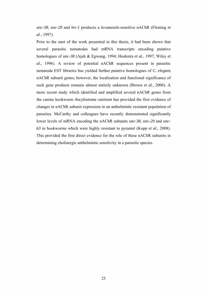
Figure 1.6: The nAChR gene family of C. elegans This diagram is taken from Brown et al. 2006. The cys-loop ligand-gated ion-
channel gene family of C. elegans contains ~100 genes, of which ~30 encode
putative nAChRs. The nAChRs are here shown grouped by sequence similarity
(coloured boxes), and have been aligned with the vertebrate nAChR sequences.
Also shown are other cys-loop LGIC sequences from C. elegans, many of
which are "orphans" for which no ligand has yet been identified.
26
1.5 Aims and Objectives The pharmacology of the native nAChRs present in muscle membrane from
Ascaris suum has been well described, but most work on the molecular biology
of nematode nAChRs has been performed using the model nematode C. elegans
(Jones & Sattelle, 2004; Qian et al., 2006). The few studies which have
investigated the molecular biology of nAChRs in parasites have utilised species
in the same phylogenetic clade as C. elegans (Kopp et al., 2008).
The aim of this project was to identify the nAChR subunits from Ascaris suum
which formed the levamisole-sensitive nAChR, in order to understand the
described pharmacology of the native nAChR in terms of the underlying
molecular biology. The initial stage of the project aimed to identify which
nAChR subunits were likely to be present by searching the genome of a related
nematode, Brugia malayi, then to amplify subunits with a putative role in the
levamisole receptor from Ascaris suum.
The next objective was to raise antisera for immunofluorescent labelling, in
order to ascertain whether the nAChR subunits were present on the Ascaris
suum muscle membrane. The final aim of the project was to produce
recombinant receptors in a heterologous expression system using the Ascaris
nAChR subunit cDNA, to allow the pharmacology of the nAChRs to be
characterised.
27

Chapter 2: Methods and Materials
28
2.1 Materials
2.1.1 Nematode tissue The nematode tissue used in the initial stages of this project was kindly
supplied by Professor Richard Martin and Dr. Alan Robertson of Iowa State
University Veterinary College. Heads and muscle strips from freshly collected
Ascaris suum adults were sent to the University of Bath packaged in dry ice.
Upon arrival at the laboratory, the nematode tissue was stored at -80˚C to
prevent RNA degradation.
Live specimens of Ascaris suum, and Ascaris suum eggs, were kindly supplied
by Emma Kidd and Professor Aaron Maule of Queen's University Belfast.
These were sent to the University of Bath in Ascaris Ringer Solution (ARS) at
ambient temperature. Upon arrival in the laboratory, eggs were aliquoted into
2ml cryo-vial tubes and snap frozen then stored in liquid nitrogen. Live adult
Ascaris suum were maintained in the laboratory for 2-3 days in a 5 litre plastic
beaker containing 2-3 litres of ARS, kept in a 37˚C incubator with daily
changes of solution.
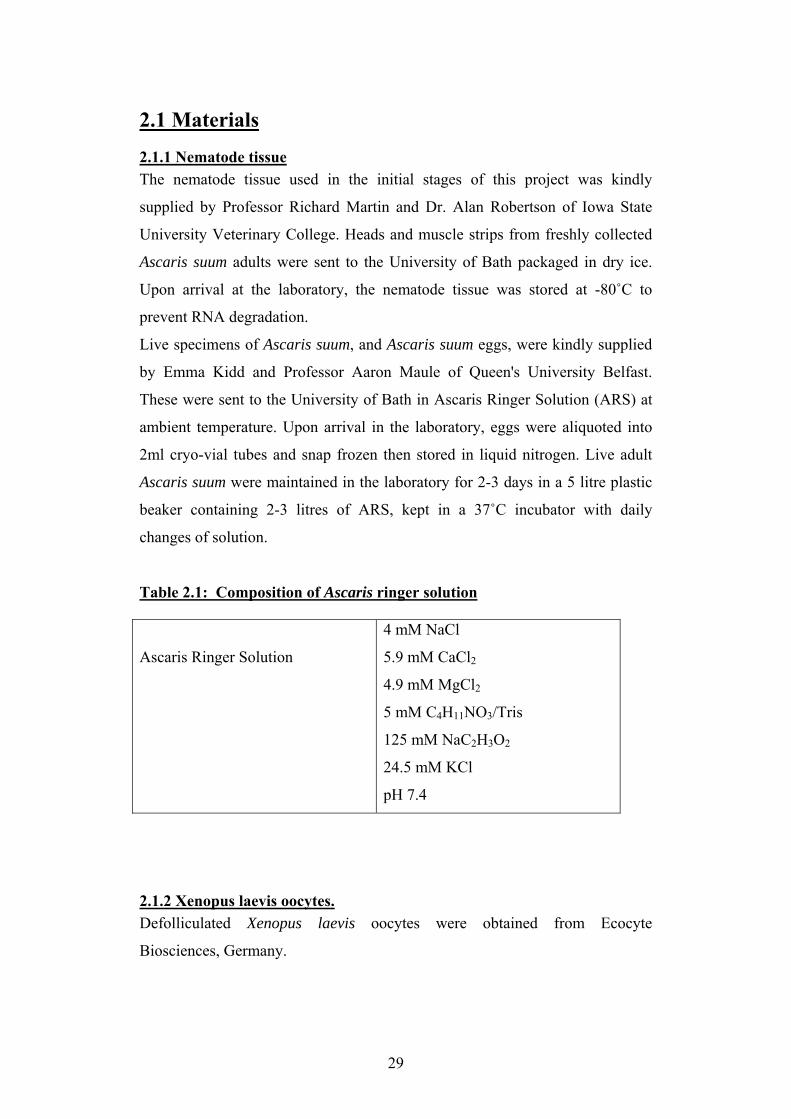
Table 2.1: Composition of Ascaris ringer solution
Ascaris Ringer Solution
4 mM NaCl
5.9 mM CaCl2
4.9 mM MgCl2
5 mM C4H11NO3/Tris
125 mM NaC2H3O2
24.5 mM KCl
pH 7.4
2.1.2 Xenopus laevis oocytes. Defolliculated Xenopus laevis oocytes were obtained from Ecocyte
Biosciences, Germany.
29
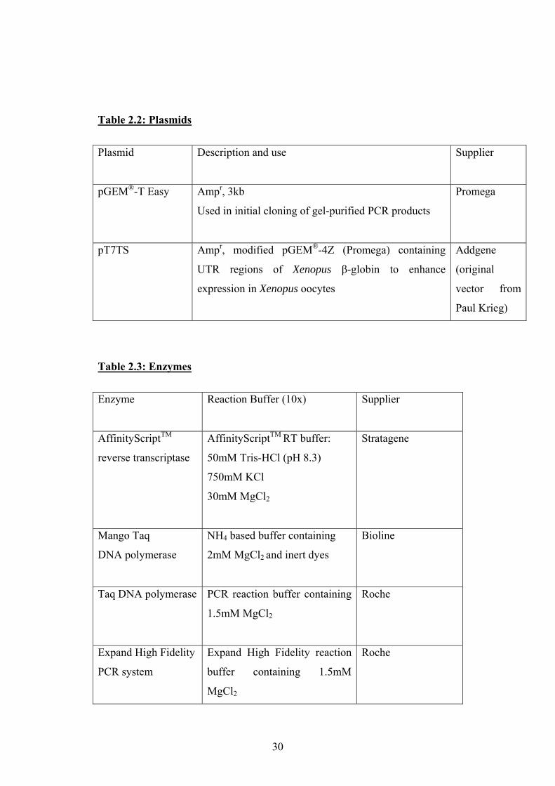
Table 2.2: Plasmids
Plasmid
Description and use Supplier
pGEM®-T Easy Ampr, 3kb
Used in initial cloning of gel-purified PCR products
Promega
pT7TS Ampr, modified pGEM®-4Z (Promega) containing
UTR regions of Xenopus β-globin to enhance
expression in Xenopus oocytes
Addgene
(original
vector from
Paul Krieg)
Table 2.3: Enzymes
Enzyme
Reaction Buffer (10x) Supplier
AffinityScriptTM
reverse transcriptase
AffinityScriptTM RT buffer:
50mM Tris-HCl (pH 8.3)
750mM KCl
30mM MgCl2
Stratagene
Mango Taq
DNA polymerase
NH4 based buffer containing
2mM MgCl2 and inert dyes
Bioline
Taq DNA polymerase
PCR reaction buffer containing
1.5mM MgCl2
Roche
Expand High Fidelity
PCR system
Expand High Fidelity reaction
buffer containing 1.5mM
MgCl2
Roche
30
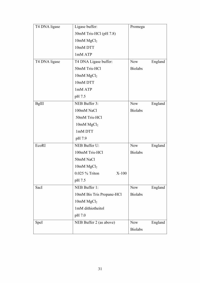
T4 DNA ligase
Ligase buffer:
30mM Tris-HCl (pH 7.8)
10mM MgCl2
10mM DTT
1mM ATP
Promega
T4 DNA ligase
T4 DNA Ligase buffer:
50mM Tris-HCl
10mM MgCl2
10mM DTT
1mM ATP
pH 7.5
New England
Biolabs
BglII
NEB Buffer 3:
100mM NaCl
50mM Tris-HCl
10mM MgCl2
1mM DTT
pH 7.9
New England
Biolabs
EcoRI
NEB Buffer U:
100mM Tris-HCl
50mM NaCl
10mM MgCl2
0.025 % Triton X-100
pH 7.5
New England
Biolabs
SacI
NEB Buffer 1:
10mM Bis Tris Propane-HCl
10mM MgCl2
1mM dithiotheitol
pH 7.0
New England
Biolabs
SpeI NEB Buffer 2 (as above)
New England
Biolabs
31
2.1.5 Chemicals and reagants Unless otherwise specified, all chemicals and general laboratory reagents were
of molecular biology grade, and were purchased from Sigma-Aldrich, UK.
2.2 Bioinformatics
2.2.1 Reciprocal BLAST searches Sequences for the protein-coding regions of all the described C. elegans nAChR
subunit genes (Jones & Sattelle, 2004) were obtained from the NCBI database
(http://www.ncbi.nlm.nih.gov/). These sequences were then used to perform
searches of the genome sequence of Brugia malayi
(http://www.tigr.org/tdb/e2k1/bma1/) using the TBLASTN algorithm. This
algorithm compares translated sequences at the amino acid level rather than
making comparisons at the nucleotide level, so is more likely to identify
homologous sequences from species which might have different codon usage.
The closest match to the C. elegans query sequence found in the Brugia malayi
genome was then used to perform a reciprocal TBLASTN search of the NCBI
sequence database http://www.ncbi.nlm.nih.gov/BLAST). If this search
identified the Brugia malayi sequence as most similar to the C. elegans nAChR
subunit sequence used as the original query, the sequence was identified as
genuinely homologous to this C. elegans nAChR subunit. This search strategy
was then repeated to identify the nAChR subunit genes of another parasitic
nematode, Trichinella spiralis. The Trichinella spiralis genome has been
sequenced by the Washington University Genome Sequencing Centre
(http://genome.wustl.edu/tools/blast/).
2.2.2 Sequence alignments and tree construction When all putative nAChR sequences had been obtained from the parasite
genomes, they were named according to their putative identity and listed along
with their C. elegans homologues in a WordPad text document. This format is
necessary to upload the sequences to the alignment program. The sequences
were uploaded to ClustalX (version 1.83) and a complete alignment was
32
performed; the ClustalX program was also used to produce a boot-strapped
neighbour-joined tree from the alignment. The tree of sequence similarity was
viewed using TreeView (version 1.6.6).
2.3 PCR and Cloning Methods
2.3.1 Primer Design The NCBI database (http://www.ncbi.nih.gov/ ) was searched for nicotinic
acetylcholine receptor subunit sequences from Ascaris spp. One partial
sequence was present in the database, with accession number AJ011382; this
corresponds to an alpha subunit and putative unc-38 homologue. The Ascaris
suum EST database (http://zeldia.cap.ed.ac.uk/ncbi_blast.html ) was searched
for sequences showing similarity to C. elegans nicotinic acetylcholine receptor
subunits. Three such sequences were found, but upon translation and sequence
comparison in all reading frames, two of these (ASC25431 and ASC07050_1)
were found to contain non-coding regions with many stop codons, so were
considered unlikely to encode nAChR subunits. One of the sequences from the
EST database, ASC19452_1, was successfully translated and appeared to
encode a putative nAChR subunit. Specific primers were therefore designed to
amplify the AJ011382 and ASC19452_1 sequences. Further degenerate primers
were designed to amplify additional nAChR subunits using the UNC-29 and
UNC-63 nAChR sequences from C. elegans and Brugia malayi, based on
conserved amino acids. This process is described more fully in section 3.3.
2.3.2 RNA extraction All equipment used for RNA work was rendered RNase free before use.
Pipettes, glassware and surfaces were treated with RNase away reagent (Fluka);
plastic tubes, pipette tips and distilled water were treated with diethyl
pyrocarbonate then autoclaved.
RNA was extracted from the nematode tissue using Trizol® reagent
(Invitrogen). The tissue was ground up using a cooled pestle and mortar and 1-
4ml Trizol® was added (volume depending on sample size). The Trizol® and
nematode tissue were mixed further in a homogeniser, then decanted into 1.5ml
33

microcentrifuge tubes. The homogenate was centrifuged at 12000 x g at 4˚C for
10 minutes to remove insoluble material, particularly protein from muscle strip
samples. The supernatant was transferred to clean eppendorf tubes, then 0.2ml
chloroform was added per 1ml of Trizol® used in the initial extraction. The
tubes were shaken and incubated at ~20˚C for 2 minutes, then centrifuged at
12000 x g at 4˚C for 10 minutes to separate the lower organic phase containing
DNA and protein from the upper aqueous phase containing the RNA. The
aqueous phase was transferred to a clean tube and mixed with isopropanol
(0.5ml per 1ml Trizol® used initially) to precipitate the RNA. After incubation
at ~20˚C for 10 minutes, the precipitated RNA was collected into a pellet by
centrifugation at 12000 x g and 4˚C for 10 minutes. The supernatant was
discarded, and the RNA pellet washed by resuspension in ethanol (1ml ethanol
per 1ml Trizol® used initially). The RNA was again collected by centrifugation,
at 7500 x g and 4˚C for 5 minutes. The RNA was air dried for ~5 minutes, then
dissolved in 10-40µl RNase free water.
The RNA was quantified and the purity checked by measuring the absorbance
of a 1/100 dilution at 280nm and 260nm using a spectrophotometer. An
A260/A280 ratio of ~1.8 indicated a pure RNA preparation. The amount of
RNA was calculated by:
A260 x dilution factor x 40 = amount of RNA in µg/ml.
2.3.3 Reverse Transcription The mRNA was reverse transcribed into cDNA using AffinityScript® reverse
transcriptase (Stratagene). The following components were added to an RNase-
free 0.5ml tube:
3µl oligo(dT) primer (100ng/µl)
~200ng total RNA (~1ng mRNA)
RNase-free water to 14.2µl
The reaction was heated to 65˚C for 5 minutes then slowly cooled to ~20˚C to
allow (Bektesh et al., 1988)primer annealing.
The following components were added to the reaction:
2µl AffinityScript® 10x buffer
2µl DTT
34
1µl AffinityScript® RT (50U/µl)
0.8µl 100mM dNTPs
The reaction incubated at 42˚C for 1 hour. After this time the reaction was
stopped by incubation at 70˚C for 15 minutes.
2.3.4 PCR All initial PCR reactions were carried out using Mango-Taq DNA polymerase
(Bioline); this preparation contains loading dyes for electrophoresis in the
buffer component.
50µl reactions were assembled as follows:
5µl 10x Mango-Taq reaction buffer
2.5µl MgCl2 (50mM)
1µl Mango-Taq DNA polymerase (1U/µl)
1µl dNTP mix (100mM)
1µl cDNA template
1µl forward primer (final concentration10 pmol specific primer, 100 pmol
degenerate primer)
1µl reverse primer (final concentration10 pmol specific primer, 100 pmol
degenerate primer)
37.5µl dd.H2O
PCR conditions used with specific primers were as follows:
2 minutes at 95˚C initial denaturation step
35 cycles of:
1 minute denaturation step at 95˚C
1 minute primer annealing step at 2-5˚C below primer melting temperature
1-2 minutes extension step at 72˚C (~1 minute per kilobase of product)
Then a 5 minute extension step at 72˚C, followed by cooling to 16˚C,
maintained until the programme was stopped and the reactions removed from
the thermal cycler.
PCR conditions used with degenerate primers were as follows:
2 minutes at 95˚C initial denaturation step
35
10 cycles of:
1 minute denaturation step at 95˚C
1 minute primer annealing step at 3˚C above primer melting temperature,
dropping -0.5˚C with each subsequent cycle
1-2 minutes extension step at 72˚C (~1 minute per kilobase of product)
30 cycles of:
1 minute denaturation step at 95˚C
1 minute primer annealing step at 2˚C below lower primer melting temperature
1-2 minutes extension step at 72˚C (~1 minute per kilobase of product)
Then a 5 minute extension step at 72˚C, followed by cooling to 16˚C,
maintained until the programme was stopped and the reactions removed from
the thermal cycler. A list of primers used for PCR is given in appendix i.
When problems arose due to low expression levels or non-specific
amplification, primer pairs internal to the original pair were designed, to carry
out nested PCR and reamplify the products of the original PCR reaction.
2.2.5 5’ and 3’ RACE When a specific partial sequence coding for a nAChR subunit had been
obtained, 5’ and 3’ RACE (rapid amplification of cDNA ends) was carried out
to obtain full length nAChR subunit sequences. 5’ RACE used a forward primer
specific to the splice leader sequence SL1 known to precede most nematode
coding sequences (Bektesh et al., 1988), and a reverse primer complementary to
the nAChR partial sequence. 3’ RACE used a reverse primer complementary to
a known anchor sequence added to the poly-A 3’ end of the mRNA during the
reverse transcription, and a forward primer specific to the partial nAChR
sequence.
2.2.6 Agarose gel electrophoresis 1% w/v gels were made from agarose dissolved in TBE buffer (see appendix i
for buffer compositions). The mixture was microwaved until the agarose had
dissolved, then allowed to cool to ~40˚C before addition of ethidium bromide to
a final concentration of 0.5µg/ml. The gel was poured and allowed to solidify,
36

placed in an electrophoresis tank containing TBE buffer, then 20µl samples of
PCR product and 100bp molecular weight markers (Promega) mixed with 5µl
6x loading dye (Promega) were loaded. A current of 50-100mA was applied,
and separation of the DNA by molecular weight tracked by migration of the
loading dyes down the gel.
2.3.7 Gel extraction If PCR products of the correct size were observed on the agarose gel, they were
carefully excised with a clean razor blade, and the DNA extracted using a gel
extraction kit (Qiagen). The gel slice was dissolved in 3 volumes of buffer to 1
volume of gel by incubation at 50˚C for ~10 minutes. 1 volume of isopropanol
to 1 volume of gel was added, then the mixture was applied to a DNA binding
spin column and centrifuged at 13000 x g for 1 minute. The bound DNA on the
spin column was then washed with 750µl ethanol-based buffer and
subsequently dried by further centrifugations for 1 minute at 13000 x g. 30µl
nuclease-free d.H2O was applied to the spin column, and the eluted DNA
collected by a final centrifugation step at 13000 x g for 1 minute.
2.3.8 Ligation reactions The gel extracted DNA was then ligated into pGEM-T Easy vector (Promega).
Ligation reactions were assembled as follows:
5µl 2x Rapid Ligation Buffer
1µl pGEM-T Easy vector (50ng)
3µl gel-extracted DNA
1µl T4 DNA ligase (3U/µl)
The reactions were incubated for either 1 hour at room temperature or
overnight at 4˚C.
2.3.9 Preparation of competent cells A scraping of cells from a permanent culture of stock XL1 blue strain E.coli
was inoculated into 100ml of LB broth in a 1 litre conical flask and incubated at
37
37˚C with moderate agitation for ~3 hours until the OD600 reached ~0.3
(indicating that the number of viable cells < 108/ml).
The culture was transferred to sterile 50ml tubes and cooled on ice for 10
minutes, then centrifuged at 2700 x g at 4˚C for 10 minutes. The supernatant
was discarded and the tube inverted to remove any remaining media from pellet
of cells. The bacterial cells were resuspended in 30 ml of 80mM MgCl2-20mM
CaCl2 solution which had previously been cooled on ice, then collected by a
further centrifugation at 2700 x g at 4˚C for 10 minutes. After again discarding
the supernatant and draining by inversion of the tube, the cells were
resuspended in 2ml 0.1M CaCl2 (cooled on ice) per 50ml of original culture.
Glycerol was added as a cryoprotectant (15-25% by volume) then the
competent cells were dispensed into 200µl aliquots and snap-frozen in liquid N2
prior to storage at -80˚C.
2.3.10 Transformation of competent cells An aliquot of competent cells was thawed on ice. 200µl cells were added to the
DNA insert/vector product of the ligation reaction, then left on ice for 30
minutes. The cells were heat-shocked at 42˚C for 5 minutes, then added to 1ml
of LB medium and incubated at 37˚C for 1 hour. LB/ampicillin agar plates were
spread with 10µl IPTG (20%w/v) and 50µl X-Gal (2%, Promega), which was
left to absorb at 37˚C for 1 hour. The transformed cells were collected by
centrifugation (13000 x g for 1 minute), resuspended in residual media, then
spread onto the prepared agar plates. The plates were incubated overnight at
37˚C.
Colonies containing recombinant plasmids with the DNA insert of interest were
white in colour; these were used to inoculate LB/ampicillin broth and cultured
overnight at 37˚C.
2.3.11 Plasmid extraction and digest Plasmids were extracted from overnight cultures of transformed E. coli using
the GenElute Plasmid Miniprep Kit (Sigma-Aldrich). 1.5ml of overnight culture
was centrifuged at 13000 x g for 1 minute. The cells were resuspended in 200µl
resuspension solution, then lysed by addition of 200µl alkaline-SDS lysis
38
buffer. After 5 minutes, 350µl high salt neutralisation/binding buffer was
added, then the cell debris removed by centrifugation at 13000 x g for 10
minutes. The supernatant (containing plasmids) was added to a DNA binding
spin column (silica membrane which binds DNA in the presence of high salt
concentrations, prepared according to the manufacturer’s instructions) and
centrifuged for 1 minute at 13000 x g. The DNA bound to the spin column was
then washed by addition of 750µl ethanol-based wash solution and
centrifugation at 13000 x g for 1 minute; an additional centrifugation step was
then performed for the same time and speed to remove all traces of wash
solution from the spin column. The spin column was transferred to a clean
collection tube, and the DNA eluted in 100µl nuclease-free dd.H2O.
To confirm the presence of an insert in the extracted plasmids, a restriction
enzyme digest was performed, using the enzyme EcoR1, as the vector used has
restriction sites for this enzyme at either side of the insert site. Reactions were
assembled as follows:
5µl buffer (x10)
1-5µl plasmid preparation
0.2µl BSA
0.5µl enzyme
dd.H2O to 20µl
The reactions were incubated at 37˚C for 1-4 hours, then 5µl loading buffer was
added and the sample separated by agarose gel electrophoresis. If a DNA insert
band corresponding to the size of the initial PCR product could be seen, the
plasmid preparation was sent for sequencing, using M13 forward and reverse
sequencing primers. A BLAST search was then performed to compare the
resulting sequence with those in the NCBI database
(http://www.ncbi.nih.gov/BLAST), to determine whether the cDNA sequence
which had been amplified and cloned coded for a nicotinic acetylcholine
receptor subunit.
2.3.12 Proof -reading and sequence deposition Following successful amplification and cloning of a complete nAChR subunit
sequence, the PCR reaction was repeated using new specific primers and the
39
Expand High Fidelity PCR system (Roche) to ensure the accuracy of the
sequence amplification. The PCR product was then cloned as described
previously, and plasmid from 3 or 4 separate clones was sequenced to ensure
consensus. The sequences were then deposited in the NCBI database, and
assigned accession numbers.
2.3.13 Sub-cloning into expression vector pT7TS The pT7TS plasmid is an expression vector optimised for Xenopus oocyte
expression by the insertion of the Xenopus β-globin 5' and 3' UTR sequence.
This plasmid originated in the laboratory of Paul Krieg, University of Texas,
and can be obtained from Addgene (plasmid 17091).
The sequences of Ascaris suum unc-38 and unc-29 were analysed using
software from Biology Workbench 3.2 (http://seqtool.sdsc.edu/CGI/BW.cgi) to
ensure that the restriction sites needed for sub-cloning into pT7TS, BglII and
SpeI, were not present in the cDNA sequences. Primers were designed to
amplify the full-length nAChR subunit cDNAs and add the appropriate
restriction sites to the ends of the sequences; a full list of primers used is given
in Appendix i. PCR was performed as described in section 2.2.4, using 1µl of
pGEM-T Easy plasmid containing the nAChR subunit sequence as template.
The PCR product was then cloned into pGEM-T Easy as described in sections
2.2.10 and 2.2.11.
1-3 µg of both the pGEM-T Easy plasmids containing the nAChR cDNAs plus
restriction sites, and the pT7TS vector, were digested using the restriction
enzymes BglII and SpeI as described on the New England Biolabs double-
digest finder website
(http://www.neb.com/nebecomm/DoubleDigestCalculator.asp). Both the
digested vector and nAChR sequences were purified by agarose gel
electrophoresis (section 2.2.6). Both the plasmid and the insert were quantified
by obtaining the A260 value using a spectrophotometer, then they were ligated
together in a 1:3 vector:insert ratio using New England Biolabs T4 DNA ligase
as described in the manufacturer's instructions. Competent cells were
transformed using the product of the ligation reaction, then plated out and
colonies picked and grown overnight as described in section 2.2.10. Plasmids
40
were purified (section 2.2.11) and sequenced to ensure that the pT7TS plasmids
now contained the Ascaris suum unc-38 and unc-29 sequences.
2.3.14 Real-time PCR Primers were designed to amplify ~200bp regions of the N-terminal regions of
the Ascaris suum nAChR subunits unc-38, unc-29 and unc-63. An additional
primer set was designed to amplify a region of similsr length from the Ascaris
suum β-tubulin sequence obtained from the GenBank database (accession
number FE918860). All primers had a similar length, GC content and melting
temperature, and the primer sequences are listed in Appendix i. RNA was
extracted from Ascaris heads and body wall sections as described in section
2.3.2. Reverse transcription reactions were set up as described in section 2.3.3,
ensuring that all reactions contained the same amount of RNA. The primers and
cDNA were first tested by ordinary PCR, using the same reaction conditions
which would be used for real-time PCR. Real-time PCR was performed using a
Stratagene Mx3000P thermal cycler, and amplification plots and analysis were
generated using the accompanying computer software. Each real-time PCR run
included negative control reactions in which the cDNA template had been
omitted, to control for any potential contamination.
Real-time PCR reactions were set up as follows:
25µl iTaqTM SYBR® Green supermix with ROX (Bio-Rad)
22µl nuclease free d.H2O
1µl forward primer
1µl reverse primer
1µl cDNA template
PCR conditions used with real-time PCR primers:
2 minutes at 95˚C initial denaturation step
40 cycles of:
1 minute denaturation step at 95˚C
45 second primer annealing step at 58˚C
1 minute extension step at 72˚
41
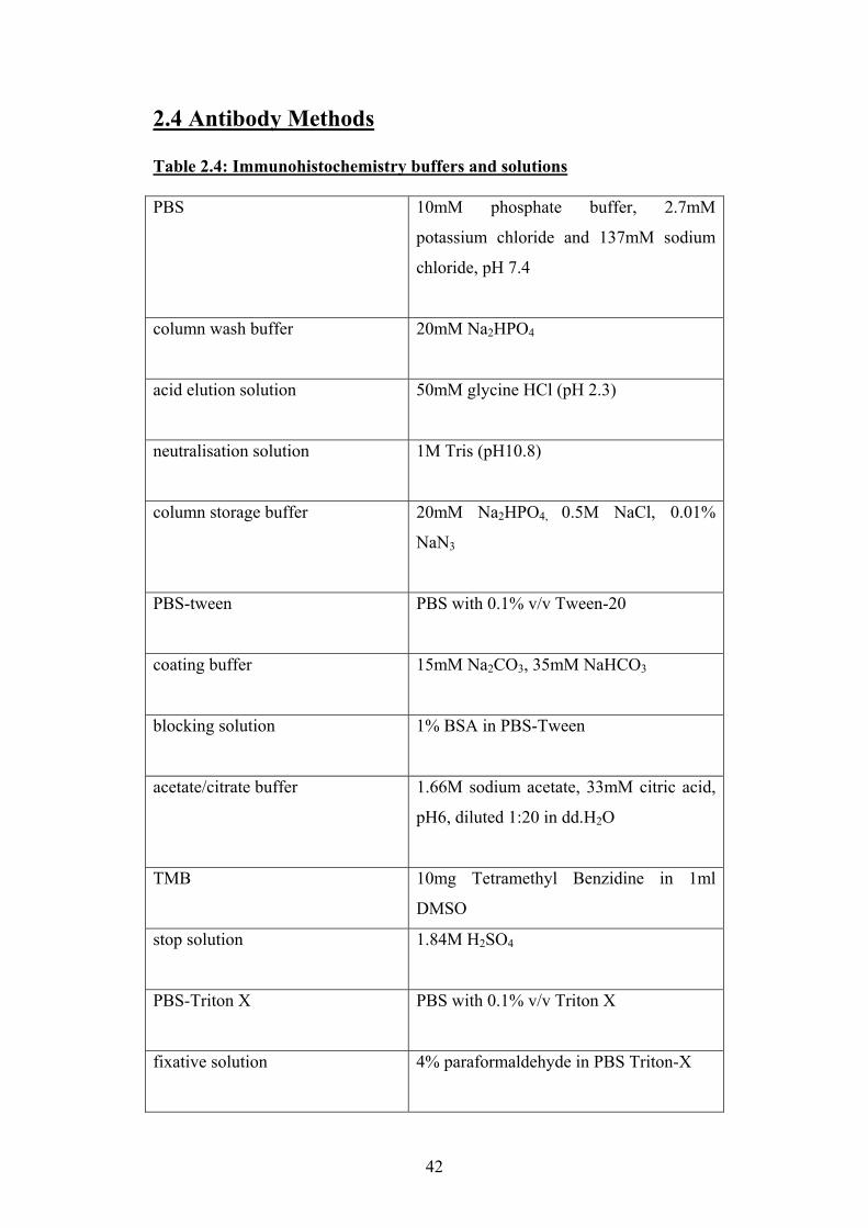
2.4 Antibody Methods Table 2.4: Immunohistochemistry buffers and solutions PBS 10mM phosphate buffer, 2.7mM
potassium chloride and 137mM sodium
chloride, pH 7.4
column wash buffer 20mM Na2HPO4
acid elution solution 50mM glycine HCl (pH 2.3)
neutralisation solution 1M Tris (pH10.8)
column storage buffer 20mM Na2HPO4, 0.5M NaCl, 0.01%
NaN3
PBS-tween PBS with 0.1% v/v Tween-20
coating buffer 15mM Na2CO3, 35mM NaHCO3
blocking solution 1% BSA in PBS-Tween
acetate/citrate buffer 1.66M sodium acetate, 33mM citric acid,
pH6, diluted 1:20 in dd.H2O
TMB 10mg Tetramethyl Benzidine in 1ml
DMSO
stop solution 1.84M H2SO4
PBS-Triton X PBS with 0.1% v/v Triton X
fixative solution 4% paraformaldehyde in PBS Triton-X
42
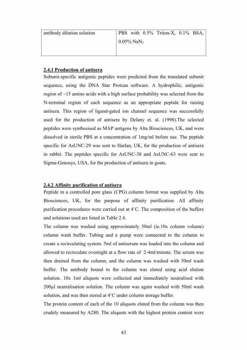
antibody dilution solution PBS with 0.5% Triton-X, 0.1% BSA,
0.05% NaN3
2.4.1 Production of antisera Subunit-specific antigenic peptides were predicted from the translated subunit
sequence, using the DNA Star Protean software. A hydrophilic, antigenic
region of ~15 amino acids with a high surface probability was selected from the
N-terminal region of each sequence as an appropriate peptide for raising
antisera. This region of ligand-gated ion channel sequence was successfully
used for the production of antisera by Delany et. al. (1998).The selected
peptides were synthesised as MAP antigens by Alta Biosciences, UK, and were
dissolved in sterile PBS at a concentration of 1mg/ml before use. The peptide
specific for AsUNC-29 was sent to Harlan, UK, for the production of antisera
in rabbit. The peptides specific for AsUNC-38 and AsUNC-63 were sent to
Sigma-Genosys, USA, for the production of antisera in goats.
2.4.2 Affinity purification of antisera Peptide in a controlled pore glass (CPG) column format was supplied by Alta
Biosciences, UK, for the purpose of affinity purification. All affinity
purification procedures were carried out at 4˚C. The composition of the buffers
and solutions used are listed in Table 2.4.
The column was washed using approximately 50ml (ie.10x column volume)
column wash buffer. Tubing and a pump were connected to the column to
create a recirculating system. 5ml of antiserum was loaded into the column and
allowed to recirculate overnight at a flow rate of 2-4ml/minute. The serum was
then drained from the column, and the column was washed with 50ml wash
buffer. The antibody bound to the column was eluted using acid elution
solution. 10x 1ml aliquots were collected and immediately neutralised with
200µl neutralisation solution. The column was again washed with 50ml wash
solution, and was then stored at 4˚C under column storage buffer.
The protein content of each of the 10 aliquots eluted from the column was then
crudely measured by A280. The aliquots with the highest protein content were
43
assumed to contain most of the eluted antibody, and were pooled then dialysed
overnight against PBS.
2.4.3 ELISA A 96-well plate was coated with 100µl of peptide in each well, diluted to
10µg/ml in coating buffer (see Table 2.4) and incubated at 37˚C for 1 hour, then
at 4˚C overnight. The plate was washed 3x 15 minutes with PBS-Tween, with
rigorous expulsion of liquid from the wells between washes. The wells were
filled with 200µl blocking solution (Table 2.4), and incubated on a shaker for 1
hour. The plate was then washed with PBS-Tween, 3x 15 minute washes.
Primary antisera (antiserum from terminal bleed, affinity purified antibody, and
pre-immune serum) was placed in the first row of wells at 1:100 dilution in PBS
Tween. This was serially diluted in PBS-Tween across the plate. The plate was
incubated on a shaker for 2 hours at cool room temperature, then washed for 3x
15 minutes in PBS-Tween, with rigorous expulsion of liquid from the wells
between washes.
Peroxidase-conjugated secondary antiserum (anti-rabbit or anti-goat IgG HRP)
was diluted 1:2000 in PBS-Tween, and 100µl added to each well. The plate was
incubated on a shaker for 2 hours at cool room temperature, then washed for 3x
15 minutes in PBS-Tween, and for 1x 15 minutes in PBS.
The plate was developed using a solution of 0.5ml TMB and 10µl H2O2 (30%
v/v) in 49.5ml of acetate/citrate buffer. 100µl of this solution was added to each
well, and the reaction observed until the colour developed. The reaction was
then stopped by addition of 50µl stop solution to each well.
A plate reader was used to measure the A450 values for each well. These values
were the plotted as a graph of A450 against dilution.
2.4.4 Preparation of muscle cells for immunofluorescence Muscle cells were loosened from the cuticle using an adaptation of the
collagenase treatment protocol described by Johnson and Stretton (1985). The
worms were pinned in a dissection tray, injected with 500µl 0.5% w/v crude
bacterial collagenase (Sigma) in ARS (100µl at 5 equidistant points along the
worm), and incubated at 37˚C for 1 hour. Worms were then rinsed and the
44
cuticle opened under PBS, allowing the muscle cells to be rinsed out and
collected using a Pasteur pipette. The muscle cells were allowed to settle slowly
out of suspension, and the PBS was gently removed and replaced by fixative
solution. The cells were fixed for ~12 hours, the washed 3 times in PBS-Triton
X, allowing the cells to settle slowly out of solution as centrifugation can
damage cell structure. Sections of cuticle with nerve cords attached were also
dissected from the specimen after collagenase digestion, and were flat-fixed for
~24 hours between glass slides then washed 3 times in PBS-Triton X.
2.4.5 Immunofluorescence Fixed Ascaris suum material was labelled with antibodies for indirect
immunofluorescence using an adaptation of the methods described by Brownlee
et al. (1993). Affinity purified antibody was diluted 1:100 in antibody dilution
solution (see Table 2.4), and 5ml was added to muscle cells or cuticle/nerve
cord preparations in 15ml tubes. The muscle cells were incubated in primary
antibody overnight on a roller at 4˚C. The cuticle strips bearing nerve cords
were incubated in the same manner for ~24 hours. The specimens were then
washed for 3x 15 minutes in PBS Triton-X.
Secondary antibody (either FITC conjugated anti-goat IgG or Alexafluor 594
conjugated anti-rabbit IgG) was diluted 1:200 in antibody dilution solution and
added to muscle cells or cuticle/nerve cord preparations in 15ml tubes. After 4-
5 hours incubation at 4˚C on a roller, the muscle cells were washed 3x15
minutes in PBS Triton-X, and mounted on slides under coverslips. Cuticle
strips bearing nerve cords were subjected to a longer incubation in secondary
antibody, ~12 hours, before washing and mounting. All prepared and mounted
Ascaris suum material was viewed using a Zeiss 510 confocal laser scanning
microscope.
45
2.5 Xenopus oocyte expression and electrophysiology
2.5.1 Linearisation of plasmid DNA . The Ascaris suum unc-38 and unc-29 sequences were analysed using software
from the Biology Workbench 3.2 website (http://seqtool.sdsc.edu/CGI/BW.cgi)
to match restriction sites from the pT7TS plasmid downstream of the Xenopus
globin 3' UTR to enzymes which would not cut the insert sequence itself. SacI
was found to be appropriate for the linearisation of both unc-38 and unc-29
containing plasmids.
Restriction digests were set up in nuclease free 1.5ml microcentrifuge tubes:
74µl nuclease free H2O
10µl 10x reaction buffer
1µl BSA (10mg/ml)
5µl restriction enzyme
10µl plasmid (~1µg/µl)
The reactions were incubated at 37˚C for 2-3 hours.
The linear DNA was then purified by phenol-chloroform extraction. 200µl
nuclease-free H2O and 300µl phenol:chloroform:isoamyl alcohol (25:24:1,
Sigma-Aldritch) were added to the digest reaction mixture, and vortex mixed.
Centrifugation at 13000 x g for 2 minutes was carried out to allow phase
separation. The upper aqueous layer was transferred to another nuclease-free
tube, and an equal volume of chloroform added. The sample was again vortex
mixed then centrifuged at 13000g for 2 minutes. The aqueous layer containing
the DNA was removed to another nuclease-free tube, and 30µl 3M sodium
acetate and 720µl of absolute ethanol were added. The tube was placed at -20˚C
overnight to allow the DNA to precipitate. The DNA was recovered by
centrifugation at 13000 x g and 4˚C for 15 minutes. The supernatant was
removed, and the DNA allowed to dry for ~5 minutes before resuspension in
10µl nuclease-free H2O.
2.5.2 In vitro transcription In vitro transcription of the linear DNA was carried out using the T7
mMESSAGE mMACHINE kit (Ambion). This kit transcribes the sequence
starting from the T7 site, which in pT7TS is upstream of the insert and the
46
Xenopus β-globin UTRs. Transcription reactions were set up in RNase-free
microcentrifuge tubes:
3µl nuclease-free H2O
10µl 2x NTP/Cap
2µl 10x Reaction Buffer
3µl linear template DNA
2µl enzyme mix
The reactions were incubated at 37˚C for 2 hours, then the DNA template
removed by addition of 1µl DNaseI and a further incubation at 37˚C for 15
minutes. The reaction was then stopped by addition of 15µl ammonium acetate
stop solution, and the volume was made up to 150µl by addition of nuclease-
free H2O.
The cRNA was then purified by phenol-chloroform extraction. An equal
volume of phenol:chloroform (5:1, Sigma-Aldrich) was added to the reaction
mixture, vortex mixed, then centrifuged at 13000 x g for 2 minutes to allow
phase separation. The upper aqueous phase was removed to a clean nuclease-
free microcentrifuge tube, and an equal volume of chloroform added. The
sample was again vortex mixed then centrifuged at 13000 x g for 2 minutes.
The aqueous layer containing the RNA was removed to another nuclease-free
tube, and 150µl isopropanol added. The RNA was precipitated by placing at -
20˚C overnight, then recovered by centrifugation at 13000 x g and 4˚C for 15
minutes. The supernatant was removed and the RNA dried at room temperature
for ~5 minutes before resuspension in 10µl nuclease-free H2O. The
concentration of RNA was quantified by A260 using a spectrophotometer. The
concentration of each subunit cRNA was adjusted to 1µg/µl by dilution with
nuclease free water.
2.5.5 Microinjection of oocytes The oocytes were incubated in incubation medium (table 2.5). All
manipulations of oocytes were carried out using 3ml plastic pasteur pipettes and
a fine paintbrush. The oocytes were stabilised during injection procedures by
placing them in 35mm petri dishes onto which grooves of the correct width to
47
accommodate oocytes had been scored using a heated razor blade. Injection
needles were pulled from 3.5nl glass capillary tubes (World Precision
Instruments, USA) using a p-2000 needle puller (Sutter Instrument Company,
USA) with the settings heat=335, fil.=4, vel.=45, del.=250, pull=150. The
needle tips were broken off using fine forceps, and the needles back-filled with
mineral oil and attached to the Drummond ‘Nanoject’ microinjector. The needle
was filled with the appropriate cRNA solution, and each oocyte was injected
with 50nl of cRNA (approximately 40ng). The oocytes were placed in 35mm
petri dishes in fresh incubation medium and incubated at 18 ºC for 48-72 hours,
with daily changes of medium. After 72 hours, oocytes were maintained at 4 ºC
up to a maximum time of 5 days post-injection.
2.5.6 Two-electrode voltage clamp Recordings were made 2-4 days after RNA injection. Microelectrodes were
pulled from 1.17mm interior diameter borosilicate capillary tubing (Warner
instruments, order no. 64-0805) using a p-2000 needle puller (Sutter Instrument
Company, USA) with the settings heat=355, fil.=4, vel.=50, del.=225, pull=75.
The microelectrodes were backfilled with 3M KCl solution using a long fine
needle. After attachment to the headstage, the resistance of the electrodes was
checked to ensure it was between 0.5 and 5Ω, and the voltage of each electrode
was zeroed.
The eggs were placed in the bath, impaled with the electrodes, and continuously
perfused with SOS to which 0.5µM atropine had been added, to inhibit any
endogenous muscarinic receptors which may also respond to acetylcholine.
Current was applied sufficient to hold the membrane potential at -60mV,
applied and measured using a GeneClamp 500 amplifier (Axon Instruments).
Perfusion was delivered by a ValveLink 8.2 digitally controlled perfusion
system (AutoMate Scientific, USA). Agonists and antagonists (obtained from
Sigma) were dissolved to the required concentrations in SOS. Agonists were
perfused for 5 second intervals, then washed off by perfusion of SOS.
Antagonists were perfused for 30 seconds prior to application of agonist; wash
off was achieved by further perfusion of SOS. Agonist doses were applied at 3
48
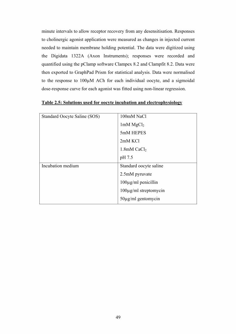
minute intervals to allow receptor recovery from any desensitisation. Responses
to cholinergic agonist application were measured as changes in injected current
needed to maintain membrane holding potential. The data were digitized using
the Digidata 1322A (Axon Instruments); responses were recorded and
quantified using the pClamp software Clampex 8.2 and Clampfit 8.2. Data were
then exported to GraphPad Prism for statistical analysis. Data were normalised
to the response to 100µM ACh for each individual oocyte, and a sigmoidal
dose-response curve for each agonist was fitted using non-linear regression.
Table 2.5: Solutions used for oocyte incubation and electrophysiology
Standard Oocyte Saline (SOS) 100mM NaCl
1mM MgCl2
5mM HEPES
2mM KCl
1.8mM CaCl2
pH 7.5
Incubation medium Standard oocyte saline
2.5mM pyruvate
100µg/ml penicillin
100µg/ml streptomycin
50µg/ml gentomycin
49

Chapter 3: Bioinformatics
50
3.1 Introduction The model nematode C. elegans has the largest known nAChR gene family,
with ~30 nAChR subunit genes (Brown et al., 2006). However, prior to the start
of the work presented here, little was known about the nAChR gene families of
other nematodes, though putative homologues of unc-38, which encodes an α-
subunit of C. elegans levamisole-sensitive nAChR, had been identified in a few
nematode parasites (Ajuh & Egwang, 1994; Hoekstra et al., 1997; Walker et
al., 2001). In insects, the nAChR gene family found in the model dipteran
Drosophila melanogaster has provided a basis from which the genomes of
other insects can be searched to identify nAChR subunit genes based on their
homology to Drosophila sequences. This approach has been successfully
applied to insect groups phylogenetically distinct from flies, such as the honey
bee Apis mellifera, the flour beetle Tribolium castaneum (Jones & Sattelle,
2006; Jones & Sattelle, 2007).
As the genome sequence of Ascaris suum was unavailable at the beginning of
this project, it was decided that a search of the genome of a related parasite,
Brugia malayi, may provide useful information about which nAChR subunit
genes were likely to be present in Ascaris suum. Brugia malayi and Ascaris
suum both belong to the clade III phylogenetic group, whereas C. elegans
belongs to the phylogenetically distinct clade V (Blaxter et al., 1998). As the
nAChR gene family of the model nematode C. elegans has been extremely well
described, C. elegans nAChR subunit sequences were used as query sequences
for performing searches of the parasite genome (Jones et al., 2007; Jones &
Sattelle, 2004). When this search had been completed, the search for nAChR
subunit genes was extended to the genome of another phylogenetically distinct
parasite, the clade I nematode Trichinella spiralis. A summary of the steps
involved in performing a reciprocal BLAST search to identify putative nAChR
subunit genes is shown in Figure 3.1. The methods are described more fully in
section 2.2.
51
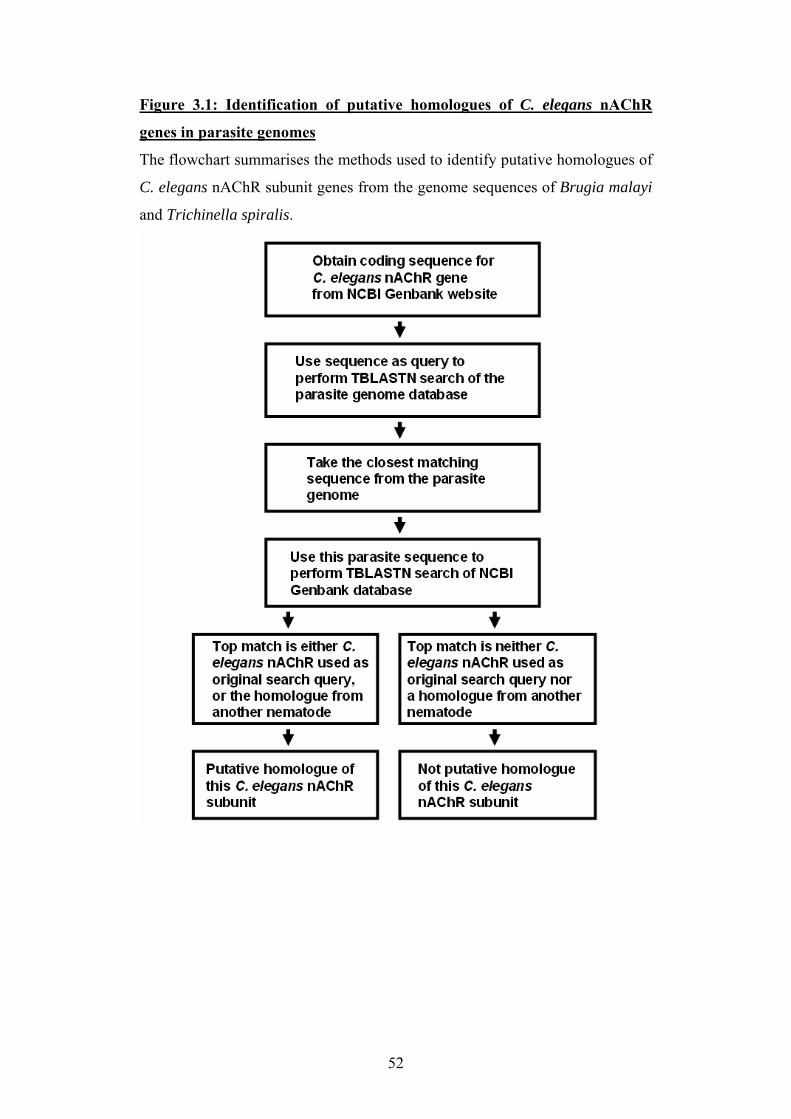
Figure 3.1: Identification of putative homologues of C. elegans nAChR
genes in parasite genomes
The flowchart summarises the methods used to identify putative homologues of
C. elegans nAChR subunit genes from the genome sequences of Brugia malayi
and Trichinella spiralis.
52
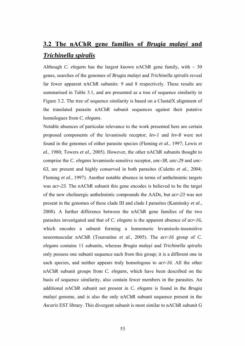
3.2 The nAChR gene families of Brugia malayi and
Trichinella spiralis
Although C. elegans has the largest known nAChR gene family, with ~ 30
genes, searches of the genomes of Brugia malayi and Trichinella spiralis reveal
far fewer apparent nAChR subunits: 9 and 8 respectively. These results are
summarised in Table 3.1, and are presented as a tree of sequence similarity in
Figure 3.2. The tree of sequence similarity is based on a ClustalX alignment of
the translated parasite nAChR subunit sequences against their putative
homologues from C. elegans.
Notable absences of particular relevance to the work presented here are certain
proposed components of the levamisole receptor; lev-1 and lev-8 were not
found in the genomes of either parasite species (Fleming et al., 1997; Lewis et
al., 1980; Towers et al., 2005). However, the other nAChR subunits thought to
comprise the C. elegans levamisole-sensitive receptor, unc-38, unc-29 and unc-
63, are present and highly conserved in both parasites (Culetto et al., 2004;
Fleming et al., 1997). Another notable absence in terms of anthelmintic targets
was acr-23. The nAChR subunit this gene encodes is believed to be the target
of the new cholinergic anthelmintic compounds the AADs, but acr-23 was not
present in the genomes of these clade III and clade I parasites (Kaminsky et al.,
2008). A further difference between the nAChR gene families of the two
parasites investigated and that of C. elegans is the apparent absence of acr-16,
which encodes a subunit forming a homomeric levamisole-insensitive
neuromuscular nAChR (Touroutine et al., 2005). The acr-16 group of C.
elegans contains 11 subunits, whereas Brugia malayi and Trichinella spiralis
only possess one subunit sequence each from this group; it is a different one in
each species, and neither appears truly homologous to acr-16. All the other
nAChR subunit groups from C. elegans, which have been described on the
basis of sequence similarity, also contain fewer members in the parasites. An
additional nAChR subunit not present in C. elegans is found in the Brugia
malayi genome, and is also the only nAChR subunit sequence present in the
Ascaris EST library. This divergent subunit is most similar to nAChR subunit G
53
from the pond snail Lymnaea stagnalis (van Nierop et al., 2006), and is
apparently conserved amongst clade III nematodes, though it was not found in
the genome of the clade I nematode Trichinella spiralis. With the agreement of
the annotators of the C. elegans nAChR subunits (Andrew Jones and Jonathon
Hodgkin, personal communication) this sequence was named acr-26; acr- is the
prefix given the nAChR subunits not already named by phenotype, and 26 is the
logical designation for a nAChR subunit additional to the 25 acr's already
named in C. elegans (Jones et al., 2007).
54
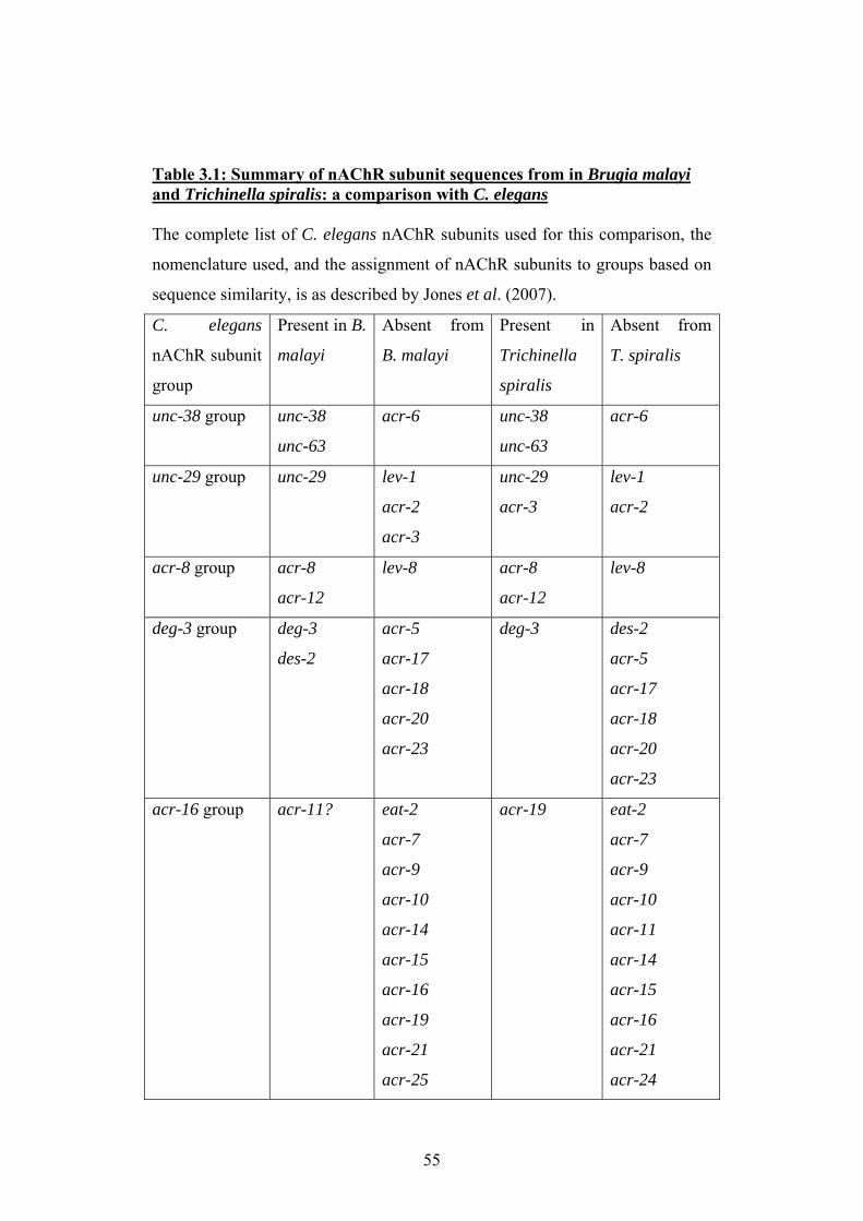
Table 3.1: Summary of nAChR subunit sequences from in Brugia malayi and Trichinella spiralis: a comparison with C. elegans The complete list of C. elegans nAChR subunits used for this comparison, the
nomenclature used, and the assignment of nAChR subunits to groups based on
sequence similarity, is as described by Jones et al. (2007).
C. elegans
nAChR subunit
group
Present in B.
malayi
Absent from
B. malayi
Present in
Trichinella
spiralis
Absent from
T. spiralis
unc-38 group unc-38
unc-63
acr-6 unc-38
unc-63
acr-6
unc-29 group unc-29 lev-1
acr-2
acr-3
unc-29
acr-3
lev-1
acr-2
acr-8 group acr-8
acr-12
lev-8 acr-8
acr-12
lev-8
deg-3 group deg-3
des-2
acr-5
acr-17
acr-18
acr-20
acr-23
deg-3 des-2
acr-5
acr-17
acr-18
acr-20
acr-23
acr-16 group acr-11? eat-2
acr-7
acr-9
acr-10
acr-14
acr-15
acr-16
acr-19
acr-21
acr-25
acr-19 eat-2
acr-7
acr-9
acr-10
acr-11
acr-14
acr-15
acr-16
acr-21
acr-24
55
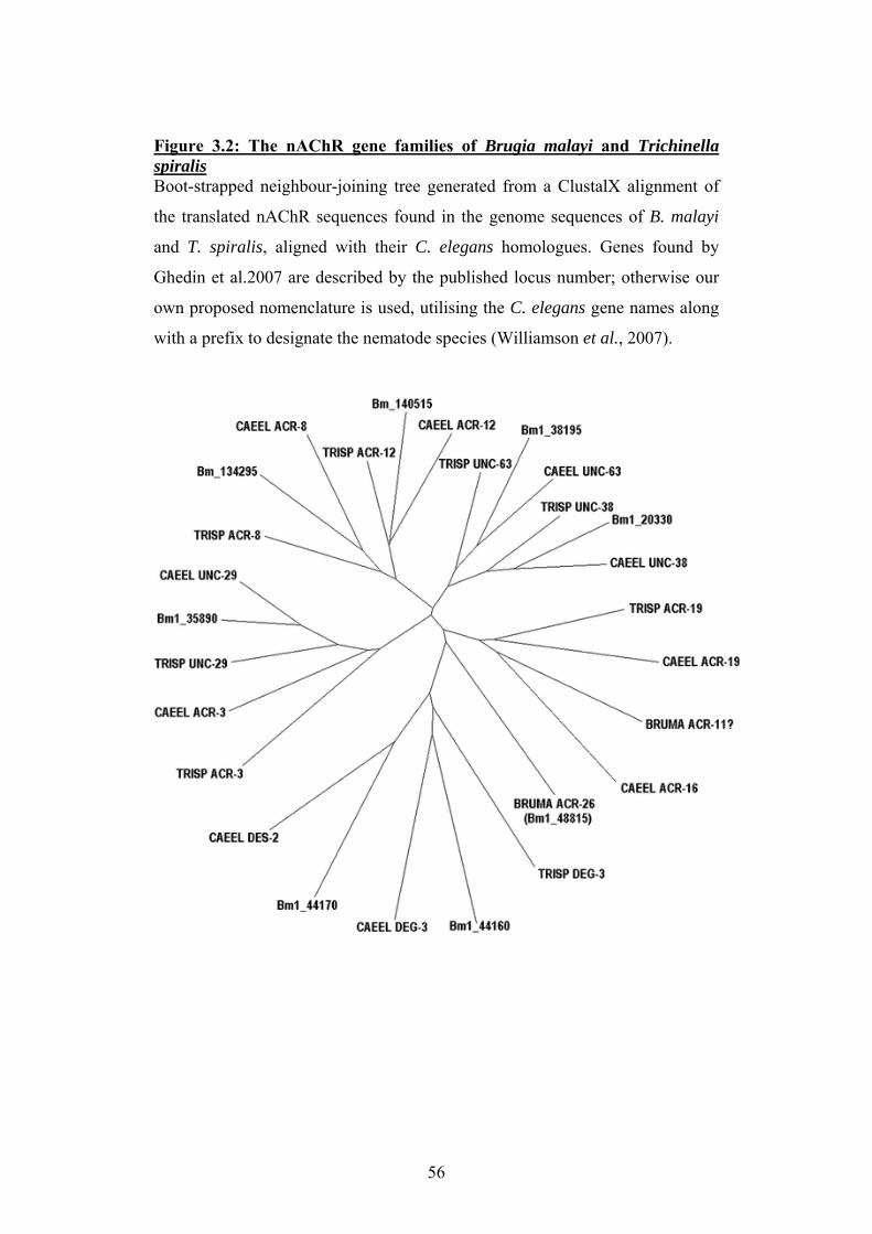
Figure 3.2: The nAChR gene families of Brugia malayi and Trichinella spiralis Boot-strapped neighbour-joining tree generated from a ClustalX alignment of
the translated nAChR sequences found in the genome sequences of B. malayi
and T. spiralis, aligned with their C. elegans homologues. Genes found by
Ghedin et al.2007 are described by the published locus number; otherwise our
own proposed nomenclature is used, utilising the C. elegans gene names along
with a prefix to designate the nematode species (Williamson et al., 2007).
56
3.3 The use of consensus nAChR sequences for primer design The primary reason for carrying out searches for nAChR subunit sequences in
the genome of Brugia malayi was to predict which nAChR subunits were
likely to be present in Ascaris suum, so attempts could be made to amplify these
using PCR. Amplification of nAChR subunits for which partial sequences were
not already present in the databases would require the use of degenerate
primers, which are designed on regions of amino acid sequence conserved
between species. Alignments were made of the translated sequences of well-
conserved nAChR subunits from C. elegans and Brugia malayi using ClustalW.
Figure 3.3 shows ClustalW alignments used for the design of degenerate
primers designed to amplify unc-29 and unc-63. Conserved regions featuring
amino acids with the fewest possible combinations of codons were chosen for
primer design, to minimise the degeneracy.
57
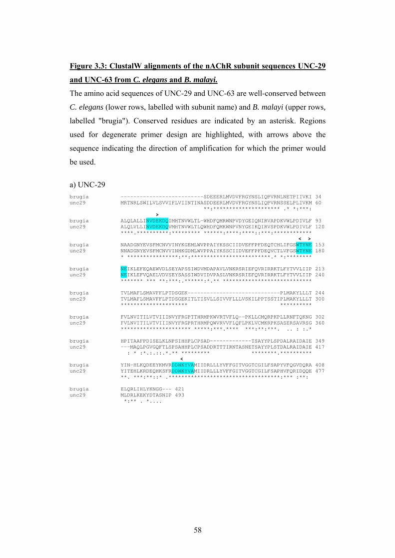
Figure 3.3: ClustalW alignments of the nAChR subunit sequences UNC-29
and UNC-63 from C. elegans and B. malayi.
The amino acid sequences of UNC-29 and UNC-63 are well-conserved between
C. elegans (lower rows, labelled with subunit name) and B. malayi (upper rows,
labelled "brugia"). Conserved residues are indicated by an asterisk. Regions
used for degenerate primer design are highlighted, with arrows above the
sequence indicating the direction of amplification for which the primer would
be used.
a) UNC-29 brugia --------------------------SDEEERLMVDVFRGYNSLIQPVRNLNETPIIVKI 34 unc29 MRTNRLSWILVLSVVIFLVIINTINASDDEERLMVDVFRGYNSLIQPVRNSSELPLIVKM 60 **:********************* .* *:***: > brugia ALQLALLINVDEKDQIMHTNVWLTL-WHDFQMRWNPVDYGEIQNIRVAPDKVWLPDIVLF 93 unc29 ALQLVLLINVDEKDQVMHTNVWLTLQWHDFQMKWNPVNYGEIKQIRVSPDKVWLPDIVLF 120 ****.**********:********* ******:****:****::***:************ < > brugia NAADGNYEVSFMCNVVINYKGEMLWVPPAIYKSSCIIDVEFFPFDEQTCHLIFGSWTYNE 153 unc29 NNADGNYEVSFMCNVVINHKGDMLWVPPAIYKSSCIIDVEFFPFDEQVCTLVFGSWTYNE 180 * ****************:**:*************************.* *:******** brugia NEIKLEFEQAEWVDLSEYAPSSIWDVMDAPAVLVNKRSRIEFQVRIRRKTLFYTVVLIIP 213 unc29 NEIKLEFVQAELVDVSEYSASSIWDVIDVPASLVNKRSRIEFQVRIRRKTLFYTVVLIIP 240 ******* *** **:***:.******:*.** **************************** brugia TVLMAFLSMAVFFLPTDSGEK-----------------------------PLMAKYLLLT 244 unc29 TVLMAFLSMAVFFLPTDSGEKITLTISVLLSIVVFLLLVSKILPPTSSTIPLMAKYLLLT 300 ********************* ********** brugia FVLNVITILVTVIIINVYFRGPTTHRMPKWVRTVFLQ--PKLLCMQRPKPLLRNFTQKNG 302 unc29 FVLNVITILVTVIIINVYFRGPRTHRMPQWVRVVFLQFLPKLVCMKRPKSASERSAVRSG 360 ********************** *****:***.**** ***:**:***. .. : :.* brugia HPITAAFPDISELKLNPSIHHPLCPSAD-------------TSAYYPLSPDALRAIDAIE 349 unc29 ---MAQLPGVGQFTLSPSAHHPLCPSADDRTTTIRNTASNETSAYYPLSTDALRAIDAIE 417 : * :*.:.::.*.** ********* ********.********** < brugia YIN-HLKQDEEYKMVRDDWKYVAMIIDRLLLYVFFGITVGGTCGILFSAPYVFQGVDQRA 408 unc29 YITEHLKRDEQHKSFRDDWKYVAMIIDRLLLYVFFGITVGGTCGILFSAPHVFQRIDQQE 477 **. ***:**::* .***********************************:*** :**: brugia ELQRLIHLYKNGG--- 421 unc29 MLDRLKEKYDTASNIP 493 *:** . *....
58
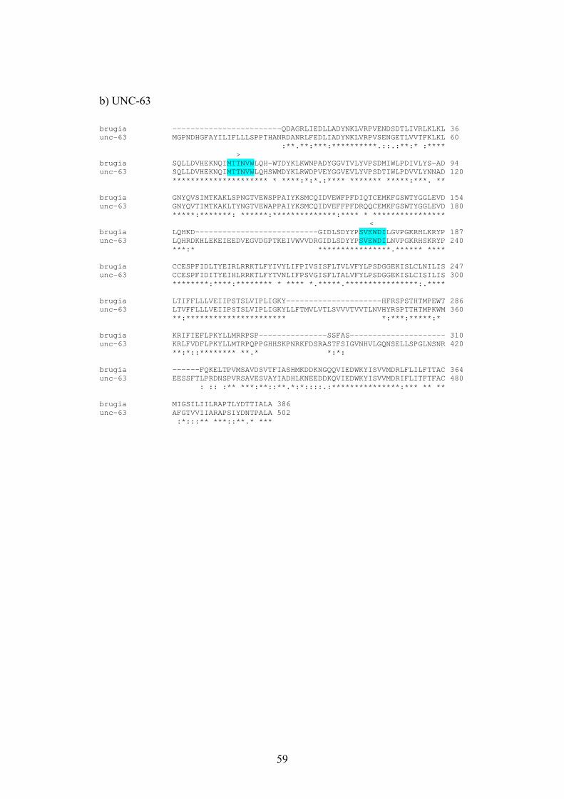
b) UNC-63 brugia ------------------------QDAGRLIEDLLADYNKLVRPVENDSDTLIVRLKLKL 36 unc-63 MGPNDHGFAYILIFLLLSPPTHANRDANRLFEDLIADYNKLVRPVSENGETLVVTFKLKL 60 :**.**:***:**********.::.:**:* :**** > brugia SQLLDVHEKNQIMTTNVWLQH-WTDYKLKWNPADYGGVTVLYVPSDMIWLPDIVLYS-AD 94 unc-63 SQLLDVHEKNQIMTTNVWLQHSWMDYKLRWDPVEYGGVEVLYVPSDTIWLPDVVLYNNAD 120 ********************* * ****:*:*.:**** ******* *****:***. ** brugia GNYQVSIMTKAKLSPNGTVEWSPPAIYKSMCQIDVEWFPFDIQTCEMKFGSWTYGGLEVD 154 unc-63 GNYQVTIMTKAKLTYNGTVEWAPPAIYKSMCQIDVEFFPFDRQQCEMKFGSWTYGGLEVD 180 *****:*******: ******:**************:**** * **************** < brugia LQHKD---------------------------GIDLSDYYPSVEWDILGVPGKRHLKRYP 187 unc-63 LQHRDKHLEKEIEEDVEGVDGPTKEIVWVVDRGIDLSDYYPSVEWDILNVPGKRHSKRYP 240 ***:* ****************.****** **** brugia CCESPFIDLTYEIRLRRKTLFYIVYLIFPIVSISFLTVLVFYLPSDGGEKISLCLNILIS 247 unc-63 CCESPFIDITYEIHLRRKTLFYTVNLIFPSVGISFLTALVFYLPSDGGEKISLCISILIS 300 ********:****:******** * **** *.*****.****************:.**** brugia LTIFFLLLVEIIPSTSLVIPLIGKY---------------------HFRSPSTHTMPEWT 286 unc-63 LTVFFLLLVEIIPSTSLVIPLIGKYLLFTMVLVTLSVVVTVVTLNVHYRSPTTHTMPKWM 360 **:********************** *:***:*****:* brugia KRIFIEFLPKYLLMRRPSP---------------SSFAS--------------------- 310 unc-63 KRLFVDFLPKYLLMTRPQPPGHHSKPNRKFDSRASTFSIGVNHVLGQNSELLSPGLNSNR 420 **:*::******** **.* *:*: brugia ------FQKELTPVMSAVDSVTFIASHMKDDKNGQQVIEDWKYISVVMDRLFLILFTTAC 364 unc-63 EESSFTLPRDNSPVRSAVESVAYIADHLKNEEDDKQVIEDWKYISVVMDRIFLITFTFAC 480 : :: :** ***:**::**.*:*::::.:***************:*** ** ** brugia MIGSILIILRAPTLYDTTIALA 386 unc-63 AFGTVVIIARAPSIYDNTPALA 502 :*:::** ***::**.* ***
59
3.4 Discussion
A bioinformatics approach to investigating the nAChR gene families of parasitic
nematodes for which good quality genome sequence is available has revealed a
surprisingly small nAChR gene family in Brugia malayi and Trichinella spiralis
compared to C. elegans. The close correlation between the nAChR subunits found in
both parasites, and the fairly complete and assembled nature of these genome
sequences, suggests that this is indeed the full extent of these nAChR gene families.
Support for this comes from work using the genome of Haemonchus contortus, a
clade V nematode quite closely related to C. elegans, which has also been carried out
in our laboratory. Although the genome sequencing project of Haemonchus contortus
is much less complete than the parasite genome sequences used here, the majority of
the C. elegans nAChR subunit genes (~25) were easily identified (Thomas Walsh,
personal communication). The results presented here also broadly agree with the
annotation accompanying the Brugia malayi genome in terms of the number of
nAChR subunits present, which was published after the work described in this chapter
had been completed. However, the annotators of the Brugia malayi genome utilised a
rather confusing and inconsistent nomenclature when describing the nAChR subunits,
where C. elegans gene names were assigned to non-homologous Brugia malayi
sequences (Ghedin et al., 2007). Figure 3.2 utilises the nomenclature we proposed in
Williamson et al. 2007, where C. elegans gene names are assigned to parasite nAChR
subunit sequences which are clearly homologous to C. elegans sequences, and a
prefix is added to denote the genus and species to which the sequence belongs. This
nomenclature has the support of the C. elegans nAChR community, and a similar
strategy has been used to ensure consistency when annotating the insect nAChR gene
families (Andrew Jones and Jonathon Hodgkin, personal communication; Jones et al.
2007).
The degree of conservation between putatively homologous nAChR subunits from the
different nematode species was variable both within and between nAChR subunits;
the ligand-binding motifs and the transmembrane regions were usually quite similar,
but the cytoplasmic region was too dissimilar to be identified from genomic sequence
in the majority of cases. It would therefore be misleading to attempt to quantify the
similarity of homologous nAChR sequences from the regions which could be
60
identified in silico; a more complete comparison would require the amplification and
sequencing of all these parasite nAChRs, which is beyond the scope of the work
presented here. The most highly conserved nAChR subunit sequences (in terms of the
amount of matching sequence which could be easily identified in the parasite
genomes) appeared to be unc-38, unc-29 and unc-63; this provides a good basis for
the design of primers to amplify these subunits from other nematode species. The high
degree of conservation suggests these subunits have an essential role in cholinergic
signalling in nematodes. When the proposed role of these subunits as components of
the levamisole receptor and their changing expression levels in pyrantel resistant
hookworms are considered, investigating the function of these nAChR subunits in
parasites is clearly key to understanding both sensitivity and resistance to this class of
cholinergic anthelmintic (Fleming et al., 1997; Kopp et al., 2008). In contrast to this,
the lack of acr-23 in Brugia malayi and Trichinella spiralis suggests that if the AAD
compounds do in fact have activity against these nematodes, they must act via a
different target to the one described from C. elegans (Kaminsky et al., 2008).
The findings presented here suggest that either the nematode nAChR gene family
underwent a dramatic expansion in the clade V nematodes, or that nAChR genes have
been lost from B. malayi and T. spiralis. Either hypothesis may suggest that this lack
of nAChR diversity is linked to the different neurotransmission requirements of a
free-living versus a parasitic lifestyle; it should be noted that larvae of many parasitic
clade V species must survive outside a host while they develop to an infective stage,
whereas transmission from host to host is usually more direct for parasites from
clades III and I (Dorris et al., 1999). An alternative explanation for the smaller
number of nAChR genes present in these parasites could also be a greater degree of
alternative splicing; though insects possess only a small number of nAChR genes,
they are known to increase their receptor repertoire by splicing and editing mRNA
(Sattelle et al., 2005). No evidence to support this hypothesis has yet emerged from
the parasite genomes, but this cannot be entirely ruled out without performing
amplification of all the nAChR subunit transcripts generated by the nematodes.
While the dramatic difference in the number of nAChR gene family members
between C. elegans and the parasites Brugia malayi and Trichinella spiralis is
certainly intriguing, it should be noted that it is currently unknown whether the
genomes of these parasites are typical of the clades to which they belong. A more
61

complete comparison of the gene families of nematodes from different clades cannot
be carried out until many more nematode genomes have been successfully sequenced.
62

Chapter 4: Amplification of nAChR subunits from Ascaris suum
63
4.1 Introduction To investigate the molecular basis of cholinergic anthelmintic sensitivity in a parasitic
nematode, the starting point must be to amplify sequences encoding nAChR subunits
which are potential components of the relevant receptor. For the model nematode C.
elegans, a comprehensively sequenced and annotated genome, and the ease with
which drug resistant mutants can be produced and maintained in vitro, has greatly
facilitated the identification of nAChR subunits forming anthelmintic drug targets.
For parasitic nematodes, a relative paucity of genomic information, and the
difficulties involved in producing or isolating drug resistant worms, makes the task of
investigating the molecular basis of drug sensitivity and resistance much more
difficult. It should be noted that even when some sequence information is available
for a parasite in the form of an EST library, this is often biased towards the most
abundant transcripts, and rarely provides more than two or three partial sequences for
ligand-gated ion channels. However, several previous studies have succeeded in
amplifying a limited number of nAChR subunits from nematode parasites, using two
main approaches. Parasite homologues of unc-38 were obtained from both
Haemonchus contortus and Trichostrongylus colubriformis by constructing and
screening genomic DNA libraries (Hoekstra et al., 1997; Wiley et al., 1996).
Degenerate PCR using primers based on the amino acid sequence of C. elegans
nAChR subunits yielded the Teladorsagia circumcincta unc-38 homologue, and was
more recently employed to facilitate amplification of several nAChR sequences from
Ancylostoma caninum which could not be located by searches of the A. caninum
sequence database (Kopp et al., 2008; Walker et al., 2001). Both these approaches
rely heavily on a high degree of sequence similarity between the sequence being used
as a probe or for primer design and the sequence the experiment is designed to obtain;
all the parasites mentioned in the previous examples are from clade V and are
therefore closely related to each other and to the model nematode C. elegans. In the
work presented here, it was hoped that degenerate PCR utilising primers designed
using the sequence information from the clade III parasite Brugia malayi would allow
amplification of nAChR subunits from Ascaris suum (described in section 3.3). This
approach was used to amplify partial sequence of Ascaris suum unc-29 and unc-63.
64
An existing partial sequence in the GenBank database and the Ascaris suum EST
database was used to design primers specific to Ascaris suum unc-38, and an
additional novel nAChR subunit. After partial nAChR subunit sequences had been
obtained, RACE techniques were used to amplify the 5' and 3' ends of the sequences
(Frohman et al., 1988). RACE techniques have previously been used successfully to
obtain full-length transcripts from Ascaris spp. (Timinouni & Bazzicalupo, 1997). As
a modification to the RACE protocol, the nematode splice leader sequence SL1 was
used as the 5' RACE primer. SL1 is a splice-leader element found at the 5' end of the
many of nematode mRNA sequences, and has previously been successfully used as a
5' RACE primer for amplification of nematode transcripts (Bektesh et al., 1988; Pape
et al., 1999).
4.2 Potential components of the levamisole-sensitive nAChR
from Ascaris suum
Full length sequences encoding homologues of unc-38 and unc-29 were amplified
from Ascaris suum body wall muscle. The process by which these sequences were
obtained is summarised in Figure 4.1. These sequences were deposited in the
GenBank database, with the accession numbers EU053155 and EU006073. Figure 4.2
shows the translated sequences of Ascaris suum UNC-38 and UNC-29, where some
key features are highlighted. Like the homologous C. elegans sequence, Ascaris suum
unc-38 encodes a nAChR α subunit, containing amino acid residues from the principal
binding loops A, B and C known to contribute to ligand binding (see Figure 1.4,
which is taken from Changeux & Taly, 2008) . The loop C motif for both the C.
elegans and Ascaris suum unc-38 is Y-X-X-C-C, rather than the more usual Y-X-C-C.
This additional loop C residue is a proline known to be involved in the differential
sensitivity of nematode and mammalian muscle nAChRs; another residue conferring
this differential levamisole sensitivity is a glutamic acid present on loop B, which is
again conserved in both C. elegans and Ascaris suum (Rayes et al., 2004). The
Ascaris suum unc-29 sequence, like the homologous sequence from C. elegans,
encodes a non-α subunit, lacking the principal binding components but possessing the
ligand-binding residues of the complementary binding loops D, E and F (see Figure
1.4, which is taken from Changeux & Taly, 2008). Both Ascaris suum UNC-38 and
65
UNC-29 possess the charged glutamic acid and aspartic acid residues near the base of
the second transmembrane domain indicating that they are components of a cation
channel (see Figure 1.5, taken from Corringer 2000). This is an important distinction
for a nematode nAChR, as C. elegans is known to possess acetylcholine-gated anion
channels (Putrenko et al., 2005).
66
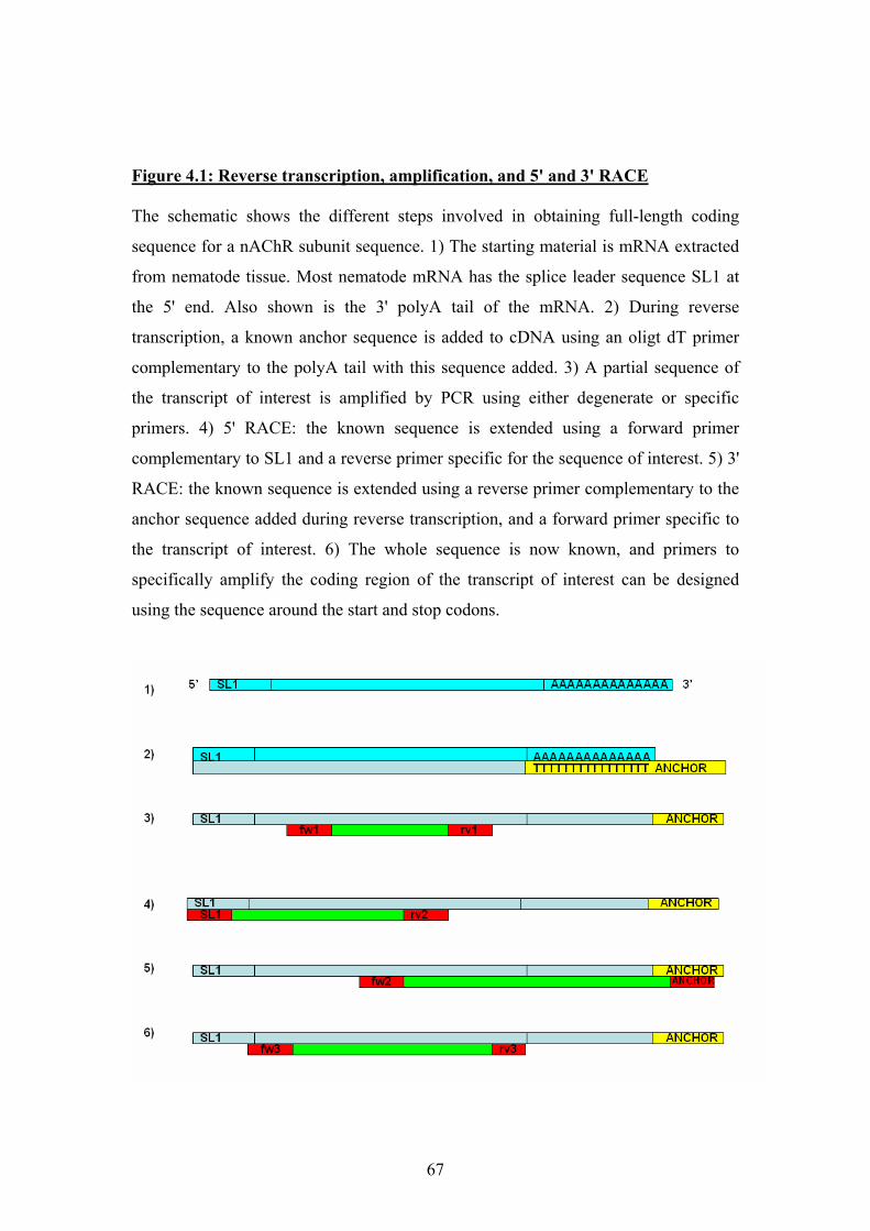
Figure 4.1: Reverse transcription, amplification, and 5' and 3' RACE The schematic shows the different steps involved in obtaining full-length coding
sequence for a nAChR subunit sequence. 1) The starting material is mRNA extracted
from nematode tissue. Most nematode mRNA has the splice leader sequence SL1 at
the 5' end. Also shown is the 3' polyA tail of the mRNA. 2) During reverse
transcription, a known anchor sequence is added to cDNA using an oligt dT primer
complementary to the polyA tail with this sequence added. 3) A partial sequence of
the transcript of interest is amplified by PCR using either degenerate or specific
primers. 4) 5' RACE: the known sequence is extended using a forward primer
complementary to SL1 and a reverse primer specific for the sequence of interest. 5) 3'
RACE: the known sequence is extended using a reverse primer complementary to the
anchor sequence added during reverse transcription, and a forward primer specific to
the transcript of interest. 6) The whole sequence is now known, and primers to
specifically amplify the coding region of the transcript of interest can be designed
using the sequence around the start and stop codons.
67
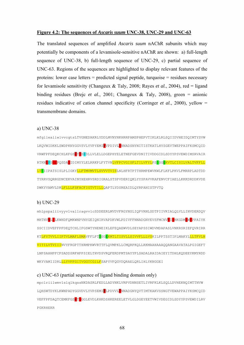
Figure 4.2: The sequences of Ascaris suum UNC-38, UNC-29 and UNC-63 The translated sequences of amplified Ascaris suum nAChR subunits which may
potentially be components of a levamisole-sensitive nAChR are shown: a) full-length
sequence of UNC-38, b) full-length sequence of UNC-29, c) partial sequence of
UNC-63. Regions of the sequences are highlighted to display relevant features of the
proteins: lower case letters = predicted signal peptide, turquoise = residues necessary
for levamisole sensitivity (Changeux & Taly, 2008; Rayes et al., 2004), red = ligand
binding residues (Brejc et al., 2001; Changeux & Taly, 2008), green = anionic
residues indicative of cation channel specificity (Corringer et al., 2000), yellow =
transmembrane domains.
a) UNC-38 mfglleallwlvvcgtsLTVGNEDAKRLYDDLMVNYNKHRRPAMSPHEPVTIKLKLRLSQIIDVHEIDQIMTYSVW LKQVWIDKKLSWDPKNYGGVSVLYVPYEMIWVPDIVLYNNADSNYNITISTKATLHYSGEVTWEPPAIFKSMCQID VRWFPFDEQKCHLKFGSWTYSEDLLVLELLDGEPHYELETNEFGEVDNITIVDDGIDLSDYYPSVEWDIMSRVAIR RTKNYPSCCPQSDAYIDIMYYLELRRKPLFYTVNLVFPCVGISFLTILVFYLPSDSGEKVTLCISILVALTVFFLL LTEIIPATSISLPLIGKYLLFTMVMVTLSVVVTVISLNLHFRTPTTHRMPEWVKWLFLKFLPKVLFMRRPLADTDD TYRRVSQRRGDNCEKVAINYHEHRVSRDIGRALSTSPVDERIQKLYYSPAVVKAFENVCFIAELLKKKDRDDKVDE DWKYVAMVLDRLFLLLFSFACFIGTVTILLQAPTLYDSREAIDLQYRPANISTPVTQ
b) UNC-29 mhlpspaliivyyvlvallragvvlcSDDEERLMVDVFRGYNSLIQPVRNLSDTPIIVKIALQLVLLINVDERDQV MHTNVWLTLKWHDFQMKWNPVNYGEIQNIRVAPDKVWLPDIVFFNNADGNYEVSFMCNVVISHKGDMLWVPPAIYK SSCIIDVEFFPFDEQTCHLIFGSWTYNENEIKLEFEQAEWVDLSEYAPSSIWDVMDAPASLVNKRSRIEFQVRIRR KTLFYTVVLIIPTVLMAFLSMAVFFLPTDSGEKMTLTISVLLSIVVFLLLVSKILPPTSSTIPLMAKYLLLTFVLN VITILVTVIIINVYFRGPTTHRMPKWVRTTFLQVMPKLLCMQRPKQLLRKMAAHAAAQQANGAAVATALPGIGEFT LNPSAHHPFCPSADDSRFHPPSIELTHVDSVRQFERDPMTSAYYPLSADALRAIDAIEYITDHLKQDEEYKMYRDD WKYVAMIIDRLLLYVFFGITVGGTCGILFSAPYVFQGVDQRAELQRLIHLYKNGGEI
c) UNC-63 (partial sequence of ligand binding domain only) mpclriilwmvlslqlkqssNKDASRLFEDLLADYNKLVRPVDNNSETLIVRFKLKLSQLLDVHEKNQIMTTNVW LQHSWTDYKLKWNPADYGGVDVLYVPSEMIWLPDVVLYNNADGNYQVTIMTKAKVSSNGTVEWAPPAIYKSMCQID VEFFPFDAQTCEMKFGSWTYGGLEVDLKHKDSHKEREELETVLGLDGEYEETVWIVDEGIDLSDYYPSVEWDILRV PGKRHEKR
68
Only a partial sequence of Ascaris suum unc-63 was obtained, which was initially
interpreted as being caused by due to mis-priming of the mRNA polyA tail during
reverse transcription, as the nucleotide sequence obtained had regions containing
many adjacent adenine bases. This possibility was eliminated by altering the reverse
transcription primers for a mixture of random hexamers; it was still not possible to
amplify any further unc-63 sequence from Ascaris cDNA despite using several
different degenerate PCR primers. The sequence obtained did however appear to
encode a partial ligand-binding domain of a nAChR α subunit homologous to C.
elegans unc-63. This partial sequence is also shown in Figure 4.2.
Tables 4.1, 4.2 and 4.3 show the similarity of the Ascaris suum UNC-38, UNC-29 and
UNC-63 to other nAChR subunits at the amino acid level. The top 5 most similar
sequences returned from a search of the GenBank database using the TBLASTN
algorithm are included in each table. In the majority of cases, homologous sequences
from other nematode species were returned by the search, in which case only the
nematode genus and species is included in the table. Where one of the matching
sequences was not from a nematode, the genus and species of the organism
concerned, and the designated name for the nAChR subunit using nomenclature
appropriate for that species, are included. It should be noted that bias may be
introduced into these results by partial sequences, either used as a search query, or as
a returned matching sequence from the database. In summary however, there is
clearly much greater similarity between homologous nAChR subunits from different
nematodes than there is similarity with nAChR subunits from different phyla. As a
point of comparison, a similar BLAST search using the Ascaris suum sequences was
performed but the results restricted to human nAChR sequences. These generally
showed roughly 40% identity to the nematode subunits, though surprisingly, UNC-38
and UNC-63 appeared slightly more similar to human α6 and α4, and UNC-29 to
human β2, than to the human muscle nAChR subunits. The similarity of UNC-29 to
β2 is also apparent in the conserved ligand binding residues (Brejc et al., 2001).
69
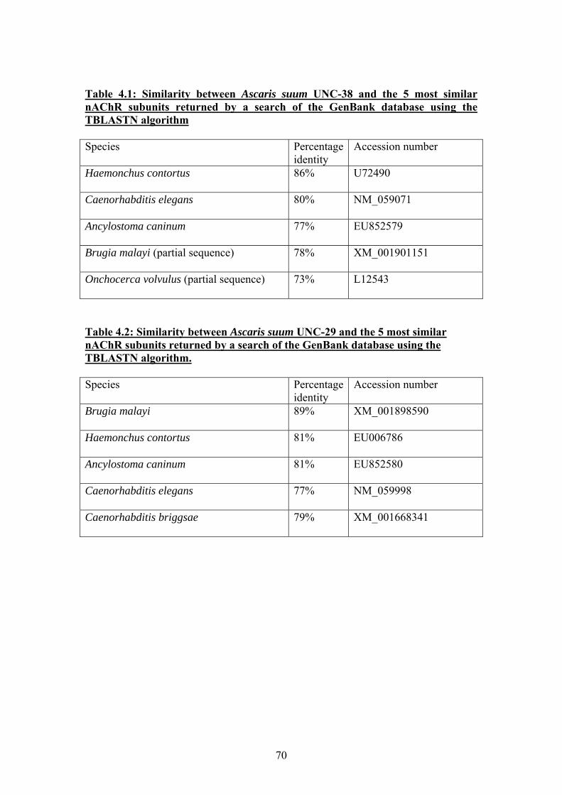
Table 4.1: Similarity between Ascaris suum UNC-38 and the 5 most similar nAChR subunits returned by a search of the GenBank database using the TBLASTN algorithm Species Percentage
identity Accession number
Haemonchus contortus
86% U72490
Caenorhabditis elegans
80% NM_059071
Ancylostoma caninum
77% EU852579
Brugia malayi (partial sequence)
78% XM_001901151
Onchocerca volvulus (partial sequence)
73% L12543
Table 4.2: Similarity between Ascaris suum UNC-29 and the 5 most similar nAChR subunits returned by a search of the GenBank database using the TBLASTN algorithm. Species Percentage
identity Accession number
Brugia malayi
89% XM_001898590
Haemonchus contortus
81% EU006786
Ancylostoma caninum
81% EU852580
Caenorhabditis elegans
77% NM_059998
Caenorhabditis briggsae
79% XM_001668341
70
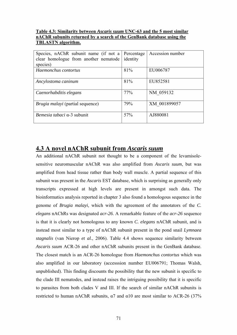
Table 4.3: Similarity between Ascaris suum UNC-63 and the 5 most similar nAChR subunits returned by a search of the GenBank database using the TBLASTN algorithm. Species, nAChR subunit name (if not a clear homologue from another nematode species)
Percentage identity
Accession number
Haemonchus contortus
81% EU006787
Ancylostoma caninum
81% EU852581
Caenorhabditis elegans
77% NM_059132
Brugia malayi (partial sequence)
79% XM_001899057
Bemesia tabaci α-3 subunit
57% AJ880081
4.3 A novel nAChR subunit from Ascaris suum An additional nAChR subunit not thought to be a component of the levamisole-
sensitive neuromuscular nAChR was also amplified from Ascaris suum, but was
amplified from head tissue rather than body wall muscle. A partial sequence of this
subunit was present in the Ascaris EST database, which is surprising as generally only
transcripts expressed at high levels are present in amongst such data. The
bioinformatics analysis reported in chapter 3 also found a homologous sequence in the
genome of Brugia malayi, which with the agreement of the annotators of the C.
elegans nAChRs was designated acr-26. A remarkable feature of the acr-26 sequence
is that it is clearly not homologous to any known C. elegans nAChR subunit, and is
instead most similar to a type of nAChR subunit present in the pond snail Lymnaea
stagnalis (van Nierop et al., 2006). Table 4.4 shows sequence similarity between
Ascaris suum ACR-26 and other nAChR subunits present in the GenBank database.
The closest match is an ACR-26 homologue from Haemonchus contortus which was
also amplified in our laboratory (accesssion number EU006791; Thomas Walsh,
unpublished). This finding discounts the possibility that the new subunit is specific to
the clade III nematodes, and instead raises the intriguing possibility that it is specific
to parasites from both clades V and III. If the search of similar nAChR subunits is
restricted to human nAChR subunits, α7 and α10 are most similar to ACR-26 (37%
71
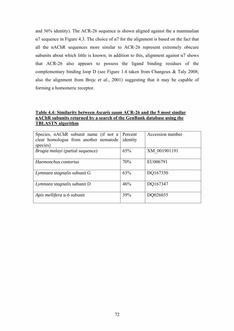
and 36% identity). The ACR-26 sequence is shown aligned against the a mammalian
α7 sequence in Figure 4.3. The choice of α7 for the alignment is based on the fact that
all the nAChR sequences more similar to ACR-26 represent extremely obscure
subunits about which little is known; in addition to this, alignment against α7 shows
that ACR-26 also appears to possess the ligand binding residues of the
complementary binding loop D (see Figure 1.4 taken from Changeux & Taly 2008;
also the alignment from Brejc et al., 2001) suggesting that it may be capable of
forming a homomeric receptor.
Table 4.4: Similarity between Ascaris suum ACR-26 and the 5 most similar nAChR subunits returned by a search of the GenBank database using the TBLASTN algorithm Species, nAChR subunit name (if not a clear homologue from another nematode species)
Percent identity
Accession number
Brugia malayi (partial sequence)
65% XM_001901191
Haemonchus contortus
70% EU006791
Lymnaea stagnalis subunit G
63% DQ167350
Lymnaea stagnalis subunit D
46% DQ167347
Apis mellifera α-6 subunit
39% DQ026035
72
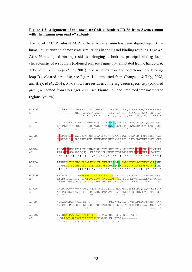
Figure 4.3: Alignment of the novel nAChR subunit ACR-26 from Ascaris suum with the human neuronal α7 subunit The novel nAChR subunit ACR-26 from Ascaris suum has been aligned against the
human α7 subunit to demonstrate similarities in the ligand binding residues. Like α7,
ACR-26 has ligand binding residues belonging to both the principal binding loops
characteristic of α subunits (coloured red, see Figure 1.4; annotated from Changeux &
Taly, 2008, and Brejc et al., 2001), and residues from the complementary binding
loop D (coloured turquoise, see Figure 1.4; annotated from Changeux & Taly, 2008,
and Brejc et al., 2001). Also shown are residues confering cation specificity (coloured
green; annotated from Corringer 2000, see Figure 1.5) and predicted transmembrane
regions (yellow).
ACR26 MATRRRELILLUFIHGVVDVVLADSSITDLSEIDGTATDEQKLLYHLLRQYEKAVRPVRN a7 ----------MRCSPGGVWLALAAS----LLHVSLQGEFQRKLYKELVKNYNPLERPVAN : * * :.** * * .:. . ::** .*:::*: *** * ACR26 ASHTVTVKLGMTMTNIFEMDEKNQVLTINVWLDQEWKDELLRWDPKEFGGIQSIRIPCDL a7 DSQPLTVYFSLSLLQIMDVDEKNQVLTTNIWLQMSWTDHYLQWNVSEYPGVKTVRFPDGQ *:.:** :.::: :*:::******** *:**: .*.*. *:*: .*: *::::*:* . ACR26 IWLPDIVLYNNADDYTAGYMRSRAMVFYDGTVFWPPPTQLRSTCKIDVTYFPFDSQHCAL a7 IWKPDILLYNSADERFDATFHTNVLVNSSGHCQYLPPGIFKSSCYIDVRWFPFDVQHCKL ** ***:***.**: . :::..:* .* : ** ::*:* *** :**** *** * ACR26 KFGSWTYHGFQVDITNRSDNVDLSNYVVSGEFDLVRVHQKRRVVKYTCCPEPYPDVTFFI a7 KFGSWSYGGWSLDLQMQ--EADISGYIPNGEWDLVGIPGKRSERFYECCKEPYPDVTFTV *****:* *:.:*: : :.*:*.*: .**:*** : ** * ** ******** : ACR26 HIRRKTLYYLYNVVFPCMMMSVLTLLVFLLPPDSGEKIALGITVLLAFSVFVLAIAEKMP a7 TMRRRTLYYGLNLLIPCVLISALALLVFLLPADSGEKISLGITVLLSLTVFMLLVAEIMP :**:**** *:::**:::*.*:*******.******:*******:::**:* :** ** ACR26 ETSDSMPLIGIYLTVVMAMTSVSVVMTVMVLNFHHRGPFNQAVPKWVHRLVLNRLRRALC a7 ATSDSVPLIAQYFASTMIIVGLSVVVTVIVLQYHYHDPDGGKMPKWTRVILLNWCAWFLR ****:***. *:: .* :..:***:**:**::*::.* . :***.: ::** * ACR26 MRLPYTG------WKDNGFCSANGMTKTISIGLAMDDFNVDSFEELDNQFLQMQETELVN a7 MKRPGEDKVRPACQHKQRRCSLASVEMSAVVPPPASNGNLLYIGFRGLDGVHCVPTPDSG *: * . :.: ** .: : : . .: *: : . : :: * . ACR26 VDDAAALRKKKCRPNELHS-----------RLLKTLQVLIRRQEMEDLYQTLANEWRQVA a7 VVCGRMACSPTHDEHLLHGGQPPEGDPDLAKILEEVRYIANRFRCQDESEAVCSEWKFAA * . . . : **. ::*: :: : .* . :* :::..**: .* ACR26 QVIDRLLFWVFLVCTVIITLILLIIIPAVHRSMESDVFDESLYGLH a7 CVVDRLCLMAFSVFTIICTIGILMSAPNFVEAVSKDFA-------- *:*** : .* * *:* *: :*: * . .::..*.
73
4.4 Real-time PCR Real-time PCR was performed to ascertain whether relatively low expression levels of
Ascaris suum unc-63 were causing difficulty in amplifying the full-length transcript.
This technique also allowed a comparison to be made of expression levels of unc-38,
unc-29 and unc-63 in tissue taken from Ascaris suum head and body wall. Head and
muscle tissue were taken from 3 different individual nematodes and the RNA
extracted. Reverse transcription reactions were performed on each batch of RNA,
using the same amount of total RNA in the reaction each time. A reference gene
expressed at the same levels in both head and muscle tissue was also amplified
alongside the nAChR subunit genes to ensure the amplification in each reaction was
comparable. Two replicates were performed for each amplification reaction from each
batch of cDNA. Examples of amplification plots are shown in Figure 4.4. The real-
time PCR data is also reported as mean critical threshold (Ct) values in Table 4.5;
critical threshold values indicate the number of PCR cycles necessary before the
amplification of a transcript reaches the detection threshold.
A comparison of relative abundance of the nAChR subunits in tissue taken from
Ascaris suum head and body wall showed that there is very little difference in
expression levels between these different regions of the nematode. This finding can
only be preliminary however, as only very crude dissections were performed to obtain
the tissue used this experiment. The head preparations would have contained
pharyngeal tissue, sensory amphids, and the nerve ring in addition to muscle tissue;
the body wall muscle would also have contained motorneurons and regions of the
dorsal and ventral nerve cords. A more thorough comparison of nAChR subunit
expression levels in different tissues would therefore require a more detailed
dissection which separated the muscle from the nervous system and other tissues, and
would require more fresh parasite tissue than was available during this investigation.
When the relative abundance of the different nAChR subunits were compared,
amplification of unc-63 (as shown by the Ct values in Table 4.5) occurred consistently
later than amplification of unc-38 and unc-29. The Ct values suggest that unc-38 and
unc-29 transcripts are perhaps equally adundant, whereas unc-63 is expressed at a
slightly lower level. However, the relative expression level of unc-63 did not appear
74
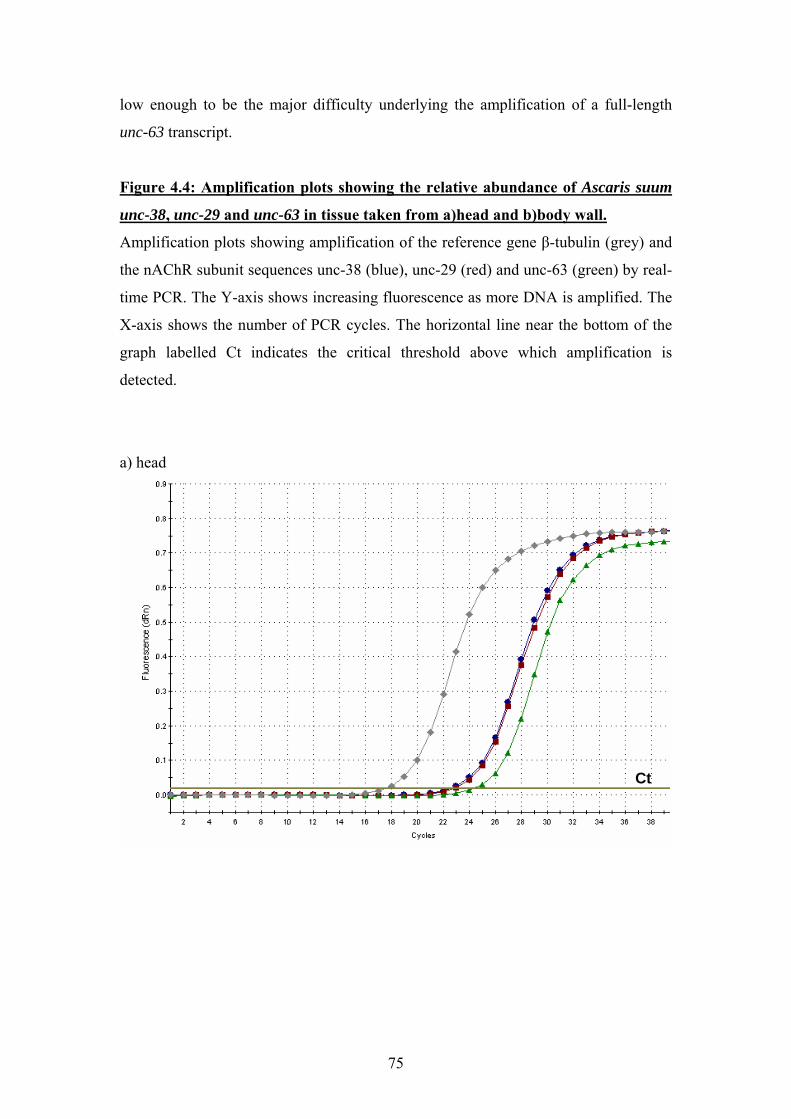
low enough to be the major difficulty underlying the amplification of a full-length
unc-63 transcript.
Figure 4.4: Amplification plots showing the relative abundance of Ascaris suum
unc-38, unc-29 and unc-63 in tissue taken from a)head and b)body wall.
Amplification plots showing amplification of the reference gene β-tubulin (grey) and
the nAChR subunit sequences unc-38 (blue), unc-29 (red) and unc-63 (green) by real-
time PCR. The Y-axis shows increasing fluorescence as more DNA is amplified. The
X-axis shows the number of PCR cycles. The horizontal line near the bottom of the
graph labelled Ct indicates the critical threshold above which amplification is
detected.
a) head
Ct
75
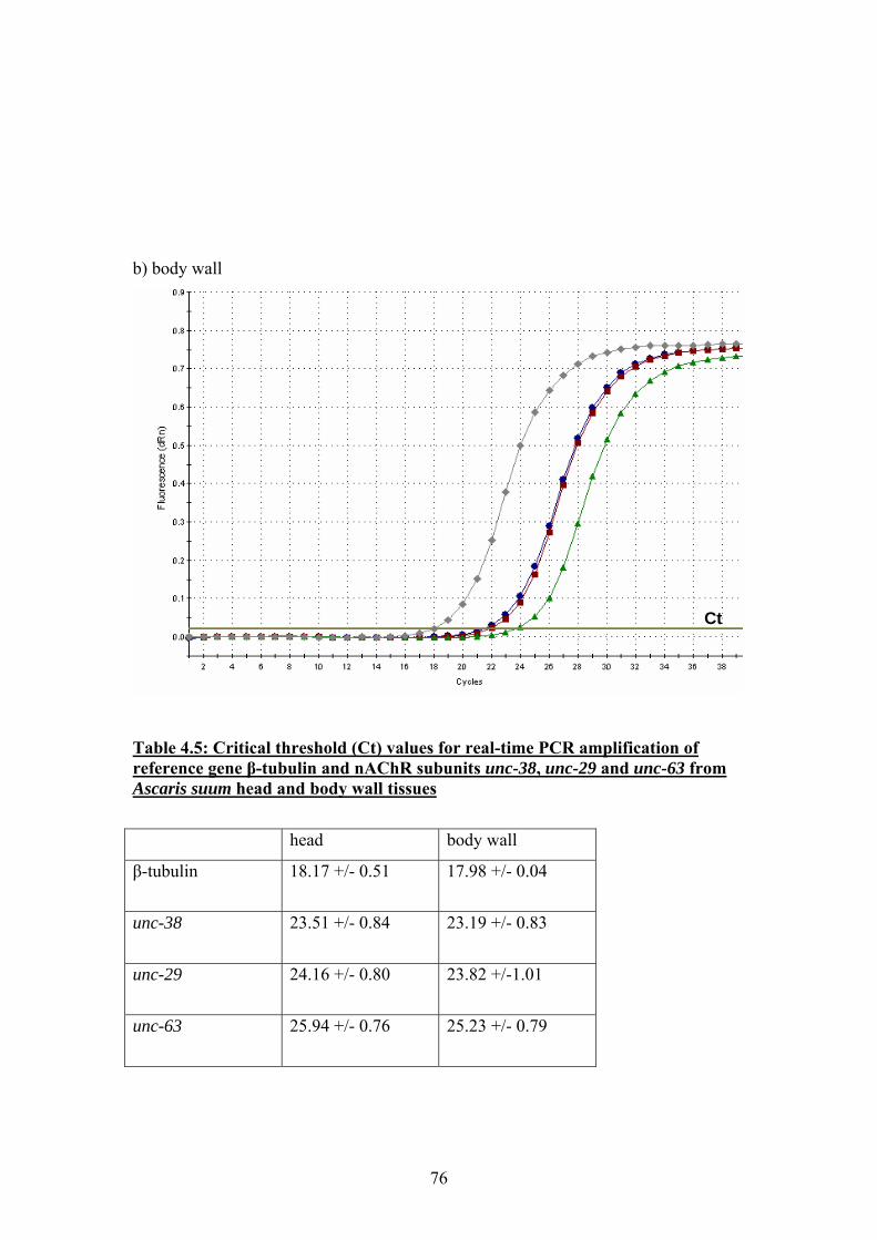
b) body wall
Ct
Table 4.5: Critical threshold (Ct) values for real-time PCR amplification of reference gene β-tubulin and nAChR subunits unc-38, unc-29 and unc-63 from Ascaris suum head and body wall tissues
head body wall
β-tubulin 18.17 +/- 0.51 17.98 +/- 0.04
unc-38 23.51 +/- 0.84
23.19 +/- 0.83
unc-29 24.16 +/- 0.80
23.82 +/-1.01
unc-63 25.94 +/- 0.76
25.23 +/- 0.79
76
4.5 Discussion In the course of this investigation, 3 full-length nAChR subunit sequences have been
cloned from Ascaris suum cDNA; two of these were clear homologues of the C.
elegans nAChR subunits unc-38 and unc-29, and are therefore good candidates for
components of a levamisole-sensitive nAChR (Fleming et al., 1997). The additional
full-length subunit, named acr-26, has no clear homologue in C. elegans but has been
shown to be present in the parasites Brugia malayi and Haemonchus contortus,
demonstrating that this subunit may be worthy of further investigation as a potential
new anthelmintic target. The amplification of a partial sequence of unc-63 from
Ascaris suum supports the findings described in chapter 3 that unc-63 appears to be
present in other parasitic nematodes; this is further supported by the amplification of
unc-63 from Ancylostoma caninum by McCarthy and colleagues. However, the failure
to successfully extend the partial sequence to full length using either additional
degenerate primers designed on conserved regions of other unc-63 sequences, or
using a primer linked to the polyA tail of the mRNA during the reverse transcription,
cannot be satisfactorily explained by the work presented here. Although unc-63
appears to be expressed at lower levels than unc-38 and unc-29, it is clearly detectable
by PCR. It is however a possibility that in Ascaris suum, the unc-63 transcript may be
alternatively spliced, or simply truncated; such matters cannot be resolved until the
genome sequence of Ascaris suum is completely sequenced, assembled and available
to search.
The amplification of cDNA sequence from Ascaris suum encoding these nAChR
subunits provides a basis for further investigations into the molecular biology
underlying the pharmacology of Ascaris suum nAChRs, in particular the basis of
levamisole and pyrantel sensitivity. If Ascaris suum populations resistant to these
drugs were to become available, the sequence of unc-38 and unc-29 could easily be
checked for polymorphisms which may underly this differential drug sensitivity. In
addition to this, if drug resistance was caused by a change in the expression levels of
nAChR subunits, we have demonstrated that expression levels of unc-38, unc-29 and
unc-63 from Ascaris suum can be quantified using real-time PCR. Such a difference
in expression levels was found between hookworms resistant and susceptible to
77

pyrantel, and studies such as these can provide the basis for diagnostic assays to
assess the level of drug resistance in a parasite population (Kopp et al., 2008; Walsh
et al., 2007).
78
Chapter 5: Immunohistochemical localisation of Ascaris suum UNC-38, UNC-29 and UNC-63
79
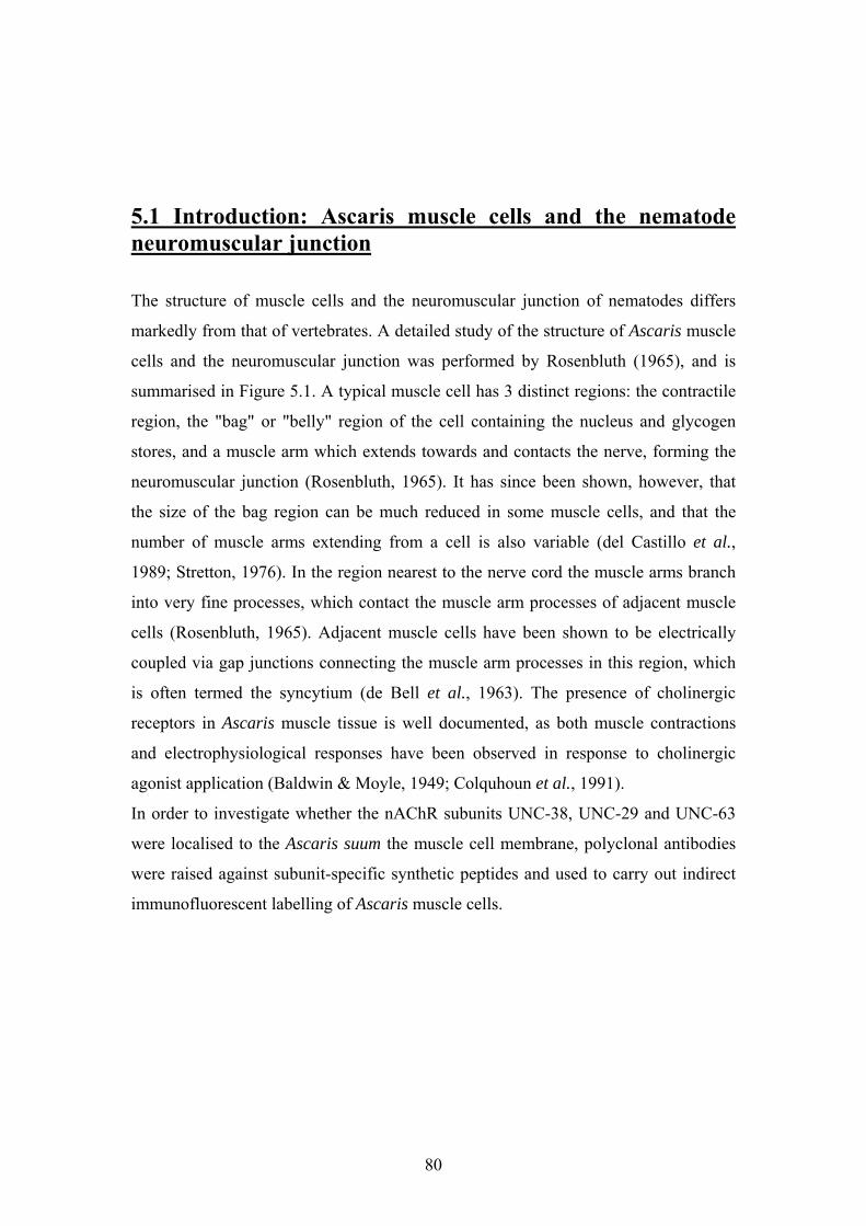
5.1 Introduction: Ascaris muscle cells and the nematode neuromuscular junction
The structure of muscle cells and the neuromuscular junction of nematodes differs
markedly from that of vertebrates. A detailed study of the structure of Ascaris muscle
cells and the neuromuscular junction was performed by Rosenbluth (1965), and is
summarised in Figure 5.1. A typical muscle cell has 3 distinct regions: the contractile
region, the "bag" or "belly" region of the cell containing the nucleus and glycogen
stores, and a muscle arm which extends towards and contacts the nerve, forming the
neuromuscular junction (Rosenbluth, 1965). It has since been shown, however, that
the size of the bag region can be much reduced in some muscle cells, and that the
number of muscle arms extending from a cell is also variable (del Castillo et al.,
1989; Stretton, 1976). In the region nearest to the nerve cord the muscle arms branch
into very fine processes, which contact the muscle arm processes of adjacent muscle
cells (Rosenbluth, 1965). Adjacent muscle cells have been shown to be electrically
coupled via gap junctions connecting the muscle arm processes in this region, which
is often termed the syncytium (de Bell et al., 1963). The presence of cholinergic
receptors in Ascaris muscle tissue is well documented, as both muscle contractions
and electrophysiological responses have been observed in response to cholinergic
agonist application (Baldwin & Moyle, 1949; Colquhoun et al., 1991).
In order to investigate whether the nAChR subunits UNC-38, UNC-29 and UNC-63
were localised to the Ascaris suum the muscle cell membrane, polyclonal antibodies
were raised against subunit-specific synthetic peptides and used to carry out indirect
immunofluorescent labelling of Ascaris muscle cells.
80
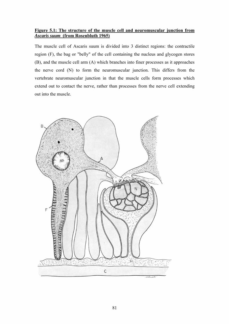
Figure 5.1: The structure of the muscle cell and neuromuscular junction from Ascaris suum (from Rosenbluth 1965) The muscle cell of Ascaris suum is divided into 3 distinct regions: the contractile
region (F), the bag or "belly" of the cell containing the nucleus and glycogen stores
(B), and the muscle cell arm (A) which branches into finer processes as it approaches
the nerve cord (N) to form the neuromuscular junction. This differs from the
vertebrate neuromuscular junction in that the muscle cells form processes which
extend out to contact the nerve, rather than processes from the nerve cell extending
out into the muscle.
81
5.2 Choice of peptides for antisera production and ELISA results
In order to determine regions of the nAChR subunits suitable for use as peptides for
the production of antisera, the translated N-terminal domain sequence of UNC-38,
UNC-29 and UNC-63 was analysed using ProteAN software. The results of this are
shown in Figure 5.2.1. The chosen peptides were predicted to be hydrophilic,
antigenic, and had a high probability of being present on the surface of the protein in
its folded conformation. The selected peptides were located at the extreme N-termini
of the subunits just after the predicted signal peptide cleavage site, a strategy
successfully used previously to raise antisera against nematode glutamate-gated
chloride channels (Delany et al., 1998).
The antisera were affinity purified, and enzyme-linked immunosorbent assays were
performed to ensure that the antibodies had activity against the peptides used for
antisera production. The results are shown in Figure 5.2.2, and confirm that the
terminal antisera and affinity purified antibody solutions all show a greater titre of
antibodies which react against the peptides used for antisera production than the pre-
immunw serum. The affinity purified antibody solutions also appeared to show less
non-specific reactivity at greater dilutions than the terminal antiserum. The ELISA
results were also used to determine an appropriate dilution of antibody for use in the
subsequent indirect immunofluorescence procedures.
82
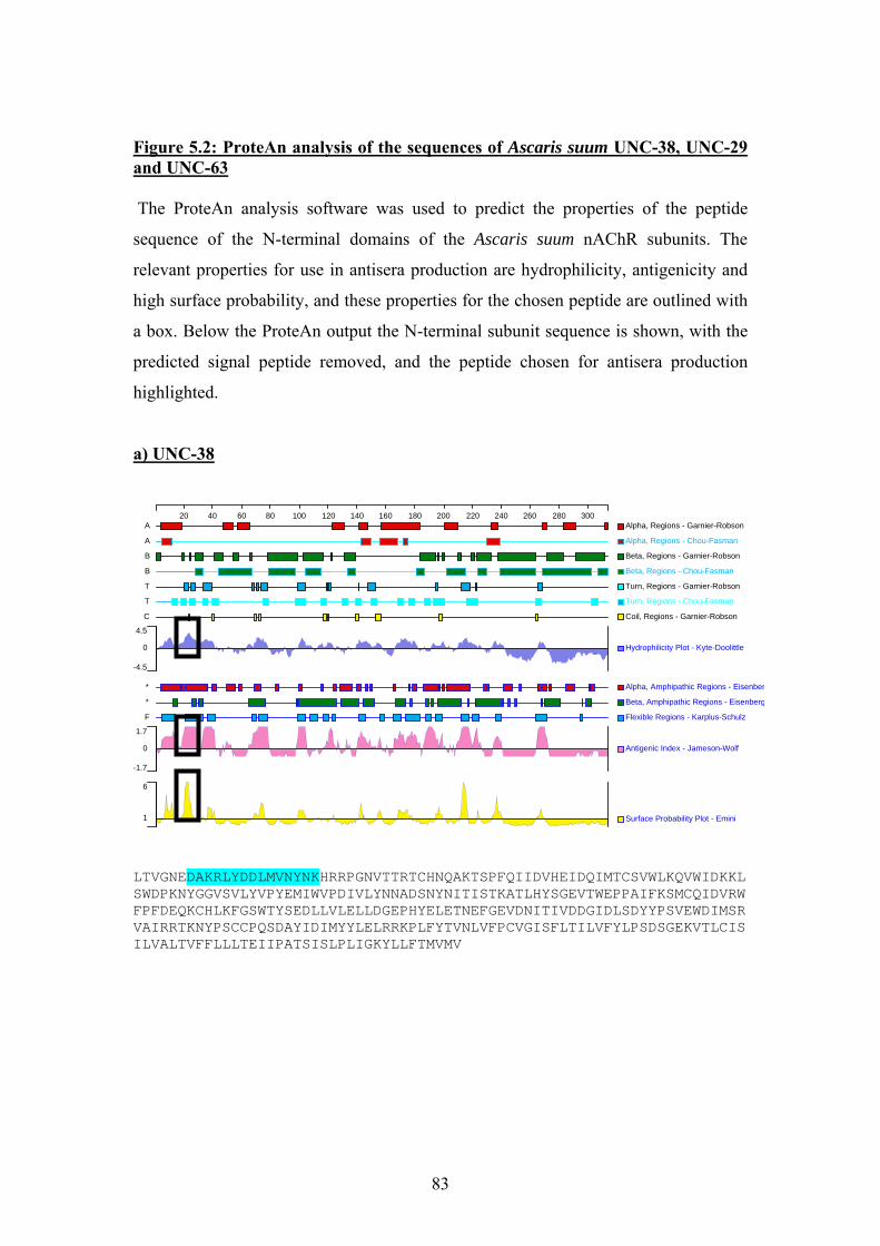
Figure 5.2: ProteAn analysis of the sequences of Ascaris suum UNC-38, UNC-29 and UNC-63 The ProteAn analysis software was used to predict the properties of the peptide
sequence of the N-terminal domains of the Ascaris suum nAChR subunits. The
relevant properties for use in antisera production are hydrophilicity, antigenicity and
high surface probability, and these properties for the chosen peptide are outlined with
a box. Below the ProteAn output the N-terminal subunit sequence is shown, with the
predicted signal peptide removed, and the peptide chosen for antisera production
highlighted.
a) UNC-38
20 40 60 80 100 120 140 160 180 200 220 240 260 280 300Alpha, Regions - Garnier-RobsonA
Alpha, Regions - Chou-FasmanA
Beta, Regions - Garnier-RobsonB
Beta, Regions - Chou-FasmanB
Turn, Regions - Garnier-RobsonT
Turn, Regions - Chou-FasmanT
Coil, Regions - Garnier-RobsonC
Hydrophilicity Plot - Kyte-Doolittle0
4.5
-4.5
Alpha, Amphipathic Regions - Eisenber*
Beta, Amphipathic Regions - Eisenberg*
Flexible Regions - Karplus-SchulzF
Antigenic Index - Jameson-Wolf0
1.7
-1.7
Surface Probability Plot - Emini1
6
LTVGNEDAKRLYDDLMVNYNKHRRPGNVTTRTCHNQAKTSPFQIIDVHEIDQIMTCSVWLKQVWIDKKLSWDPKNYGGVSVLYVPYEMIWVPDIVLYNNADSNYNITISTKATLHYSGEVTWEPPAIFKSMCQIDVRWFPFDEQKCHLKFGSWTYSEDLLVLELLDGEPHYELETNEFGEVDNITIVDDGIDLSDYYPSVEWDIMSRVAIRRTKNYPSCCPQSDAYIDIMYYLELRRKPLFYTVNLVFPCVGISFLTILVFYLPSDSGEKVTLCISILVALTVFFLLLTEIIPATSISLPLIGKYLLFTMVMV
83
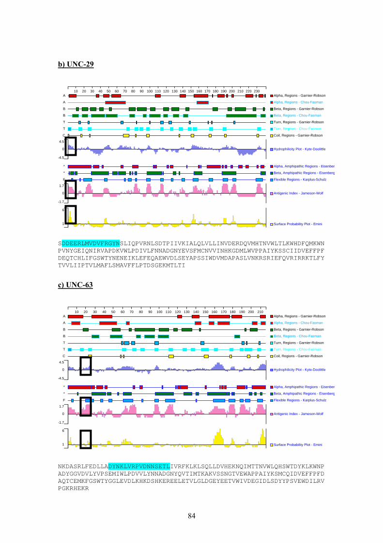
b) UNC-29
10 20 30 40 50 60 70 80 90 100 110 120 130 140 150 160 170 180 190 200 210 220 230Alpha, Regions - Garnier-RobsonA
Alpha, Regions - Chou-FasmanA
Beta, Regions - Garnier-RobsonB
Beta, Regions - Chou-FasmanB
Turn, Regions - Garnier-RobsonT
Turn, Regions - Chou-FasmanT
Coil, Regions - Garnier-RobsonC
Hydrophilicity Plot - Kyte-Doolittle0
4.5
-4.5
Alpha, Amphipathic Regions - Eisenber*
Beta, Amphipathic Regions - Eisenberg*
Flexible Regions - Karplus-SchulzF
Antigenic Index - Jameson-Wolf0
1.7
-1.7
Surface Probability Plot - Emini1
6
SDDEERLMVDVFRGYNSLIQPVRNLSDTPIIVKIALQLVLLINVDERDQVMHTNVWLTLKWHDFQMKWNPVNYGEIQNIRVAPDKVWLPDIVLFNNADGNYEVSFMCNVVINHKGDMLWVPPAIYKSSCIIDVEFFPFDEQTCHLIFGSWTYNENEIKLEFEQAEWVDLSEYAPSSIWDVMDAPASLVNKRSRIEFQVRIRRKTLFYTVVLIIPTVLMAFLSMAVFFLPTDSGEKMTLTI
c) UNC-63
10 20 30 40 50 60 70 80 90 100 110 120 130 140 150 160 170 180 190 200 210Alpha, Regions - Garnier-RobsonA
Alpha, Regions - Chou-FasmanA
Beta, Regions - Garnier-RobsonB
Beta, Regions - Chou-FasmanB
Turn, Regions - Garnier-RobsonT
Turn, Regions - Chou-FasmanT
Coil, Regions - Garnier-RobsonC
Hydrophilicity Plot - Kyte-Doolittle0
4.5
-4.5
Alpha, Amphipathic Regions - Eisenber*
Beta, Amphipathic Regions - Eisenberg*
Flexible Regions - Karplus-SchulzF
Antigenic Index - Jameson-Wolf0
1.7
-1.7
Surface Probability Plot - Emini1
6
NKDASRLFEDLLADYNKLVRPVDNNSETLIVRFKLKLSQLLDVHEKNQIMTTNVWLQHSWTDYKLKWNPADYGGVDVLYVPSEMIWLPDVVLYNNADGNYQVTIMTKAKVSSNGTVEWAPPAIYKSMCQIDVEFFPFDAQTCEMKFGSWTYGGLEVDLKHKDSHKEREELETVLGLDGEYEETVWIVDEGIDLSDYYPSVEWDILRVPGKRHEKR
84
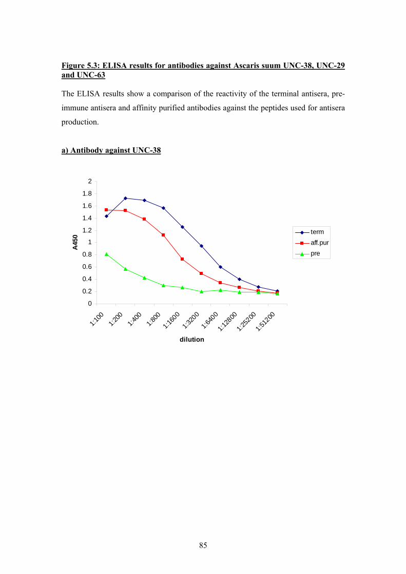
Figure 5.3: ELISA results for antibodies against Ascaris suum UNC-38, UNC-29 and UNC-63 The ELISA results show a comparison of the reactivity of the terminal antisera, pre-
immune antisera and affinity purified antibodies against the peptides used for antisera
production.
a) Antibody against UNC-38
0
0.2
0.4
0.6
0.8
1
1.2
1.4
1.6
1.8
2
1:100
1:200
1:400
1:800
1:160
0
1:320
0
1:640
0
1:128
00
1:252
00
1:512
00
dilution
A45
0 termaff.purpre
85
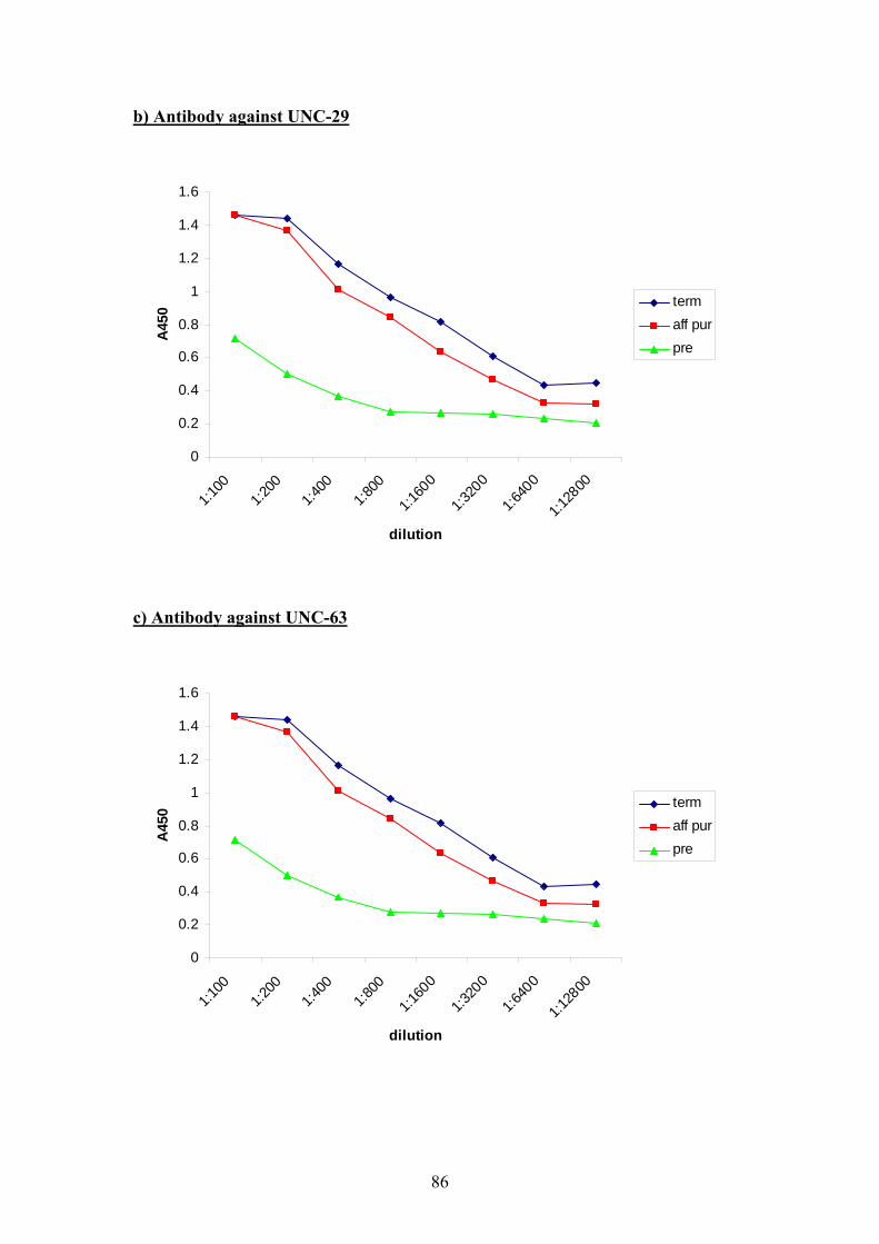
b) Antibody against UNC-29
0
0.2
0.4
0.6
0.8
1
1.2
1.4
1.6
1:100
1:200
1:400
1:800
1:160
0
1:320
0
1:640
0
1:128
00
dilution
A45
0 termaff purpre
c) Antibody against UNC-63
0
0.2
0.4
0.6
0.8
1
1.2
1.4
1.6
1:100
1:200
1:400
1:800
1:160
0
1:320
0
1:640
0
1:128
00
dilution
A45
0 termaff purpre
86
5.3 Indirect Immunofluorescence
Muscle cells and cuticle-mounted nerve cords were prepared by collagenase digestion
of fresh Ascaris suum adult females as described in section 2.4.4 (Johnson & Stretton,
1985). The material was then fixed and sequentially incubated with primary and
secondary antibody solutions as descibed in section 2.4.5 (Brownlee et al., 1993).
Negative controls were used to demonstrate that the antibody labelling was specific to
the nAChR subunits; these are shown in Figure 5.3.1. The negative controls were pre-
absorbed primary antibody, normal goat IgG, and secondary antiserum alone. The
negative controls all showed only very low levels of immunofluorescence consistent
with very little non-specific antibody labelling. Unfortunately the microscope used
did not have a phase-contrast feature which would have allowed clear images of the
specimens to be obtained using visible light.
Initially, Ascaris suum muscle cells, and strips of cuticle with attached nerve cord,
were incubated with antibody against UNC-38. The purpose of this was to investigate
whether the nAChRs containing this subunit were neuromuscular or neuronal. The
results of this are shown in Figure 5.3.2. UNC-38 is clearly localised to the muscle
cell membrane, and is present over the entire muscle cell surface. The neuronal tissue
itself was not labelled with the antibody against UNC-38, but the nerve-cord
preparation did reveal details of the muscle arm processes contacting the nerve cord to
form the neuromuscular junctions.
Muscle cells were then incubated with primary antibodies against both UNC-38 and
UNC-29, followed by secondary antibodies conjugated to different fluorescent labels,
to ascertain whether UNC-38 and UNC-29 co-localised. The results of this are shown
in Figure 5.3.3. Both UNC-38 and UNC-29 were localised to the muscle cell
membrane, and clearly co-localised, especially in the region of the muscle cell arm.
At high magnification, the co-localised fluorescence has a punctate appearance, which
may represent receptor clusters in the distal region of the muscle cell arm.
Finally, muscle cells were incubated with antibodies against UNC-63, the results of
which are shown in Figure 5.3.4. UNC-63 is also clearly localised to the muscle cell
arms of Ascaris suum. However, attempts to perform co-localisation experiments
using antibodies against UNC-63 and UNC-29 together were unsuccessful, with only
the UNC-29 labelling being visible (results not shown). Possible reasons for this are
discussed in section 5.4.
87
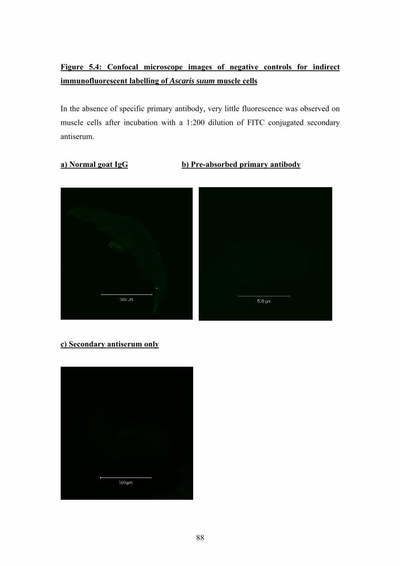
Figure 5.4: Confocal microscope images of negative controls for indirect
immunofluorescent labelling of Ascaris suum muscle cells
In the absence of specific primary antibody, very little fluorescence was observed on
muscle cells after incubation with a 1:200 dilution of FITC conjugated secondary
antiserum.
a) Normal goat IgG b) Pre-absorbed primary antibody
c) Secondary antiserum only
88
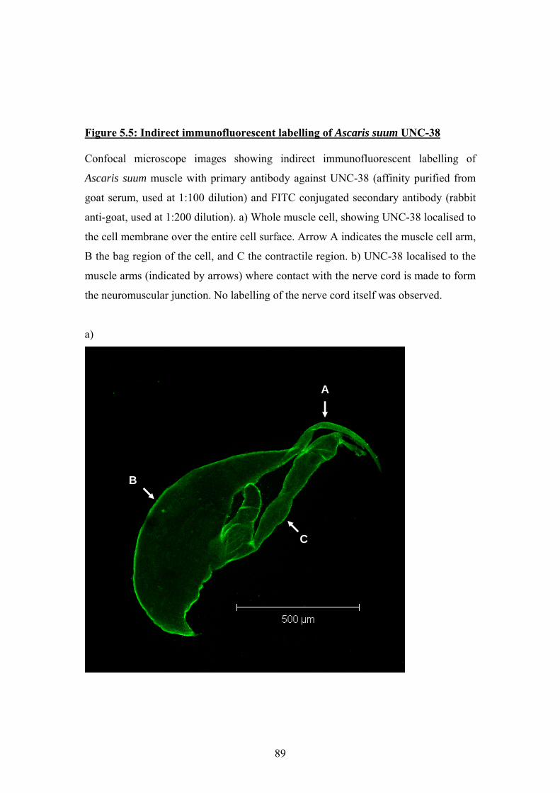
Figure 5.5: Indirect immunofluorescent labelling of Ascaris suum UNC-38 Confocal microscope images showing indirect immunofluorescent labelling of
Ascaris suum muscle with primary antibody against UNC-38 (affinity purified from
goat serum, used at 1:100 dilution) and FITC conjugated secondary antibody (rabbit
anti-goat, used at 1:200 dilution). a) Whole muscle cell, showing UNC-38 localised to
the cell membrane over the entire cell surface. Arrow A indicates the muscle cell arm,
B the bag region of the cell, and C the contractile region. b) UNC-38 localised to the
muscle arms (indicated by arrows) where contact with the nerve cord is made to form
the neuromuscular junction. No labelling of the nerve cord itself was observed.
a)
A
B
C
89
b)
90
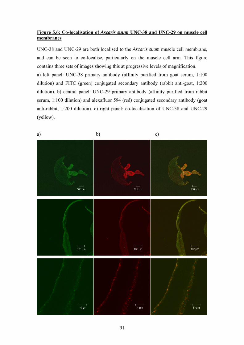
Figure 5.6: Co-localisation of Ascaris suum UNC-38 and UNC-29 on muscle cell membranes
NC-38 and UU NC-29 are both localised to the Ascaris suum muscle cell membrane,
100
) b) c)
and can be seen to co-localise, particularly on the muscle cell arm. This figure
contains three sets of images showing this at progressive levels of magnification.
a) left panel: UNC-38 primary antibody (affinity purified from goat serum, 1:
dilution) and FITC (green) conjugated secondary antibody (rabbit anti-goat, 1:200
dilution). b) central panel: UNC-29 primary antibody (affinity purified from rabbit
serum, 1:100 dilution) and alexafluor 594 (red) conjugated secondary antibody (goat
anti-rabbit, 1:200 dilution). c) right panel: co-localisation of UNC-38 and UNC-29
(yellow).
a
91
Figure 5.7: Indirect immunofluorescent labelling of Ascaris suum UNC-63 Indirect immunofluorescent labelling of the Ascaris suum muscle cell arm using
primary antibody against UNC-63 (affinity purified from goat serum, 1:100 dilution)
and FITC conjugated secondary antibody (rabbit anti-goat, 1:200 dilution). A
greyscale image was obtained, and is shown at two different magnifications.
92
5.4 Discussion
The strategy of raising antisera against short N-terminal subunit-specific peptides
from the nAChR subunits UNC-38, UNC-29 and UNC-63 appears to have been
successful; the ELISA results confirm that in all cases, immunisation resulted in the
production of antibodies directed against the peptides. The negative controls carried
out alongside the immunofluorescent labelling did not show any fluorescent labelling
in the absence of specific primary antibody, and non-specific IgG did not bind to the
Ascaris suum muscle cells. However, the absolute specificity of these antibodies for
the target nAChR subunit can not be taken for granted; in the case of mammalian
nAChRs it has been demonstrated that antibodies against α7 also recognise other
roteins, and will label tissue from the α7-/- mouse (Jones & Wonnacott, 2005). In the
ase of a parasitic nematode for which transgenic techniques are unavailable, it is very
ifficult to confirm antibody specificity in such a manner. The specificity of our
antibodies for the target nAChR has to some extent been confirmed by our
State University, who were able to partially inhibit levamisole-
duced contractions of Ascaris suum muscle by prior incubation with the antibodies
p
c
d
collaborators at Iowa
in
directed against UNC-38 and UNC-29 (Alan Robertson, personal communication).
No such effect was seen using the antibody directed against UNC-63, and it is
interesting that this is the antibody for which we failed to achieve co-localisation with
the antibody directed against UNC-29. This could suggest that UNC-63 is perhaps not
a component of either the UNC-29 containing nAChR or the levamisole-receptor.
However, any negative results obtained by our collaborators may also be explained by
the difficulties involved in sufficiently permeabilising the Ascaris muscle tissue to
allow antibody penetration while still retaining enough integrity that the muscle will
contract (Alan Robertson, personal communication). Similarly, our failure to
demonstrate co-localisation may result from some interaction between the UNC-63
and UNC-29 antibodies, or may have been due to the presence of less UNC-63
protein than UNC-29, which might have led to the stronger fluorescence from UNC-
29 masking weaker fluorescence from UNC-63. Unfortunately it was not possible to
obtain sufficient fresh Ascaris suum specimens to make repeated attempts to
demonstrate co-localisation of UNC-63 and UNC-29; indeed, a shortage of fresh, live
93
Ascaris specimens limited the number of times any of the immunofluorescence
experiments could be carried out to 3 replicates of the UNC-38 and UNC-29 labelling,
and two replicates of the UNC-63 labelling.
The results shown here demonstrate that Ascaris suum UNC-38 and UNC-29 are
localised to the muscle cell membrane, and clearly co-localise. This is consistent with
the proposed role for UNC-38 and UNC-29 as components of the same levamisole-
sensitive receptor present at the C. elegans neuromuscular junction (Fleming et al.,
1997). The distribution of receptors over the whole surface of the Ascaris suum
muscle cell differs from the receptor distribution in C. elegans, where the receptors
are confined to clusters at the neuromuscular junction (Qian et al., 2008). However,
the presence of nAChRs on the bag region of the Ascaris suum muscle cell is well
described, and single-channel electrophysiological recordings of Ascaris cholinergic
receptors are routinely performed using the bag region of the muscle cell (Brading &
Caldwell, 1971; Pennington & Martin, 1990). It is therefore likely that the distribution
of fluorescent labelling observed on the Ascaris muscle cell does indeed represent the
distribution of actual nAChRs.
94

Chapter 6: Electrophysiology
95
6.1 Introduction
The oocytes of the African clawed frog Xenopus laevis have been used for the
expression of exogenous proteins since the 1970's. Initially it was discovered that
injection of mRNA encoding globins into oocytes resulted in translation of the
exogenous protein (Gurdon et al., 1974). Subsequently, it was demonstrated that
injection of mRNA or cDNA extracted from vertebrate brain resulted in the
expression of neuronal membrane proteins and receptors (Ballivet et al., 1988;
Tomaselli et al., 1989). Of particular relevance to the work presented here, it was
shown that injection of Torpedo electric organ mRNA into Xenopus oocytes resulted
in the production of functional nAChRs (Sumikawa et al., 1984). Other studies
showed that cloning, in vitro transcription and oocyte injection of Torpedo nAChR
subunits also produced functional recombinant nAChRs (Mishina et al., 1984). The
Xenopus oocyte system therefore has a long established history as a useful tool for
heterologous expression of ligand-gated ion channels.
The most widely used electrophysiological technique employed to study exogenous
receptors and channels expressed in Xenopus oocytes is two-electrode voltage clamp.
The principal of this technique is to control the cell membrane potential in order to
allow measurements of current to be made; initially this procedure was developed to
study the giant axons of squid (Hodgkin et al., 1952; Marmont, 1949). The large size
of the Xenopus oocyte makes it particularly amenable to two-electrode voltage-clamp
experiments. A voltage electrode inserted through the cell membrane holds the
membrane potential constant, while a second intracellular electrode applies the
necessary current to maintain this membrane potential. The current is applied and the
holding potential maintained by a feedback amplifier connected to the electrodes
(summarised in Hille 2001). The changes in current needed to maintain the membrane
potential correspond to changes in ion flow through the membrane, which occur when
the ion channels under investigation open. These changes in current are converted to a
digital signal and recorded by a computer equipped with appropriate software.
The Xenopus oocyte expression system and the two-electrode voltage clamp
technique have been used to study ligand-gated ion channels of both vertebrates and
invertebrates. The system allows the proposed function of cloned cDNAs showing
sequence homology with other LGICs to be confirmed (Schofield et al., 1987).
96
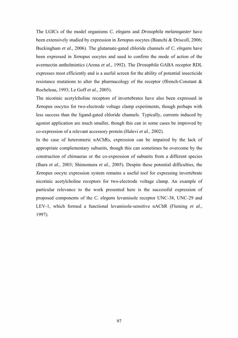
The LGICs of the model organisms C. elegans and Drosophila melanogaster have
been extensively studied by expression in Xenopus oocytes (Bianchi & Driscoll, 2006;
Buckingham et al., 2006). The glutamate-gated chloride channels of C. elegans have
been expressed in Xenopus oocytes and used to confirm the mode of action of the
avermectin anthelmintics (Arena et al., 1992). The Drosophila GABA receptor RDL
expresses most efficiently and is a useful screen for the ability of potential insecticide
resistance mutations to alter the pharmacology of the receptor (ffrench-Constant &
Rocheleau, 1993; Le Goff et al., 2005).
The nicotinic acetylcholine receptors of invertebrates have also been expressed in
Xenopus oocytes for two-electrode voltage clamp experiments, though perhaps with
less success than the ligand-gated chloride channels. Typically, currents induced by
agonist application are much smaller, though this can in some cases be improved by
co-expression of a relevant accessory protein (Halevi et al., 2002).
In the case of heteromeric nAChRs, expression can be impaired by the lack of
appropriate complementary subunits, though this can sometimes be overcome by the
construction of chimaeras or the co-expression of subunits from a different species
(Ihara et al., 2003; Shimomura et al., 2005). Despite these potential difficulties, the
Xenopus oocyte expression system remains a useful tool for expressing invertebrate
nicotinic acetylcholine receptors for two-electrode voltage clamp. An example of
particular relevance to the work presented here is the successful expression of
proposed components of the C. elegans levamisole receptor UNC-38, UNC-29 and
LEV-1, which formed a functional levamisole-sensitive nAChR (Fleming et al.,
1997).
97
6.2: Initial experiments to co-express Ascaris unc-38 and unc-29
Preliminary experiments were performed to demonstrate that Ascaris unc-38 and unc-
29 did indeed co-express to form a heteromeric receptor. Oocytes were injected either
with 25ng of Ascaris unc-38 cRNA, 25ng of Ascaris unc-29 cRNA, or 25ng Ascaris
unc-38+25ng Ascaris unc-29 cRNA. All injection volumes were adjusted to 50nl
using nuclease-free water. The results of this are shown in Figure 6.1, and clearly
demonstrate that both Ascaris unc-38 and unc-29 are required to produce a functional
nAChR which responds to ACh. The response to 100µM ACh was ~600-800nA,
varying between different oocytes.
Preliminary pharmacological experiments were then performed to discover which
agonists the heterologously expressed Ascaris nAChR responded to, and to ensure
that such responses could be blocked with an appropriate antagonist to confirm a
genuine nAChR response. The agonists used were nicotine, ACh and levamisole; it
has been demonstrated that in C. elegans, UNC-38 and UNC-29 are components of
the levamisole-sensitive nAChR (Fleming et al., 1997). The antagonist chosen was
mecamylamine; this is a broad-spectrum non-competitive antagonist of nAChRs, and
has additionally been shown to inhibit ACh-induced contractions and currents in
native Ascaris muscle preparations (Colquhoun et al., 1991; Natoff, 1969). The
results are shown in Figure 6.2. The receptors composed of Ascaris unc-38 and unc-
29 clearly respond to 100µM concentrations of acetylcholine, nicotine and
levamisole; the response to levamisole appeared slightly larger than the response to
ACh, which was in turn slightly larger than the response to nicotine. The responses to
all these agonists can be fully blocked by application of 10µM mecamylamine, and
the response recovers when the mecamylamine is washed off. The response to
nicotine, and the blockade by mecamylamine, confirm that the ACh response is
nicotinic in nature, rather than muscarinic. The relatively large response to levamisole
confirms that in Ascaris suum, UNC-38 and UNC-29 are components of a levamisole-
sensitive nAChR, as they are in C. elegans.
98
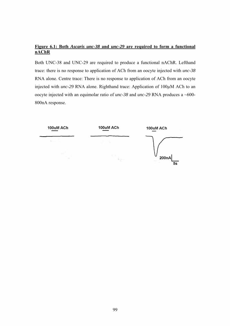
Figure 6.1: Both Ascaris unc-38 and unc-29 are required to form a functional nAChR Both UNC-38 and UNC-29 are required to produce a functional nAChR. Lefthand
trace: there is no response to application of ACh from an oocyte injected with unc-38
RNA alone. Centre trace: There is no response to application of ACh from an oocyte
injected with unc-29 RNA alone. Righthand trace: Application of 100µM ACh to an
oocyte injected with an equimolar ratio of unc-38 and unc-29 RNA produces a ~600-
800nA response.
99
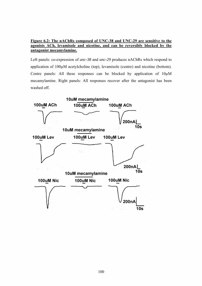
Figure 6.2: The nAChRs composed of UNC-38 and UNC-29 are sensitive to the agonists ACh, levamisole and nicotine, and can be reversibly blocked by the antagonist mecamylamine. Left panels: co-expression of unc-38 and unc-29 produces nAChRs which respond to
application of 100µM acetylcholine (top), levamisole (centre) and nicotine (bottom).
Centre panels: All these responses can be blocked by application of 10µM
mecamylamine. Right panels: All responses recover after the antagonist has been
washed off.
100
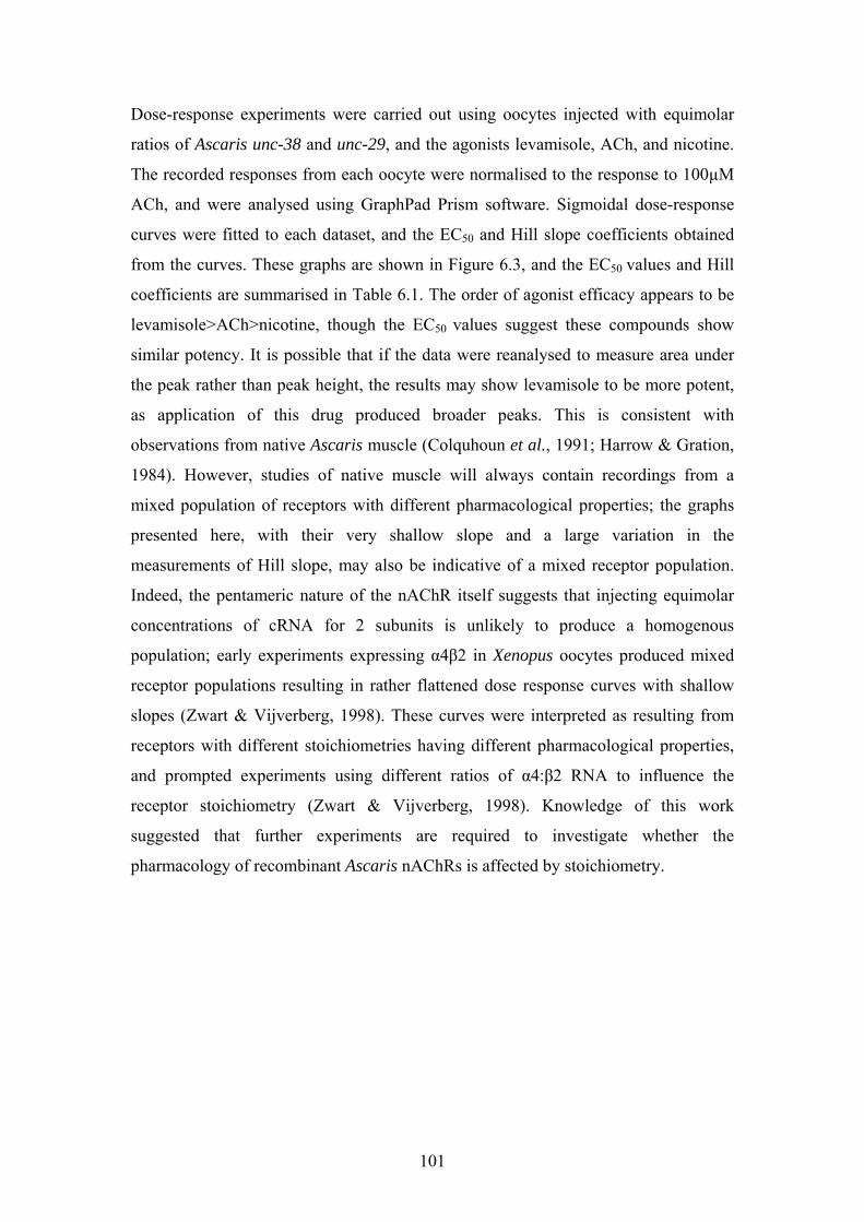
Dose-response experiments were carried out using oocytes injected with equimolar
ratios of Ascaris unc-38 and unc-29, and the agonists levamisole, ACh, and nicotine.
The recorded responses from each oocyte were normalised to the response to 100µM
ACh, and were analysed using GraphPad Prism software. Sigmoidal dose-response
curves were fitted to each dataset, and the EC50 and Hill slope coefficients obtained
from the curves. These graphs are shown in Figure 6.3, and the EC50 values and Hill
coefficients are summarised in Table 6.1. The order of agonist efficacy appears to be
levamisole>ACh>nicotine, though the EC50 values suggest these compounds show
similar potency. It is possible that if the data were reanalysed to measure area under
the peak rather than peak height, the results may show levamisole to be more potent,
as application of this drug produced broader peaks. This is consistent with
observations from native Ascaris muscle (Colquhoun et al., 1991; Harrow & Gration,
1984). However, studies of native muscle will always contain recordings from a
mixed population of receptors with different pharmacological properties; the graphs
presented here, with their very shallow slope and a large variation in the
measurements of Hill slope, may also be indicative of a mixed receptor population.
Indeed, the pentameric nature of the nAChR itself suggests that injecting equimolar
concentrations of cRNA for 2 subunits is unlikely to produce a homogenous
population; early experiments expressing α4β2 in Xenopus oocytes produced mixed
receptor populations resulting in rather flattened dose response curves with shallow
slopes (Zwart & Vijverberg, 1998). These curves were interpreted as resulting from
receptors with different stoichiometries having different pharmacological properties,
and prompted experiments using different ratios of α4:β2 RNA to influence the
receptor stoichiometry (Zwart & Vijverberg, 1998). Knowledge of this work
suggested that further experiments are required to investigate whether the
pharmacology of recombinant Ascaris nAChRs is affected by stoichiometry.
101
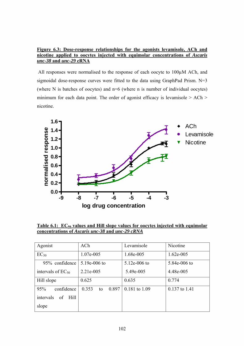
Figure 6.3: Dose-response relationships for the agonists levamisole, ACh and nicotine applied to oocytes injected with equimolar concentrations of Ascaris unc-38 and unc-29 cRNA All responses were normalised to the response of each oocyte to 100µM ACh, and
sigmoidal dose-response curves were fitted to the data using GraphPad Prism. N=3
(where N is batches of oocytes) and n=6 (where n is number of individual oocytes)
minimum for each data point. The order of agonist efficacy is levamisole > ACh >
nicotine.
-9 -8 -7 -6 -5 -4 -30.00.20.40.60.81.01.21.41.6
AChLevamisoleNicotine
log drug concentration
norm
alis
ed r
espo
nse
Table 6.1: EC50 values and Hill slope values for oocytes injected with equimolar concentrations of Ascaris unc-38 and unc-29 cRNA
Agonist ACh Levamisole Nicotine
EC50 1.07e-005 1.68e-005 1.62e-005
95% confidence
intervals of EC50
5.19e-006 to
2.21e-005
5.12e-006 to
5.49e-005
5.84e-006 to
4.48e-005
Hill slope 0.625 0.635 0.774
95% confidence
intervals of Hill
slope
0.353 to 0.897
0.181 to 1.09 0.137 to 1.41
102
6.3 Experiments using oocytes injected with 1:5 and 5:1 ratios of Ascaris unc-38 and unc29 cRNA
Oocytes were injected with Ascaris unc-38 + unc-29 cRNA in the ratios 1:5 and 5:1
unc-38:unc-29. The total amount of RNA injected was kept constant at 50ng per
oocyte, in an injection volume of 50nl. It was hoped that this would drive the
stoichiometry of the expressed receptors to either 3:2 UNC-38:UNC-29 or 2:3 UNC-
38:UNC-29, to allow any difference in pharmacology between the two types to be
further investigated. Although potentially receptors with a 4:1 or 1:4 stoichiometry
could also be generated, it has previously been demonstrated by transfection of cells
with α4 and β2 cDNA that the stoichiometry of such heteromeric receptors composed
of just 2 subunits is 3:2 or 2:3, and that adjusting the subunit cDNA ratios drove the
receptors to one stoichiometry or the other without introducing new combinations
(Nelson et al., 2003). Preliminary experiments suggested that the 1:5 and 5:1 RNA
ratios were optimal for this experiment; although studies of the human α4β2 receptor
have used 10:1 or 9:1 ratios to drive changes in receptor stoichiometry (Moroni et al.,
2006; Nelson et al., 2003; Zwart & Vijverberg, 1998). 1:10 and 10:1 ratios of Ascaris
unc-38:unc-29 RNA either did not produce functional receptors, or resulted in very
low expression levels where the maximal current was <20nA.
Dose-response experiments using the agonists levamisole and nicotine were carried
out on the receptors expressed from injection of the different RNA ratios. Maximal
responses from oocytes injected with the 1:5 ratio were ~300-400nA; for the 5:1 ratio,
~100-200nA. The responses from each oocyte were normalised to the response to
100µM ACh, and were again analysed using GraphPad Prism as described in Results
section 1. Dose response relationships and examples of electrophysiology traces for
oocytes injected with a 1:5 ratio of unc-38:unc-29 RNA are shown in Figure 6.4. Dose
response relationships and examples of electrophysiology traces for oocytes injected
with a 5:1 ratio of unc-38:unc-29 RNA are shown in Figure 6.5. The EC50 and Hill
slope values are presented in Table 6.2.
Changing the stoichiometry of the Ascaris nAChR by using different RNA ratios
clearly has an effect on the pharmacology. The 1:5 ratio produces a receptor
population which is more sensitive to levamisole, whereas nicotine is only a partial
agonist . In contrast to this, the 5:1 ratio produces a receptor population which is most
sensitive to nicotine, whereas levamisole is only a partial agonist. It cannot be inferred
103

from the data collected here whether the partial activity of the less efficacious
compound resulted from a true partial agonism of an almost homogeneous receptor
population, or from full agonist activity at a sub-population of receptors with the
opposite stoichiometry to that of the majority in a heterogeneous population. Once
again, the relatively shallow slope of the graphs may suggest a mixed receptor
population, but attempts to fit two separate curves to each dataset using Graphpad
Prism were unsuccessful.
104
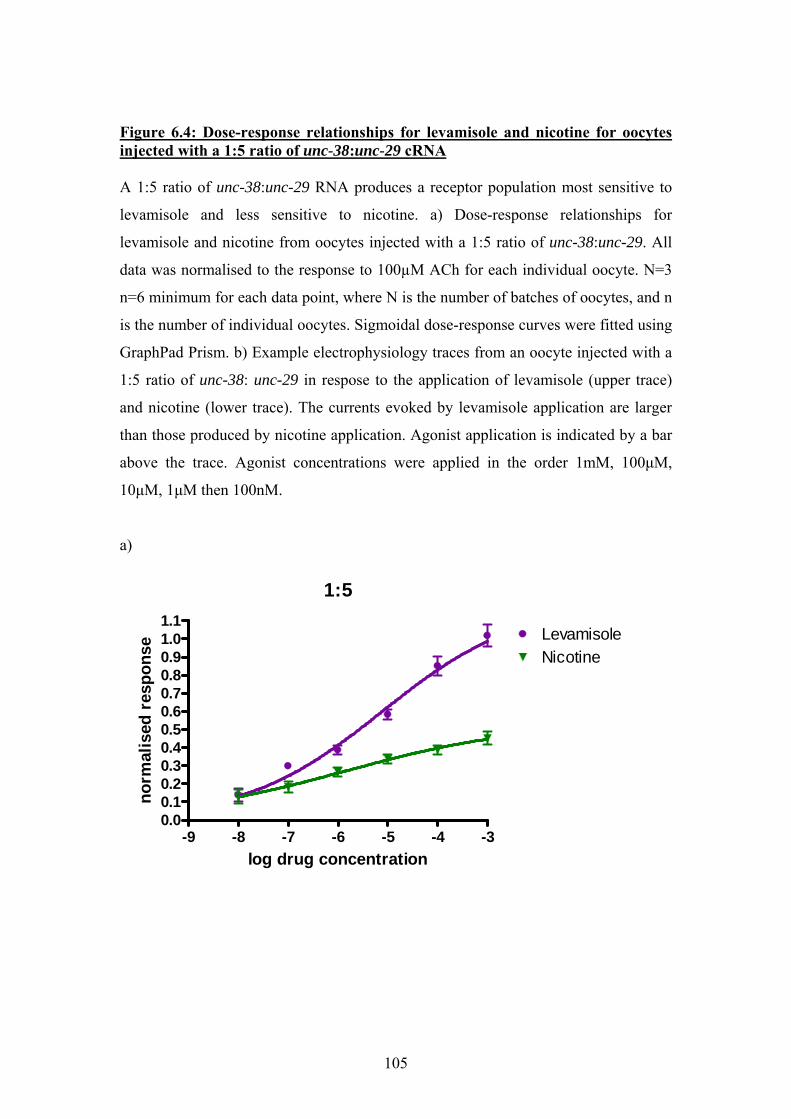
Figure 6.4: Dose-response relationships for levamisole and nicotine for oocytes injected with a 1:5 ratio of unc-38:unc-29 cRNA A 1:5 ratio of unc-38:unc-29 RNA produces a receptor population most sensitive to
levamisole and less sensitive to nicotine. a) Dose-response relationships for
levamisole and nicotine from oocytes injected with a 1:5 ratio of unc-38:unc-29. All
data was normalised to the response to 100µM ACh for each individual oocyte. N=3
n=6 minimum for each data point, where N is the number of batches of oocytes, and n
is the number of individual oocytes. Sigmoidal dose-response curves were fitted using
GraphPad Prism. b) Example electrophysiology traces from an oocyte injected with a
1:5 ratio of unc-38: unc-29 in respose to the application of levamisole (upper trace)
and nicotine (lower trace). The currents evoked by levamisole application are larger
than those produced by nicotine application. Agonist application is indicated by a bar
above the trace. Agonist concentrations were applied in the order 1mM, 100µM,
10µM, 1µM then 100nM.
a)
1:5
-9 -8 -7 -6 -5 -4 -30.00.10.20.30.40.50.60.70.80.91.01.1
LevamisoleNicotine
log drug concentration
norm
alis
ed r
espo
nse
105
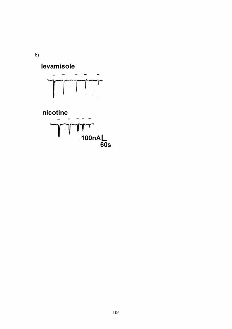
b)
106
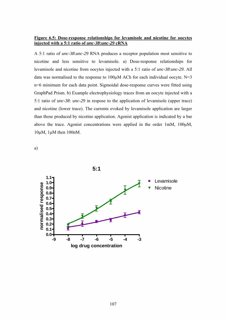
Figure 6.5: Dose-response relationships for levamisole and nicotine for oocytes injected with a 5:1 ratio of unc-38:unc-29 cRNA A 5:1 ratio of unc-38:unc-29 RNA produces a receptor population most sensitive to
nicotine and less sensitive to levamisole. a) Dose-response relationships for
levamisole and nicotine from oocytes injected with a 5:1 ratio of unc-38:unc-29. All
data was normalised to the response to 100µM ACh for each individual oocyte. N=3
n=6 minimum for each data point. Sigmoidal dose-response curves were fitted using
GraphPad Prism. b) Example electrophysiology traces from an oocyte injected with a
5:1 ratio of unc-38: unc-29 in respose to the application of levamisole (upper trace)
and nicotine (lower trace). The currents evoked by levamisole application are larger
than those produced by nicotine application. Agonist application is indicated by a bar
above the trace. Agonist concentrations were applied in the order 1mM, 100µM,
10µM, 1µM then 100nM.
a)
5:1
-9 -8 -7 -6 -5 -4 -30.00.10.20.30.40.50.60.70.80.91.01.1
LevamisoleNicotine
log drug concentration
norm
alis
ed r
espo
nse
107
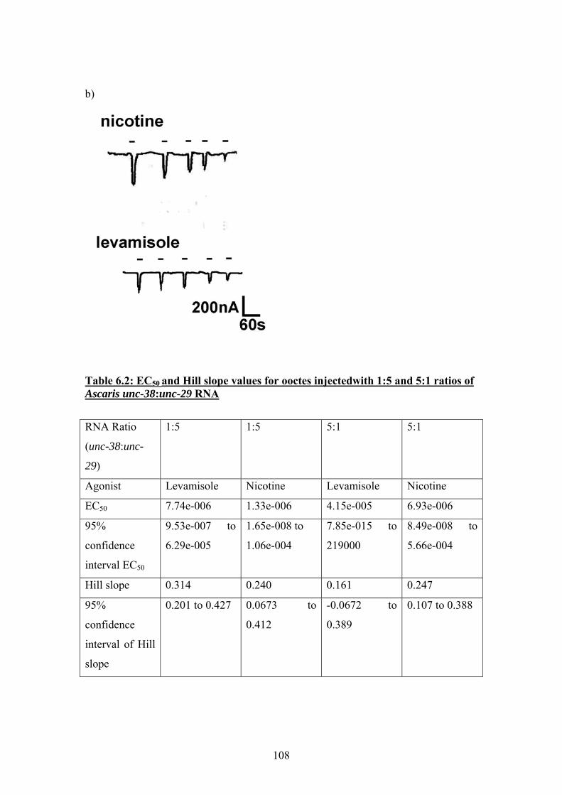
b)
Table 6.2: EC50 and Hill slope values for ooctes injectedwith 1:5 and 5:1 ratios of Ascaris unc-38:unc-29 RNA
RNA Ratio
(unc-38:unc-
29)
1:5 1:5 5:1 5:1
Agonist Levamisole Nicotine Levamisole Nicotine
EC50 7.74e-006 1.33e-006 4.15e-005 6.93e-006
95%
confidence
interval EC50
9.53e-007 to
6.29e-005
1.65e-008 to
1.06e-004
7.85e-015 to
219000
8.49e-008 to
5.66e-004
Hill slope 0.314 0.240 0.161 0.247
95%
confidence
interval of Hill
slope
0.201 to 0.427 0.0673 to
0.412
-0.0672 to
0.389
0.107 to 0.388
108
Although changing the receptor pharmacology by adjusting the stoichiometry is well
described for the mammalian α4β2 receptor (Moroni & Bermudez, 2006; Moroni et
al., 2006; Nelson et al., 2003), this has not previously been demonstrated in
invertebrates. The striking difference in sensitivity to levamisole and nicotine
observed between receptor populations driven to different stoichiometry also raised
the possibility that two of the observed pharmacological subtypes found in native
Ascaris muscle (the L- and N- subtypes) could in fact be generated using just these
two nAChR subunits, UNC-38 and UNC-29 (Levandoski et al., 2005). It was
therefore necessary to test this hypothesis using different agonist compounds known
to be specific for these different pharmacological nAChR subtypes.
6.4 Further pharmacological studies on oocytes injected with 1:5 and 5:1 ratios of Ascaris unc-38 and unc29 cRNA
Oocytes were again injected with 1:5 or 5:1 ratios of Ascaris unc-38:unc-29 cRNA, to
produce receptor populations with predominantly 2:3 or 3:2 UNC-38:UNC-29
stoichiometry. Of the available agonist compounds, pyrantel and oxantel were chosen
for comparative pharmacology because in addition to their respective specificity for
the L- and N-subtypes (Martin et al., 2004; Qian et al., 2006; Robertson et al., 1994)
they are anthelmintic drugs of medical and veterinary relevance. Dose-response
experiments were performed on the oocytes injected with 1:5 and 5:1 ratios of Ascaris
unc-38:unc-29 cRNA using pyrantel and oxantel. The action of these compounds is
more complex than the simple agonist properties of nicotine, levamisole and ACh;
pyrantel and oxantel are known to be agonists of the native Ascaris nAChRs at low
concentrations, but cause open channel block at higher concentrations, producing an
asymmetric bell-shaped dose-response relationship (Dale & Martin, 1995; Harrow &
Gration, 1984). The oocytes injected with a 1:5 ratio of Ascaris unc-38:unc29 cRNA
were much more sensitive to pyrantel, showing very little response to the application
of oxantel. The responses for this experiment were all normalised to the maximal
response to pyrantel, which occurred at a concentration of 1µM. Dose-response
relationships for pyrantel and oxantel from oocytes injected with a 1:5 ratio of Ascaris
unc-38:unc-29 cRNA are shown in Figure 6.6a; examples of the electrophysiology
traces obtained from these experiments are shown in Figure 6.6b. The oocytes
109
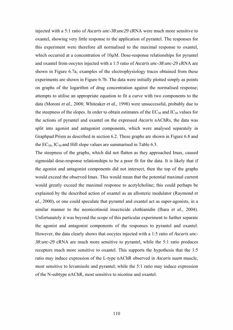
injected with a 5:1 ratio of Ascaris unc-38:unc29 cRNA were much more sensitive to
oxantel, showing very little response to the application of pyrantel. The responses for
this experiment were therefore all normalised to the maximal response to oxantel,
which occurred at a concentration of 10µM. Dose-response relationships for pyrantel
and oxantel from oocytes injected with a 1:5 ratio of Ascaris unc-38:unc-29 cRNA are
shown in Figure 6.7a; examples of the electrophysiology traces obtained from these
experiments are shown in Figure 6.7b. The data were initially plotted simply as points
on graphs of the logarithm of drug concentration against the normalised response;
attempts to utilise an appropriate equation to fit a curve with two components to the
data (Moroni et al., 2008; Whiteaker et al., 1998) were unsuccessful, probably due to
the steepness of the slopes. In order to obtain estimates of the EC50 and IC50 values for
the actions of pyrantel and oxantel on the expressed Ascaris nAChRs, the data was
split into agonist and antagonist components, which were analysed separately in
Graphpad Prism as described in section 6.2. These graphs are shown in Figure 6.8 and
the EC50, IC50 and Hill slope values are summarised in Table 6.3.
The steepness of the graphs, which did not flatten as they approached Imax, caused
sigmoidal dose-response relationships to be a poor fit for the data. It is likely that if
the agonist and antagonist components did not intersect, then the top of the graphs
would exceed the observed Imax. This would mean that the potential maximal current
would greatly exceed the maximal response to acetylcholine; this could perhaps be
explained by the described action of oxantel as an allosteric modulator (Raymond et
al., 2000), or one could speculate that pyrantel and oxantel act as super-agonists, in a
similar manner to the neonicotinoid insecticide clothianidin (Ihara et al., 2004).
Unfortunately it was beyond the scope of this particular experiment to further separate
the agonist and antagonist components of the responses to pyrantel and oxantel.
However, the data clearly shows that oocytes injected with a 1:5 ratio of Ascaris unc-
38:unc-29 cRNA are much more sensitive to pyrantel, while the 5:1 ratio produces
receptors much more sensitive to oxantel. This supports the hypothesis that the 1:5
ratio may induce expression of the L-type nAChR observed in Ascaris suum muscle,
most sensitive to levamisole and pyrantel; while the 5:1 ratio may induce expression
of the N-subtype nAChR, most sensitive to nicotine and oxantel.
110
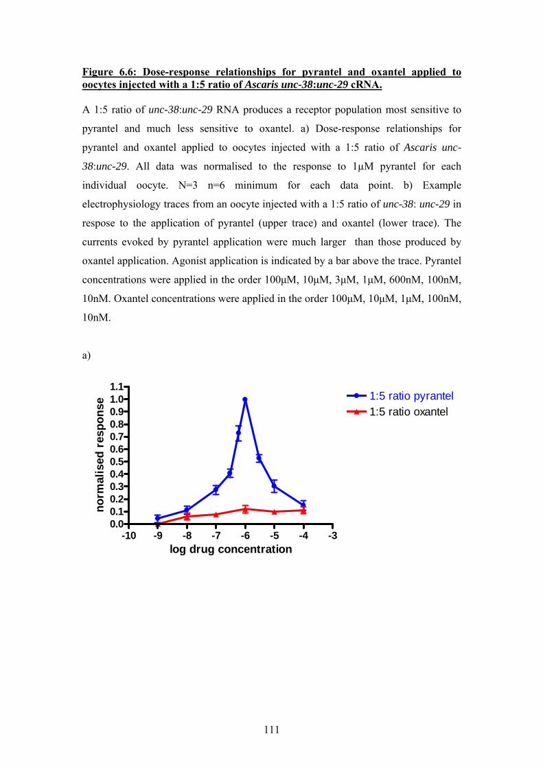
Figure 6.6: Dose-response relationships for pyrantel and oxantel applied to oocytes injected with a 1:5 ratio of Ascaris unc-38:unc-29 cRNA. A 1:5 ratio of unc-38:unc-29 RNA produces a receptor population most sensitive to
pyrantel and much less sensitive to oxantel. a) Dose-response relationships for
pyrantel and oxantel applied to oocytes injected with a 1:5 ratio of Ascaris unc-
38:unc-29. All data was normalised to the response to 1µM pyrantel for each
individual oocyte. N=3 n=6 minimum for each data point. b) Example
electrophysiology traces from an oocyte injected with a 1:5 ratio of unc-38: unc-29 in
respose to the application of pyrantel (upper trace) and oxantel (lower trace). The
currents evoked by pyrantel application were much larger than those produced by
oxantel application. Agonist application is indicated by a bar above the trace. Pyrantel
concentrations were applied in the order 100µM, 10µM, 3µM, 1µM, 600nM, 100nM,
10nM. Oxantel concentrations were applied in the order 100µM, 10µM, 1µM, 100nM,
10nM.
a)
-10 -9 -8 -7 -6 -5 -4 -30.00.10.20.30.40.50.60.70.80.91.01.1
1:5 ratio pyrantel1:5 ratio oxantel
log drug concentration
norm
alis
ed r
espo
nse
111
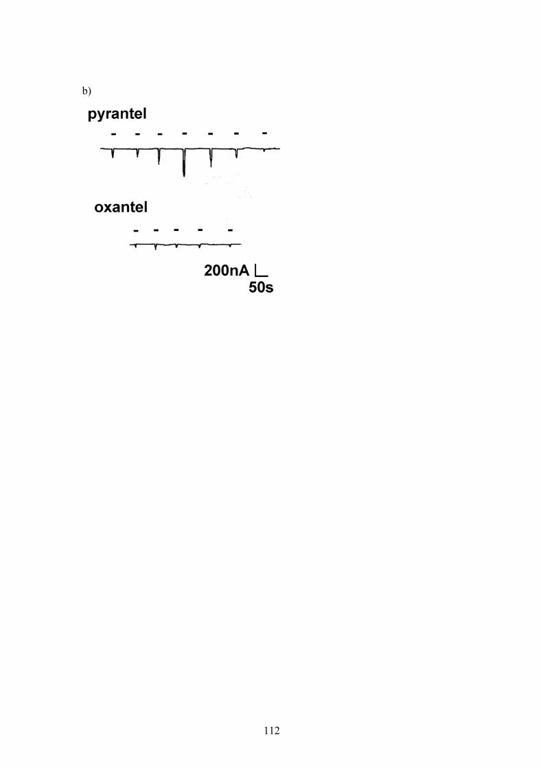
b)
112
Figure 6.7: Dose-response relationship for oxantel and pyrantel applied to oocytes injected with a 5:1 ratio of Ascaris unc-38:unc-29 cRNA. A 5:1 ratio of Ascaris unc-38:unc-29 cRNA produces a receptor population most
sensitive to oxantel and much less sensitive to pyrantel. a) Dose-response
relationships for pyrantel and oxantel applied to oocytes injected with a 5:1 ratio of
Ascaris unc-38:unc-29. All data was normalised to the response to 10µM oxantel for
each individual oocyte. N=3 n=6 minimum for each data point. b) Example
electrophysiology traces from an oocyte injected with a 5:1 ratio of unc-38: unc-29 in
respose to the application of oxantel (upper trace) and pyrantel (lower trace). The
currents evoked by oxantel application were much larger than those produced by
pyrantel application. Agonist application is indicated by a bar above the trace. Oxantel
concentrations were applied in the order100nM, 1µM, 6µM, 10µM, 60µM, 100µM,
600µM. Pyrantel concentrations were applied in the order 100µM, 10µM, 1µM,
100nM, 10nM.
a)
-10 -9 -8 -7 -6 -5 -4 -30.00.10.20.30.40.50.60.70.80.91.01.1
oxantelpyrantel
log drug concentration
norm
alis
ed r
espo
nse
113
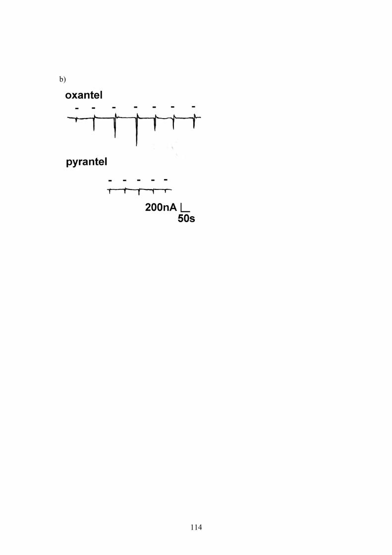
b)
114
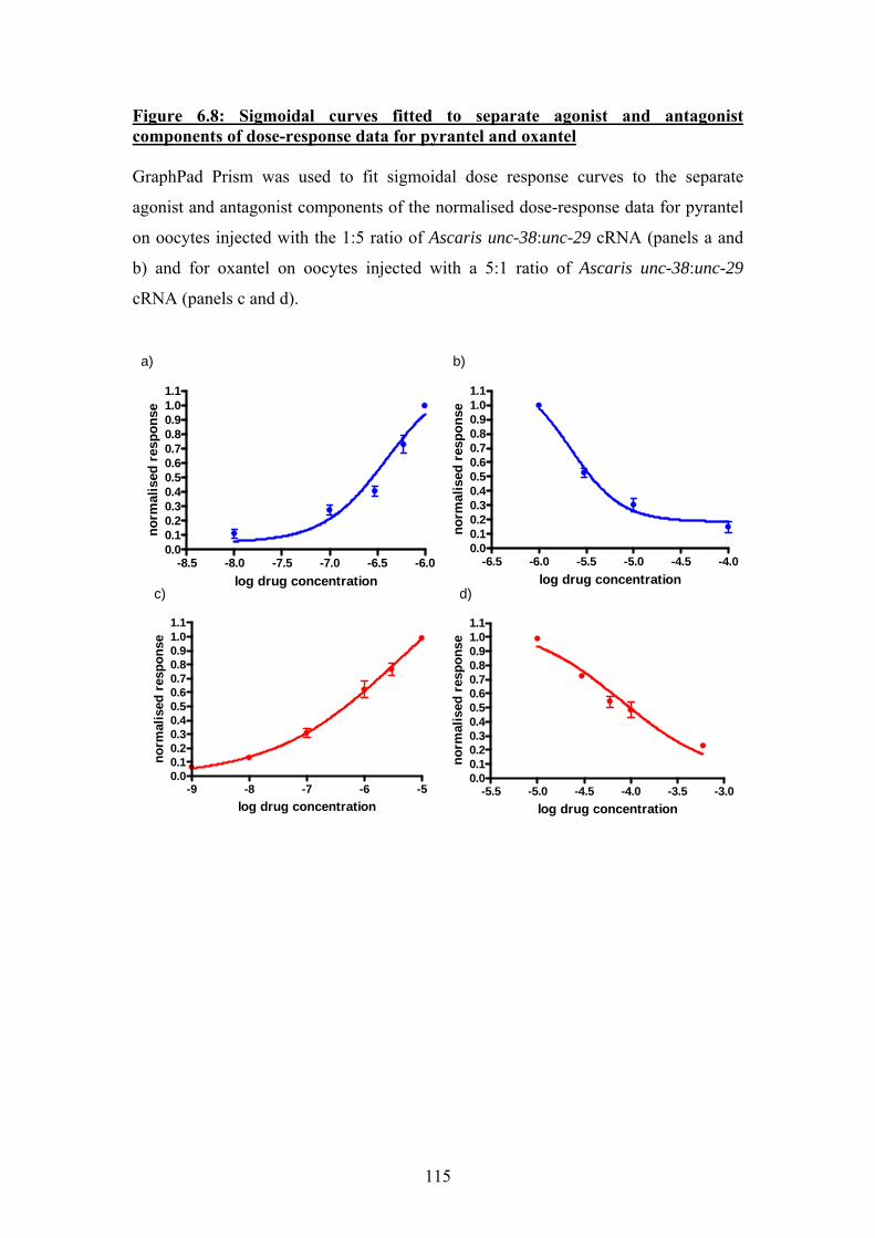
Figure 6.8: Sigmoidal curves fitted to separate agonist and antagonist components of dose-response data for pyrantel and oxantel GraphPad Prism was used to fit sigmoidal dose response curves to the separate
agonist and antagonist components of the normalised dose-response data for pyrantel
on oocytes injected with the 1:5 ratio of Ascaris unc-38:unc-29 cRNA (panels a and
b) and for oxantel on oocytes injected with a 5:1 ratio of Ascaris unc-38:unc-29
cRNA (panels c and d).
a) b)
-5.5 -5.0 -4.5 -4.0 -3.5 -3.00.00.10.20.30.40.50.60.70.80.91.01.1
log drug concentration
norm
alis
ed r
espo
nse
-9 -8 -7 -6 -50.00.10.20.30.40.50.60.70.80.91.01.1
log drug concentration
norm
alis
ed r
espo
nse
-8.5 -8.0 -7.5 -7.0 -6.5 -6.00.00.10.20.30.40.50.60.70.80.91.01.1
log drug concentration
norm
alis
ed r
espo
nse
-6.5 -6.0 -5.5 -5.0 -4.5 -4.00.00.10.20.30.40.50.60.70.80.91.01.1
log drug concentration
norm
alis
ed r
espo
nse
c) d)
115
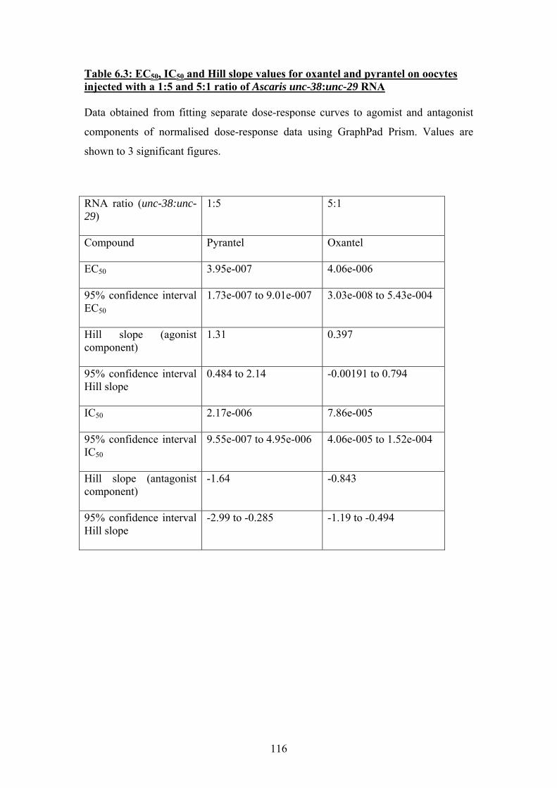
Table 6.3: EC50, IC50 and Hill slope values for oxantel and pyrantel on oocytes injected with a 1:5 and 5:1 ratio of Ascaris unc-38:unc-29 RNA Data obtained from fitting separate dose-response curves to agomist and antagonist
components of normalised dose-response data using GraphPad Prism. Values are
shown to 3 significant figures.
RNA ratio (unc-38:unc-29)
1:5 5:1
Compound
Pyrantel Oxantel
EC50
3.95e-007 4.06e-006
95% confidence interval EC50
1.73e-007 to 9.01e-007 3.03e-008 to 5.43e-004
Hill slope (agonist component)
1.31 0.397
95% confidence interval Hill slope
0.484 to 2.14 -0.00191 to 0.794
IC50
2.17e-006 7.86e-005
95% confidence interval IC50
9.55e-007 to 4.95e-006 4.06e-005 to 1.52e-004
Hill slope (antagonist component)
-1.64 -0.843
95% confidence interval Hill slope
-2.99 to -0.285 -1.19 to -0.494
116
6.5 Discussion
The heterologous expression of functional levamisole-sensitive nAChRs using the
Ascaris suum homologues of unc-38 and unc-29 confirms that in Ascaris, as in C.
elegans, these subunits are likely to be components of the native levamisole-sensitive
nAChR (Fleming et al., 1997). However, in C. elegans, it has been reported that 3
other nAChR subunits are necessary for the levamisole response: unc-63, lev-1, and
lev-8 (Culetto et al., 2004; Fleming et al., 1997; Towers et al., 2005). We have shown
using a bioinformatics approach and the genome of Brugia malayi and a parasite
closely related to Ascaris suum that lev-1 and lev-8 are apparently absent in this group
of nematodes; however unc-63 is apparently present and well-conserved. The
functional significance of unc-63 in Ascaris is currently unknown, and further
experiments, where Ascaris unc-63 would be co-expressed with unc-38 and unc-29,
are necessary to elucidate what effect this subunit would have on receptor
pharmacology. It is possible that the third pharmacological nAChR subtype described
from Ascaris, the B-subtype, most sensitive to bephenium, may contain the UNC-63
subunit (Qian et al., 2006; Robertson et al., 2002).
If the pharmacology of Ascaris nAChRs can be altered simply by generating different
combinations of two subunits, this may help to explain how sufficient
pharmacological and neurological complexity can be found in parasites with
remarkably few nAChR genes compared to the model nematode C. elegans.
Pharmacological diversity generated by alternate stoichiometries have not previously
been described from invertebrates, but are well described for heterologous expression
of the mammalian neuronal nAChR subunits α4 and β2 (Nelson et al., 2003; Zwart &
Vijverberg, 1998). Receptors with the stoichiometry (α4)3(β2)2 have lower agonist
affinity and a different pharmacological profile to receptors with the stoichiometry
(α4)2(β2)3, and addition of an accessory subunit to give (for example) an
(α4)2(β2)2(α5) combination confers additional pharmacological differences (Kuryatov
et al., 2008; Moroni et al., 2006). In extrapolating the α4β2 model to interpret the
results presented here, we may speculate that in Ascaris the 3 pharmacological
subtypes may have the stoichiometry (UNC-38)2(UNC-29)3, (UNC-38)3(UNC-29)2,
and (UNC-38)2(UNC-29)2(UNC-63), where (UNC-38)2(UNC-29)3 constitutes the L-
117
subtype, (UNC-38)3(UNC-29)2 forms the N-subtype, and addition of UNC-63 as an
accessory subunit may potentially create the B-subtype. Further work is necessary to
confirm or refute this hypothesis: the stoichiometry of the receptors expressed here is
only inferred, and experiments where receptor stoichiometry is more clearly defined
would use concatamers composed of Ascaris unc-38 and unc-29 cRNAs joined
together, to which a small quantity of accessory subunit cRNA (Ascaris unc-38, unc-
29 or unc-63) would be added. Patch-clamp experiments to make single channel
recordings of the recombinant Ascaris nAChRs would also be very informative, as the
native Ascaris nAChRs have been characterised on a single-channel level
(Levandoski et al., 2005). In addition to this, the pharmacology of the heterologously
expressed putative L- and N-type nAChRs could be further tested using other agonist
and antagonist compounds; methyridine is an additional agonist which should activate
the N-subtype, while paraherquamide is an antagonist specific to the L-subtype
(Martin et al., 2004; Robertson et al., 2002). Unfortunately it was not possible to
obtain these compounds during the experiments presented here.
The physiological relevance of different pharmacological subtypes of nAChR in the
same tissue is unknown, but it is possible that if the receptors possess different
affinities for the native agonist ACh, then the more sensitive receptors may be
primarily extrasynaptic, while the less sensitive subtype may be more likely to be
found clustered at the synapse; this is suggested as the possible physiological
relevance of mammalian receptors with different agonist affinities by Nelson et al.
(2003). However, as all single channel measurements of native Ascaris nAChRs have
to date been from pools of extrasynaptic receptors, it is not known whether this
interpretation is relevant to the properties of nematode nAChRs.
In summary, we have demonstrated that the Ascaris homologues of unc-38 and unc-
29 co-express in Xenopus oocytes to form a functional nAChR, which is the first ever
successful expression of a parasite nAChR. We have also shown that changing the
receptor stoichiometry also changes the pharmacology, which has not previously been
described for invertebrate nAChRs. The nAChRs reconstituted here show similar
pharmacological profiles to the native Ascaris nAChRs described as the N-subtype
(most sensitive to nicotine and oxantel) and the L-subtype (most sensitive to
levamisole and pyrantel). The results presented here have important implications for
the development and management of parasite resistance to cholinergic anthelmintics;
it has previously been assumed that parasites resistant to levamisole or pyrantel may
118
still be susceptible to oxantel, as different receptor sub-types are involved (Martin et
al., 2004). If the target receptors of all these cholinergic anthelmintics contain the
same subunits, a point mutation in one of the subunits which affects drug binding will
be present in two distinct drug targets, increasing the likelihood of cross-resistance. If
a more serious loss of function mutation were to occur under anthelmintic selection,
the converse argument could be made: that cross-resistance is unlikely, as loss of one
functional subunit may result in the loss of 2 or 3 different receptor subtypes and
seriously affect cholinergic signalling in the nematode. However, McCarthy and
colleagues have demonstrated that in hookworms, unc-38, unc-29 and unc-63 are all
expressed at lower levels in pyrantel resistant populations, and this may be partially
compensated for by the upregulation of another nAChR subunit, acr-19 (Kopp et al.,
2008). If a similar resistance mechanism was to occur in Ascaris suum, and an
uncharacterised subunit could take over the role of cholinergic signalling at the
neuromuscular junction, then cross-resistance to levamisole, pyrantel and oxantel may
still become a problem.
119

Chapter 7: Conclusions
120
The aim of the work presented here was to interpret the described pharmacology of
the native neuromuscular nAChRs of Ascaris suum in terms of the underlying
molecular biology, in particular the levamisole-sensitive nAChR which is the target of
the cholinergic anthelmintic drugs levamisole and pyrantel. Prior to the start of this
work, such investigations had previously been confined to the model nematode
C.elegans, though during the course of the work presented here more recent studies
emerged associating altered expression levels of nAChR subunits with drug resistance
in the parasitic hookworm Ancylostoma caninum (Fleming et al.1997, Culetto et al.
2004, Kopp et al 2008). However, Ancylostoma is closely related to C.elegans, and no
other research group has to date addressed the role of specific nAChR subunits in
levamisole sensitivity and resistance in other nematode clades.
We have shown using a bioinformatics approach that nematodes of clade I and clade
III have surprisingly small nAChR gene families compared to the clade V nematode
C.elegans, a finding which could potentially simplify the task of assigning functional
and pharmacological significance to the nAChR subunits from these parasites. We
found that sequences encoding 3 proposed components of the levamisole-sensitive
nAChR from C. elegans, unc-38, unc-29 and unc-63, are well conserved between
nematodes of different clades. This provides a good basis for more directed studies on
the role of these nAChR subunits in levamisole sensitivity in parasitic nematodes
from clades I and III, and this work may also allow potential targets to be selected for
the design of broad spectrum anthelmintics which will be effective against nematode
parasites from all clades.
We have successfully cloned full-length sequences of unc-38 and unc-29 from the
clade III parasite of pigs Ascaris suum, and partial sequence of unc-63. The reasons
for failing to amplify a full-length sequence of unc-63 from Ascaris suum could not be
satisfactorily resolved during the course of the work presented here, though real-time
PCR experiments suggest that unc-63 is expressed at lower levels than unc-38 and
unc-29 in this nematode. It is expected that the sequencing of the Ascaris suum
genome, which is currently underway, will in future allow this matter to be explained.
The sequences obtained during the course of this work will provide a useful resource
for comparing against sequences from any levamisole or pyrantel resistant Ascaris
isolates as a screen for potential resistance mutations. In addition to this, the ability to
121
detect these nAChR subunit sequences by real-time PCR will allow any changes in
their expression levels in drug resistant Ascaris to be assessed, in a similar manner to
the work performed by Kopp et al., 2008.
In addition to the amplification of sequence encoding these proposed components of
the levamisole-sensitive nAChR, we have amplified a full-length sequence encoding a
novel, putatively parasite-specific nAChR subunit not present in C.elegans, which has
been assigned the name acr-26. Work currently being carried out by Hayley Bennett
in our laboratory will help to evaluate the potential of this novel receptor as a new
anthelmintic target.
Heterologous expression of Ascaris suum UNC-38 and UNC-29 in oocytes from
Xenopus laevis produced functional nAChRs which were subject to two-electrode
voltage clamp electrophysiology to elucidate their pharmacology. It was perhaps
surprising that just two nAChR subunits would express robustly to produce functional
levamisole-sensitive receptors, as UNC-63 is clearly implicated in levamisole
sensitivity in other nematode species (Culetto et al., 2004, Kopp et al., 2008).
However, this highlights once again the differences which can be found between
nematodes of different clades, and cautions against the assumption that C. elegans is a
valid model for phylogenetically distinct parasitic nematodes. Variations in the
responses recorded from oocytes injected with equimolar concentrations of unc-
38:unc-29 RNA, along with an awareness of work performed using the mammalian
α4β2 nAChR, prompted additional experiments to be performed where the receptors
produced were deliberately driven to form different stoichiometries using different
ratios of unc-38:unc-29 RNA (Moroni et al. 2006). The receptors generated showed a
strong resemblance to two of the native Ascaris nAChRs: a greater proportion of unc-
38 (presumably giving a 3:2 UNC-38:UNC-29 stoichiometry) produced receptors
most sensitive to nicotine and oxantel, matching the N-subtype described from
Ascaris suum muscle cells, whereas a greater proportion of unc-29 (presumably
giving the alternative 2:3 UNC-38:UNC-29 stoichiometry) produced receptors most
sensitive to levamisole and pyrantel, characteristic of the native L-subtype from
Ascaris suum (Martin et al., 2003; Martin et al., 2004). This is the first time that the
phenomenon of alternative stoichiometries displaying different pharmacology has
been described for an invertebrate nAChR, and may explain how sufficient signalling
complexity can be generated by nematodes with a very small nAChR subunit gene
family. We may also speculate that addition of unc-63 to give a 2:2:1 UNC-38:UNC-
122
29:UNC-63 stoichiometry would produce the third pharmacological nAChR described
from Ascaris suum, the B-subtype, defined by bephenium sensitivity (Qian et al.,
2006). Future work to confirm this hypothesis would require obtaining additional
agonist or antagonist compounds which are reported to be specific for the three
different pharmacological subtypes, such as the agonists methyridine and bephenium,
and the antagonists paraherquamide and deoxyparaherquamide (Martin et al., 2003;
Robertson et al., 2002). It would also be useful to make concatamer constructs of unc-
38 and unc-29 where the sequences and resulting subunit proteins are joined together,
in order to fix the stoichiometry of the receptors and obtain pure populations of our
putative N- and L- subtypes (Ericksen & Boileau, 2007). In the absence of a full
length cDNA sequence from Ascaris suum, it may be possible to co-express the
Ascaris unc-38 and unc-29 with the unc-63 homologue from another nematode
species to investigate whether this forms the putative B-subtype. unc-63 sequences are
already available from Ancylostoma caninum (Kopp et al., 2008) and Haemonchus
contortus (Thomas Walsh, unpublished); and the bioinformatics work presented here
has already found unc-63 sequences in silico for Brugia malayi and Trichinella
spiralis, which should theoretically make these cDNAs easy to amplify if parasite
material for these species could be obtained by our laboratory. It would also be
interesting to attempt expression of other parasite nAChRs subunits, in order to
determine whether their pharmacology and subunit composition resembles that of
Ascaris nAChRs, or is more similar to that of C. elegans.
In conclusion, the work presented here describes the first study to address the
molecular biology underlying cholinergic anthelmintic sensitivity in a clade III
nematode, and also describes the first functional expression of a nAChR from any
parasite. This provides a sound basis for future work to be undertaken to assess the
potential of novel anthelmintics targeting nematode nAChRs in an in vitro expression
system, and also provides a platform from which the effect of potential resistance
mutations in nematode nAChRs can be effectively evaluated.
123
References ABRAMSON, S. N. and TAYLOR, P. (1990). Identification of nicotinic receptor
ACh-binding subunits. Trends Neurosci, 13, 92. ACEVES, J., ERLIJ, D. and MARTINEZ-MARANON, R. (1970). The mechanism of
the paralysing action of tetramisole on Ascaris somatic muscle. Br J Pharmacol, 38, 602-607.
AJUH, P. M. and EGWANG, T. G. (1994). Cloning of a cDNA encoding a putative nicotinic acetylcholine receptor subunit of the human filarial parasite Onchocerca volvulus. Gene, 144, 127-129.
AKABAS, M. H., KAUFMANN, C., ARCHDEACON, P. and KARLIN, A. (1994). Identification of acetylcholine receptor channel-lining residues in the entire M2 segment of the alpha subunit. Neuron, 13, 919-927.
ALLEN, P. M. and GOTTLIEB, D. (1970). Mechanism of action of the fungicide thiabendazole, 2-(4'-thiazolyl) benzimidazole. Appl Microbiol, 20, 919-926.
ARENA, J. P., LIU, K. K., PARESS, P. S., SCHAEFFER, J. M. and CULLY, D. F. (1992). Expression of a glutamate-activated chloride current in Xenopus oocytes injected with Caenorhabditis elegans RNA: evidence for modulation by avermectin. Brain Res Mol Brain Res, 15, 339-348.
ARIAS, H. R. (1997). Topology of ligand binding sites on the nicotinic acetylcholine receptor. Brain Res Brain Res Rev, 25, 133-191.
ARIAS, H. R. (2000). Localization of agonist and competitive antagonist binding sites on nicotinic acetylcholine receptors. Neurochem Int, 36, 595-645.
AUBRY, M. L., COWELL, P., DAVEY, M. J. and SHEVDE, S. (1970). Aspects of the pharmacology of a new anthelmintic: pyrantel. Br J Pharmacol, 38, 332-344.
AWADZI, K., BOAKYE, D. A., EDWARDS, G., OPOKU, N. O., ATTAH, S. K., OSEI-ATWENEBOANA, M. Y., LAZDINS-HELDS, J. K., ARDREY, A. E., ADDY, E. T., QUARTEY, B. T., AHMED, K., BOATIN, B. A. and SOUMBEY-ALLEY, E. W. (2004). An investigation of persistent microfilaridermias despite multiple treatments with ivermectin, in two onchocerciasis-endemic foci in Ghana. Ann Trop Med Parasitol, 98, 231-249.
BAHU, M., BALDISSEROTO, M., CUSTODIO, C.M., GRALHA, C.Z., MANGILI, A.R. (2001). Hepatobiliary and pancreatic complications of ascariasis in children: a study of seven cases. Journal of Pediatric Gastroenterology and Nutrition, 33, 271-275.
BALDWIN, E. and MOYLE, V. (1949). A contribution to the physiology and pharmacology of Ascaris lumbricoides from the pig. Br J Pharmacol Chemother, 4, 145-152.
BALDWIN, J. G., NADLER, S. A. and ADAMS, B. J. (2004). Evolution of plant parasitism among nematodes. Annu Rev Phytopathol, 42, 83-105.
BALLIVET, M., ALLIOD, C., BERTRAND, S. and BERTRAND, D. (1996). Nicotinic acetylcholine receptors in the nematode Caenorhabditis elegans. J Mol Biol, 258, 261-269.
BALLIVET, M., NEF, P., COUTURIER, S., RUNGGER, D., BADER, C. R., BERTRAND, D. and COOPER, E. (1988). Electrophysiology of a chick
124
neuronal nicotinic acetylcholine receptor expressed in Xenopus oocytes after cDNA injection. Neuron, 1, 847-852.
BARTLEY, D. J., JACKSON, F., JACKSON, E. and SARGISON, N. (2004). Characterisation of two triple resistant field isolates of Teladorsagia from Scottish lowland sheep farms. Vet Parasitol, 123, 189-199.
BEG, A. A. and JORGENSEN, E. M. (2003). EXP-1 is an excitatory GABA-gated cation channel. Nat Neurosci, 6, 1145-1152.
BEKTESH, S., VAN DOREN, K. and HIRSH, D. (1988). Presence of the Caenorhabditis elegans spliced leader on different mRNAs and in different genera of nematodes. Genes Dev, 2, 1277-1283.
BETHONY, J., BROOKER, S., ALBONICO, M., GEIGER, S. M., LOUKAS, A., DIEMERT, D. and HOTEZ, P. J. (2006). Soil-transmitted helminth infections: ascariasis, trichuriasis, and hookworm. Lancet, 367, 1521-1532.
BIANCHI, L. and DRISCOLL, M. (2006). Heterologous expression of C. elegans ion channels in Xenopus oocytes. WormBook, 1-16.
BLACKHALL, W. J., LIU, H. Y., XU, M., PRICHARD, R. K. and BEECH, R. N. (1998). Selection at a P-glycoprotein gene in ivermectin- and moxidectin-selected strains of Haemonchus contortus. Mol Biochem Parasitol, 95, 193-201.
BLACKHALL, W. J., PRICHARD, R. K. and BEECH, R. N. (2008). P-glycoprotein selection in strains of Haemonchus contortus resistant to benzimidazoles. Vet Parasitol, 152, 101-107.
BLAXTER, M. (1998). Caenorhabditis elegans is a nematode. Science, 282, 2041-2046.
BLAXTER, M. (2003). Comparative genomics: two worms are better than one. Nature, 426, 395-396.
BLAXTER, M. L., DE LEY, P., GAREY, J. R., LIU, L. X., SCHELDEMAN, P., VIERSTRAETE, A., VANFLETEREN, J. R., MACKEY, L. Y., DORRIS, M., FRISSE, L. M., VIDA, J. T. and THOMAS, W. K. (1998). A molecular evolutionary framework for the phylum Nematoda. Nature, 392, 71-75.
BLOUNT, P. and MERLIE, J. P. (1989). Molecular basis of the two nonequivalent ligand binding sites of the muscle nicotinic acetylcholine receptor. Neuron, 3, 349-357.
BOES, J. and HELWIGH, A. B. (2000). Animal models of intestinal nematode infections of humans. Parasitology, 121 Suppl, S97-111.
BRADING, A. F. and CALDWELL, P. C. (1971). The resting membrane potential of the somatic muscle cells of Ascaris lumbricoides. J Physiol, 217, 605-624.
BREJC, K., VAN DIJK, W. J., KLAASSEN, R. V., SCHUURMANS, M., VAN DER OOST, J., SMIT, A. B. and SIXMA, T. K. (2001). Crystal structure of an ACh-binding protein reveals the ligand-binding domain of nicotinic receptors. Nature, 411, 269-276.
BREJC, K., VAN DIJK, W. J., SMIT, A. B. and SIXMA, T. K. (2002). The 2.7 A structure of AChBP, homologue of the ligand-binding domain of the nicotinic acetylcholine receptor. Novartis Found Symp, 245, 22-29; discussion 29-32, 165-168.
BRISSON, A. and UNWIN, P. N. (1985). Quaternary structure of the acetylcholine receptor. Nature, 315, 474-477.
BROWN, L. A., JONES, A. K., BUCKINGHAM, S. D., MEE, C. J. and SATTELLE, D. B. (2006). Contributions from Caenorhabditis elegans functional genetics
125
to antiparasitic drug target identification and validation: Nicotinic acetylcholine receptors, a case study. Int J Parasitol, 36, 617-624.
BROWNLEE, D. J., FAIRWEATHER, I., JOHNSTON, C. F., SMART, D., SHAW, C. and HALTON, D. W. (1993). Immunocytochemical demonstration of neuropeptides in the central nervous system of the roundworm, Ascaris suum (Nematoda: Ascaroidea). Parasitology, 106 ( Pt 3), 305-316.
BUCKETT, W. R. (1968). The pharmacology of pancuronium bromide: a new non-depolarising neuromuscular blocking agent. Ir J Med Sci, 7, 565-568.
BUCKINGHAM, S. D., PYM, L. and SATTELLE, D. B. (2006). Oocytes as an expression system for studying receptor/channel targets of drugs and pesticides. Methods Mol Biol, 322, 331-345.
BURG, R. W., MILLER, B. M., BAKER, E. E., BIRNBAUM, J., CURRIE, S. A., HARTMAN, R., KONG, Y. L., MONAGHAN, R. L., OLSON, G., PUTTER, I., TUNAC, J. B., WALLICK, H., STAPLEY, E. O., OIWA, R. and OMURA, S. (1979). Avermectins, new family of potent anthelmintic agents: producing organism and fermentation. Antimicrob Agents Chemother, 15, 361-367.
C.ELEGANS_SEQUENCING_CONSORTIUM (1998). Genome sequence of the nematode C. elegans: a platform for investigating biology. Science, 282, 2012-2018.
CHANGEUX, J. P., PODLESKI, T. R. and WOFSY, L. (1967). Affinity labeling of the acetylcholine-receptor. Proc Natl Acad Sci U S A, 58, 2063-2070.
CHANGEUX, J. P. and TALY, A. (2008). Nicotinic receptors, allosteric proteins and medicine. Trends Mol Med, 14, 93-102.
CHIARA, D. C. and COHEN, J. B. (1997). Identification of amino acids contributing to high and low affinity d-tubocurarine sites in the Torpedo nicotinic acetylcholine receptor. J Biol Chem, 272, 32940-32950.
CHIARA, D. C., MIDDLETON, R. E. and COHEN, J. B. (1998). Identification of tryptophan 55 as the primary site of [3H]nicotine photoincorporation in the gamma-subunit of the Torpedo nicotinic acetylcholine receptor. FEBS Lett, 423, 223-226.
CHITKARA, R. K. and KRISHNA, G. (2006). Parasitic pulmonary eosinophilia. Semin Respir Crit Care Med, 27, 171-184.
COLES, G. C., WARNER, A. K. and BEST, J. R. (1996). Triple resistant Ostertagia from angora goats. Vet Rec, 139, 299-300.
COLQUHOUN, L., HOLDEN-DYE, L. and WALKER, R. J. (1991). The pharmacology of cholinoceptors on the somatic muscle cells of the parasitic nematode Ascaris suum. J Exp Biol, 158, 509-530.
CONWAY-MORRIS, S. (1981). Parasites and the fossil record. Parasitology, 82, 489-509.
CORRINGER, P. J., GALZI, J. L., EISELE, J. L., BERTRAND, S., CHANGEUX, J. P. and BERTRAND, D. (1995). Identification of a new component of the agonist binding site of the nicotinic alpha 7 homooligomeric receptor. J Biol Chem, 270, 11749-11752.
CORRINGER, P. J., LE NOVERE, N. and CHANGEUX, J. P. (2000). Nicotinic receptors at the amino acid level. Annu Rev Pharmacol Toxicol, 40, 431-458.
COUTURIER, S., BERTRAND, D., MATTER, J. M., HERNANDEZ, M. C., BERTRAND, S., MILLAR, N., VALERA, S., BARKAS, T. and BALLIVET, M. (1990). A neuronal nicotinic acetylcholine receptor subunit (alpha 7) is developmentally regulated and forms a homo-oligomeric channel blocked by alpha-BTX. Neuron, 5, 847-856.
126
CRISCIONE, C. D., ANDERSON, J. D., SUDIMACK, D., PENG, W., JHA, B., WILLIAMS-BLANGERO, S. and ANDERSON, T. J. (2007). Disentangling hybridization and host colonization in parasitic roundworms of humans and pigs. Proc Biol Sci, 274, 2669-2677.
CROMPTON, D. W. (1986). Nutritional aspects of infection. Transactions of the Royal Society for Tropical Medicine and Hygeine, 80, 697-705.
CULETTO, E., BAYLIS, H. A., RICHMOND, J. E., JONES, A. K., FLEMING, J. T., SQUIRE, M. D., LEWIS, J. A. and SATTELLE, D. B. (2004). The Caenorhabditis elegans unc-63 gene encodes a levamisole-sensitive nicotinic acetylcholine receptor alpha subunit. J Biol Chem, 279, 42476-42483.
DALE, V. M. and MARTIN, R. J. (1995). Oxantel-activated single channel currents in the muscle membrane of Ascaris suum. Parasitology, 110 ( Pt 4), 437-448.
DAVIDSE, L. C. and FLACH, W. (1978). Interaction of thiabendazole with fungal tubulin. Biochim Biophys Acta, 543, 82-90.
DE BELL, J. T., DEL CASTILLO, J. and SANCHEZ, V. (1963). Electrophysiology of the Somatic Muscle Cells of Ascaris Lumbricoides. J Cell Physiol, 62, 159-177.
DE SILVA, N. R., BROOKER, S., HOTEZ, P. J., MONTRESOR, A., ENGELS, D. and SAVIOLI, L. (2003). Soil-transmitted helminth infections: updating the global picture. Trends Parasitol, 19, 547-551.
DE SILVA, N. R., GUYATT H.L., BUNDY, D.A. (1997). Morbidity and mortality due to Ascaris-induced intestinal obstruction. Transactions of the Royal Society for Tropical Medicine and Hygeine, 91, 31-36.
DEL CASTILLO, J., RIVERA, A., SOLORZANO, S. and SERRATO, J. (1989). Some aspects of the neuromuscular system of Ascaris. Q J Exp Physiol, 74, 1071-1087.
DELANY, N. S., LAUGHTON, D. L. and WOLSTENHOLME, A. J. (1998). Cloning and localisation of an avermectin receptor-related subunit from Haemonchus contortus. Mol Biochem Parasitol, 97, 177-187.
DENT, J. A. (2006). Evidence for a diverse Cys-loop ligand-gated ion channel superfamily in early bilateria. J Mol Evol, 62, 523-535.
DENT, J. A., SMITH, M. M., VASSILATIS, D. K. and AVERY, L. (2000). The genetics of ivermectin resistance in Caenorhabditis elegans. Proc Natl Acad Sci U S A, 97, 2674-2679.
DORRIS, M., DE LEY, P. and BLAXTER, M. L. (1999). Molecular analysis of nematode diversity and the evolution of parasitism. Parasitol Today, 15, 188-193.
EINARSON, B., GULLICK, W., CONTI-TRONCONI, B., ELLISMAN, M. and LINDSTROM, J. (1982). Subunit composition of bovine muscle acetylcholine receptor. Biochemistry, 21, 5295-5302.
ERICKSEN, S. S. and BOILEAU, A. J. (2007). Tandem couture: Cys-loop receptor concatamer insights and caveats. Mol Neurobiol, 35, 113-128.
EYRE, P. (1970). Some pharmacodynamic effects of the nematocides: methyridine, tetramisole and pyrantel. J Pharm Pharmacol, 22, 26-36.
FFRENCH-CONSTANT, R. H. and ROCHELEAU, T. A. (1993). Drosophila gamma-aminobutyric acid receptor gene Rdl shows extensive alternative splicing. J Neurochem, 60, 2323-2326.
FINCHAM, J. E., MARKUS, M. B. and ADAMS, V. J. (2003). Could control of soil-transmitted helminthic infection influence the HIV/AIDS pandemic. Acta Trop, 86, 315-333.
127
FLEMING, J. T., SQUIRE, M. D., BARNES, T. M., TORNOE, C., MATSUDA, K., AHNN, J., FIRE, A., SULSTON, J. E., BARNARD, E. A., SATTELLE, D. B. and LEWIS, J. A. (1997). Caenorhabditis elegans levamisole resistance genes lev-1, unc-29, and unc-38 encode functional nicotinic acetylcholine receptor subunits. J Neurosci, 17, 5843-5857.
FROHMAN, M. A., DUSH, M. K. and MARTIN, G. R. (1988). Rapid production of full-length cDNAs from rare transcripts: amplification using a single gene-specific oligonucleotide primer. Proc Natl Acad Sci U S A, 85, 8998-9002.
FRY, G. F. and MOORE, J. G. (1969). Enterobius vermicularis: 10,000-year-old human infection. Science, 166, 1620.
FUCILE, S. (2004). Ca2+ permeability of nicotinic acetylcholine receptors. Cell Calcium, 35, 1-8.
GALZI, J. L., BERTRAND, D., DEVILLERS-THIERY, A., REVAH, F., BERTRAND, S. and CHANGEUX, J. P. (1991a). Functional significance of aromatic amino acids from three peptide loops of the alpha 7 neuronal nicotinic receptor site investigated by site-directed mutagenesis. FEBS Lett, 294, 198-202.
GALZI, J. L., DEVILLERS-THIERY, A., HUSSY, N., BERTRAND, S., CHANGEUX, J. P. and BERTRAND, D. (1992). Mutations in the channel domain of a neuronal nicotinic receptor convert ion selectivity from cationic to anionic. Nature, 359, 500-505.
GALZI, J. L., REVAH, F., BLACK, D., GOELDNER, M., HIRTH, C. and CHANGEUX, J. P. (1990). Identification of a novel amino acid alpha-tyrosine 93 within the cholinergic ligands-binding sites of the acetylcholine receptor by photoaffinity labeling. Additional evidence for a three-loop model of the cholinergic ligands-binding sites. J Biol Chem, 265, 10430-10437.
GALZI, J. L., REVAH, F., BOUET, F., MENEZ, A., GOELDNER, M., HIRTH, C. and CHANGEUX, J. P. (1991b). Allosteric transitions of the acetylcholine receptor probed at the amino acid level with a photolabile cholinergic ligand. Proc Natl Acad Sci U S A, 88, 5051-5055.
GERZANICH, V., ANAND, R. and LINDSTROM, J. (1994). Homomers of alpha 8 and alpha 7 subunits of nicotinic receptors exhibit similar channel but contrasting binding site properties. Mol Pharmacol, 45, 212-220.
GHEDIN, E., WANG, S., SPIRO, D., CALER, E., ZHAO, Q., CRABTREE, J., ALLEN, J. E., DELCHER, A. L., GUILIANO, D. B., MIRANDA-SAAVEDRA, D., ANGIUOLI, S. V., CREASY, T., AMEDEO, P., HAAS, B., EL-SAYED, N. M., WORTMAN, J. R., FELDBLYUM, T., TALLON, L., SCHATZ, M., SHUMWAY, M., KOO, H., SALZBERG, S. L., SCHOBEL, S., PERTEA, M., POP, M., WHITE, O., BARTON, G. J., CARLOW, C. K., CRAWFORD, M. J., DAUB, J., DIMMIC, M. W., ESTES, C. F., FOSTER, J. M., GANATRA, M., GREGORY, W. F., JOHNSON, N. M., JIN, J., KOMUNIECKI, R., KORF, I., KUMAR, S., LANEY, S., LI, B. W., LI, W., LINDBLOM, T. H., LUSTIGMAN, S., MA, D., MAINA, C. V., MARTIN, D. M., MCCARTER, J. P., MCREYNOLDS, L., MITREVA, M., NUTMAN, T. B., PARKINSON, J., PEREGRIN-ALVAREZ, J. M., POOLE, C., REN, Q., SAUNDERS, L., SLUDER, A. E., SMITH, K., STANKE, M., UNNASCH, T. R., WARE, J., WEI, A. D., WEIL, G., WILLIAMS, D. J., ZHANG, Y., WILLIAMS, S. A., FRASER-LIGGETT, C., SLATKO, B., BLAXTER, M. L. and SCOTT, A. L. (2007). Draft genome of the filarial nematode parasite Brugia malayi. Science, 317, 1756-1760.
128
GOUDIE, A. C., EVANS, N. A., GRATION, K. A., BISHOP, B. F., GIBSON, S. P., HOLDOM, K. S., KAYE, B., WICKS, S. R., LEWIS, D., WEATHERLEY, A. J. and ET AL. (1993). Doramectin--a potent novel endectocide. Vet Parasitol, 49, 5-15.
GUEST, M., BULL, K., WALKER, R. J., AMLIWALA, K., O'CONNOR, V., HARDER, A., HOLDEN-DYE, L. and HOPPER, N. A. (2007). The calcium-activated potassium channel, SLO-1, is required for the action of the novel cyclo-octadepsipeptide anthelmintic, emodepside, in Caenorhabditis elegans. Int J Parasitol, 37, 1577-1588.
GURDON, J. B., WOODLAND, H. R. and LINGREL, J. B. (1974). The translation of mammalian globin mRNA injected into fertilized eggs of Xenopus laevis I. Message stability in development. Dev Biol, 39, 125-133.
HALEVI, S., MCKAY, J., PALFREYMAN, M., YASSIN, L., ESHEL, M., JORGENSEN, E. and TREININ, M. (2002). The C. elegans ric-3 gene is required for maturation of nicotinic acetylcholine receptors. EMBO J, 21, 1012-1020.
HALL, L. R. and PEARLMAN, E. (1999). Pathogenesis of onchocercal keratitis (River blindness). Clin Microbiol Rev, 12, 445-453.
HARDER, A., HOLDEN-DYE, L., WALKER, R. and WUNDERLICH, F. (2005). Mechanisms of action of emodepside. Parasitol Res, 97 Suppl 1, S1-S10.
HARDER, A., SCHMITT-WREDE, H. P., KRUCKEN, J., MARINOVSKI, P., WUNDERLICH, F., WILLSON, J., AMLIWALA, K., HOLDEN-DYE, L. and WALKER, R. (2003). Cyclooctadepsipeptides--an anthelmintically active class of compounds exhibiting a novel mode of action. Int J Antimicrob Agents, 22, 318-331.
HARROW, I. D. and GRATION, A. F. (1985). Mode of action of the anthelmintics morantel, pyrantel and levamisole on muscle cell membrane of the nematode Ascaris suum. Pesticide Science, 16, 662-672.
HARROW, I. D. and GRATION, K. A. (1984). Pesticide Science. HASTIE, A. C. and GEORGOPOULOS, S. G. (1971). Mutational resistance to
fungitoxic benzimidazole derivatives in Aspergillus nidulans. J Gen Microbiol, 67, 371-373.
HLAING, T. (1993). Ascariasis and childhood malnutrition. Parasitology, 107, 25-36. HODGKIN, A. L., HUXLEY, A. F. and KATZ, B. (1952). Measurement of current-
voltage relations in the membrane of the giant axon of Loligo. J Physiol, 116, 424-448.
HOEKSTRA, R., VISSER, A., WILEY, L. J., WEISS, A. S., SANGSTER, N. C. and ROOS, M. H. (1997). Characterization of an acetylcholine receptor gene of Haemonchus contortus in relation to levamisole resistance. Mol Biochem Parasitol, 84, 179-187.
HOLDEN-DYE, L. and WALKER, R. J. (1990). Avermectin and avermectin derivatives are antagonists at the 4-aminobutyric acid (GABA) receptor on the somatic muscle cells of Ascaris; is this the site of anthelmintic action? Parasitology, 101 Pt 2, 265-271.
HUGANIR, R. L. and GREENGARD, P. (1990). Regulation of neurotransmitter receptor desensitization by protein phosphorylation. Neuron, 5, 555-567.
IHARA, M., MATSUDA, K., OTAKE, M., KUWAMURA, M., SHIMOMURA, M., KOMAI, K., AKAMATSU, M., RAYMOND, V. and SATTELLE, D. B. (2003). Diverse actions of neonicotinoids on chicken alpha7, alpha4beta2 and Drosophila-chicken SADbeta2 and ALSbeta2 hybrid nicotinic acetylcholine
129

receptors expressed in Xenopus laevis oocytes. Neuropharmacology, 45, 133-144.
IHARA, M., MATSUDA, K., SHIMOMURA, M., SATTELLE, D. B. and KOMAI, K. (2004). Super agonist actions of clothianidin and related compounds on the SAD beta 2 nicotinic acetylcholine receptor expressed in Xenopus laevis oocytes. Biosci Biotechnol Biochem, 68, 761-763.
IMOTO, K., BUSCH, C., SAKMANN, B., MISHINA, M., KONNO, T., NAKAI, J., BUJO, H., MORI, Y., FUKUDA, K. and NUMA, S. (1988). Rings of negatively charged amino acids determine the acetylcholine receptor channel conductance. Nature, 335, 645-648.
IMOTO, K., METHFESSEL, C., SAKMANN, B., MISHINA, M., MORI, Y., KONNO, T., FUKUDA, K., KURASAKI, M., BUJO, H., FUJITA, Y. and ET AL. (1986). Location of a delta-subunit region determining ion transport through the acetylcholine receptor channel. Nature, 324, 670-674.
JACKSON, F. and COOP, R. L. (2000). The development of anthelmintic resistance in sheep nematodes. Parasitology, 120 Suppl, S95-107.
JOHNSON, C. D. and STRETTON, A. O. (1985). Localization of choline acetyltransferase within identified motoneurons of the nematode Ascaris. J Neurosci, 5, 1984-1992.
JONES, A. K., DAVIS, P., HODGKIN, J. and SATTELLE, D. B. (2007). The nicotinic acetylcholine receptor gene family of the nematode Caenorhabditis elegans: an update on nomenclature. Invert Neurosci, 7, 129-131.
JONES, A. K. and SATTELLE, D. B. (2004). Functional genomics of the nicotinic acetylcholine receptor gene family of the nematode, Caenorhabditis elegans. Bioessays, 26, 39-49.
JONES, A. K. and SATTELLE, D. B. (2006). The cys-loop ligand-gated ion channel superfamily of the honeybee, Apis mellifera. Invert Neurosci, 6, 123-132.
JONES, A. K. and SATTELLE, D. B. (2007). The cys-loop ligand-gated ion channel gene superfamily of the red flour beetle, Tribolium castaneum. BMC Genomics, 8, 327.
JONES, A. K. and SATTELLE, D. B. (2008). The cys-loop ligand-gated ion channel gene superfamily of the nematode, Caenorhabditis elegans. Invert Neurosci.
JONES, I. W. and WONNACOTT, S. (2005). Why doesn't nicotinic ACh receptor immunoreactivity knock out? Trends Neurosci, 28, 343-345.
KALE, O. O. (1998). Onchocerciasis: the burden of disease. Ann Trop Med Parasitol, 92 Suppl 1, S101-115.
KAMINSKY, R., DUCRAY, P., JUNG, M., CLOVER, R., RUFENER, L., BOUVIER, J., WEBER, S. S., WENGER, A., WIELAND-BERGHAUSEN, S., GOEBEL, T., GAUVRY, N., PAUTRAT, F., SKRIPSKY, T., FROELICH, O., KOMOIN-OKA, C., WESTLUND, B., SLUDER, A. and MASER, P. (2008). A new class of anthelmintics effective against drug-resistant nematodes. Nature, 452, 176-180.
KAO, P. N. and KARLIN, A. (1986). Acetylcholine receptor binding site contains a disulfide cross-link between adjacent half-cystinyl residues. J Biol Chem, 261, 8085-8088.
KERBOEUF, D., BLACKHALL, W., KAMINSKY, R. and VON SAMSON-HIMMELSTJERNA, G. (2003). P-glycoprotein in helminths: function and perspectives for anthelmintic treatment and reversal of resistance. Int J Antimicrob Agents, 22, 332-346.
130
KOPP, S. R., COLEMAN, G. T., TRAUB, R. J., MCCARTHY, J. S. and KOTZE, A. C. (2008). Acetylcholine receptor subunit genes from Ancylostoma caninum: Altered transcription patterns associated with pyrantel resistance. Int J Parasitol.
KUROSAKI, T., FUKUDA, K., KONNO, T., MORI, Y., TANAKA, K., MISHINA, M. and NUMA, S. (1987). Functional properties of nicotinic acetylcholine receptor subunits expressed in various combinations. FEBS Lett, 214, 253-258.
KURYATOV, A., ONKSEN, J. and LINDSTROM, J. (2008). Roles of accessory subunits in alpha4beta2(*) nicotinic receptors. Mol Pharmacol, 74, 132-143.
KUZMINA, T. A. and KHARCHENKO, V. O. (2008). Anthelmintic resistance in cyathostomins of brood horses in Ukraine and influence of anthelmintic treatments on strongylid community structure. Vet Parasitol, 154, 277-288.
KWA, M. S., VEENSTRA, J. G. and ROOS, M. H. (1994). Benzimidazole resistance in Haemonchus contortus is correlated with a conserved mutation at amino acid 200 in beta-tubulin isotype 1. Mol Biochem Parasitol, 63, 299-303.
LAMBSHEAD, J. (1993). Oceanis, 19. LE GOFF, G., HAMON, A., BERGE, J. B. and AMICHOT, M. (2005). Resistance to
fipronil in Drosophila simulans: influence of two point mutations in the RDL GABA receptor subunit. J Neurochem, 92, 1295-1305.
LE HESRAN, J. Y., AKIANA, J., NDIAYE EL, H. M., DIA, M., SENGHOR, P. and KONATE, L. (2004). Severe malaria attack is associated with high prevalence of Ascaris lumbricoides infection among children in rural Senegal. Trans R Soc Trop Med Hyg, 98, 397-399.
LESTER, H. A., DIBAS, M. I., DAHAN, D. S., LEITE, J. F. and DOUGHERTY, D. A. (2004). Cys-loop receptors: new twists and turns. Trends Neurosci, 27, 329-336.
LEVANDOSKI, M. M., ROBERTSON, A. P., KUIPER, S., QIAN, H. and MARTIN, R. J. (2005). Single-channel properties of N- and L-subtypes of acetylcholine receptor in Ascaris suum. Int J Parasitol, 35, 925-934.
LEWIS, J. A., WU, C. H., LEVINE, J. H. and BERG, H. (1980). Levamisole-resistant mutants of the nematode Caenorhabditis elegans appear to lack pharmacological acetylcholine receptors. Neuroscience, 5, 967-989.
LITTLETON, J. T. and GANETZKY, B. (2000). Ion channels and synaptic organization: analysis of the Drosophila genome. Neuron, 26, 35-43.
LOVERIDGE, B., MCARTHUR, M., MCKENNA, P. B. and MARIADASS, B. (2003). Probable multigeneric resistance to macrocyclic lactone anthelmintics in cattle in New Zealand. N Z Vet J, 51, 139-141.
MARMONT, G. (1949). Studies on the axon membrane; a new method. J Cell Physiol, 34, 351-382.
MARTIN, R. J. (1993). Neuromuscular transmission in nematode parasites and antinematodal drug action. Pharmacol Ther, 58, 13-50.
MARTIN, R. J., BAI, G., CLARK, C. L. and ROBERTSON, A. P. (2003). Methyridine (2-[2-methoxyethyl]-pyridine]) and levamisole activate different ACh receptor subtypes in nematode parasites: a new lead for levamisole-resistance. Br J Pharmacol, 140, 1068-1076.
MARTIN, R. J., CLARK, C. L., TRAILOVIC, S. M. and ROBERTSON, A. P. (2004). Oxantel is an N-type (methyridine and nicotine) agonist not an L-type (levamisole and pyrantel) agonist: classification of cholinergic anthelmintics in Ascaris. Int J Parasitol, 34, 1083-1090.
131
MARTIN, R. J. and ROBERTSON, A. P. (2000). Electrophysiological investigation of anthelmintic resistance. Parasitology, 120 Suppl, S87-94.
MATHERS, C. D., EZZATI, M. and LOPEZ, A. D. (2007). Measuring the burden of neglected tropical diseases: the global burden of disease framework. PLoS Negl Trop Dis, 1, e114.
MCCAVERA, S., WALSH, T. K. and WOLSTENHOLME, A. J. (2007). Nematode ligand-gated chloride channels: an appraisal of their involvement in macrocyclic lactone resistance and prospects for developing molecular markers. Parasitology, 134, 1111-1121.
MELLIN, T. N., BUSCH, R. D. and WANG, C. C. (1983). Postsynaptic inhibition of invertebrate neuromuscular transmission by avermectin B1a. Neuropharmacology, 22, 89-96.
MILLAR, N. S. and GOTTI, C. (2008). Diversity of vertebrate nicotinic acetylcholine receptors. Neuropharmacology.
MISHINA, M., KUROSAKI, T., TOBIMATSU, T., MORIMOTO, Y., NODA, M., YAMAMOTO, T., TERAO, M., LINDSTROM, J., TAKAHASHI, T., KUNO, M. and ET AL. (1984). Expression of functional acetylcholine receptor from cloned cDNAs. Nature, 307, 604-608.
MISHINA, M., TOBIMATSU, T., IMOTO, K., TANAKA, K., FUJITA, Y., FUKUDA, K., KURASAKI, M., TAKAHASHI, H., MORIMOTO, Y., HIROSE, T. and ET AL. (1985). Location of functional regions of acetylcholine receptor alpha-subunit by site-directed mutagenesis. Nature, 313, 364-369.
MITREVA, M., BLAXTER, M. L., BIRD, D. M. and MCCARTER, J. P. (2005). Comparative genomics of nematodes. Trends Genet, 21, 573-581.
MITREVA, M. and JASMER, D. P. (2006). Biology and genome of Trichinella spiralis. WormBook, 1-21.
MONGAN, N. P., BAYLIS, H. A., ADCOCK, C., SMITH, G. R., SANSOM, M. S. and SATTELLE, D. B. (1998). An extensive and diverse gene family of nicotinic acetylcholine receptor alpha subunits in Caenorhabditis elegans. Receptors Channels, 6, 213-228.
MONGAN, N. P., JONES, A. K., SMITH, G. R., SANSOM, M. S. and SATTELLE, D. B. (2002). Novel alpha7-like nicotinic acetylcholine receptor subunits in the nematode Caenorhabditis elegans. Protein Sci, 11, 1162-1171.
MORONI, M. and BERMUDEZ, I. (2006). Stoichiometry and pharmacology of two human alpha4beta2 nicotinic receptor types. J Mol Neurosci, 30, 95-96.
MORONI, M., VIJAYAN, R., CARBONE, A., ZWART, R., BIGGIN, P. C. and BERMUDEZ, I. (2008). Non-agonist-binding subunit interfaces confer distinct functional signatures to the alternate stoichiometries of the alpha4beta2 nicotinic receptor: an alpha4-alpha4 interface is required for Zn2+ potentiation. J Neurosci, 28, 6884-6894.
MORONI, M., ZWART, R., SHER, E., CASSELS, B. K. and BERMUDEZ, I. (2006). alpha4beta2 nicotinic receptors with high and low acetylcholine sensitivity: pharmacology, stoichiometry, and sensitivity to long-term exposure to nicotine. Mol Pharmacol, 70, 755-768.
NADLER, S. A., CARRENO, R. A., MEJIA-MADRID, H., ULLBERG, J., PAGAN, C., HOUSTON, R. and HUGOT, J. P. (2007). Molecular phylogeny of clade III nematodes reveals multiple origins of tissue parasitism. Parasitology, 134, 1421-1442.
132
NATOFF, I. L. (1969). The pharmacology of the cholinoceptor in muscle preparations of Ascaris lumbricoides var. suum. Br J Pharmacol, 37, 251-257.
NEJSUM, P., PARKER, E. D., JR., FRYDENBERG, J., ROEPSTORFF, A., BOES, J., HAQUE, R., ASTRUP, I., PRAG, J. and SKOV SORENSEN, U. B. (2005). Ascariasis is a zoonosis in denmark. J Clin Microbiol, 43, 1142-1148.
NELSON, M. E., KURYATOV, A., CHOI, C. H., ZHOU, Y. and LINDSTROM, J. (2003). Alternate stoichiometries of alpha4beta2 nicotinic acetylcholine receptors. Mol Pharmacol, 63, 332-341.
NODA, M., TAKAHASHI, H., TANABE, T., TOYOSATO, M., FURUTANI, Y., HIROSE, T., ASAI, M., INAYAMA, S., MIYATA, T. and NUMA, S. (1982). Primary structure of alpha-subunit precursor of Torpedo californica acetylcholine receptor deduced from cDNA sequence. Nature, 299, 793-797.
ORTELLS, M. O. and LUNT, G. G. (1995). Evolutionary history of the ligand-gated ion-channel superfamily of receptors. Trends Neurosci, 18, 121-127.
PAPE, M., VON SAMSON-HIMMELSTJERNA, G. and SCHNIEDER, T. (1999). Characterisation of the beta-tubulin gene of Cylicocyclus nassatus? Int J Parasitol, 29, 1941-1947.
PARKINSON, J., WHITTON, C., SCHMID, R., THOMSON, M. and BLAXTER, M. (2004). NEMBASE: a resource for parasitic nematode ESTs. Nucleic Acids Res, 32, D427-430.
PARTONO, F. (1987). The spectrum of disease in lymphatic filariasis. Ciba Found Symp, 127, 15-31.
PEDERSEN, S. E. and COHEN, J. B. (1990). d-Tubocurarine binding sites are located at alpha-gamma and alpha-delta subunit interfaces of the nicotinic acetylcholine receptor. Proc Natl Acad Sci U S A, 87, 2785-2789.
PENG, X., KATZ, M., GERZANICH, V., ANAND, R. and LINDSTROM, J. (1994). Human alpha 7 acetylcholine receptor: cloning of the alpha 7 subunit from the SH-SY5Y cell line and determination of pharmacological properties of native receptors and functional alpha 7 homomers expressed in Xenopus oocytes. Mol Pharmacol, 45, 546-554.
PENNINGTON, A. J. and MARTIN, R. J. (1990). A patch-clamp study of acetylcholine-activated ion channels in Ascaris suum muscle. J Exp Biol, 154, 201-221.
PHILLIPS, W. D., MAIMONE, M. M. and MERLIE, J. P. (1991). Mutagenesis of the 43-kD postsynaptic protein defines domains involved in plasma membrane targeting and AChR clustering. J Cell Biol, 115, 1713-1723.
POINAR, G. and BOUCOT, A. J. (2006). Evidence of intestinal parasites of dinosaurs. Parasitology, 1-5.
PRICHARD, R. K., HALL, C. A., KELLY, J. D., MARTIN, I. C. and DONALD, A. D. (1980). The problem of anthelmintic resistance in nematodes. Aust Vet J, 56, 239-251.
PUTRENKO, I., ZAKIKHANI, M. and DENT, J. A. (2005). A family of acetylcholine-gated chloride channel subunits in Caenorhabditis elegans. J Biol Chem, 280, 6392-6398.
QIAN, H., MARTIN, R. J. and ROBERTSON, A. P. (2006). Pharmacology of N-, L-, and B-subtypes of nematode nAChR resolved at the single-channel level in Ascaris suum. Faseb J, 20, 2606-2608.
QIAN, H., ROBERTSON, A. P., POWELL-COFFMAN, J. A. and MARTIN, R. J. (2008). Levamisole resistance resolved at the single-channel level in Caenorhabditis elegans. Faseb J, 22, 3247-3254.
133
RAFTERY, M. A., VANDLEN, R. L., REED, K. L. and LEE, T. (1976). Characterization of Torpedo californica acetylcholine receptor: its subunit composition and ligand-binding properties. Cold Spring Harb Symp Quant Biol, 40, 193-202.
RANGANATHAN, R., CANNON, S. C. and HORVITZ, H. R. (2000). MOD-1 is a serotonin-gated chloride channel that modulates locomotory behaviour in C. elegans. Nature, 408, 470-475.
RAYES, D., DE ROSA, M. J., BARTOS, M. and BOUZAT, C. (2004). Molecular basis of the differential sensitivity of nematode and mammalian muscle to the anthelmintic agent levamisole. J Biol Chem, 279, 36372-36381.
RAYMOND, V., MONGAN, N. P. and SATTELLE, D. B. (2000). Anthelmintic actions on homomer-forming nicotinic acetylcholine receptor subunits: chicken alpha7 and ACR-16 from the nematode Caenorhabditis elegans. Neuroscience, 101, 785-791.
ROBERTSON, A. P., BJORN, H. E. and MARTIN, R. J. (1999). Resistance to levamisole resolved at the single-channel level. Faseb J, 13, 749-760.
ROBERTSON, A. P., BJORN, H. E. and MARTIN, R. J. (2000). Pyrantel resistance alters nematode nicotinic acetylcholine receptor single-channel properties. European Journal of Pharmacology, 394, 1-8.
ROBERTSON, A. P., CLARK, C. L., BURNS, T. A., THOMPSON, D. P., GEARY, T. G., TRAILOVIC, S. M. and MARTIN, R. J. (2002). Paraherquamide and 2-deoxy-paraherquamide distinguish cholinergic receptor subtypes in Ascaris muscle. J Pharmacol Exp Ther, 302, 853-860.
ROBERTSON, S. J. and MARTIN, R. J. (1993). Levamisole-activated single-channel currents from muscle of the nematode parasite Ascaris suum. Br J Pharmacol, 108, 170-178.
ROBERTSON, S. J., PENNINGTON, A. J., EVANS, A. M. and MARTIN, R. J. (1994). The action of pyrantel as an agonist and an open channel blocker at acetylcholine receptors in isolated Ascaris suum muscle vesicles. Eur J Pharmacol, 271, 273-282.
ROSENBLUTH, J. (1965). Ultrastructure of somatic muscle cells in Ascaris lumbricoides. II. Intermuscular junctions, neuromuscular junctions, and glycogen stores. J Cell Biol, 26, 579-591.
ROZHKOVA, E. K., MALYUTINA, T. A. and SHISHOV, B. A. (1980). Pharmacological characteristics of cholinoreception in somatic muscles of the nematode, Ascaris suum. Gen Pharmacol, 11, 141-146.
SANSOM, M. S. (1995). Ion-channel gating. Twist to open. Curr Biol, 5, 373-375. SATTELLE, D. B., JONES, A. K., SATTELLE, B. M., MATSUDA, K., REENAN,
R. and BIGGIN, P. C. (2005). Edit, cut and paste in the nicotinic acetylcholine receptor gene family of Drosophila melanogaster. Bioessays, 27, 366-376.
SCHOFIELD, P. R., DARLISON, M. G., FUJITA, N., BURT, D. R., STEPHENSON, F. A., RODRIGUEZ, H., RHEE, L. M., RAMACHANDRAN, J., REALE, V., GLENCORSE, T. A. and ET AL. (1987). Sequence and functional expression of the GABA A receptor shows a ligand-gated receptor super-family. Nature, 328, 221-227.
SHIMOMURA, M., SATOH, H., YOKOTA, M., IHARA, M., MATSUDA, K. and SATTELLE, D. B. (2005). Insect-vertebrate chimeric nicotinic acetylcholine receptors identify a region, loop B to the N-terminus of the Drosophila Dalpha2 subunit, which contributes to neonicotinoid sensitivity. Neurosci Lett, 385, 168-172.
134
SHIMOMURA, M., YOKOTA, M., OKUMURA, M., MATSUDA, K., AKAMATSU, M., SATTELLE, D. B. and KOMAI, K. (2003). Combinatorial mutations in loops D and F strongly influence responses of the alpha7 nicotinic acetylcholine receptor to imidacloprid. Brain Res, 991, 71-77.
SILVESTRE, A. and CABARET, J. (2002). Mutation in position 167 of isotype 1 beta-tubulin gene of Trichostrongylid nematodes: role in benzimidazole resistance? Mol Biochem Parasitol, 120, 297-300.
SINE, S. M. and ENGEL, A. G. (2006). Recent advances in Cys-loop receptor structure and function. Nature, 440, 448-455.
SINE, S. M., QUIRAM, P., PAPANIKOLAOU, F., KREIENKAMP, H. J. and TAYLOR, P. (1994). Conserved tyrosines in the alpha subunit of the nicotinic acetylcholine receptor stabilize quaternary ammonium groups of agonists and curariform antagonists. J Biol Chem, 269, 8808-8816.
STEWART, T. B. and HALE, O. M. (1988). Losses to internal parasites in swine production. J Anim Sci, 66, 1548-1554.
STRETTON, A. O. (1976). Anatomy and development of the somatic musculature of the nematode Ascaris. J Exp Biol, 64, 773-788.
SUMIKAWA, K., PARKER, I., AMANO, T. and MILEDI, R. (1984). Separate fractions of mRNA from Torpedo electric organ induce chloride channels and acetylcholine receptors in Xenopus oocytes. Embo J, 3, 2291-2294.
TALY, A., DELARUE, M., GRUTTER, T., NILGES, M., LE NOVERE, N., CORRINGER, P. J. and CHANGEUX, J. P. (2005). Normal mode analysis suggests a quaternary twist model for the nicotinic receptor gating mechanism. Biophys J, 88, 3954-3965.
TASNEEM, A., IYER, L. M., JAKOBSSON, E. and ARAVIND, L. (2005). Identification of the prokaryotic ligand-gated ion channels and their implications for the mechanisms and origins of animal Cys-loop ion channels. Genome Biol, 6, R4.
THEODORIDES, V. J., SCOTT, G. C. and LADEMAN, M. S. (1970). Strains of Haemonchus contortus resistant against benzimidazole anthelmintics. Am J Vet Res, 31, 859-863.
THIENPONT, D., VANPARIJS, O. F., RAEYMAEKERS, A. H., VANDENBERK, J., DEMOEN, J. A., ALLEWIJN, F. T., MARSBOOM, R. P., NIEMEGEERS, C. J., SCHELLEKENS, K. H. and JANSSEN, P. A. (1966). Tetramisole (R 8299), a new, potent broad spectrum anthelmintic. Nature, 209, 1084-1086.
TIMINOUNI, M. and BAZZICALUPO, P. (1997). cut-1-like genes of Ascaris lumbricoides. Gene, 193, 81-87.
TOMASELLI, G. F., MARBAN, E. and YELLEN, G. (1989). Sodium channels from human brain RNA expressed in Xenopus oocytes. Basic electrophysiologic characteristics and their modification by diphenylhydantoin. J Clin Invest, 83, 1724-1732.
TOUROUTINE, D., FOX, R. M., VON STETINA, S. E., BURDINA, A., MILLER, D. M., 3RD and RICHMOND, J. E. (2005). acr-16 encodes an essential subunit of the levamisole-resistant nicotinic receptor at the Caenorhabditis elegans neuromuscular junction. J Biol Chem, 280, 27013-27021.
TOWERS, P. R., EDWARDS, B., RICHMOND, J. E. and SATTELLE, D. B. (2005). The Caenorhabditis elegans lev-8 gene encodes a novel type of nicotinic acetylcholine receptor alpha subunit. J Neurochem, 93, 1-9.
UNWIN, N. (1993). Nicotinic acetylcholine receptor at 9 A resolution. J Mol Biol, 229, 1101-1124.
135
UNWIN, N. (1995). Acetylcholine receptor channel imaged in the open state. Nature, 373, 37-43.
UNWIN, N. (1998). The nicotinic acetylcholine receptor of the Torpedo electric ray. J Struct Biol, 121, 181-190.
UNWIN, N. (2005). Refined structure of the nicotinic acetylcholine receptor at 4A resolution. J Mol Biol, 346, 967-989.
VAN NIEROP, P., BERTRAND, S., MUNNO, D. W., GOUWENBERG, Y., VAN MINNEN, J., SPAFFORD, J. D., SYED, N. I., BERTRAND, D. and SMIT, A. B. (2006). Identification and functional expression of a family of nicotinic acetylcholine receptor subunits in the central nervous system of the mollusc Lymnaea stagnalis. J Biol Chem, 281, 1680-1691.
VILLARROEL, A. and SAKMANN, B. (1992). Threonine in the selectivity filter of the acetylcholine receptor channel. Biophys J, 62, 196-205; discussion 205-198.
WALKER, J., HOEKSTRA, R., ROOS, M. H., WILEY, L. J., WEISS, A. S., SANGSTER, N. C. and TAIT, A. (2001). Cloning and structural analysis of partial acetylcholine receptor subunit genes from the parasitic nematode Teladorsagia circumcincta. Vet Parasitol, 97, 329-335.
WALSH, T. K., DONNAN, A. A., JACKSON, F., SKUCE, P. and WOLSTENHOLME, A. J. (2007). Detection and measurement of benzimidazole resistance alleles in Haemonchus contortus using real-time PCR with locked nucleic acid Taqman probes. Vet Parasitol, 144, 304-312.
WHITEAKER, P., SHARPLES, C. G. and WONNACOTT, S. (1998). Agonist-induced up-regulation of alpha4beta2 nicotinic acetylcholine receptors in M10 cells: pharmacological and spatial definition. Mol Pharmacol, 53, 950-962.
WHO (1995). Onchocerciasis and its control. World Health Organisation Technical Report Series, 1-103.
WHO (2005). Deworming for health and development. Report of the third global meeting of the partners for parasite control, World Health Organisation, Geneva.
WILEY, L. J., WEISS, A. S., SANGSTER, N. C. and LI, Q. (1996). Cloning and sequence analysis of the candidate nicotinic acetylcholine receptor alpha subunit gene tar-1 from Trichostrongylus colubriformis. Gene, 182, 97-100.
WILLIAMSON, S. M., WALSH, T. K. and WOLSTENHOLME, A. J. (2007). The cys-loop ligand-gated ion channel gene family of Brugia malayi and Trichinella spiralis: a comparison with Caenorhabditis elegans. Invert Neurosci, 7, 219-226.
WITZEMANN, V., BARG, B., CRIADO, M., STEIN, E. and SAKMANN, B. (1989). Developmental regulation of five subunit specific mRNAs encoding acetylcholine receptor subtypes in rat muscle. FEBS Lett, 242, 419-424.
WOLSTENHOLME, A. J., FAIRWEATHER, I., PRICHARD, R., VON SAMSON-HIMMELSTJERNA, G. and SANGSTER, N. C. (2004). Drug resistance in veterinary helminths. Trends Parasitol, 20, 469-476.
WOLSTENHOLME, A. J. and ROGERS, A. T. (2005). Glutamate-gated chloride channels and the mode of action of the avermectin/milbemycin anthelmintics. Parasitology, 131 Suppl, S85-95.
WYLIE, T., MARTIN, J. C., DANTE, M., MITREVA, M. D., CLIFTON, S. W., CHINWALLA, A., WATERSTON, R. H., WILSON, R. K. and MCCARTER, J. P. (2004). Nematode.net: a tool for navigating sequences from parasitic and free-living nematodes. Nucleic Acids Res, 32, D423-426.
136
WYND, S., MELROSE, W. D., DURRHEIM, D. N., CARRON, J. and GYAPONG, M. (2007). Understanding the community impact of lymphatic filariasis: a review of the sociocultural literature. Bull World Health Organ, 85, 493-498.
YASSIN, L., GILLO, B., KAHAN, T., HALEVI, S., ESHEL, M. and TREININ, M. (2001). Characterization of the deg-3/des-2 receptor: a nicotinic acetylcholine receptor that mutates to cause neuronal degeneration. Mol Cell Neurosci, 17, 589-599.
YATES, D. M., PORTILLO, V. and WOLSTENHOLME, A. J. (2003). The avermectin receptors of Haemonchus contortus and Caenorhabditis elegans. Int J Parasitol, 33, 1183-1193.
YATES, D. M. and WOLSTENHOLME, A. J. (2004). An ivermectin-sensitive glutamate-gated chloride channel subunit from Dirofilaria immitis. Int J Parasitol, 34, 1075-1081.
ZHANG, H. and KARLIN, A. (1997). Identification of acetylcholine receptor channel-lining residues in the M1 segment of the beta-subunit. Biochemistry, 36, 15856-15864.
ZWART, R. and VIJVERBERG, H. P. (1998). Four pharmacologically distinct subtypes of alpha4beta2 nicotinic acetylcholine receptor expressed in Xenopus laevis oocytes. Mol Pharmacol, 54, 1124-1131.
137
Appendix i: Primers SL1 GGTTTAATTACCCAAGTTTGAG ANCHOR (from Boehringer-Mannhein RACE kit) GACCACGCGTATCGATGTCGAC Ascaris suum unc-38 homologue S1 fw GTCGCGCTTACCGTTTTCTTCC S2 rv CCATCGCCACATATTTCCAGTCTT 5’ RACE (rv primers used with SL1 fw primers) S72 TAAAGCACGCTGACACCACC S73 ATAGCACAATATCGGGCACC Full Length S84 fw CTGCATTTATTAAGATGTTTGG S85 rv ATGTAAATTATTGAGTGACTGG Ascaris suum ASC19452_1 (acr-26) S11 fw TCAAATTCACCCGACACAACATA S10 fw CGGCAATACGAGAAGGCAGTG S12 rv AACAACGCAGACGATTACACG S9 rv GAACGATGTCGGGCAACCAGAT 3’ RACE (fw primers used with rv anchor primer) 3’EST1 TTATCATGGATTTCAAGTGG 3’EST2 TAATTATGTTGTGTCGGGTG 5’ RACE (rv primers used with fw SL1 primer 5’EST3 AACGTTTATCGTCAACACCTG 5’EST2 TCGTCATCGTCATTCCAAGC 5’EST1 TTATCATGGATTTCAAGT Full length NewESTall fw ATGATGGCAACTCGTCGG NewESTall rv TTAATGCAGACCATATAAAGAC
138
Ascaris suum unc-29 Degenerate primers used initially S55 fw ATCAA(CT)GT(AGCT)GA(CT)GA(AG)AA(AG)GA(CT)CA S56 fw TGGAC(AGCT)TA(CT)AA(CT)GA(AG)AA(AG)GA(CT)CA S57 rv AT(CT)TC(AG)TT(CT)TC(AG)TT(AG)TA(AGCT)GTCCA S58 rv CAG(AGCT)GC(AGCT)AC(AG)TA(CT)TTCCA(AG)TC(AG)TC 5’ RACE S57 rv AT(CT)TC(AG)TT(CT)TC(AG)TT(AG)TA(AGCT)GTCCA 3’ RACE S65 fw GCACTAAGAGCTATTGACGCG S66 fw CATCACTGATCATCTCAAGCAGG S67 fw CGAGACGACTGGAAATACATTGC Full Length S82 fw CACTGAGGGCAGTTATGCACC S83 rv CAGTGTGGGCGAGATATTAGATC Ascaris suum unc-63 homologue Degenerate primers used initially S52 fw ATGTG(CT)CA(AG)AT(ATC)GA(CT)GT(ATCG)GA(AG)T S53 rv A(AG)(AGT)AT(AG)TCCCA(TC)TCNAC 5’ RACE (nested reamplification) new5’63a CGACCGTGCCATTCGACGACA new5’63b GACCAAGAACAGTCTCCAAT Specific Primers for N-terminal As63fw AGGATGCCGTGCTTACGCAT As63rv TCATGGCGCTTCCCTGGCA Primers with restriction sites for sub-cloning For pT7TS: unc38 BglII fw AGATCTGTTATGTCCGACGTCGCATGCTC unc38 SpeI rv ACTAGTTTGAGTGACTGGAGTCGAG unc29 BglII fw AGATCTATGCACCTGCCGTCACC unc29 SpeI rv ACTAGTTTAGATTTCTCCTCCATT
139
Real-time PCR primers beta-tubulin fw AACTGCAGTCTGTGACATTCCG beta-tubulin rv TGTGAATTCCATCTCGTCCATTCC RT unc38 fw GTCTCTTAGAAGCATTGCTATGGCT RT unc38 rv GGACATAAAGCACGCTGACACCA RT unc29 fw CCTCATCATCGTCTACTATGTGCT RT unc29 rv CCACCCTGATGTTCTGAATCT RT unc63 fw GTGGATGGTATTGTCCCTGCAGCT RT unc63 rv GTCCGTCCAACTATGTTGCAGCCA
140
Appendix ii: Abbreviations
AAD aminoacetonitrile derivative
ACh acetylcholine
Ampr ampicillin resistance
ATP adenosine5'-triphosphate
ARS Ascaris ringer solution
BSA bovine serum albumin
CPG controlled pore glass
cDNA complementary deoxyribonucleic acid
DEPC diethyl pyrocarbonate
DTT dithiothreitol
EDTA ethylenediamine tetra acetic acid
ELISA enzyme linked immunoabsorbant assay
EST expressed sequence tag
FBS fetal bovine serum
FITC fluorescein isothiocyanate
GABA γ-aminobutyric acid
GluCl glutamate-gated chloride channel
HRP horseradish peroxidase
IgG immunoglobulin G
IPTG isopropylthio-β-D galactoside
LB luria broth
LGIC ligand-gated ion channel
nAChR nicotinic acetylcholine receptor
141
NMJ neuromuscular junction
dNTPs deoxynucleoside 5'-phosphates
PBS phosphate buffered saline
PCR polymerase chain reaction
RACE rapid amplification of cDNA ends
cRNA capped RNA
RT reverse transcriptase
SOS standard oocyte saline
SL1 splice leader 1
TBE tris-borate-EDTA buffer
TM transmembrane domain
TMB tetramethyl Benzidine
Tris-HCl tris (hydroxymethyl) methylamine hydrochloride
UTR untranslated region
X-gal 5-bromo-4-chloro-3-indoyl-β-D-galactoside
142


![Ascaris and ascariasis - Semantic Scholar · Ascaris suum is a widespread parasitic nematode that causes infection in pigs with high prevalence rates in host populations [5, 6]. The](https://img.dokumen.tips/doc/110x75/5ca0073588c99350178c8373/ascaris-and-ascariasis-semantic-scholar-ascaris-suum-is-a-widespread-parasitic.jpg)