Embed Size (px)
Citation preview

125S. Morand et al. (eds.), New Frontiers of Molecular Epidemiology of Infectious Diseases, DOI 10.1007/978-94-007-2114-2_7, © Springer Science+Business Media B.V. 2012
Abstract Tuberculosis remains a major public health problem. The pathology caused by a bacteria belonging to the Mycobacterium tuberculosis complex is responsible of more than nine million new cases and of nearly two million deaths per year. The development of molecular biology since early 1990s and recent advances according to genomics offer new opportunities to understand the epide-miological dissemination of strains from patient scale to the world scale. Molecular methods were initially developed to confirm genetic link between M. tuberculosis strains isolated in similar epidemiological circumstances such as intra familial transmission, nosocomial transmission, distinction between exogenous re-infection or relapse and to explore suspected transmission chain. Methods were first based on analysis of polymorphism of an insertion sequence IS6110 by southern blotting, which evolved to be the gold standard for genotyping. PCR-based methods were developed mainly with IS6110 as target. A method based on the analysis of the Direct Repeat (DR) region, further named spoligotyping, allows identification and typing of M. tuberculosis complex isolates at the same time. In years 2000, explora-tion of Variable Number of Tandem Repeat (VNTR), called MIRU for Mycobacterial Interspersed Repeat Unit, was developed. This method consists in amplifying poly-morphic repetitive sequences scattered throughout M. tuberculosis chromosome by PCR, in order to obtain a digit corresponding to the repetitions present at each locus. For phylogenetic purposes, all these molecular methods based on mobile genetic elements, especially insertion sequences, or repetitive DNA sequences showed limits and were supplanted by Single Nucleotide Polymorphisms (SNP), Large Sequence Polymorphism (LSP) also called Regions of Differences (RD).
P. Lanotte (*)Service de Bactériologie-Virologie, Hôpital Bretonneau, CHRU de Tours, 2 boulevard Tonnellé, Tours Cedex 37044, France
Laboratoire de Microbiologie, Faculté de Pharmacie & EA 3854, Faculté de Médecine, Université François Rabelais, Tours, Francee-mail: [email protected]
Chapter 7Molecular Epidemiology of Tuberculosis
Philippe Lanotte

126 P. Lanotte
7.1 Introduction
Tuberculosis is a scourge shared by humankind since at least thirty-five thousand years (Gutierrez et al. 2005; Daniel 2006). This pathology exploded especially in Europe in nineteenth–twentieth centuries and a quarter of Europeans died from tuberculosis at this period (Daniel 2006). With the improvement of hygiene, preven-tion by BCG and anti-tuberculosis therapies in sixties, it was guessed than this dis-ease would disappear by 2000. This objective was not reached due to global poverty and apparition of HIV/AIDS pandemic.
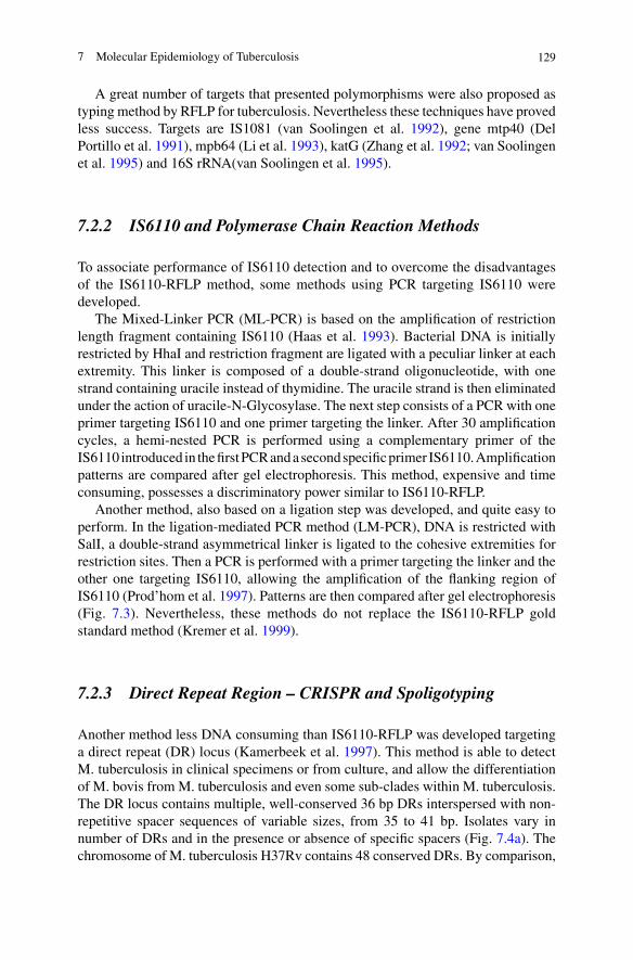
Actually, tuberculosis is still a major cause of illness and death worldwide, espe-cially in Asia and Africa. Globally, 9.2 million new cases and 1.7 million deaths from tuberculosis occurred in 2006, of which 0.7 million cases and 0.2 million deaths concerned HIV-positive people. Population growth has boosted these numbers compared with those previously reported by the World Health Organization (WHO report 2009). India, China, Indonesia, South Africa and Nigeria rank in top countries in terms of absolute numbers of cases. The African region has the highest incidence rate (363 per 100,000 population, Fig. 7.1). There was an estimation of 14.4 million prevalent cases of tuberculosis in 2006, with 0.5 million cases of multidrug-resistant (MDR) tuberculosis, 1.5 million deaths from tuberculosis in HIV-negative people and 0.2 million among people infected with HIV. Africa, South-East Asia and Western Pacific regions accounted for 83% of total notification cases. Several new public health problems emerged, such as VIH (especially in Africa) with 80% people co-infected tuberculosis-VIH in some countries, and the extension of MDR (multidrug resistant) and XDR (ultra-drug resistant). 440,000 cases of MDR tuberculosis were estimated, which corresponds to 3.6% of tubercu-losis cases. Almost 50% of these MDR cases occurred in China and India. Moreover MDR-tuberculosis caused 150,000 deaths in 2008. Among MDR tuberculosis, 5.4% were considered XDR tuberculosis.
Mycobacterium tuberculosis is the main representative member of the Mycobacterium tuberculosis complex (MTBC). MTBC includes M. tuberculosis, M. africanum which are pathogens restricted to humans and Mycobacterium bovis has a wide host spectrum including humans. Mycobacterium canettii is also pathogen for human but is exceptionally isolated, Mycobacterium microti is known as infecting rodents. MTBC members are characterized by 99.9% similarity at the nucleotide level and identical for their 16S rRNA sequences (Boddinghaus et al. 1990; Sreevatsan et al. 1997) but they differ widely in terms of their host tropisms, pheno-types, and pathogenicity.
In recent years, with the increasing knowledge on M. tuberculosis genome, molecular methods were developed for different objectives: molecular typing using genetic markers to explore chain of transmission, to distinguish recent exogenous from reactivation of latent tuberculosis, to study MDR outbreaks, to identify species among MTBC, to conduct phylogenetic studies. The choice of markers is then essential given the limitations of certain methods and their acces-sibility and applicability.

1277 Molecular Epidemiology of Tuberculosis
7.2 Genetic Markers and Molecular Methods in the Field of Tuberculosis
7.2.1 IS6110 and Restriction Length Fragment Polymorphism (RFLP)
Among the genetic markers used, insertion sequence (IS) and specifically IS6110 was the first marker used in a typing objective (Thierry et al. 1990a; Thierry et al. 1990b; Coros et al. 2008). IS6110 is member of the family IS3. Four to 20 copies of this 1,361 bp element have been found scattered throughout the genome of M. tuberculosis, while strains of M. bovis contain only few copies of IS6110 (Cousins et al. 1998). With the data obtained from the sequencing of M. tuberculosis H37Rv, most of the IS6110 copies were found in intergenic or non-coding region, near tRNA genes. The distribution of IS6110 is not homogeneous on the chromosome (Cole et al. 1998). Initial studies confirmed that, even if IS6110 is a transposable element, transposition frequency is low and no polymorphism was detected in experimental infection of guinea pigs with M. tuberculosis strains over a period of 3 months. The stability is probably maintained for many years. The number of copies of IS6110 is variable for an isolate to another with variable localisation on the chromosome. Due to this second polymorphism level, the distribution of IS6110 in M. tuberculosis strains isolated from different patients can reveal
Fig. 7.1 Estimated tuberculosis incidence rates, 2008 (WHO report 2009)
128 P. Lanotte
different genomic arrangements of the sequences, whereas strains isolated from patients of the same tuberculosis outbreak may show identical distribution pat-terns (van Embden et al. 1993).
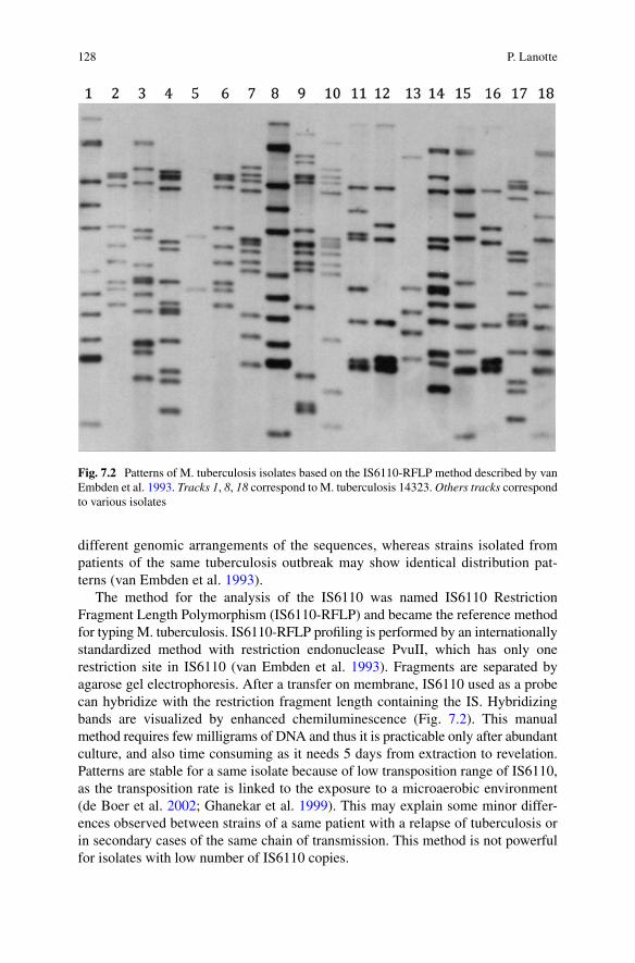
The method for the analysis of the IS6110 was named IS6110 Restriction Fragment Length Polymorphism (IS6110-RFLP) and became the reference method for typing M. tuberculosis. IS6110-RFLP profiling is performed by an internationally standardized method with restriction endonuclease PvuII, which has only one restriction site in IS6110 (van Embden et al. 1993). Fragments are separated by agarose gel electrophoresis. After a transfer on membrane, IS6110 used as a probe can hybridize with the restriction fragment length containing the IS. Hybridizing bands are visualized by enhanced chemiluminescence (Fig. 7.2). This manual method requires few milligrams of DNA and thus it is practicable only after abundant culture, and also time consuming as it needs 5 days from extraction to revelation. Patterns are stable for a same isolate because of low transposition range of IS6110, as the transposition rate is linked to the exposure to a microaerobic environment (de Boer et al. 2002; Ghanekar et al. 1999). This may explain some minor differ-ences observed between strains of a same patient with a relapse of tuberculosis or in secondary cases of the same chain of transmission. This method is not powerful for isolates with low number of IS6110 copies.
Fig. 7.2 Patterns of M. tuberculosis isolates based on the IS6110-RFLP method described by van Embden et al. 1993. Tracks 1, 8, 18 correspond to M. tuberculosis 14323. Others tracks correspond to various isolates
1297 Molecular Epidemiology of Tuberculosis
A great number of targets that presented polymorphisms were also proposed as typing method by RFLP for tuberculosis. Nevertheless these techniques have proved less success. Targets are IS1081 (van Soolingen et al. 1992), gene mtp40 (Del Portillo et al. 1991), mpb64 (Li et al. 1993), katG (Zhang et al. 1992; van Soolingen et al. 1995) and 16S rRNA(van Soolingen et al. 1995).
7.2.2 IS6110 and Polymerase Chain Reaction Methods
To associate performance of IS6110 detection and to overcome the disadvantages of the IS6110-RFLP method, some methods using PCR targeting IS6110 were developed.
The Mixed-Linker PCR (ML-PCR) is based on the amplification of restriction length fragment containing IS6110 (Haas et al. 1993). Bacterial DNA is initially restricted by HhaI and restriction fragment are ligated with a peculiar linker at each extremity. This linker is composed of a double-strand oligonucleotide, with one strand containing uracile instead of thymidine. The uracile strand is then eliminated under the action of uracile-N-Glycosylase. The next step consists of a PCR with one primer targeting IS6110 and one primer targeting the linker. After 30 amplification cycles, a hemi-nested PCR is performed using a complementary primer of the IS6110 introduced in the first PCR and a second specific primer IS6110. Amplification patterns are compared after gel electrophoresis. This method, expensive and time consuming, possesses a discriminatory power similar to IS6110-RFLP.
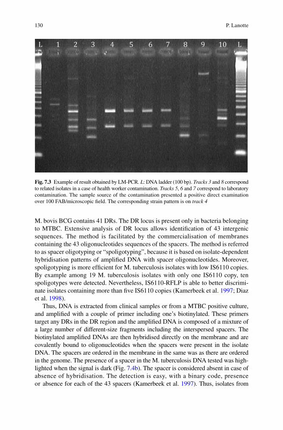
Another method, also based on a ligation step was developed, and quite easy to perform. In the ligation-mediated PCR method (LM-PCR), DNA is restricted with SalI, a double-strand asymmetrical linker is ligated to the cohesive extremities for restriction sites. Then a PCR is performed with a primer targeting the linker and the other one targeting IS6110, allowing the amplification of the flanking region of IS6110 (Prod’hom et al. 1997). Patterns are then compared after gel electrophoresis (Fig. 7.3). Nevertheless, these methods do not replace the IS6110-RFLP gold standard method (Kremer et al. 1999).
7.2.3 Direct Repeat Region – CRISPR and Spoligotyping
Another method less DNA consuming than IS6110-RFLP was developed targeting a direct repeat (DR) locus (Kamerbeek et al. 1997). This method is able to detect M. tuberculosis in clinical specimens or from culture, and allow the differentiation of M. bovis from M. tuberculosis and even some sub-clades within M. tuberculosis. The DR locus contains multiple, well-conserved 36 bp DRs interspersed with non-repetitive spacer sequences of variable sizes, from 35 to 41 bp. Isolates vary in number of DRs and in the presence or absence of specific spacers (Fig. 7.4a). The chromosome of M. tuberculosis H37Rv contains 48 conserved DRs. By comparison,
130 P. Lanotte
M. bovis BCG contains 41 DRs. The DR locus is present only in bacteria belonging to MTBC. Extensive analysis of DR locus allows identification of 43 intergenic sequences. The method is facilitated by the commercialisation of membranes containing the 43 oligonucleotides sequences of the spacers. The method is referred to as spacer oligotyping or “spoligotyping”, because it is based on isolate-dependent hybridisation patterns of amplified DNA with spacer oligonucleotides. Moreover, spoligotyping is more efficient for M. tuberculosis isolates with low IS6110 copies. By example among 19 M. tuberculosis isolates with only one IS6110 copy, ten spoligotypes were detected. Nevertheless, IS6110-RFLP is able to better discrimi-nate isolates containing more than five IS6110 copies (Kamerbeek et al. 1997; Diaz et al. 1998).
Thus, DNA is extracted from clinical samples or from a MTBC positive culture, and amplified with a couple of primer including one’s biotinylated. These primers target any DRs in the DR region and the amplified DNA is composed of a mixture of a large number of different-size fragments including the interspersed spacers. The biotinylated amplified DNAs are then hybridised directly on the membrane and are covalently bound to oligonucleotides when the spacers were present in the isolate DNA. The spacers are ordered in the membrane in the same was as there are ordered in the genome. The presence of a spacer in the M. tuberculosis DNA tested was high-lighted when the signal is dark (Fig. 7.4b). The spacer is considered absent in case of absence of hybridisation. The detection is easy, with a binary code, presence or absence for each of the 43 spacers (Kamerbeek et al. 1997). Thus, isolates from
Fig. 7.3 Example of result obtained by LM-PCR. L: DNA ladder (100 bp). Tracks 3 and 8 correspond to related isolates in a case of health worker contamination. Tracks 5, 6 and 7 correspond to laboratory contamination. The sample source of the contamination presented a positive direct examination over 100 FAB/microscopic field. The corresponding strain pattern is on track 4

1317 Molecular Epidemiology of Tuberculosis
Fig. 7.4 (a) A-Schematic illustrating the principles of the CRISPR- and VNTR-based genotyping in MTBC. These genotyping methods are known as ‘spoligotyping’ and ‘MIRU-VNTR-typing’, respectively. Spoligotyping is based on the detection of 43 unique spacers located between direct repeats at a specific locus of the MTBC genome known as the direct repeat (DR) locus. Spoligotyping patterns are commonly represented by black and white squares indicating presence or absence of particular spacers, respectively. The deletion of some of these 43 spacers allows to differentiate between strains. MIRU-VNTR analysis relies on the identification of different number of repeats at several loci scattered around the bacterial genome (marked by (A), (B), (C), and (D) in the fig-ure). The number of repeats at each locus is combined to generate a unique numerical code used to establish phylogenetic and epidemiological links between strains (from Comas et al. 2009). (b) Example of spoligotype patterns

132 P. Lanotte
epidemiologically-related cases share same hybridisation patterns. An international spoligotyping database (SpolDB4) has been recently made accessible via internet, which facilitates molecular epidemiological studies at a global scale (Brudey et al. 2006).
The Direct Repeat locus of the MTBC is a member of the CRISPR (Clustered regularly interspaced short palindromic repeats) sequences family recently described (Brudey et al. 2006; Pourcel et al. 2005). Spoligotyping is a widely used PCR-based reverse-hybridization blotting technique that assays the genetic diversity of this locus and useful for clinical laboratory, molecular epidemiology, evolutionary and population genetics. Recently a new format of this method has been proposed as a test that uses microbeads-based techniques with new spacers to increase the dis-criminative power of the method (Zhang et al. 2010).
7.2.4 Short Repetitive DNA Sequence and Polymorphic GC-Rich Sequence
Short repetitive DNA sequences associated with genetic diversity were also used as typing methods. Repeat of a triplet GTG was detected by southern blotting after HinfI digestion (Wiid et al. 1994). Polymorphic GC-rich repeat sequence (PGRS) also called Pro-Glu (Prolin-Glutamic acid or PE) repeat sequence is the target of southern-blotting methods after AluI digestion (Ross et al. 1992). In M. tuberculo-sis, PGRS appears to be present in at least 30 copies varying in number and distribu-tion from strain to strain.
Double Repetitive Element amplification (DRE-PCR) targets PGRS and IS6110 (Friedman et al. 1995). This technique is based on the fact that the distance between the copy numbers of IS6110 and PGRS may vary from strain to strain. These varia-tions allow different sizes and numbers of DNA fragments to be amplified.
7.2.5 Analysis of Tandem Repeats, VNTR-MIRU Method
Major Polymorphic Tandem Repeat (MPTR), present at multiple chromosomal loci, was used initially to compare isolates (Hermans et al. 1992). Eleven loci of tandem repeats were then explored by Frothingham et al. (Frothingham et al. 1998). Among them, six Exact Tandem Repeats (ETR), ETR-A to ETR-F and one among five MPTR (MPTR-A) present a sufficient polymorphism. All together, these markers were able to distinguish 22 of 25 strains of M. tuberculosis and five on 23 M. bovis BCG tested. Each ETR locus had multiple alleles in the panel. Polymorphism corresponds to insertions or deletions of tandem repeats. Allele profiles were repro-ducible and stable, as demonstrated by analyses of multiple isolates of particular reference strains (Frothingham et al. 1998). Supply et al. described 41 Variable Number of Tandem Repeat (VNTR) called MIRU (Mycobacterial Interspersed
1337 Molecular Epidemiology of Tuberculosis
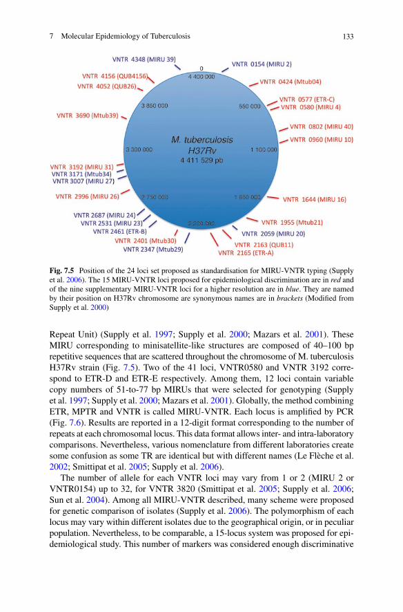
Repeat Unit) (Supply et al. 1997; Supply et al. 2000; Mazars et al. 2001). These MIRU corresponding to minisatellite-like structures are composed of 40–100 bp repetitive sequences that are scattered throughout the chromosome of M. tuberculosis H37Rv strain (Fig. 7.5). Two of the 41 loci, VNTR0580 and VNTR 3192 corre-spond to ETR-D and ETR-E respectively. Among them, 12 loci contain variable copy numbers of 51-to-77 bp MIRUs that were selected for genotyping (Supply et al. 1997; Supply et al. 2000; Mazars et al. 2001). Globally, the method combining ETR, MPTR and VNTR is called MIRU-VNTR. Each locus is amplified by PCR (Fig. 7.6). Results are reported in a 12-digit format corresponding to the number of repeats at each chromosomal locus. This data format allows inter- and intra-laboratory comparisons. Nevertheless, various nomenclature from different laboratories create some confusion as some TR are identical but with different names (Le Flèche et al. 2002; Smittipat et al. 2005; Supply et al. 2006).
The number of allele for each VNTR loci may vary from 1 or 2 (MIRU 2 or VNTR0154) up to 32, for VNTR 3820 (Smittipat et al. 2005; Supply et al. 2006; Sun et al. 2004). Among all MIRU-VNTR described, many scheme were proposed for genetic comparison of isolates (Supply et al. 2006). The polymorphism of each locus may vary within different isolates due to the geographical origin, or in peculiar population. Nevertheless, to be comparable, a 15-locus system was proposed for epi-demiological study. This number of markers was considered enough discriminative
Fig. 7.5 Position of the 24 loci set proposed as standardisation for MIRU-VNTR typing (Supply et al. 2006). The 15 MIRU-VNTR loci proposed for epidemiological discrimination are in red and of the nine supplementary MIRU-VNTR loci for a higher resolution are in blue. They are named by their position on H37Rv chromosome are synonymous names are in brackets (Modified from Supply et al. 2000)

134 P. Lanotte
for routine investigations, but a 24-locus system was considered as a high-resolution tool for phylogenetic studies (Supply et al. 2006).
MIRU-VNTR is easy to perform and accessible method for genetic studies (Martin et al. 2007). The initial method can be performed on automated sequencer with a peculiar tool to identify the PCR product size when fluorescent dye primers are used. Moreover, using automated sequencer, three or four VNTR can be explored simultaneously (Supply et al. 2001).
7.2.6 Single Nucleotide Polymorphisms, the SNP Method
Genetic polymorphism can be detected also at the nucleotide level. Single nucle-otide polymorphisms (SNP) are decomposed in non synonymous (nsSNP) and in synonymous SNPs (sSNP). nsSNP indicate amino acid changes and are implicated by example, in resistance to anti-tuberculosis drugs (Gutacker et al. 2006). These sSNP are able to divide mycobacterial populations in genetic groups. Moreover, sSNP are theoretically evolutionarily neutral and then can be used for population-genetics and for examination of phylogenetic relationships among bacterial isolates. Combination of sSNP, nsSNP and intergenic SNP was shown to be representative of phylogenetic lineages over the world (Gutacker et al. 2006).
Phylogenetic studies undergone with this method seem to be robust (Comas et al. 2009). Among 212 SNPs tested, Filliol et al. (2006) identified six SNP markers that were sufficient to classify a global M. tuberculosis collection into seven phyloge-netically distinct “SNP cluster groups” (SCGs). Using some others SNPs enabled to subdivide the SCGs into subgroups (SC subgroups) giving then a more discriminative method for phylogenetic analysis (Alland et al. 2007).
Recently, an approach combining two multiplex allele-specific minisequencing assays was proposed and permits detection of eight species- and eight lineage-specific
Fig. 7.6 Example of patterns obtained for MIRU 26. L: DNA Ladder (100 pb). Tracks 1–5: clinical isolates, track 6: M. tuberculosis H37Rv, track 7: negative control

1357 Molecular Epidemiology of Tuberculosis
single nucleotide polymorphisms (SNP) (Bouakaze et al. 2010). This method uses a commercial kit SNaPshot™ Multiplex Kit (Applied Biosystems) followed by an eight-plex minisequencing reaction and finally analysis of extension products by capillary electrophoresis. Distinction between principal genetic groups defined by SNPs was then obtained for 56 MTBC strains (Bouakaze et al. 2010).
7.2.7 Large Sequence Polymorphisms or Regions of Difference, Comparative Genomic and DNA Microarrays
Comparative genomics studies, available since the first publication of a complete genome of M. tuberculosis H37Rv strain (Coros et al. 2008), revealed that large genomic insertions or deletions are important sources of genome plasticity in MTBC (Fleischmann et al. 2002; Brosch et al. 2002). These genomic modifications are also known as large sequence polymorphisms (LSPs) or regions of difference (RD) (Brosch et al. 2002; Tsolaki et al. 2004). More than 60 different LSPs were found in clinical isolates in comparison with M. tuberculosis H37Rv representing around 4% of the genome (Tsolaki et al. 2004). Deletions are not randomly distributed. Some LSPs are associated with genes encoding members of the proline-glutamic acid or proline-proline-glutamic acid protein families and represent polymorphism that occurs under selective pressure while others may reflect phylogeny (Alland et al. 2007). RD were also used for identification of species within MTBC (Brosch et al. 2002; Huard et al. 2006; Pinsky and Banaei 2008). DNA microarrays were used to analyze these LSPs and to improve evolutionary knowledge of M. tuberculosis (Comas et al. 2009; Tsolaki et al. 2004; Hirsh et al. 2004; Gagneux et al. 2006).
7.3 Applications of Molecular Typing for Tuberculosis
Applications of molecular typing techniques are various from inter-human trans-mission with several objectives (MDR transmission, intra-familial transmission, re-infection/relapse, nosocomial transmission) to evolution of M. tuberculosis strains at global scale, including laboratory contamination and patho-physiology among others. The main applications of these methods are discussed below.
7.3.1 Inter-Human Transmission
Initial genetic studies were performed to confirm outbreaks, to identify index cases in outbreak, to trace chain of transmission in specific groups (like prisoners), and to identify unsuspected transmission. Studies have explored epidemiology within a town or at the level of a country (Diaz et al. 1998; Small et al. 1994;

136 P. Lanotte
Lopez-Calleja et al. 2009). IS6110-RFLP, Spoligotyping and MIRU-VNTR were the predominantly methods used for this purpose. Molecular methods also showed that evolution of M. tuberculosis in vivo is characterized by periods of relative genomic stability followed by bursts of mutation (Schurch et al. 2010). Molecular epidemiology improved knowledge on the dissemination of the bacteria. For example, in Germany, Barniol et al. (2009) demonstrated that few exchanges of strains between foreign-born cases in comparison to exchanges within local popu-lations (Barniol et al. 2009).
7.3.2 Multi-Drug Resistant Transmission
Many studies have focused on the transmission of MDR strains. In 1993, a major outbreak was observed in New York and 60% of the cases were linked to MDR strains (Valway et al. 1994; Frieden et al. 1996). Cases diagnosed accounted for nearly one fourth of the cases of multidrug-resistant tuberculosis in the United States over a 3 years period. Most patients had nosocomial-acquired disease, and were also infected by HIV (86%), with an important mortality rate (83%). IS6110-RFLP typing method used to characterized this peculiar strain (“strain W”) and some variants allowed the follow-up of the dissemination of the strain widely in the community over years (Munsiff et al. 2003) across the United States (Agerton et al. 1999) and was also isolated in Europe (Bifani et al. 1996; Schwoebel et al. 1998). Variants observed on molecular patterns presented additional drug resistance probably linked to nsSNP as demonstrated by other studies (Bifani et al. 2008; Post et al. 2004). Moreover, new insights concerning local phenomenon in lung lesions lead to the emergence of heterogeneous population of bacilli with different drug-susceptibilities (Post et al. 2004; Kaplan et al. 2003).
A well-known worldwide cluster associated with anti-tuberculosis drug resis-tance is the Beijing genotype, which can be identified routinely by spoligotyping. This peculiar genotype, genetically linked with the “strain W”, is mainly responsible of MDR and XDR strains over the world. For example, in a recent study in Taiwan, 44.4% of the XDR M. tuberculosis isolates and 56.7% of the MDR isolates belonged to the Beijing genotypes’ family (Lai et al. 2010).
7.3.3 Exogenous Re-infection and Relapse
It was admitted that initial tuberculosis protects against re-infection. Molecular methods were able to demonstrate that re-infection exists and that the relative importance of re-infection is likely depending on the epidemiological context. In geographical areas with a low incidence of tuberculosis, recurrent tuberculosis is generally due to reactivation of the disease. An increased risk for re-infection was
1377 Molecular Epidemiology of Tuberculosis
observed in immigrant patients compared to inhabitants in low tuberculosis incidence areas (Mathema et al. 2006; Warren et al. 2004).
A molecular epidemiology study performed in Italy, based on more than 2,100 patients with tuberculosis, identified that 32 patients (1.5%) had two distinct epi-sodes of tuberculosis (with a cure as outcome of the first episode and 6 months between the two episodes) (Bandera et al. 2001). In five patients (16%), the DNA fingerprinting patterns of M. tuberculosis strains responsible of the second episode did not match those corresponding to isolates of the first episode, indicating exog-enous re-infection (Bandera et al. 2001). Episodes of re-infection in areas with low incidences of tuberculosis are, however, rare compared to those in high-incidence geographical regions. It was also admitted that tuberculosis may result from a single infection with a single M. tuberculosis strain. A study suggested that multiple infec-tions are frequent in high-incidence regions, implying high re-infection rates and lack of efficient protective immunity conferred by initial infection (Warren et al. 2004). In populations that have emigrated from high-risk areas, re-infection may represent a considerable contributor to the rate of recurrent tuberculosis. Moreover, drug susceptible and drug-resistant strains may also coexist (Warren et al. 2004).
Relapse is not an anecdotal aspect of tuberculosis. Between 1% and 11% of cases have a second recurrent episode (Martin et al. 2007). Relapse could be associated with non observance of therapy, immune-suppressive therapy or infec-tion in the elderly (Hocking and Choi 1997; Mathema et al. 2006; Comas and Gagneux 2009).
Identification of recurrences caused by exogenous re-infection could influence therapeutic and epidemiological decisions because susceptibility could be different and the patient should be considered as a new case. When a case is assumed to be a relapse, rapid information on exogenous re-infection by a strain spreading into the community could indicate new recent transmission routes and ongoing transmission events. Moreover, higher risk of relapse rather than re-infection may be observed in HIV-positive subjects and in patients infected with multidrug-resistant tuberculosis.
M. tuberculosis isolates exhibiting identical DNA fingerprinting patterns can harbor substantial genomic diversity. Because this variability may not be captured by traditional genotyping methods of MTBC, some important aspects of the trans-mission dynamics could be missed or misinterpreted (Niemann et al. 2009).
7.3.4 Nosocomial Transmission
During the New York’s outbreak of 1990–1993, the great majority of cases were acquired at hospital (Valway et al. 1994; Frieden et al. 1996). Dissemination of the “W strain” in other states was associated with the use of bronchoscope (Agerton et al. 1999). In France, M. tuberculosis cross-contamination due to the use of bron-choscope was also confirmed by molecular technique (Carricajo et al. 1999).
138 P. Lanotte
The risk of transmission of M. tuberculosis from patients to health-care workers is a neglected problem in many countries of low- and middle incomes especially when the clinical form is not typical (Joshi et al. 2006; Menzies et al. 2007). A higher risk of acquiring TB disease was associated with certain work locations (inpatient TB facility, laboratory, internal medicine, and emergency facilities) and occupational categories (radiology technicians, patient attendants, nurses, ward attendants, paramedics, and clinical officers) (Joshi et al. 2006).
In high-income countries, risk can be higher for health-care workers if the infec-tion control measures are ineffective (Menzies et al. 2007).
7.3.5 Laboratory Contamination
The false diagnosis of tuberculosis due to laboratory cross-contamination is a well-known event, which has been reported to occur in 0.1–3% of cases (de Boer et al. 2002; Ruddy et al. 2002; Small et al. 1993). Laboratory contamination is suspected (i) when only one sample of a patient is culture positive with a small number of colonies, (ii) when M. tuberculosis is cultivated from a sample processed together with a smear-positive specimen, and (iii) when clinician considered an alternative diagnosis as more probable. Suspicion of false positivity is increased when these conditions are associated (Martinez et al. 2006). Microbiologists should rule out with clinicians the epidemiological links between suspected cases. The final confir-mation of false positivity requires the application of molecular tools to prove that MTBC isolates from co-processed specimens share identical genotypic patterns (Martin et al. 2008). MIRU-VNTR appears to be more adequate than RFLP for analyzing cross-contamination alerts. It is faster than RFLP and the correlation with RFLP diagnosis is high. A permanent suspicious attitude from clinical bacteriologist and an access to fast resolution of cross-contamination alerts could enable more rapid management of suspected false positive cases (Mathema et al. 2006).
7.3.6 Phylogeny of M. tuberculosis at Global Scale
As mentioned above, molecular tools and appropriate uses of new markers allow the determination of the emergence of peculiar clones over the world.
Although strain W was characterized in New York City using IS6110-RFLP (Agerton et al. 1999), a similar group of predominant isolates was identified with the same molecular characteristics in China. IS6110-RFLP showed that 86% of the isolates belonged to a genetically closely-related group (van Soolingen et al. 1995; Bifani et al. 2002). Because the majority of these strains originated from the province of Beijing, the cluster was named the “Beijing family”. Strains of this family were also found to dominate in neighboring countries such as Mongolia, South Korea, and Thailand, where this cluster represents respectively 50%, 43%, 37%
1397 Molecular Epidemiology of Tuberculosis
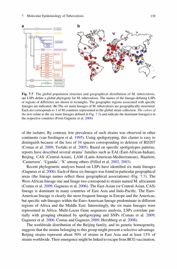
of the isolates. By contrast, low prevalence of such strains was observed in other continents (van Soolingen et al. 1995). Using spoligotyping, this cluster is easy to distinguish because of the loss of 34 spacers corresponding to deletion of RD207 (Comas et al. 2009; Tsolaki et al. 2005). Based on specific spoligotypes patterns, reports have described several strains’ families such as EAI (East-African-Indian), Beijing, CAS (Central-Asian), LAM (Latin-American-Mediterranean), Haarlem, ‘Cameroon’, ‘Uganda’, ‘X’ among others (Filliol et al. 2002, 2003).
Recent phylogenetic analyses based on LSPs have identified six main lineages (Gagneux et al. 2006). Each of these six lineages was found in particular geographical areas (the lineage names reflect these geographical associations) (Fig. 7.7). The West-African lineage one and linage two correspond to strains named M. africanum (Comas et al. 2009; Gagneux et al. 2006). The East-Asian (or Central-Asian, CAS) lineage is dominant in many countries of East Asia and Indo-Pacific. The Euro-American lineage is clearly the most frequent lineage in Europe and the Americas, but specific sub-lineages within the Euro-American lineage predominate in different regions of Africa and the Middle East. Interestingly, the six main lineages were represented in Africa. Multi-Locus Gene sequences analysis, LSPs correlate par-tially with grouping obtained by spoligotyping and SNPs (Comas et al. 2009; Gagneux et al. 2006; Comas and Gagneux 2009; Hershberg et al. 2008).
The worldwide distribution of the Beijing family, and its genetic homogeneity, suggests that the strains belonging to this group might present a selective advantage. Beijing strains represent about 50% of strains in East Asia and at least 13% of strains worldwide. Their emergence might be linked to escape from BCG vaccination,
Fig. 7.7 The global population structure and geographical distribution of M. tuberculosis. (a) LSPs define a global phylogeny for M. tuberculosis. The names of the lineage-defining LSPs or regions of difference are shown in rectangles. The geographic regions associated with specific lineages are indicated. (b) The six main lineages of M. tuberculosis are geographically structured. Each dot corresponds to 1 of 80 countries represented in the global strain collection. The colors of the dots relate to the six main lineages defined in Fig. 7.7a and indicate the dominant lineage(s) in the respective countries (From Gagneux et al. 2006)

140 P. Lanotte
and to multi-drug resistance in many areas. Finally, the Beijing family has been linked to polymorphisms in immune genes, suggesting the possibility of human–mycobacterial co-evolution. These results suggest that M. tuberculosis Beijing strains have a high capacity to withstand tuberculosis treatment, even in the absence of drug-resistance (Parwati et al. 2010).
Another facet of phylogenetic studies deals with M. tuberculosis species and members of the MTBC. LSPs (RDs) helped at confirming a common ancestor, although still in debate (Brosch et al. 2002; Huard et al. 2006; Fabre et al. 2004; Gutierrez et al. 2005). Notion of ancestral (basal) and modern (derived) lineages is based on the lack of deletion in the TbD1 region containing a leucine (CTG) at katG in position 463, which characterised ancestral (basal) M. tuberculosis strains (Fig. 7.8) (Brosch et al. 2002; Gordon et al. 2009). A study has suggested that ancestral mycobacteria may have affected early hominids in East Africa around three million years ago (Gutierrez et al. 2005).
7.3.7 Pathophysiology of Tuberculosis
Molecular tools provide further insights on pathophysiology of tuberculosis. Nearly 20% of patients with a smear-negative are responsible of tuberculosis transmission (Behr et al. 1999).
Fig. 7.8 Working model of the evolutionary scheme of tubercle bacilli illustrating successive loss of DNA in certain lineages (grey boxes). The scheme is based on the presence or absence of con-served RDs (TbD1) and on sequence polymorphisms in five selected genes. Note that the distances between certain branches may not correspond to actual phylogenetic differences calculated by other methods (From S. V. Gordon et al. 2009)

1417 Molecular Epidemiology of Tuberculosis
Moreover, histological examination of different lung lesions revealed variability in morphology and distribution of acid-fast bacilli. Molecular characterization of different isolates from different anatomical locations (for example at the surface of cavities, in granulomas) suggests that a single infected strain may undergo genetic changes during treatment, which leads to acquisition of additional drug resistance independently in these different locations resulting in parallel evolution of hetero-geneous drug-resistant sub-populations (Kaplan et al. 2003).
As mentioned previously, repetitive sequences of PE-PGRS are used in some molecular methods. PE-PGRS may have role in pathophysiology of tuberculosis. PE-PGRS16 and PE-PGRS26 can be implicated in latency phenomenon (Talarico et al. 2008). Moreover, sequence variations in the PE-PGRS33 protein with large insertion, deletion or mutation may result in the lack of cavity formation in the lungs (Talarico et al. 2007).
Some particular groups such as the Beijing/W strains may present some specific biological features. This cluster can be associated with the development of extra-thoracic localization of tuberculosis (Kong et al. 2006). Moreover, the genotype of M. tuberculosis, defined by LSPs or SNPs, may influence features of pulmo-nary and meningitis tuberculosis. The association between Beijing lineage and disease progression suggests that this lineage may influence intra-cerebral inflam-matory response. In addition, increased drug resistance among bacteria of the East Asian Beijing lineage might influence the response to treatment (Thwaites et al. 2008). Beijing strains appear to be more virulent in animal models, and to cause more histo-pathological changes, higher growth, and increased mortality. At a molecular level, Beijing strains have specific properties in terms of protein and lipid structures and interaction with the immune system (Parwati et al. 2010). All these studies suggest that the genetic diversity of M. tuberculosis has impor-tant clinical consequences.
7.3.8 Molecular Methods Adapted to Other Members of the Mycobacterium tuberculosis Complex
Molecular methods are applied for all members of MTBC. Nevertheless, some adaptations are necessary to increase the discriminatory power of markers used (Allix et al. 2006; Roring et al. 2002). Indeed, M. bovis contains few copies of IS6110 and RFLP based on this IS was found sufficiently discriminative. New loci for VNTR were identified (Roring et al. 2002); their analyses and spoligotypes pat-terns helped at identifying person-to-person transmission (Evans et al. 2007; Sunder et al. 2009).
In M. africanum, a new generation of spoligotyping based on a 68-spacer format defined new additional patterns, which helped at better understanding the evolution of M. africanum (Brudey et al. 2004). Complementary studies also allowed reclassification of some M. africanum lineages, initially defined as sub-type (Brudey et al. 2004).
142 P. Lanotte
7.4 Conclusion
The standard molecular epidemiological typing techniques are based on mobile (e.g., IS6110-RFLP) or repetitive (e.g., spoligotyping and MIRU-VNTR) DNA elements that evolve fast, resulting in high discriminatory power, an important prerequisite for detecting ongoing transmission, identifying laboratory cross-contaminations, or differentiating disease relapse from re-infection.
However, because of rapid changes at these loci, identical finger printing patterns can emerge in unrelated strain lineages (homoplasy) as a result of convergent evolu-tion, making it difficult to define deep phylogenetic structures unambiguously. Moreover, these methods could present some limits in phylogenetic studies. SNP analysis or comparative genomic studies, identifying regions of difference, seem to be more congruent and robust for this purpose.
All together, these methods allow new insights in the understanding the various facets of tuberculosis. Molecular typing methods lead to improvements in epide-miological studies in association with classical epidemiologic approaches. Genotyping methods afford greater resolution in the detection of unsuspected transmission and in the differentiation between exogenous re-infection and relapse. New genomics technologies allow the improvement of large population genetics studies and in understanding MTBC strains evolution. Identification of particular lineages or strains exhibiting specific virulence properties or epidemic potential can be described. Molecular studies have also challenged our view on the animal origin of human tuberculosis but also on the possibility of co-infection or re-infection after cure.
Acknowledgement The author thanks MC Gutierrez for providing some pictures.
References
Agerton TB, Valway SE, Blinkhorn RJ et al (1999) Spread of strain W, a highly drug-resistant strain of Mycobacterium tuberculosis, across the United States. Clin Infect Dis 29:85–92
Alland D, Lacher DW, Hazbón MH et al (2007) Role of large sequence polymorphisms (LSPs) in generating genomic diversity among clinical isolates of Mycobacterium tuberculosis and the utility of LSPs in phylogenetic analysis. J Clin Microbiol 45:39–46
Allix C, Walravens K, Saegerman C et al (2006) Evaluation of the epidemiological relevance of variable-number tandem-repeat genotyping of Mycobacterium bovis and comparison of the method with IS6110 restriction fragment length polymorphism analysis and spoligotyping. J Clin Microbiol 44:1951–1962
Bandera A, Gori A, Catozzi L et al (2001) Molecular epidemiology study of exogenous reinfection in an area with a low incidence of tuberculosis. J Clin Microbiol 39:2213–2218
Barniol J, Niemann S, Louis VR et al (2009) Transmission dynamics of pulmonary tuberculosis between autochthonous and immigrant sub-populations. BMC Infect Dis 9:197
Behr MA, Warren SA, Salamon H et al (1999) Transmission of Mycobacterium tuberculosis from patients smear-negative for acid-fast bacilli. Lancet 353:444–449
Bifani PJ, Plikaytis BB, Kapur V et al (1996) Origin and interstate spread of a New York City multidrug-resistant Mycobacterium tuberculosis clone family. JAMA 275:452–457

1437 Molecular Epidemiology of Tuberculosis
Bifani PJ, Mathema B, Kurepina NE et al (2002) Global dissemination of the Mycobacterium tuberculosis W-Beijing family strains. Trends Microbiol 10:45–52
Bifani P, Mathema B, Kurepina N et al (2008) The evolution of drug resistance in Mycobacterium tuberculosis: from a mono-rifampicin-resistant cluster into increasingly multidrug-resistant variants in an HIV-seropositive population. J Infect Dis 198:90–94
Boddinghaus B, Rogall T, Flohr T et al (1990) Detection and identification of mycobacteria by amplification of rRNA. J Clin Microbiol 28:1751–1759
Bouakaze C, Keyser C, de Martino SJ et al (2010) Identification and genotyping of Mycobacterium tuberculosis complex species using a SNaPshot minisequencing-based assay. J Clin Microbiol 48:1758–1766
Brosch R, Gordon SV, Marmiesse M et al (2002) A new evolutionary scenario for the Mycobacterium tuberculosis complex. Proc Natl Acad Sci USA 99:3684–3689
Brudey K, Gutierrez MC, Vincent V et al (2004) Mycobacterium africanum genotyping using novel spacer oligonucleotides in the direct repeat locus. J Clin Microbiol 42:5053–5057
Brudey K, Driscoll J, Rigouts L et al (2006) Mycobacterium tuberculosis complex genetic diver-sity: mining the fourth international spoligotyping database (SpolDB4) for classification, popu-lation genetics and epidemiology. BMC Microbiol 6:23
Carricajo A, Vincent V, Berthelot P et al (1999) Mycobacterial cross-contamination of broncho-scope detected by molecular techniques. J Hosp Infect 42:252–253
Cole ST, Brosch R, Parkhill J et al (1998) Deciphering the biology of Mycobacterium tuberculosis from the complete genome sequence. Nature 393:537–544
Comas I, Gagneux S (2009) The past and future of tuberculosis research. PLoS Pathog 5(10):e1000600
Comas I, Homolka S, Niemann S et al (2009) Genotyping of genetically monomorphic bacteria: DNA sequencing in Mycobacterium tuberculosis highlights the limitations of current method-ologies. PLoS One 4:e7815
Coros A, DeConno E, Derbyshire KM (2008) IS6110, a Mycobacterium tuberculosis complex-specific insertion sequence, is also present in the genome of Mycobacterium smegmatis, sug-gestive of lateral gene transfer among mycobacterial species. J Bacteriol 190:3408–3410
Cousins DV, Skuce RA, Kazwala RR et al (1998) Towards a standardized approach to DNA finger-printing of Mycobacterium bovis. International union against tuberculosis and lung disease, tuberculosis in animals subsection. Int J Tuberc Lung Dis 2:471–478
Daniel TM (2006) The history of tuberculosis. Respir Med 100:1862–1870de Boer AS, Blommerde B, de Haas PEW et al (2002) False-positive mycobacterium tuberculosis
cultures in 44 laboratories in The Netherlands (1993 to 2000): incidence, risk factors, and con-sequences. J Clin Microbiol 40:4004–4009
Del Portillo P, Murillo LA, Patarroyo ME (1991) Amplification of a species-specific DNA frag-ment of Mycobacterium tuberculosis and its possible use in diagnosis. J Clin Microbiol 29:2163–2168
Diaz R, Kremer K, de Haas PE et al (1998) Molecular epidemiology of tuberculosis in Cuba out-side of Havana, July 1994-June 1995: utility of spoligotyping versus IS6110 restriction frag-ment length polymorphism. Int J Tuberc Lung Dis 2:743–750
Evans JT, Smith EG, Banerjee A et al (2007) Cluster of human tuberculosis caused by Mycobacterium bovis: evidence for person-to-person transmission in the UK. Lancet 369:1270–1276
Fabre M, Koeck JL, Le Fleche P et al (2004) High genetic diversity revealed by variable-number tandem repeat genotyping and analysis of hsp65 gene polymorphism in a large collection of“ Mycobacterium canettii” strains indicates that the M. tuberculosis complex is a recently emerged clone of “M. canettii”. J Clin Microbiol 42:3248–3255
Filliol I, Driscoll JR, Van Soolingen D et al (2002) Global distribution of Mycobacterium tubercu-losis spoligotypes. Emerg Infect Dis 8:1347–1349
Filliol I, Driscoll JR, van Soolingen D et al (2003) Snapshot of moving and expanding clones of Mycobacterium tuberculosis and their global distribution assessed by spoligotyping in an inter-national study. J Clin Microbiol 41:1963–1970
144 P. Lanotte
Filliol I, Motiwala AS, Cavatore M et al (2006) Global phylogeny of Mycobacterium tuberculosis based on single nucleotide polymorphism (SNP) analysis: insights into tuberculosis evolution, phylogenetic accuracy of other DNA fingerprinting systems, and recommendations for a minimal standard SNP set. J Bacteriol 188:759–772
Fleischmann RD, Alland D, Eisen JA et al (2002) Whole-genome comparison of Mycobacterium tuberculosis clinical and laboratory strains. J Bacteriol 184:5479–5490
Frieden TR, Sherman LF, Maw KL et al (1996) A multi-institutional outbreak of highly drug-resistant tuberculosis: epidemiology and clinical outcomes. JAMA 276:1229–1235
Friedman CR, Stoeckle MY, Johnson WD et al (1995) Double-repetitive-element PCR method for subtyping Mycobacterium tuberculosis clinical isolates. J Clin Microbiol 33:1383–1384
Frothingham R, Meeker-O’Connell WA et al (1998) Genetic diversity in the Mycobacterium tuberculosis complex based on variable numbers of tandem DNA repeats. Microbiology 144:1189–1196
Gagneux S, DeRiemer K, Van T et al (2006) Variable host–pathogen compatibility in Mycobacterium tuberculosis. Proc Natl Acad Sci USA 103:2869–2873
Ghanekar K, McBride A, Dellagostin O et al (1999) Stimulation of transposition of the Mycobacterium tuberculosis insertion sequence IS6110 by exposure to a microaerobic envi-ronment. Mol Microbiol 33:982–993
Gordon SV, Bottai D, Simeone R et al (2009) Pathogenicity in the tubercle bacillus: molecular and evolutionary determinants. Bioessays 31:378–388
Gutacker MM, Mathema B, Soini H et al (2006) Single-nucleotide polymorphism-based popula-tion genetic analysis of Mycobacterium tuberculosis strains from 4 geographic sites. J Infect Dis 193:121–128
Gutierrez MC, Brisse S, Brosch R et al (2005) Ancient origin and gene mosaicism of the progeni-tor of Mycobacterium tuberculosis. PLoS Pathog 1(1):e5
Haas WH, Butler WR, Woodley CL et al (1993) Mixed-linker polymerase chain reaction: a new method for rapid fingerprinting of isolates of the Mycobacterium tuberculosis complex. J Clin Microbiol 31:1293–1298
Hermans PW, van Soolingen D, van Embden JD (1992) Characterization of a major polymorphic tandem repeat in Mycobacterium tuberculosis and its potential use in the epidemiology of Mycobacterium kansasii and Mycobacterium gordonae. J Bacteriol 174:4157–4165
Hershberg R, Lipatov M, Small PM et al (2008) High functional diversity in Mycobacterium tuber-culosis driven by genetic drift and human demography. PLoS Biol 6(12):e311
Hirsh AE, Tsolaki AG, DeRiemer K et al (2004) Stable association between strains of Mycobacterium tuberculosis and their human host populations. Proc Natl Acad Sci USA 101:4871–4876
Hocking TL, Choi C (1997) Tuberculosis: a strategy to detect and treat new and reactivated infec-tions. Geriatrics 52:52–54
Huard RC, Fabre M, de Haas P et al (2006) Novel genetic polymorphisms that further delineate the phylogeny of the Mycobacterium tuberculosis complex. J Bacteriol 188:4271–4287
Joshi R, Reingold AL, Menzies D et al (2006) Tuberculosis among health-care workers in low- and middle-income countries: a systematic review. PLoS Med 3(12):e494
Kamerbeek J, Schouls L, Kolk A et al (1997) Simultaneous detection and strain differentiation of Mycobacterium tuberculosis for diagnosis and epidemiology. J Clin Microbiol 35:907–914
Kaplan G, Post FA, Moreira AL et al (2003) Mycobacterium tuberculosis growth at the cavity surface: a microenvironment with failed immunity. Infect Immun 71:7099–7108
Kong Y, Cave MD, Zhang L et al (2006) Association between Mycobacterium tuberculosis Beijing/W lineage strain infection and extrathoracic tuberculosis: insights from epidemiologic and clinical characterization of the three principal genetic groups of M. tuberculosis clinical isolates. J Clin Microbiol 45:409–414
Kremer K, van Soolingen D, Frothingham R et al (1999) Comparison of methods based on differ-ent molecular epidemiological markers for typing of Mycobacterium tuberculosis complex strains: interlaboratory study of discriminatory power and reproducibility. J Clin Microbiol 37:2607–2618

1457 Molecular Epidemiology of Tuberculosis
Lai CC, Tan CK, Lin SH et al (2010) Clinical and genotypic characteristics of extensively drug-resistant and multidrug-resistant tuberculosis. Eur J Clin Microbiol Infect Dis 29:597–600
Le Flèche P, Fabre M, Denoeud F et al (2002) High resolution, on-line identification of strains from the Mycobacterium tuberculosis complex based on tandem repeat typing. BMC Microbiol 2:37
Li H, Ulstrup JC, Jonassen TO et al (1993) Evidence for absence of the MPB64 gene in some substrains of Mycobacterium bovis BCG. Infect Immun 61:1730–1734
López-Calleja AI, Gavín P, Lezcano MA et al (2009) Unsuspected and extensive transmission of a drug-susceptible Mycobacterium tuberculosis strain. BMC Pulm Med 9:3
Martín A, Herránz M, Serrano MJR et al (2007) Rapid clonal analysis of recurrent tuberculosis by direct MIRU-VNTR typing on stored isolates. BMC Microbiol 7:73
Martín A, Herranz M, Lirola MM et al (2008) Optimized molecular resolution of cross-contami-nation alerts in clinical mycobacteriology laboratories. BMC Microbiol 8:30
Martínez M, García de Viedma D, Alonso M et al (2006) Impact of laboratory cross-contamination on molecular epidemiology studies of tuberculosis. J Clin Microbiol 44:2967–2969
Mathema B, Kurepina NE, Bifani PJ et al (2006) Molecular epidemiology of tuberculosis: current insights. Clin Microbiol Rev 19:658–685
Mazars E, Lesjean S, Banuls AL et al (2001) High-resolution minisatellite-based typing as a por-table approach to global analysis of Mycobacterium tuberculosis molecular epidemiology. Proc Natl Acad Sci USA 98:1901–1906
Menzies D, Joshi R, Pai M (2007) Risk of tuberculosis infection and disease associated with work in health care settings. Int J Tuberc Lung Dis 11:593–605
Munsiff SS, Nivin B, Sacajiu G et al (2003) Persistence of a highly resistant strain of tuberculosis in New York city during 1990–1999. J Infect Dis 188:356–363
Niemann S, Köser CU, Gagneux S et al (2009) Genomic diversity among drug sensitive and mul-tidrug resistant isolates of Mycobacterium tuberculosis with identical DNA fingerprints. PLoS One 4(10):e7407
Parwati I, Alisjahbana B, Apriani L et al (2010) Mycobacterium tuberculosis Beijing genotype is an independent risk factor for tuberculosis treatment failure in Indonesia. J Infect Dis 201:553–557
Pinsky BA, Banaei N (2008) Multiplex real-time PCR assay for rapid identification of Mycobacterium tuberculosis complex members to the species level. J Clin Microbiol 46:2241–2246
Post FA, Willcox PA, Mathema B et al (2004) Genetic polymorphism in Mycobacterium tuber-culosis isolates from patients with chronic multidrug-resistant tuberculosis. J Infect Dis 190:99–106
Pourcel C, Salvignol G, Vergnaud G (2005) CRISPR elements in Yersinia pestis acquire new repeats by preferential uptake of bacteriophage DNA, and provide additional tools for evolu-tionary studies. Microbiology 151:653–663
Prod’hom G, Guilhot C, Gutierrez MC et al (1997) Rapid discrimination of Mycobacterium tuber-culosis complex strains by ligation-mediated PCR fingerprint analysis. J Clin Microbiol 35:3331–3334
Roring S, Scott A, Brittain D et al (2002) Development of variable-number tandem repeat typing of Mycobacterium bovis: comparison of results with those obtained by using existing exact tandem repeats and spoligotyping. J Clin Microbiol 40:2126–2133
Ross BC, Raios K, Jackson K et al (1992) Molecular cloning of a highly repeated DNA element from Mycobacterium tuberculosis and its use as an epidemiological tool. J Clin Microbiol 30:942–946
Ruddy M, McHugh TD, Dale JW et al (2002) Estimation of the rate of unrecognized cross-con-tamination with Mycobacterium tuberculosis in London microbiology laboratories. J Clin Microbiol 40:4100–4104
Schürch AC, Kremer K, Kiers A et al (2010) The tempo and mode of molecular evolution of Mycobacterium tuberculosis at patient-to-patient scale. Infect Genet Evol 10:108–114
146 P. Lanotte
Schwoebel V, Decludt B, de Benoist AC et al (1998) Multidrug resistant tuberculosis in France 1992–1994: two case-control studies. BMJ 317:630–631
Small PM, McClenny NB, Singh SP et al (1993) Molecular strain typing of Mycobacterium tuber-culosis to confirm cross-contamination in the mycobacteriology laboratory and modification of procedures to minimize occurrence of false-positive cultures. J Clin Microbiol 31:1677–1682
Small PM, Hopewell PC, Singh SP et al (1994) The epidemiology of tuberculosis in San Francisco. A population-based study using conventional and molecular methods. N Engl J Med 330:1703–1709
Smittipat N, Billamas P, Palittapongarnpim M et al (2005) Polymorphism of variable-number tandem repeats at multiple loci in Mycobacterium tuberculosis. J Clin Microbiol 43:5034–5043
Sreevatsan S, Pan X, Stockbauer KE et al (1997) Restricted structural gene polymorphism in the Mycobacterium tuberculosis complex indicates evolutionarily recent global dissemination. Proc Natl Acad Sci USA 94:9869–9874
Sun Y, Bellamy R, Lee ASG et al (2004) Use of mycobacterial interspersed repetitive unit-variable-number tandem repeat typing to examine genetic diversity of Mycobacterium tuber-culosis in Singapore. J Clin Microbiol 42:1986–1993
Sunder S, Lanotte P, Godreuil S et al (2009) Human-to-human transmission of tuberculosis caused by Mycobacterium bovis in immunocompetent patients. J Clin Microbiol 47:1249–1251
Supply P, Magdalena J, Himpens S et al (1997) Identification of novel intergenic repetitive units in a mycobacterial two-component system operon. Mol Microbiol 26:991–1003
Supply P, Mazars E, Lesjean S et al (2000) Variable human minisatellite-like regions in the Mycobacterium tuberculosis genome. Mol Microbiol 36:762–771
Supply P, Lesjean S, Savine E et al (2001) Automated high-throughput genotyping for study of global epidemiology of Mycobacterium tuberculosis based on mycobacterial interspersed repetitive units. J Clin Microbiol 39:3563–3571
Supply P, Allix C, Lesjean S et al (2006) Proposal for standardization of optimized mycobacterial interspersed repetitive unit-variable-number tandem repeat typing of Mycobacterium tubercu-losis. J Clin Microbiol 44:4498–4510
Talarico S, Donald Cave M, Foxman B et al (2007) Association of Mycobacterium tuberculosis PE_PGRS33 polymorphism with clinical and epidemiological characteristics. Tuberculosis 87:338–346
Talarico S, Zhang L, Marrs CF et al (2008) Mycobacterium tuberculosis PE_PGRS16 and PE_PGRS26 genetic polymorphism among clinical isolates. Tuberculosis 88:283–294
Thierry D, Brisson-Noël A, Vincent-Lévy-Frébault V et al (1990a) Characterization of a Mycobacterium tuberculosis insertion sequence, IS6110, and its application in diagnosis. J Clin Microbiol 28:2668–2673
Thierry D, Cave MD, Eisenach KD et al (1990b) IS6110, an IS-like element of Mycobacterium tuberculosis complex. Nucleic Acids Res 18:188
Thwaites G, Caws M, Chau TTH et al (2008) Relationship between Mycobacterium tuberculosis genotype and the clinical phenotype of pulmonary and meningeal tuberculosis. J Clin Microbiol 46:1363–1368
Tsolaki AG, Hirsh AE, DeRiemer K et al (2004) Functional and evolutionary genomics of Mycobacterium tuberculosis: insights from genomic deletions in 100 strains. Proc Natl Acad Sci USA 101:4865–4870
Tsolaki AG, Gagneux S, Pym AS, de la Salmoniere G et al (2005) Genomic deletions classify the Beijing/W strains as a distinct genetic lineage of Mycobacterium tuberculosis. J Clin Microbiol 43:3185–3191
Valway SE, Greifinger RB, Papania M et al (1994) Multidrug-resistant tuberculosis in the New York State prison system, 1990–1991. J Infect Dis 170:151–156
van Embden JD, Cave MD, Crawford JT et al (1993) Strain identification of Mycobacterium tuber-culosis by DNA fingerprinting: recommendations for a standardized methodology. J Clin Microbiol 31:406–409

1477 Molecular Epidemiology of Tuberculosis
van Soolingen D, Hermans PW, de Haas PE et al (1992) Insertion element IS1081-associated restriction fragment length polymorphisms in Mycobacterium tuberculosis complex species: a reliable tool for recognizing Mycobacterium bovis BCG. J Clin Microbiol 30:1772–1777
van Soolingen D, Qian L, de Haas PE et al (1995) Predominance of a single genotype of Mycobacterium tuberculosis in countries of east Asia. J Clin Microbiol 33:3234–3238
Warren RM, Victor TC, Streicher EM et al (2004) Patients with active tuberculosis often have dif-ferent strains in the same sputum specimen. Am J Respir Crit Care Med 169:610–614
WHO report (2009) Global tuberculosis control: a short update to the 2009 report. http://www.who.int/tb/publications/global_report/2009/update/en/index.html
Wiid IJ, Werely C, Beyers N et al (1994) Oligonucleotide (GTG)5 as a marker for Mycobacterium tuberculosis strain identification. J Clin Microbiol 32:1318–1321
Zhang Y, Heym B, Allen B et al (1992) The catalase-peroxidase gene and isoniazid resistance of Mycobacterium tuberculosis. Nature 358:591–593
Zhang J, Abadia E, Refregier G et al (2010) Mycobacterium tuberculosis complex CRISPR geno-typing: improving efficiency, throughput and discriminative power of ‘spoligotyping’ with new spacers and a microbead-based hybridization assay. J Med Microbiol 59:285–294