Embed Size (px)
Citation preview
Palaeodiversity 7: 167–183; Stuttgart 30 December 2014. 167
1. Introduction
Archaeognathans (=Microcoryphia) are considered to exhibit numerous plesiomorphic characters or characters states. Among Insects, they are the sister group of Dicon-dylia (Zygentoma + Pterygota) and therefore of major interest for inferring for systematics and reconstructing the evolution of Hexapoda and its ingoups. As such it is an important task to reconstruct the ground pattern of Archaeognatha, i.e. the morphology of the ancestor (stem-species) of archaeognathans. As fossil species can possess plesiomorphic traits no longer found in modern represent-atives (e.g., DONOGHUE et al. 1989; RUST 2006; EDGECOMBE 2010; HAUG JT et al. 2010) they are especially valuable for such an approach.
STURM & BACH (1993) provided the first attempt to phylogenetic relationships in Archaeognatha using princi-ples of phylogenetic systematics (e.g., SUDHAUS & REHFELD 1993), but this study was exclusively based on extant species. BITSCH & NEL (1999) investigated the relation-ships among Archaeognatha including fossil representa-tives. The authors group extant Machilidae together with Meinertellidae as sister group of extinct Triassomachilis uralensis. Yet, RASNITSYN (2002) considered T. uralensis as mayfly nymph (see also SINITSHENKOVA 2000). STURM & MACHIDA (2001) considering only extant representa-tives, resolve Ditrigoniophthalmus as the sistergroup of Machilidae (restr.) + Meinertellidae. Besides Ditrigoni-ophthalmus, the authors mention additional ‘palaeoforms’ (Mesomachilis and Charimachilis). The analysis of KOCH (2003) supports Ditrigoniophthalmus as sister group of other extant archaeognaths. Furthermore, he proposes the
extinct Dasyleptidae (=Monura sensu SHAROV 1957; Car-boniferous to Triassic according to BECHLY & STOCKAR (2011) as sister group of Ditrigoniophthalmus + extant Archaeognatha.
Apart from describing archaeognathan ichnofossils GETTY et al. (2013, see also MENDES & WUNDERLICH 2013) gave a detailed summary of fossil Archaeognatha (imprints and amber inclusions). The most diverse fossil archaeog-nathan fauna (so far) can be found in Baltic amber.
First note on Archaeognatha preserved in Baltic amber was given by KOCH & BERENDT (1854). Some additional inclusions were described by V. OLFERS (1907). Together these authors described 36 fossil species, but SILVESTRI (1912) revised the specimens leaving only 11 species. Recent authors (WEITSCHAT & WICHARD 2002; MENDES & WUNDERLICH 2013) demand a revision of archaeoganthan fossils in Baltic amber.
We describe two fossil representatives of Archae-ognatha from Baltic amber, deposited at the Bayer-ische Staatssammlung für Paläontologie und Geologie in Munich. We use modern imaging techniques to document all available aspects of morphology and discuss the most important characters in the context of the insects’ ovipos-itor evolution.
A c k n o w l e d g e m e n t sMARTIN NOSE (Bayerische Staatssammlung für Paläontolo-
gie und Geologie, Munich) is thanked for his help with the spec-imens. MATTHES KENNING (Greifswald) collected the samples of Petrobius maritimus in Sweden. CWH is supported by a fellow-ship of the Landesgraduiertenstiftung Mecklenburg-Vorpom-mern. CWH and MKH thank STEFFEN HARZSCH (Greifswald)
New data on fossil Archaeognatha from Baltic amber and the origin of the insect ovipositor
CHRISTIAN W. HÄDICKE, MARIE K. HÖRNIG, JOACHIM T. HAUG & CAROLIN HAUG
A b s t r a c tThe primary wingless insect group Archaeognatha (=Microcoryphia) is the sister group of dicondylian insects
(Zygentoma + Pterygota). Therefore knowledge of character states in Archaeognatha is essential for the discussion of insect evolution as a whole. The incorporation of data from the fossil record is necessary for evolutionary biol-ogy as well as systematics, to introduce possible more ancestral character states. In the last years, fossil archaeog-nathans were described from amber deposits around the world, yet most data from Baltic amber dates more than hundred years back. In this paper we describe two new specimens of fossil Archaeognatha from Baltic amber. The ovipositor of one specimen, hence a female, is well-preserved showing four valves each representing an elongate multi-annulated structure distantly resembling the cerci. In pterygote insects the valves, as the name suggests, are usually solid simple structures. We therefore take the morphology of the aracheognathan ovipositor as a starting point to discuss potential homologies of the insect ovipositor also in comparison to non-insect arthropods.
K e y w o r d s : Amber, Baltic, Petrobius, ovipositor.

168 PALAEODIVERSITY 7, 2014
for his support. JTH is currently kindly funded by the German Research Foundation (DFG; HA 63000/3-1). JTH and CH thank J. MATTHIAS STARCK (Munich) for his support. We also thank all people involved in providing open source, open access or low cost software, such as OpenOffice, Gimp, Inkscape, Blender, CombineZM, CombineZP, Image Analyzer, ImageJ or Micro-soft Image Composite Editor. We also thank LUIS F. MENDES, (Lisbon), and an anonymous reviewer for useful comments on the manuscript.
2. Material and methods
2.1. Material
We examined two inclusions in Baltic amber assigned to the Eocene (40–50 mya). Both specimens are deposited in the Bayerische Staatssammlung für Paläontologie und Geologie, Munich, with the collection number BSPG 1967 XX 4 and BSPG 1995 I 53.
Fig. 1. Habitus of BSPG 1995 I 53 A: dorsal and ventral view. Background cleared manually B: red-blue stereo anaglyph of a virtual surface reconstruction in ventral view. Please use red-cyan glasses to view. Abbreviations: H: Head, P: prothorax, MS: mesothorax, MT: metathorax, I-XI: abdominal metameres 1-11.

HÄDICKE ET AL.: FOSSIL ARCHAEOGNATHA FROM BALTIC AMBER 169
BSPG 1967 XX 4 was part of the collection “SCHEELE”, with the origin given as “Samland”. BSPG 1995 I 53 was donated in 1995 by MIKE BÄÄTJER (Hamburg) and originated from the Tertiary of Poland. Further aspects remain unknown.
2.2. Documentation methods
Overview and close-up images were taken with a Zeiss Axiophot compound microscope equipped with a Sco-petek DCM 510 ocular camera. 2.5x and 4x objectives were used, which equals a magnification of 25 respec-tively 40 times. Light was cross-polarised to reduce reflex-ions (e.g., HAUG C et al. 2011, 2012; HAUG JT et al. 2012, 2013a; HÖRNIG et al. 2013). To compensate surface irreg-ularities of the amber, glycerine was applied and covered by a cover slip (e.g., HAUG JT et al. 2013b; HÄDICKE et al. 2013). To generate consistently sharp images in all arrays, stacks up to 60 images along the z-axis were taken and fused with Image Analyzer (http://logicnet.dk/Analyzer/). Fused single images were stitched with the Photomerge function of Adobe Photoshop CS3.
Stereo images (red/blue) of specimen BSPG 1967 XX 4 were processed with ImageAnalyzer (http://logicnet.dk/Analyzer/), based on the image stacks (HAUG JT et al. 2012, 2013b; HAUG C et al. 2013).
For stereo images of specimen BSPG 1995 I 53, we used a Canon Eos Rebel T3i camera equipped with a Canon MP-E 65 mm macro objective and a Canon Macro Twin Lite MT-24 EX flashlight with polarisation filters. Two images from different angles were taken and arranged and edited with Adobe Photoshop CS3.
2.3. Method of descriptions
Morphological descriptions follow the concepts pre-sented by HAUG JT et al. (2012). Hence first the general
body organisation is mentioned, then each body seg-ment is described strictly from anterior to posterior. Each appendage is described from proximal to distal, element for element. Terminology is kept in a neutral way, to allow also non-specialists to understand it, but specialist terms are additionally given. This is thought to enhance com-munication also beyond the archaeognath community. Additionally for this case measurements are given also for “non-standard” structures as these might be relevant in a broader comparisons. Measurements were taken on the images. As a remark this also applies to names of larger monophyletic groups. While Microcoryphia VERHOEFF, 1904 seems now to be used within the specialists’ com-munity, we use here Archaeognatha BÖRNER, 1904 as it seems to be the more generally known term.
3. Results
3.1. Description of BSPG 1995 I 53
H a b i t u s :Total length approximately 17 mm (body with terminal filament), female. Body with (presumably) 20 segments (or metameres), the ocular segment and 19 post-ocular segments (Fig. 1). Ocular segment and post-ocular segments 1–5 form the head capsule. Post-ocular segments 6–8 with walking legs; ‘thorax’. Post-ocular segments 9–16 with a pair of lateral styli and median eversible vesicles. Post-ocular segments 17 and 18 with a pair of valves. Post-ocular segment 19 with paired lateral cerci (approx. 5 mm long) and median terminal filament (approx. 7.6 mm long) (Fig. 1).
H e a d ( o c u l a r s e g m e n t + p o s t - o c u l a r s e g m e n t 1 – 5 ) : Head capsule ellipsoid, broader
Fig. 2. Dorsal view of the head of BSPG 1995 I 53. A: Colour image, B: stereo anayglyph of the same region. Please use red-cyan glasses to view.
170 PALAEODIVERSITY 7, 2014
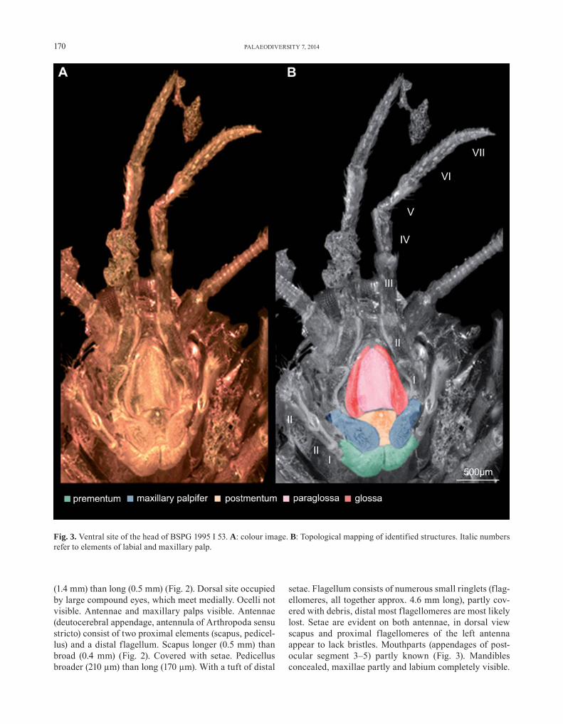
(1.4 mm) than long (0.5 mm) (Fig. 2). Dorsal site occupied by large compound eyes, which meet medially. Ocelli not visible. Antennae and maxillary palps visible. Antennae (deutocerebral appendage, antennula of Arthropoda sensu stricto) consist of two proximal elements (scapus, pedicel-lus) and a distal flagellum. Scapus longer (0.5 mm) than broad (0.4 mm) (Fig. 2). Covered with setae. Pedicellus broader (210 μm) than long (170 μm). With a tuft of distal
setae. Flagellum consists of numerous small ringlets (flag-ellomeres, all together approx. 4.6 mm long), partly cov-ered with debris, distal most flagellomeres are most likely lost. Setae are evident on both antennae, in dorsal view scapus and proximal flagellomeres of the left antenna appear to lack bristles. Mouthparts (appendages of post-ocular segment 3–5) partly known (Fig. 3). Mandibles concealed, maxillae partly and labium completely visible.
Fig. 3. Ventral site of the head of BSPG 1995 I 53. A: colour image. B: Topological mapping of identified structures. Italic numbers refer to elements of labial and maxillary palp.

HÄDICKE ET AL.: FOSSIL ARCHAEOGNATHA FROM BALTIC AMBER 171
Maxilla only known by the maxillary palp. Maxillary palp consists of seven elements (palpomeres). All but the first maxillary palpomere with setae and scales. Maxillary pal-pomere 1, proximally articulated to stipes, club-shaped, 0.4 mm long, proximally 0.2 mm and distally 0.4 mm broad. Maxillary palpomere 2 tube-shaped, 0.5 mm long and 0.2 mm broad. Maxillary palpomere 3 tube-shaped, 0.7 mm long and 0.2 mm broad. Maxillary palpomere 4 tube-shaped, 0.5 mm long and 0.2 mm broad. Maxillary palpomere 5 tube-shaped, 0.5 mm long and 0.2 mm broad. Maxillary palpomere 6 tube-shaped, 0.7 mm long and 0.2 mm broad. Maxillary palpomere 7 peg-like, 0.4 mm long and 0.2 mm broad. Labium (medially conjoined appendages of post-ocular segment 5) known with, distal central part (prementum), bearing three antero-median, shovel-like enditic protrusions (unpaired glossa and paired paraglossae) and distally with two labial palps. Labial palp consists of three elements (palpomeres), setae are evident on the 3. palpomere labial palpomere 1, proximally articu-lated to prementum tube-shaped, 150 μm long and 125 μm broad. Labial palpomere 2 tube-shaped, 0.3 mm long and 0.1 mm broad. Labial palpomere 3 tube-shaped, 0.5 mm long and 0.2 mm broad.
T h o r a x ( p o s t - o c u l a r s e g m e n t s 6 – 8 ) : Dorsally each of the three segments with a sclerotised tergite (pronotum, mesonotum, metanotum). Pronotum broader (1.6 mm) than long (0.6 mm), except for a lateral patch of scales no details are visible due to distortion of the amber. Appendages of post-ocular segment 6 (propo-dia) consist of six elements (coxa, trochanter, femur, tibia, tarsus, pretarsus) (Fig. 4). All covered with setae, scales are not visible. Procoxa tube-shaped, 0.8 mm long and 0.2 mm broad. Protrochanter covered by debris (left one)
or concealed due to orientation. Profemur tube-shaped, 0.7 mm long and 0.2 mm broad. Left one partly cov-ered with debris. Protibia tube-shaped, 0.5 mm long and 0.1 mm broad. Left one partly covered with debris. Protar-sus consists of three elements (tarsomeres): 1st tarsomere: 0.3 mm long and 0.1 mm broad, 2nd tarsomere: 0.4 m long and 0.1 mm broad, 3rd tarsomere: 0.2 mm long and 0.1 mm broad. The propretarsus bears paired claws, together they are 0.1 mm long. Mesonotum broader (2 mm) than long (0.6 mm) dorsally. No details but laterally located scales are visible. Appendages of post-ocular segment 7 (mes-opodia) consist of six elements (coxa, trochanter, femur, tibia, tarsus, pretarsus). All covered with setae, scales are not visible. Mesocoxa tube-shaped, 0.6 mm long and 0.2 mm broad. Mesotrochanter: right one covered with secretion (Verlumung) and left one only partly visible due to orientation. Mesofemur tube-shaped, 0.6 mm long and 0.2 mm broad. Mesotibia tube-shaped, 0.6 mm long and 0.2 mm broad. Mesotarsus with three elements (tar-someres), 1st tarsomere: 0.3 mm long and 0.1 mm broad, 2nd tarsomere: 0.2 mm long and 0.1 mm broad, 3rd tar-somere: 0.3 mm long and 0.1 mm broad. The mesopre-tarsus bears paired claws, together about 0.1 μm long. Metanotum broader (2.4 mm) than long (1.2 mm), except for a lateral patch of scales no surface features are visible. Metapodia consist of coxa, trochanter, femur, tibia, tar-sus, pretarsus. Occupied by setae, scales are not visible. Metacoxa tube-shaped, 0.8 mm long and 0.4 mm broad and bearing a coxal stylet. Metatrochanter tube-shaped, 0.7 mm long and 0.2 mm broad. Metafemur tube-shaped, 0.8 mm long and 0.3 mm broad. Metatibia tube-shaped, 0.8 mm long and 0.3 mm broad. Metatarsus consists of three elements (tarsomeres), 1st tarsomere: 0.3 mm long and 0.1 mm broad, 2nd tarsomere: 0.3 mm long and
Fig. 4. Ventral view of the thorax of BSPG 1995 I 53 A: colour image. B: Topological mapping of identified structures. Proximal ele-ments of right pro- and mesopodia are covered by escaped body fluid.

172 PALAEODIVERSITY 7, 2014
0.1 mm broad, 3rd tarsomere: 0.3 mm long and 0.1 mm broad. Metapretarsus equipped with paired claws, together 0.2 mm long.
A b d o m e n ( p o s t - o c u l a r s e g m e n t s 9 – 1 9 ) : Abdomen consists of eleven metameres (Figs. 5, 6). Abdominal metamere 1, dorsally with a well-sclero-tised tergite (notum). Notum broader (2.4 mm) than long 0.7 mm), covered with scales (visible laterally) and setae.
Abdominal metamere 1 ventrally partly concealed by metapodia, with a median pair of coxopodal vesicle, stylus visible on the right margin. Abdominal metamere 2 dor-sally with a well sclerotised tergite (notum). Notum broader (2.4 mm) than long (0.4 mm), covered with scales and setae. Abdominal metamere 2 ventrally with median and lateral pair of coxopodal vesicles as well as left stylus are evident. Coxopodites not visible. Abdominal metamere 3, dorsally with a well sclerotised tergite (notum). Notum broader
Fig. 5. Ventral view of the abdomen of BSPG 1995 I 53 A: colour image. B: Topological mapping of identified structures. Retracted coxopodal vesicles are difficult to acertain, so maybe not all are marked.

HÄDICKE ET AL.: FOSSIL ARCHAEOGNATHA FROM BALTIC AMBER 173
(2.3 mm) than long (0.5 mm), covered with scales and setae. Abdominal metamere 3, ventrally with median coxopodal vesicles perhaps collapsed, lateral pair of coxopodal vesi-cles and right stylus visible. Distal portion of coxopodites recognizable. Abdominal metamere 4, dorsally with a well sclerotised tergite (notum). Notum broader (2.4 mm) than long 0.4 mm), covered with scales and setae. Abdominal metamere 4, ventrally with one median coxopodal vesi-cle evaginated, two visible styli, coxopodites distinctly
separated. Abdominal metamere 5, dorsally with a well-sclerotised tergite (notum). Notum broader 2.3 mm) than long (0.5 mm), covered with scales and setae. Abdominal metamere 5 ventrally with one median coxopodal vesicle evaginated, lateral coxopodal vesicle and both styli visi-ble. Abdominal metamere 6 dorsally with a well-sclero-tised tergite (notum). Notum broader (2.2 mm) than long (0.6 mm), covered with scales and setae. Abdominal met-amere 6 ventrally with only median coxopodal vesicles
Fig. 6. Ventral view of the terminal portion of the abdomen in BSPG 1995 I 53 A: colour image. B: Topological mapping of identi-fied structures. C: Stereo image of the distal portion of the ovipositor.

174 PALAEODIVERSITY 7, 2014
are evident, both styli are recognizable, coxopodites dis-tinct distally. Abdominal metamere 7 dorsally with a well-sclerotised tergite (notum). Notum broader (2 mm) than long (0.7 mm), covered with scales and setae. Abdomi-nal metamere 7 ventrally with only median coxopodal vesicles, both styli are recognizable, coxopodites indis-tinct. Abdominal metamere 8, dorsally with a well-scler-otised tergite (notum). Notum broader (1.9 mm) than long (0.7 mm), covered with scales and setae. Abdominal met-amere 8 ventrally with styli inserted on the distal tip of then coxopodites, both coxopodites separated from base to tip. Abdominal metamere 9 dorsally with a well scler-otised tergite (notum) notum broader (1.7 mm) than long (0.6 mm), covered with scales and setae. Abdominal met-amere 9 ventrally with styli are evident, coxopodites not visible. Abdominal metamere 10 dorsally with a well scle-rotised tergite (notum). Notum broader (1.3 mm) than long (0.5 mm), covered with scales and setae. Abdominal met-amere 11 dorsally with a well-sclerotised tergite (notum). Notum broader (1.1 mm) than long (0.4 mm), bears termi-nal filament (7.5 long, 78 presumably elements) and cerci (5 mm long, presumably 45 elements), covered with scales and setae.
G e n i t a l i a : Ovipositor consists of four valves; valves 1 3.5 mm long; valves 2 3.7 mm; annulated bearing several setae; number of annuli not detectable due to res-olution (Fig. 6).
3.2. Description of BSPG 1967 XX 4
H a b i t u s : Total length approximately 12.8 mm (body with broken terminal filament), perhaps male (no ovipositor visible) (Fig. 7). Ventral side of the abdomen largely covered by secretion (Fig. 7 B). Body with (pre-sumably) 20 segments, the ocular segment and 19 post-ocular segments. Numerous scales are located around the specimen. Ocular segment and post-ocular segments 1–5 form the head capsule. Post-ocular segments 6–8 with walking legs; ‘thorax’’ Post-ocular segments 9–16 with a pair of lateral styli and median eversible vesicles. Post-ocular segment 19 with paired lateral cerci (approx. 4.1 mm; likely broken) and median terminal filament (approx. 3.2 mm long; broken).
H e a d ( o c u l a r s e g m e n t + p o s t - o c u l a r s e g m e n t 1 – 5 ) : Head capsule ellipsoid, broader (1.7 mm) than long 0.6 mm). Dorsal site occupied by large compound eyes, which meet medially (Fig. 7 C). Left com-pound eye collapsed. Both eyes partly covered distortions of the amber. Ocelli not visible. Antennae and maxillary palps visible. Antennae (deutocerebral appendage, anten-nula of Arthropoda senus stricto) consists of scapus, pedi-
cellus and flagellum. Scapus longer (1.9 mm) than broad (1 mm). Scales visible but no setae (Fig. 7C). Pedicellus broader (0.8 mm) than long (0.6 mm). Neither setae nor scales visible. Flagellum right one lost. Left one consist of numerous elements (flagellomeres). Setae are evident on the flagellum, scales are visible. Mouthparts concealed by whitish clouding (Fig. 7D). Mandibles not visible. Max-illa represented by the maxillary palp, ventrally. Maxil-lary palp consists of seven elements (palpomeres). At least the distal palpomeres equipped with setae. Scales are not visible. Maxillary palpomere 1: left one concealed by right one visible, club-shaped. Approx. 400 μm long and 216 μm broad, distally. Maxillary palpomere 2 tube-shaped, 0.8 mm long and 0.2 mm broad. Maxillary pal-pomere 3 tube-shaped, 0.6 mm long and 0.2 mm broad. Maxillary palpomere 4 tube-shaped, 1.4 mm long and 0.3 mm broad. Maxillary palpomere 5 tube-shaped, 0.6 mm long and 0.3 mm broad. Maxillary palpomere 6 tube-shaped, 0.9 mm long and 0.2 mm broad. Maxillary palpomere 7 peg-like, 0.4 mm long and 0.1 mm broad. Labium represented by labial palps only. Left one covered by whitish clouding. Labial palp consists of three elements (palpomeres), setae are evident on the palpomere 2 and 3. Labial palpomere 1 both not visible. Labial palpomere 2 tube-shaped, 0.5 mm long and 0.2 mm broad. Labial pal-pomere 3 poorly clavate, 0.2 mm broad length not measur-able due to orientation.
T h o r a x ( p o s t - o c u l a r s e g m e n t s 6 – 8 ) : Pronotum broader (1.7) than long (1 mm), likely covered with scales (conceal by distortions of the amber). Dark col-oured with a pair of pale bands (Fig. 7A). Propodia with femur, tibia, tarsus and pretarsus visible. Covered with setae and scales. Left propodium completely and the prox-imal podomeres of the right propodium covered by white clouding and dislocated. Profemur tube-shaped, 0.8 mm long and 0.3 mm broad. Protibia tube-shaped, 0.7 mm long and 0.2 mm broad. Protarsus no segmentation visible, is tube-shaped, approx. 0.8 mm long and 0.1 mm broad. The propretarsus bears paired claws, together they are 0.1 mm long. Mesonotum broader (1.9 mm) than long (0.5 mm) dorsally. Covered with scales, Dark coloured with four pale bands. Mesopodia consists of femur, tibia, tarsus and pretarsus. Covered with setae and scales. Left propo-dium completely and the proximal podomeres of the right propodium covered by white clouding. Mesotrochanter tube-shaped, 1 mm long and 0.4 mm broad. Mesofemur tube-shaped, 0.3 mm broad and length not measurable due to orientation. Mesotibia tube-shaped, 0.2 mm broad and length not measurable due to orientation. Mesotarsus no segmentation visible, tube-shaped, approx. 0.7 mm long and 0.2 mm broad. Mesopretarsus bears paired claws, together about 0.2 mm long. Metanotum broader (3 mm) than long (1.1 mm), Covered with scales. Six longitudinal

HÄDICKE ET AL.: FOSSIL ARCHAEOGNATHA FROM BALTIC AMBER 175
pale bands are evident on a dark background. Metapodia consist of coxa, trochanter, femur, tibia, tarsus, pretarsus. Pretarsus not visible. Left podomere and proximal right podomeres largely covered by white clouding. Metacoxa tube-shaped, 0.3 mm broad and bearing a stylus. Length
not measurable due to orientation. Metatrochanter tube-shaped, 1.1 mm long and 0.4 mm broad. Metafemur tube-shaped, 1.1 mm long and 0.3 mm broad. Metatarsus and metatibia are not separable. Tube-shaped and together 1 mm long and 0.1 mm broad.
Fig. 7. Habitus and details of BSPG 1967 XX 4. A: Dorsal view, note the detached scales. The anterior portion is blurry due to distor-tions of surrounding resin and the thorax is partly covered by other inclusions. B: Ventral view, note the massive Verlumung (whit-ish clouding) of the ventral portion and appendages. C: Dorsal portion of head. Note left antenna equipped with scales. D: Ventral side of the head.

176 PALAEODIVERSITY 7, 2014
A b d o m e n ( p o s t - o c u l a r s e g m e n t s 9 – 1 9 ) : Abdomen consists of eleven metameres. Clouding cov-ers the right margins of abdominal nota 3–5 and the left margin of abdominal nota 10 and 11 (Fig. 7A). Abdom-inal metamere 1 notum broader (2.4 mm) than long (0.6 mm), covered with scales. The notum exhibits alter-nating dark and pale longitudinal bands. Abdominal met-amere 2 notum broader (2.2 mm) than long (0.4 mm), covered with scales. The notum exhibits alternating dark and pale longitudinal bands. Abdominal metamere 3 notum broader (2 mm) than long (0.4 mm), covered with scales. The notum exhibits alternating dark and pale lon-gitudinal bands. Abdominal metamere 4 notum broader (2 mm) than long (0.4 mm), covered with scales. The notum exhibits alternating dark and pale longitudinal bands. Abdominal metamere 5 notum broader (1.9 mm) than long (0.4 mm), covered with scales. The notum exhib-its alternating dark and pale longitudinal bands. Abdom-inal metamere 6 notum broader (2.1 mm) than long (0.5 mm), covered with scales. The notum exhibits alter-nating dark and pale longitudinal bands. Abdominal met-amere 7 notum broader (2.3 mm) than long (0.5 mm), covered with scales. The notum exhibits alternating dark and pale longitudinal bands. Setae are evident on the right margin. Abdominal metamere 8 notum broader (2.3 mm) than long (0.6 mm), covered with scales. The notum exhib-its alternating dark and pale longitudinal bands. Abdom-inal metamere 9 notum broader (1.8 mm) than long (0.5 mm), covered with scales. The notum exhibits alter-nating dark and pale longitudinal bands. Abdominal met-amere 10 notum broader (1.5mm) than long (0.5 mm), covered with scales. The notum exhibits alternating dark and pale longitudinal bands. Setae are evident on the right margin. Abdominal metamere 11 notum broader (1.1mm) than long (0.7 mm), covered with scales. The notum exhib-its alternating dark and pale longitudinal bands. Setae are evident on the right margin. Bearing an unpaired terminal filament (3.2 mm long; broken) and lateral cerci (approx. 3.9–4.2 mm, distally broken).
4. Discussion
Knowledge of fossil Archaeognatha preserved in Bal-tic amber stagnated for more than hundred years. With the present study we provide detailed descriptions of hitherto undescribed specimens assigned to Archaeognatha.
4.1. Phylogenetic interpretation of BSPG 1995 I 53
The long maxillary palps, consisting of seven pal-pomeres, places this fossil within Archaeognatha. Large eyes, touching each other at the median axis of the head is a synapomorphy with the sister group Ditrigoniophtalmus
+ (“Machilidae” + Meinertellidae). Other apomorphies quoted by KOCH (2003) are not visible. Meso- and meta-coxae with styli, head and appendages covered with scales etc. suggest affinities with Machilidae. Scales on the fla-gellum are absent, suggesting a placement in Petrobiinae (STURM & MACHIDA 2001).
The doubled pair of coxal vesicles, occurring also on the abdominal coxopodites 2 to 5, as observed in this spec-imen is in favour of a close affinity to Machilis macrura, M. corusca or M. diastatica of SILVESTRI (1912).
Although the terminal filament is damaged, the cerci are likely more than half of the length of the terminal fil-ament. Therefore, the specimen is perhaps a representa-tive of M. corusca or M. diastatica. Yet, as outlined above this specimen most likely belongs to Petrobiinae. Descrip-tions for the last two species are given by V. OLFERS (1907, M. diastatica as Machilodes diastatica), KOCH & BERENDT (1854, M. corusca as Petrobius coruscus) and SILVESTRI (1912, both species). Both species are smaller than this specimen (but it is not clear, whether the measurements of other studies also include the terminal filament). They share the presence of two pairs of coxopodal vesicles on the abdominal coxopodites 2–5 with our specimen. There are some more similarities between the larger (9–10 mm long) M. diastatica and the present specimen, oviposi-tor longer than gonostyli of the ninth abdominal coxopo-dites and meso- and metocoxae with styli. In the smaller (7.5 mm) M. corusca, the ovipositor is as long as the gon-ostyli. However, according to previous description scales are present on the antennae, thus both species do not belong to Petrobiinae. STURM & MACHIDA (2001) distinguish four genera groups in Petrobiinae, among these only represent-atives of Petrobius occur in Europe, thus a comparison with this group is obvious. The present specimen shares the following characters with Petrobius: flagellum with-out scales, two pairs of coxal vesicles on abdominal cox-opodites 2–5, styli on meso- and metacoxae (?). But these features are also shared by other representatives of Petro-biinae (MENDES 1990). The primary ovipositor observed in these specimens, however, suggests close affinities to the Petrobius group of STURM & MACHIDA (2001). This group comprises Petrobius and Parapetrobius azoricus, the later exhibits only one pair of coxopodal vesicles on the abdom-inal coxopodites. Therefore, the present specimen likely belongs to Petrobius. In particular two extant species are known from Northern Europe: P. brevistylis and P. mariti-mus. The ovipositor of P. brevistylis is quoted to be as long as the styli of the ninth abdominal coxites (CARPENTER 1913).
Hence the specimen exhibits a mosaic of characters. It quite likely represents a new species, based on the dis-tinctly longer ovipositor which distinguishes it from other known representatives of Petrobius. Yet, a clear differ-ential diagnosis would demand a revision of all known archaeognathan species known from Baltic amber. As

HÄDICKE ET AL.: FOSSIL ARCHAEOGNATHA FROM BALTIC AMBER 177
Fig. 8. Schematic drawing of an archaeognathan abdomen, showing possible homologies of insect appendages to those of other crus-taceans as proposed in this paper. Colour scheme following WALOSSEK (1993).
such an approach is clearly beyond this study, we can cur-rently not erect a new species.
4.2. Phylogenetic interpretation of BSPG 1967 XX 4
In this specimen, scales are present in the antennae. It thus is undoubted a representative of Machilinae. Fur-ther details are difficult to observe due to the state of preservation. This demonstrates a general problem with Archaeogatha in amber. Although, the specimen appears well-preserved, most taxonomically important characters are not available. Therefore we hope that the exhaustive description given here for both specimens will facilitate a future recognition of new diagnostic characters which are more easily accessible also in fossil specimens.
5. Evolutionary origin of the ovipositor
A key character of insects (Ectognatha of most Ger-man authors) well exhibited in one of the specimens is the ovipositor, consisting of two pairs of so-called gonapophy-ses or valves. This apomorphy of the Insects ( BEUTEL et al. 2014) is maybe another feature responsible for the profound success of this group. The evolution of this structure is cou-pled to the behaviour to lay the eggs inside a substrate (e.g., soil, plants, animals) instead of onto a surface. This strat-egy appears to be coupled to the following advantages:
1) More stable environmental conditions for the eggs. Within the substrate external factors such as temperature
and humidity are likely more stable or at least less varia-ble than on a surface.
2) Protection from predators and parasitoids. If the egg is laid within a substrate the possible predators will have more difficulties in locating it compared to an egg on the surface.
Usually a long posteriorly extending structure should also be a disadvantage, hence have evolutionary costs (see e.g. discussion in HÖRNIG et al. 2013). Yet, as archaeogna-thans also possess the terminal filament which extends even further, there seems to be virtually no costs in this case.
While the function of an oviposition “tool” can readily be reconstructed the exact origin of this structure remains partly problematical. The available data still give a very incomplete picture, different authors interpret these more or less scarce data differently and also the present authors do not share a common opinion on this issue. Hence it seems to be worthwhile to briefly discuss two opposing views on the ovipositor origin.
5.1. Ovipositor origin: an entomologist’s view by CWH
5.1.1. Ovipositor morphology: historical sketch
The evolutionary origin of the insect ovipositor is con-troversially discussed for more than hundred years. Orig-inally a sternal origin of external genitalia in insects was proposed by LACAZE-DUTHERIERS (1849, 1850, 1853) based on comparative morphology.

178 PALAEODIVERSITY 7, 2014
This interpretation was questioned by subsequent embryological and morphological studies (WEISMANN 1866; GANIN 1869; WOOD-MASON 1879; WHEELER 1893), in which the ovipositor was interpreted as deviated append-ages without sound arguments (ZANDER 1899).
HEYMONS (1876, 1897) revived LACAZE DUTHERIES’ hypothesis by outlining that anlagen abdominal podia dif-fer from genitalia ones in spatial and time origin. This view was again opposed by VERHOEFF (1896, 1903), who based on comparative morphology favoured an appendic-ular origin (but see MATSUDA 1958).
The idea that gonapophyses are homologous of coxal vesicles was introduced by SILVESTRI (1905); evidences were given by GUSTAFSON (1950), WYGODZINSKY (1961) and BITSCH (1974). Coxal vesicles were considered to be homologous with legs by several authors (SHAROV 1966; SNODGRASS 1933). However, the interpretation of homol-ogous structures varies. SMITH (1969) summarised four views regarding the evolutionary origin of insect exter-nal genitalia:
1) Derived from appendages;2) derived from parts of appendages;3) derived from sternal surface as outgrowth.The main problem for sound conclusions on the evo-
lution of the insect ovipositor arises from the embryolog-ical paradox, that in many insect representatives putative appendicular anlagen on the sternum disappear during subsequent developmental phases (MATSUDA 1958, 1976). The relevance of this reflects in used terminology, for example, the term ‘gonapophyses’ is only appropriate in case of a sternal origin, while, if originated from append-ages, the term ‘gonopods’ should be used (ESCHERICH 1905). In the subsequent sections we will explore that the evolutionary origin of insect genitalia (or coxopodal vesi-cles) is still open for debate.
5.1.2. Ovipositor morphology: comparative morphology with other arthropods
MATSUDA (1976) stated: “… any theory that derives the whole genitalia in insects from apparent counterparts in other arthropods is invalid.” This fundamental position presupposes a sternal origin of insect genitalia. In Archae-ognatha, however, anlagen of styli and putative valves are situated on a coxopodite, offering a different starting point for evolutionary hypotheses.
1) Proximal appendage region. The proximal part of the thoracopods is termed ‘coxa’, and ‘coxopodite’ in abdom-inal appendages of Hexapoda. There has been accumu-lating support for the hypothesis that the insect coxa is homologous to the basipod of crustaceans (SHAROV 1966; HENNIG 1969):– Based on motoneuron innervation (WIENS & WOLF 1993);
– homology of the insect sub-coxa with the coxa of crus-taceans (BÄCKER et al. 2008).
The position and shape of the insect coxa and coxo-podite suggest serial homology (MACHIDA 1981; UCHIFUNE & MACHIDA 2005). Consequently, the coxopodite are also homologous to the crustacean basipod. Observations on the cerci (LARINK 1969; MACHIDA 1981) suggest the same for the proximal elements of these structures.
In conclusion, the proximal elements of thoracopods and abdominal appendages can be interpreted as serially homologous and as representing the basipod.
2) Latero-distal appendage parts and medio-dis-tal appendage parts. In euarthropods, the postantennal appendages are biramous, the basipod distally carries two rami: the endopod medio-distally, and the exopod latero-distally (e.g., BOXSHALL 2004; HAUG JT et al. 2013c). Due to position alone, the walking legs represent the endopod, and styli the exopod.
This arrangement is evident in Eucrustacea (e.g., BOXSHALL 2004, fig. 5C) and machilid Archaeognatha. In Myriapoda, structures similar to thoracic styli occur in Symphyla (EISENBEIS & WICHARD 1985, fig. 103) and Pauropoda (BOXSHALL 2004, fig. 7E and ‘Keulenhaar’ on panel 74 in EISENBEIS & WICHARD 1985). Conditions of the endopod will be discussed in details in the subsequent sec-tions, thus we here focus on lateral appendages.
In Symphyla, the styli-like structures, however, are located medially, suggesting a homology to the endo-pod due to the positional criterion alone. Yet, significant differences in shape of endopod and styli clearly falsify this interpretation (structural criterion), but this exam-ple shows, that position alone is not enough to postulate homology. Indeed, thoracic and abdominal styli in mach-ilid Archaeognatha exhibit structural differences. KLASS & KRISTENSEN (2001; compare BOXHALL 2004, 2013 for recent discussions of the annuli vs. ‘true’ segments) dis-tinguished two categories of styli:
1) Abdominal, (perhaps) long multi-segmented in Car-boniferous groups (KUKALOVÁ-PECK 1983 1991) otherwise unsegmented, intrinsic muscles only in Archaognatha (BRIKET-SMITH 1974), otherwise supported by muscles of the coxopodite,
2) Thoracic, unsegmented (compare STURM & MACHIDA 2001, fig. 8.15), without musculature, in extant hexap-oda only Machiloidea (Archaeognatha) possess this type of styli, probably present in fossil Diaphanopterodea ( KUKALOVÁ-PECK 1983).
While this as well as the separation of podomere and annulus bases on typological reasoning, they encourage asking, how reliable a homology of styli and coxopodal vesicles (or insect genitalia) with either exo- or endopod is.
Styli also show external similarities with other lateral structures in crustaceans (exites, epipods; see BOXSHALL &

HÄDICKE ET AL.: FOSSIL ARCHAEOGNATHA FROM BALTIC AMBER 179
JAUME 2009; MAAS et al. 2009). In particular for a non-car-cinologists the use of these terms is bewildering, assigned to structures locating lateral on the protopod (coxa and basis). Thus, homology of styli and these structures can be postulate based on the positional and structural crite-rion (external shape). Furthermore, these structures share the absence of intrinsic muscles with thoracic styli. Thus based on primary homology either exopod, exites or epi-pod can be hypothesized as homologues of styli in machi-lid Archaeognatha. In contrast to exopods, thoracic styli as well as exites and epipods lack intrinsic muscles. Assume homology of thoracic styli and exopods, an additional evolutionary step (reduction of intrinsic muscle) needs to be considered. According to the congruence criterion it is parsimonious to assume homology between exopod and abdominal styli, while thoracic styli are homologous to other propodial structures. An evolutionary novelty of thoracic styli as proposed by BOUDREAUX (1979) is possi-ble, but not likely.
5.1.3. Ovipositor morphology: comparative morphology and serial homology within Hexapoda
The presence of appendicular remnants in different Hexapoda (AX 1999), leaves no doubt, that abdominal limbs occur in ancestral Hexapoda. The ovipositor has been argued to be derived from coxopodal vesicles (WYGODZIN-SKY 1961; BITSCH 1974) or styli (DURDEN 1978). The pres-ence of styli and ovipositor valves on the same metameres, clearly indicates, that styli are not the ovipositor’s origin.Among Hexapoda, only insects and Protura possess exter-nal genitalia (CHAPMAN 1998), while the remaining groups (Collembola and Diplura) lack external genitalia. Exter-nal genitalia in Protura and insects are located on differ-ent metameres (e.g., JANETSCHEK 1970), contradicting their homology. So-called eversible sacs can be found in:– Archaeognatha (coxopodal vesicles of this study),– Zygentoma (including Tricholepideon gertschi) (cox-opodal vesicles on abdominal metameres 2 to 7; WYGOD-ZINSKY 1961; AX 1999),– Diplura (on intersegmental membrans of 2. to 8. abdominal metamere, DENIS 1949),– Protura (coxopodal vesicles on either 1st to 3rd, 1st and 2nd or 1st abdominal metamere only, JANETSCHEK 1970).
Potential homology to abdominal appendages in Col-lembola (ventral tube, tenaculum) is difficult to ascer-tain due to the highly deviated state of these structures. Diplura and Collembola devoid separated coxopods. That makes it difficult to seek for potential evolutionary pre-cursors. In Protura, abdominal appendages consist of two element or three elements (RUSEK 1974). The proximal element resembles the insects’ coxa (i.e. basis of Crusta-cea) due to intrinsic muscles. Distally, the coxa bears a so-called terminal article and in some proturan repre-
sentatives a second element (without intrinsic muscles). The terminal article bears coxopodal vesicles, but styli are absent. In conclusion, the arrangement observed in Archaeognatha is likely closer to the hexapod ground pat-tern than those in other basal hexapods, while conditions in Ellipura (Protura+Collembola) and Diplura are poten-tial apomorphies, respectively.
For statements on serial homology please consult the section of JTH below. Here, I will encourage to question an obvious serial homology of thoracic and abdominal styli (already proposed by VERHOEFF 1903), in light of the above discussion.
5.1.4. Ovipositor morphology: embryonic development in extant Archaeognatha
A 300 days old embryo of Petrobius brevistylis exhib-its anlagen for styli on the 3rd to 10th abdominal meta-meres, while coxopodal vesicles are only present on the 3rd to 5th abdominal metamere (LARINK 1969, fig. 18). Sub-sequently, anlagen for coxopodal vesicles (‘rundlichen Wulst’) appear on the remaining abdominal metameres (1st to 9th; LARINK 1969, fig. 19). In contrast to these find-ings, MACHIDA (1981) described single anlagen (his fig. 14), which differentiate into anlagen of styli and coxopo-dal vesicles (his figs. 18 and 26) in Pedetontus unimacu-latus (“biramous appendages”). However, MACHIDA (1981) also quoted that anlagen of the 8th and 9th abdominal met-ameres do not separate and only differentiated into styli (compare his figs. 26, 29, 34 and 35). In summary, contra-dicting statements on the ontogeny of abdominal append-ages in Archaeognatha occur. Appearance of styli und coxopodale vesicles can either be spatially and temporally different (P. brevistylis) or not (P. unimaculatus). Obser-vations on the brachiopod Orchestia cavimana (WOLF & SCHOLZ 2008) correspond to MACHIDA’s (1981) interpreta-tion of “biramous” abdominal appendages, but differ from descriptions of LARINK (1969). Thus we cannot decide which conditions represent the archaeognathan ground pattern. Notwithstanding, it is apparent that the ontogentic origin of the gentialia in Archaeognatha is temporally (and spatially?) different from the ontogeny of previous abdominal metameres.
This brings us back to the embryological paradox introduced above. Seemingly, conditions in Archaeogna-tha mirror the dispute on insect genitalia in small scales. While adult morphology supports an appendicular origin (compare section of JTH), ontogeny does not support this position. In my view, the disagreement with the princi-ple of “congruence with developmental patterns“ sensu SZUCSICH & WIRKNER (2007), if not refutes an appendic-ular origin of insects genitalia, at least cause ‘reasonable’ doubts. First steps to solve this causal gap are additional studies on the embryonic development in Archaeognatha,
180 PALAEODIVERSITY 7, 2014
with special reference to the contradicting observations introduced above.
5.2. Ovipositor origin: the carcinologists’ view by JTH
Insects are an ingroup of Crustacea sensu lato (e.g., ZHANG et al. 2007; HAUG JT et al. 2010). Hence the origin of certain structures in insects, in this case the ovipositor can also be discussed in the light of their wider relation-ships. As a kind of “first try” this will be outlined in the following.
5.2.1. Ovipositor morphology: phylogenetic consideration
At first the morphology of the ovipositor valves is partly surprising with all of them being annulated, occur-ring in Archaeognatha (STURM & MACHIDA 2001; KLASS & MATUSHKINA 2012) and different Zygentoma (ESCHERICH 1905). Annulated valves therefore seem to be plesiomor-phic (apomorphic for insects) while solid valves are apo-morphic for Pterygota and an ingroup of Zygentoma.
5.2.2. Interpretation of proximal appendage region
The proximal part of insect thoracopods is termed ‘coxa’, and ‘coxopodite’ in abdominal appendages. There has been accumulating support for the hypothesis that the insect coxa (of the thoracopods) is homologous to the basi-pod of crustaceans (as outlined above).
As, based on position and shape and ontogeny, the insect coxa and coxopodite appear to be serially homol-ogous (see above), coxopodites represent basipods of the abdominal appendages (including ovipositor forming appendages). Based on observations on the late embryos and early larvae of archaeognathans (MACHIDA 1981) cerci also arise from such a proximal portion, also representing the basipod. Hence all the proximal elements of thoraco-pods and abdominal appendages, including the genitalia and cerci are interpreted as being serially homologous and as representing the basipod. An appendicular origin of the genitalia is therefore seen as unequivocal.
5.2.3. Latero-distal appendage parts
The basipod distally carries two rami (e.g., HAUG JT et al. 2013c and references therein): endopod (medio-dis-tally), and exopod (distally). The best candidates to rep-resent the exopod are the styli, based on their position (also proposed by HENNIG 1994). Yet, among insects, only archaeognathans possess these structures on the thora-copods, all other insects seem to lack these. Alternative views put forward by KUKALOVÁ-PECK have been ques-tioned (e.g., BÉTHOUX & BRIGGS 2008; HAUG JT et al. early view). Hence one could argue that these structures are a specialisation of Archaeognatha.
Different crustaceans usually possess an exopod in that very position. Therefore, it seems more parsimonious to assume a loss of these structures two times, in Entog-natha (here possibly coupled to paedomorphosis?) and Dicondylia.
Based on position and shape also the serial homol-ogy of thoracic styli and abdominal styli is proposed. There have been arguments against this homology based on muscle innervations (see above). Yet, differences are not an exclusion of homology. Both structures occur in the corresponding position and are extremely similar in outer appearance. An independent (non-homologous) evo-lution of two structures in exactly corresponding position with similar outer morphology seems very unlikely. Both structures, thoracic and abdominal styli, were likely more similar ancestrally but became altered during further evo-lution. The shape and position of the cerci in the embry-onic stages (MACHIDA 1981) furthermore clearly indicate a serial homology with the styli.
Exopods of malacostracan crustaceans are often elon-gate in shape and annulated (HAUG JT et al. 2013a, b) hence distantly resembling cerci, demonstrating that exopods in principal can acquire such a shape. While a relative elon-gate shape and annulated condition could well represent a plesiomorphic state (depending on the outgroup of Hexap-oda), the extreme elongation of these structures is most likely an apomorphy.
It should also be considered for the styli if these could be derived from a multi-annulated condition via paedo-morphosis. In fact, they very much appear like undevel-oped precursors of a multi-annulated exopod. Undeveloped cerci which still lack subdivision into separate elements are indeed very similar to styli in appearance.
It seems unlikely that the styli, thoracic and abdomi-nal represent the epipod. Epipods are usually rather soft, do not bear setae and, most importantly, represent exites. Exites arise laterally and abaxially. Styli appear to arise latero-distally and hence can be best interpreted as being in the orientation of the main axis. The split into endo-pod and exopod (WOLFF & SCHOLTZ 2008) has not been observed. Yet, this split has been observed in amphipod crustaceans only based on stainings, not on external struc-tures of the embryo. Hence we cannot know whether such a split occurs in Arachaeognatha as such types of stain-ings have not yet been performed.
5.2.4. Medio-distal appendage parts
As styli most likely represent exopods, the further median structures are likely derivatives of the endopod. For the thoracopods the morphology of the distal part of the leg (starting from the trochanter) is perfectly compati-ble with such an assumption. More problematic is the con-dition of the anterior abdominal appendage. Here only the

HÄDICKE ET AL.: FOSSIL ARCHAEOGNATHA FROM BALTIC AMBER 181
so-called coxopodal vesicles occupy a position that could be identified as the insertion area of the endopod. There-fore, we must consider it as likely that these tiny structures are remains of the endopod or possibly only of the mem-brane between the former endopod and the basipod. Seri-ally homologous position in the abdominal appendage 8 and 9 is occupied by the valves. Hence, these should be considered as representing the endopod.
Annulated endopods of elongate shape are also known from different malacostracan crustaceans. Hence, the morphology of the valves fits also well the interpretation that these represent endopods. The elongate shape and the annulation could therefore be a retained feature (depend-ing on the outgroup of insects), while the use as an ovi-positor is a novelty of insects. We would need to assume a single loss of annulated, elongate endopods at abdominal appendages 8 and 9 in Entognatha. Yet, again depending on the outgroup, such a loss should be more likely than the re-evolution of a functional endopod. It is therefore also not likely that the valves evolved from coxopodal vesicles. It is more likely that coxal vesicles are reduced endopods and valves are specialised endopods.
To conclude the carcinologist’s view: The here pre-sented homologisation has to remain a working hypoth-esis. Yet it must be seen as a plausible explanation of the available data.
To speculate a bit further on this: The insect ances-tor might have had abdominal appendages with two branches (endopod and exopod) which were elongate and multi-annulated. This should be true at least for the very posterior segments, but might well apply for the entire abdominal segments (compare Fig. 8).
5.3. Ovipositor origin: a solution in sight?
As can be seen by the two opposing views outlined above the exact evolutionary origin of the insects ovi-positor must currently remains unclear. A larger-scaled comparison with discrete bit-by-bit comparisons will be necessary to resolve this issue more reliably.
6. Conclusions
To briefly summarise: We presented two exception-ally preserved specimens of Arachaeognatha found in Baltic amber. One specimen is preserved perfectly with all details of the ventral morphology including abdomi-nal appendages and genitalia. The evolutionary origin of these structures appears currently not resolvable unequiv-ocally even when incorporating fossil and embryological evidence. We still lack a lot of details of the early evolu-tion of insects and of the early insect groups. Hence, find-ing exceptionally preserved fossils like the here presented
ones add significant new pieces to the still ongoing recon-struction of these groups.
7. References
AX, P. (1999): Das System der Metazoa II. 384 pp.; Stuttgart, Jena, Lübeck & Ulm (G. Fischer).
BÄCKER, H., FANENBRUCK, M. & WÄGELE, J. W. (2008): A forgot-ten homology supporting the monophyly of Tracheata: The subcoxa of insects and myriapods re-visited. – Zoologischer Anzeiger, 247: 185–207.
BÉTHOUX, O. & BRIGGS, D. E. G. (2008): How Gerarus lost its head: stem-group Orthoptera and Paraneoptera revisited. – Systematic Entomology, 33: 529–547.
BEUTEL, R. G., FRIEDRICH, F., YANG, X.-K. & GE, S.-Q. (2014): Insect Morphology and Phylogeny. 516 pp.; Berlin (De Gruyter).
BITSCH, J. (1974): Morphologie abdominale des machilides (Thysanura)-II. Squelette et musculature des segments géni-taux femelles. – International Journal of Insect Morphology and Embryology, 3: 101–120.
BITSCH, J. (1994): The morphological groundplan of Hexapoda: critical review of recent concepts. – Annales de la Société Entomologique de France, 30: 103–129.
BITSCH, J. & NEL, A. (1999): Morphology and classification of the extinct Archaeognahta and related taxa (Hexapoda). – Annales de la Société Entomologique de France, 35: 17–29.
BOUDREAUX, H. B. (1979): Arthropod phylogeny with special ref-erence to insects. 328 pp.; New York (Wiley & Sons).
BOXSHALL, G. A. (2004): The evolution of arthropod limbs. – Biological Reviews, 79: 253–300.
BOXSHALL, G. A. (2013): Arthropod Limbs and their Develop-ment. – In: MINELLI, A., BOXSHALL, G. & FUSCO, G. (eds.): Arthropod Biology and Evolution: 241–267; Berlin & Hei-delberg (Springer).
BOXSHALL, G. A. & JAUME, D. (2009): Exopodites, epipodites and gills in crustaceans. – Arthropod Systematics & Phylogeny, 67: 229–254.
BRIKET-SMITH, S. J. R. (1974): On the abdominal morphology of Thysanura (Archaeognatha and Thysanura s. str.). – Ento-mologica Scandinavica, Supplements, 6: 67pp.
CARPENTER, G. H. (1913): The Irish species of Petrobius. – The Irish Naturalist, 22: 227–232.
CHAPMAN, R. F. (1998): The Insects Structrure and Function. 788 pp.; Cambridge (Cambridge University Press).
DENIS, R. (1949): Sous-ordre des Zygentomes. – In GRASSÉ, P.-P. (ed.): Traité des Zoologie, Insectes, 9: 246–275; Paris (Mas-son et Cie).
DONOGHUE, M. J., DOYLE, J. A., GAUTHIER, J., KLUGE, A. G. & ROWE, T. (1989): The importance of fossils in phylogeny reconstruction. – The Annual Review of Ecology, Evolution, and Systematics, 20: 431–460.
DURDEN, C. J. (1978): A dasyleptid from the Permian of Kansas, Lepidodasypus sharov n. gen., n. sp. (Insecta: Thysanura: Monura). – The Pearce-Sellards Series, 30: 1–9.
EDGECOMBE, G. D. (2010): Arthropod phylogeny: An overview from the perspectives of morphology, molecular data and the fossil record. – Arthropod Structure & Development, 39: 74–87.
EISENBEIS, G. & WICHARD, W. (1985): Atlas zur Biologie der Bode-narthropoden. 448 pp.; Stuttgart & New York (G. Fischer).
ESCHERICH, K. (1905): Das System der Lepismatidae. – Zoolo-gica, 18: 164 pp.

182 PALAEODIVERSITY 7, 2014
GANIN, M. (1869): Über die Embryonalhülle der Hymenopteren- und Lepidopteren-Embryonen. – Mémories de l’Aacadémie impériale des Sciences de St.-Pétersbourg, (7), 14: 1–18.
GETTY, P. R., SPROULE, R., WAGNER, D. L. & BUSH, A. M. (2013): Variation in Wingless Insect trace fossils: Insights from neoichnology and the Pennsylvanian of Massachussetts. – Palaios, 28: 243–258.
GRIMALDI, D. & ENGEL, M. S. (2005): Evolution of the Insects. 772 pp.; New York (Cambridge University Press).
GUSTAFSON, J. R. (1950): The origin and evolution of the genitalia of the Insecta. – Microentomology, 15: 35–67.
HÄDICKE, C. W., HAUG, C. & HAUG, J. T. (2013): Adding to the few: a tomocerid collembolan from Baltic amber. – Palaeo-diversity, 6: 149–156.
HAUG, C., MAYER, G., KUTSCHERA, V., WALOSZEK, D., MAAS, A. & HAUG, J. T. (2011): Imaging and Documentating Gamarid-eans. – International Journal of Zoology, 2011: Article ID 380829
HAUG, C., VAN ROY, P., LEIPNER, A., FUNCH, P., RUDKIN, D. M., SCHÖLLMANN, L. & HAUG, J. T. (2012): A holomorph approach to xiphosuran evolution – a case study on the ontogeny of Euproops. – Development, Genes and Evolution, 222: 253–268.
HAUG, C., NYBORG, T. & VEGA, F. J. (2013): An exceptionally pre-served upogebiid (Decapoda: Reptantia) from the Eocene of California. – Boletín de la Sociedad Geológica Mexicana, 65: 235–248.
HAUG, J. T., MAAS, A. & WALOSZEK, D. (2010): †Henningsmoen-icaris scutula, †Sandtorpia vestrogothiensis gen. et sp. nov. and heterochronic events in early crustacean evolution. – Earth and Environmental Science Transactions of the Royal Society of Edinburgh, 100: 311–350.
HAUG, J. T., MAYER, G., HAUG, C. & BRIGGS, D. E. G. (2012): A Carboniferous non-onychophoran lobopodian reveals long-term survival of a Cambrian morphotype. – Current Biol-ogy 22: 1673–1675.
HAUG, J. T., LEIPNER, A., WAPPLER, T. & HAUG, C. (2013b): Pal-aeozoic insect nymphs: new finds from the Piesberg quarry (Upper Carboniferous, Germany). – Bulletin of Geo-sciences, 88: 779–791.
HAUG, J. T., MAAS, A., HAUG, C. & WALOSZEK, D. (2013c): Evo-lution of crustacean appendages. – In: WATLING, L. & THIEL, M. (eds.): The natural history of the Crustacea, Vol. 1. Func-tional morphology and diversity, 34–73; Oxford (Oxford University Press).
HAUG, J. T., MÜLLER, C. H. G. & SOMBKE, A. (2013a): A centi-pede nymph in Baltic amber and a new approach to docu-ment amber fossils. – Organisms Diversity and Evolution, 13: 425–432.
HAUG, J. T., HAUG, C. & GARWOOD, R. (early view): Evolution of insect wings and development – new details from Palaeozoic nymphs. – Biological Reviews.
HENNIG, W. (1969): Die Stammesgeschichte der Insekten. 436 pp.; Frankfurt am Main (Kramer).
HENNIG, W. (1994): Wirbellose, 2: 335 pp.; Stuttgart (G. Fischer).HEYMONS, R. (1876): Zur Morphologie der Abdominalanhänge
bei den Insekten. – Morphologisches Jahrbuch, 24: 1876–1902.
HEYMONS, R. (1897): Entwicklungsgeschichte der Untersuchun-gen an Lepisma saccharina L. – Zeitschrift für wissen-schaftliche Zoologie, 62: 583–631.
HÖRNIG, M. K., HAUG, J. T. & HAUG, C. (2013): New details of Santanmantis axelrodi and the evolution of the mantodean morphotype. – Palaeodiversity, 6: 157–168.
JANETSCHEK, H. (1970): Protura. – In: HELMCKE, J.-G., STARK, D. & WERMUTH, H. (eds.): Handbuch Der Zoologie, 4: Arthro-poda 2. Hälfe: Insecta. 72 pp.; Berlin (De Gruyter).
KLASS, K.-D. & KRISTENSEN, N. P. (2001): The ground plan and affinities of hexapods: recent progress and open problems. – Annales de la Société Entomologique de France, 37: 265–298.
KLASS, K.-D. & MATUSHKINA, N. A. (2012): The exoskeleton of the female genitalic region in Petrobiellus takunagae (Insecta: Archaeognatha): insect-wide terminology, homol-ogies, and functional interpretations. – Arthropod Structure & Development, 41: 575–591.
KOCH, C. L. & BERENDT, G. C. (1854): Die im Bernstein befindli-chen Crustacean, Myriapoden, Arachniden, und Apterygo-ten der Vorwelt. 124 pp.; Berlin (Nicolaische Buchhandlung).
KOCH, M. (2003): Character evolution in the Archaeognatha: con-sensus and conflict. – In KLASS, K.-D. (ed.): Proceedings of the 1st Dresden Meeting on Insect Phylogeny “Phylogenetic Relationships within Insect Orders” (Dresden, Sept. 19–21, 2003): 120–122. – Entomologische Abhandlungen, 61: 2.
KUKALOVÁ-PECK, J. (1983): Origin of the insect wing and wing articulation from the arthropodan leg. – Canadian Journal of Zoology, 61: 1618–1669.
KUKALOVÁ-PECK, J. (1991): Fossil history and the evolution of hexapod structures. – In: The Insects of Australia: Text-book for students and research workers: 141–179; Canberra (CSIRO).
LACAZE-DUTHERIERS, M. (1849): Recherches sur l’armature géni-tale des insectes. – Annales des Sciences naturelle, série 3, Zoologie, 12: 353–375.
LACAZE-DUTHERIERS, M. (1850): Recherches sur l’armature géni-tale des insectes suite (2). – Annales des Sciences naturelle, série 3, Zoologie, 14: 47–52.
LACAZE-DUTHERIERS, M. (1853): De l’amure génitale femelle des insectes. – Annales des sciences naturelle, série 3, Zoolo-gie, 19: 215–237.
LARINK, O. (1969): Zur Entwicklungsgeschichte von Petrobius brevistylis (Thysanura, Insecta). – Helgoländer wissen-schaftliche Meeresuntersuchungen, 19: 111–155.
MACHIDA, R. (1981): External features of embryonic develop-ment of a Jumping Bristletail, Pedetontus unimaculatus MACHIDA (Insecta, Thysanura, MachiIidae). – Journal of Morphology, 168: 339–355.
MAAS, A., HAUG, C., HAUG, J. T., OLESEN, J., ZHANG, X. & WALO-SZEK, D. (2009): Early crustacean evolution and the appear-ance of epipodites and gills. – Arthropod Systematics & Phylogeny, 67: 255– 273.
MATSUDA, R. (1958): On the origin of the genitalia of insects. – Annals of the Entomological Society of America, 51: 84–94.
MATSUDA, R. (1976): Morphology and evolution of the insect abdomen. 534 pp.; Oxford, New York, Toronto, Sydney, Paris & Frankfurt (Pergamon Press).
MATUSHKINA, N. A. (2011): Ovipositor internal microsculpture in the relic Silverfish Tricholepidion gertschi (Insecta: Zygen-toma). – Psyche, 2011: 1–8.
MENDES, L. F. (1990): An annotated list of generic and specific names of Machilidae (Microcoryphia, Insecta) with identi-fication keys for the genera and geographical notes. – Min-isterio do Planeamento e da Administração do Território, Secretaria de Estado da Ciência e Tecnologia, Instituto de Investigação Científica Tropical, 155: 127 pp.
MENDES, L. F. & WUNDERLICH, J. (2013): New data on thysa-nurans preserved in Burmese amber (Microcoryphia and Zygentoma Insecta). – Soil Organisms, 85: 11–22.

HÄDICKE ET AL.: FOSSIL ARCHAEOGNATHA FROM BALTIC AMBER 183
OLFERS, E. W. M. V. (1907): Die „Ur-Insecten“. (Thysanura und Collembola im Bernstein.). – Schriften der Physikalisch-Ökonomischen Gesellschaft zu Königsberg, 48: 1–34.
RASNITSYN, A. P. (2002): Subclass Lepismatona LATREILLE, 1802. The Wingless Insects (=Thysanura LATREILLE 1796, s.l.). – In: RASNITSYN, A. P. & QUICKE, D. L. J. (eds.): History of Insects: 69–74; Dordrecht (Springer).
RUSEK, J. (1974): Zur Morphologie und Phylogenesis der Abdo-minalbeine der Protura. – Pedobiologia. 14: 134–137.
RUST, J. (2006): Die Bedeutung von Fossilien für phylogeneti-sche Rekonstruktionen. – Species, Phylogeny and Evolution, 1: 75–87.
SHAROV, A. G. (1966): Basic Arthropodan stock: with special ref-erence to insects. 271 pp.; Oxford (Pergamon Press).
SILVESTRI, F. (1905): Thysanura. – Zoologische Jahrbücher, Sup-plementhefte, 6 (3): 773–801.
SILVESTRI, F. (1912): Die Thysanuren des baltischen Bernsteins. – Schriften der Physikalisch-Ökonomischen Gesellschaft zu Königsberg, 53: 42–66.
SINITSHENKOVA, N. D. (2000): A review of Triassic mayflies, with a description of new species from Western Siberia and Ukraine (Ephemerida=Ephemeroptera). – Paleontological Journal, 34: 275–383.
SMITH, E. L. (1969): Evolutionary morphology of external Insect genitalia. 1. Origin and relationships to other appendages. – Annals of the Entomological Society of America, 62: 1051–1079.
SMITH, E. L. (1970): Biology and structure of some California bristletails and silverfish. – Pan-Pacific Entomologis, 46: 212–225.
SNODGRASS, R. E. (1933): Morphology of the insect abdomen Part II. The genital ducts and the ovipositor. – Smithsonian Mis-cellaneous Collections, 89: 1–148.
STURM, H. & BACH, C. (1993): On the systematics of the Archae-ognatha (Insecta). – Entomologia Generalis, 18: 55– 90.
STURM, H. & MACHIDA, R. (2001): Archaeognatha. – In: KRISTENSEN, N. P. & BEUTEL, R. G. (eds.): Handbook of Zool-ogy, 4 (37): 213 pp.; Berlin & New York (De Gruyter).
SUDHAUS, W. & REHFELD, K. (1993): Einführung in die Phyloge-netik und Systematik. 241 pp.; Stuttgart, Jena & New York (G. Fischer).
SZUCSICH, N. U. & WIRKNER, C. S. (2007): Homology: a synthetic concept of evolutionary robustness of patterns. – Zoologica Scripta, 36: 281–289.
UCHIFUNE, T. & MACHIDA R. (2005): Embryonic development of Galloisiana yuasai ASAHINA, with special reference to exter-nal morphology (Insecta: Grylloblattodea). – Journal of Morphology, 266: 182–207.
VERHOEFF, C. (1896): Zur Morphologie der Segmentanhänge bei Insecten und Myriopoden. –Zoologischer Anzeiger, 19: 378-383.
VERHOEFF, C. (1903): Zur vergleichenden Morphologie der Coxalorgane und Genitalanhänge der Tracheata. – Zoologi-scher Anzeiger, 26: 60–214.
VILHELMSEN, L. (2000): The ovipositor apparatus of basal Hyme-noptera (Insecta): phylogenetic implications and functional morphology. – Zoologica Scripta, 29: 319–345.
WALOSSEK, D. (1993): The Upper Cambrian Rehbachiella and the phylogeny of Branchiopoda and Crustacea. – Fossils & Strata, 32: 1–202.
WALOSSEK, D. (1999): On the cambrian diversity of Crustacea. – In: SCHRAM, F. R. & VON VAUPEL KLEIN, J. C. (eds.): Crusta-ceans and the Biodiveristy Crisis – Proceedings of the Fourth International Crustacean Congress: 1-27; Leiden (Brill).
WEISMANN, A. (1866): Die Metamorphose der Corethra plumi-cornis, ein weiterer Beitrag zur Entwicklungsgeschichte der Insecten. – Zeitschrift für wissenschaftliche Zoologie, 16: 45–127.
WEITSCHAT, W. & WICHARD, W. (2002): Atlas of plants and ani-mals in Baltic amber. 256 pp.; München (Pfeil).
WHEELER, W. M. (1893): A contribution to insect embryology. – Journal of Morphology, 8: 1–160.
WIENS, T. J. & WOLF, H. (1993): The inhibitory motoneurons of crayfish thoracic limbs: identification, structures, and homology with insect common inhibitors. – Journal of Com-parative Neurology, 336: 261–78.
WOOD-MASON, J. (1879): Morphological Notes bearing on the Origin of Insects. – Transactions of the Royal Entomolog-ical Society of London, 27: 145–167.
WOLFF, C. & SCHOLTZ, G. (2008): The clonal composition of bira-mous and uniramous arthropod limbs. – Proceedings of the Royal Society, (B), 275: 1023–1028.
WYGODZINSKY, P. (1961): On a surviving representative of the Lepidotrichidae (Thysanura). – Annals of the Entomologi-cal Society of America; 54: 621–627.
ZANDER, E. (1899): Beiträge zur Morphologie des Stachelappa-rates der Hymenopteren. – Zeitschrift für wissenschaftliche Zoologie, 66: 289–233.
ZHANG X.-G., SIVETER, DA. J., WALOSZEK, D. & MAAS, A. (2007): An epipod-bearing crown-group crustacean from the Lower Cambrian. – Nature, 449: 595–599.
ZRZAVÝ, J. (2008): Four chapters about the monophyly of insect “orders”: A review of recent phylogenetic contributions. – Acta Entomologica Musei Nationalis Pragae, 48: 217–232.
Addresses of the authors:CHRISTIAN W. HÄDICKE (corresponding author), MARIE K. HÖRNIG, Ernst-Moritz-Arndt-University of Greifswald, Zoological Institute and Museum, Cytology and Evolutionary Biology, Soldmannstr. 23, 17489 Greifswald, Germany,JOACHIM T. HAUG, CAROLIN HAUG, LMU Munich, Department of Biology II and GeoBio-Center, Großhaderner Str. 2, 82152 Planegg-Martinsried, Germany,E-mail (corresponding author): [email protected]
Manuscript received: 27 July 2014, revised version accepted: 17 November 2014.