Embed Size (px)
Citation preview

Nervous System: Part IVThe Central Nervous System
The Brain

2
Can you survive when part of your brain is destroyed?

3
2. Cells communicate with each other through direct contact with other cells or from a distance via
chemical signaling.
Essential Knowledge 3.D.2


Central nervoussystem (CNS)
Brain
Spinal cord
Peripheral nervoussystem (PNS)
Cranial nerves
Ganglia outsideCNS
Spinal nerves

Gray matter
Whitematter
Ventricles
What accounts for the difference between white and gray matter?

• The central canal of the spinal cord and the ventricles of the brain are hollow and filled with cerebrospinal fluid
• The cerebrospinal fluid is filtered from blood and functions to cushion the brain and spinal cord as well as to provide nutrients and remove wastes
Cerebrospinal Fluid

Glia• Glia have numerous functions
including to nourish, support, and regulate neurons– Embryonic radial glia form
tracks along which newly formed neurons migrate
– Astrocytes induce cells lining capillaries in the CNS to form tight junctions, resulting in a blood-brain barrier and restricting the entry of most substances into the brain

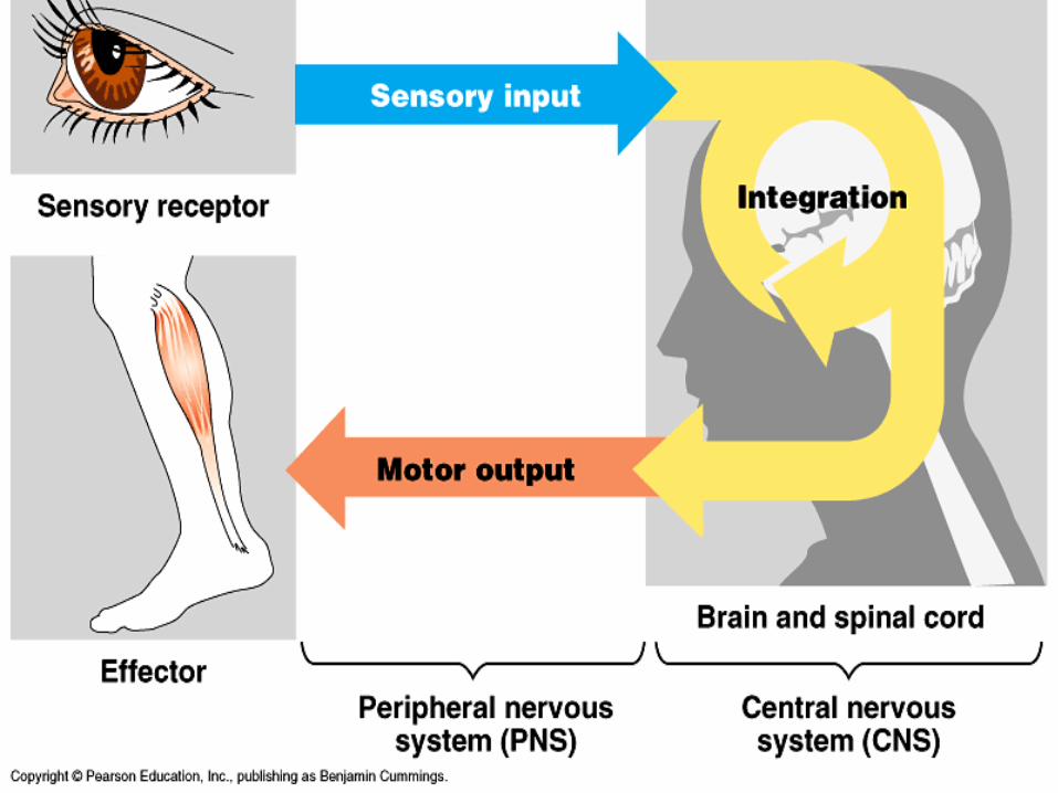
Efferent neuronsAfferent neurons
Central NervousSystem
(information processing)
Peripheral NervousSystem
Sensoryreceptors
Internaland external
stimuli
Autonomicnervous system
Motorsystem
Control ofskeletal muscle
Sympatheticdivision
Parasympatheticdivision
Entericdivision
Control of smooth muscles,cardiac muscles, glands

The Vertebrate Brain Is Regionally Specialized
• Specific brain structures are particularly specialized for diverse functions
• These structures arise during embryonic development

Embryonic brain regions Brain structures in child and adult
Forebrain
Midbrain
Hindbrain
Telencephalon
Diencephalon
Mesencephalon
Metencephalon
Myelencephalon
Cerebrum (includes cerebral cortex, whitematter, basal nuclei)
Diencephalon (thalamus, hypothalamus,epithalamus)
Midbrain (part of brainstem)
Pons (part of brainstem), cerebellum
Medulla oblongata (part of brainstem)
Midbrain
Forebrain
Hindbrain
Telencephalon
Diencephalon
Mesencephalon
Metencephalon
Myelencephalon
Spinal cord
Cerebrum Diencephalon
Midbrain
PonsMedullaoblongata
CerebellumSpinal cord
ChildEmbryo at 5 weeksEmbryo at 1 month

Brain structures in child and adult
Forebrain
Midbrain
HindbrainMyelencephalon
Cerebrum (includes cerebral cortex, whitematter, basal nuclei)
Diencephalon (thalamus, hypothalamus,epithalamus)
Midbrain (part of brainstem)
Pons (part of brainstem), cerebellum
Medulla oblongata (part of brainstem)
Midbrain
Forebrain
Hindbrain
Telencephalon
Diencephalon
Mesencephalon
Metencephalon
Myelencephalon
Spinal cord
Cerebrum Diencephalon
Midbrain
PonsMedullaoblongata
CerebellumSpinal cord
ChildEmbryo at 5 weeksEmbryo at 1 month

Cerebrum Diencephalon
Midbrain
Pons
Medullaoblongata
Cerebellum
Spinal cord
Child

Adult brain viewed from the rear
Cerebellum
Basal nucleiCerebrum
Left cerebralhemisphere
Right cerebralhemisphere
Cerebral cortex
Corpus callosum

Diencephalon
ThalamusPineal glandHypothalamus
Pituitary gland
Spinal cord
Brainstem
Midbrain
Pons
Medullaoblongata

Motor cortex(control ofskeletal muscles)
Frontal lobe
Prefrontal cortex(decision making,planning)
Broca’s area(forming speech)
Temporal lobe
Auditory cortex (hearing)
Wernicke’s area(comprehending language)
Somatosensory cortex(sense of touch)
Parietal lobe
Sensory associationcortex (integration ofsensory information)
Visual associationcortex (combiningimages and objectrecognition)
Occipital lobe
CerebellumVisual cortex(processing visualstimuli and patternrecognition)

Language and Speech
• Studies of brain activity have mapped areas responsible for language and speech
• Broca’s area in the frontal lobe is active when speech is generated
• Wernicke’s area in the temporal lobe is active when speech is heard
• These areas belong to a larger network of regions involved in language

Information Processing• The cerebral cortex receives input from sensory
organs and somatosensory receptors• Somatosensory receptors provide information about
touch, pain, pressure, temperature, and the position of muscles and limbs
• The thalamus directs different types of input to distinct locations

Frontal lobe Parietal lobe
Primarymotor cortex
Primarysomatosensorycortex
GenitaliaToes
Abdominalorgans
Tongue
JawLips
Face
EyeBrow
Neck
Thumb
FingersHand
Wrist
ForearmElbow
ShoulderTrunk
Hip
KneeTonguePharynx
JawGumsTeeth
Lips
Face
Nose
EyeThumb
FingersHand
ForearmElbow
Upper arm
Head
Neck
TrunkH
ipLeg

Primarymotor cortex
Toes
Tongue
JawLips
Face
EyeBrowNeck
Thumb
FingersHand
Wrist
ForearmElbow
ShoulderTrunk
Hip
Knee

Primarysomatosensorycortex
Genitalia
Abdominalorgans
TonguePharynx
JawGumsTeeth
Lips
Face
Nose
Eye
ThumbFingers
HandForearmElbow
Upper arm
Head
Neck
TrunkH
ipLeg

Frontal Lobe Function
• Frontal lobe damage may impair decision making and emotional responses but leave intellect and memory intact
• The frontal lobes have a substantial effect on “executive functions” of thinking making decisions.

Created by:
Debra RichardsCoordinator of Secondary Science ProgramsBryan ISDBryan, TX
![The Nervous System. Divisions of the Nervous System Central Nervous System [CNS] = Spinal Cord Brain Peripheral Nervous System [PNS]= Spinal Nerves](https://img.dokumen.tips/doc/110x75/56649d6c5503460f94a4c71d/the-nervous-system-divisions-of-the-nervous-system-central-nervous-system.jpg)