Embed Size (px)
Citation preview

During the transition from fetus to neonate somebabies will need active intervention andresuscitation. Neonatal resuscitation and followup care provided by knowledgeable and skilledhealth care professionals is critical to a healthyoutcome for these babies. Early intervention andprevention of complications is the basic principlein neonatal resuscitation and stabilization.
In Saskatchewan there are approximately 12,000births each year and it is estimated 1300 babiesmight require intensive care.
The ideal way to transport a potentially sickinfant is in utero. This involves a consultationwith a Perinatal/Neonatal Center to arrange fortransfer of the mother. Unfortunately, not allneonatal problems can be recognized to allowtransport before birth.
The Neonatal Resuscitation Program (NRP)presented in the ”Textbook of NeonatalResuscitation” 4th edition, is recommended asthe standard for neonatal resuscitation. NRPInstructors throughout Saskatchewan areavailable to provide this educational program tohealth care providers caring for newborns. ThePost Resuscitation Care of the Sick Newborn(STABLE) and Acute Care of the At-RiskNewborn (ACoRN) education programs areoffered to provide additional information on careof the ill newborn. For more information aboutthese programs contact the Perinatal EducationProgram, Division of Continuing ProfessionalLearning and Continuing Nursing Education,University of Saskatchewan or the PerinatalOutreach Program, Regina General Hospital.
Neonatal Post-Resuscitation Stabilization and
Preparation for Transport
Perinatal Education Program - 1
February, 2006
Some babies require special observation afterresuscitation or in the first days of life, whileothers may require immediate management andtransfer depending on their needs and thefacilities available at the centre of birth.
Consultation with neonatologists at the NeonatalIntensive Care Units is available 24 hours per day,7 days per week. Consultation may be sought foradvice regarding management of care or forarrangement of transport.
Infants must be resuscitated and stabilized priorto transport. The neonatal transport team isusually dispatched after a baby is born and isidentified as requiring transport, particularly insituations where viability is questionable. Uponarrival, the team will assess the infant anddetermine further management.
Good communication is essential between careproviders and hospitals. Parents and theirfamilies need to be involved in the discussionsand exchange of information regarding their illnewborn. Give the family as much information aspossible about the infant’s condition and arrangefor contact with the baby before transport. Ifpossible, a picture of the baby should be given tothe parents before the baby is transferred. Parentsshould be encouraged to visit & telephone theNICU and referral centres are encouraged toinquire about the clinical progress of the infant.
Consider transferring the mother to the tertiarycenter if the baby will be admitted to the NICU.To make arrangements for admission to theobstetrical unit, it is necessary to contact anObstetrician or Family Physician at the receivingcenter.
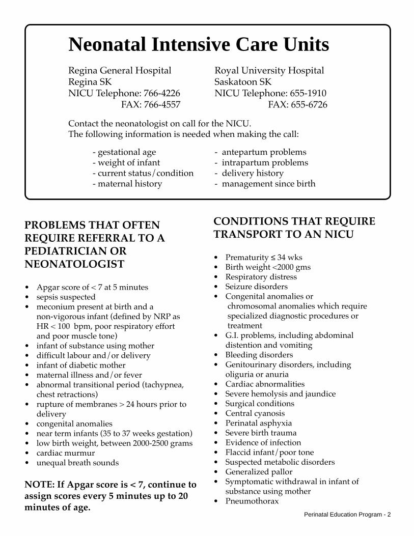
Neonatal Intensive Care UnitsRegina General Hospital Royal University HospitalRegina SK Saskatoon SKNICU Telephone: 766-4226 NICU Telephone: 655-1910
FAX: 766-4557 FAX: 655-6726
Contact the neonatologist on call for the NICU.The following information is needed when making the call:
- gestational age - antepartum problems- weight of infant - intrapartum problems- current status/condition - delivery history- maternal history - management since birth
PROBLEMS THAT OFTENREQUIRE REFERRAL TO APEDIATRICIAN ORNEONATOLOGIST
• Apgar score of < 7 at 5 minutes• sepsis suspected• meconium present at birth and a
non-vigorous infant (defined by NRP asHR < 100 bpm, poor respiratory effortand poor muscle tone)
• infant of substance using mother• difficult labour and/or delivery• infant of diabetic mother• maternal illness and/or fever• abnormal transitional period (tachypnea,
chest retractions)• rupture of membranes > 24 hours prior to
delivery• congenital anomalies• near term infants (35 to 37 weeks gestation)• low birth weight, between 2000-2500 grams• cardiac murmur• unequal breath sounds
NOTE: If Apgar score is < 7, continue toassign scores every 5 minutes up to 20minutes of age.
CONDITIONS THAT REQUIRETRANSPORT TO AN NICU
• Prematurity ≤ 34 wks• Birth weight <2000 gms• Respiratory distress• Seizure disorders• Congenital anomalies or
chromosomal anomalies which require specialized diagnostic procedures or treatment
• G.I. problems, including abdominaldistention and vomiting
• Bleeding disorders• Genitourinary disorders, including
oliguria or anuria• Cardiac abnormalities• Severe hemolysis and jaundice• Surgical conditions• Central cyanosis• Perinatal asphyxia• Severe birth trauma• Evidence of infection• Flaccid infant/poor tone• Suspected metabolic disorders• Generalized pallor• Symptomatic withdrawal in infant of
substance using mother• Pneumothorax
Perinatal Education Program - 2

Perinatal Education Program - 3
Observe continuously and do not leave the infantunattended. Handle gently. Complete the“Transport of Newborn Record” and ensure that theinfant has proper identification.
Any infant who is unwell or shows signs ofcompromise should NOT BE GIVEN ORAL FEEDS.
IV access should be established.
Vital SignsRecord every 30 minutes, depending on theinfant’s condition.
• Heart rate - Normal 110 -160 bpm
• Respiratory rate - Normal 40-60 per minute.An open airway can be maintained with theneck slightly extended & suction as necessary.
• Temperature - Maintain axillary temperatureof 36.5-37.2º C
• Blood pressure is assessed in newborns witha neonatal cuff and B.P. monitor. In general,Mean Blood Pressure correlates well with thegestational age (term infant 38 to 42 mm Hg).The signs of adequate perfusion includegood capillary refill, color, brachial andfemoral pulses, adequate urinary output andalertness. Determine capillary refilltime to assess perfusion by blanching theareas over the sternum and upper thigh usingdigital pressure. Normal refill is 2-4 seconds ina normothermic infant.
• Oximetry: Adjust oxygen flow rate to keepoxygen saturation at 88-95%. Some newbornssuch as those with cardiac or respiratoryproblems require oxygen saturation levels thatare less than or higher than this range.Consultation should be sought for thesenewborns to identify an appropriate range.
Thermoregulation
Provide warmth to maintain a normal bodytemperature. The environmental temperature inwhich an infant uses the least energy to maintainbody temperature ( neutral thermal zone )
depends on the infant’s weight, gestation, andpostnatal age. Prolonged cold stress results inincreased oxygen consumption and abnormalglucose utilization leading to hypoglycemia,hypoxemia, and acidosis.
Heat is lost by:
• Evaporation - Minimize by drying thebaby, removing wet linen and keeping theenvironment warm and humid.
• Radiation - Minimize by maintaining awarm room temperature, keeping theinfant away from cold windows, and usingdouble-walled incubators or radiant heaters
• Convection - Minimize with draft-freeenvironment.
• Conduction - Minimize by warming linenin contact with the baby.
Control environment by using:
• Incubator - Regulate by air-mode, or byservo-control using a skin probe (baby-mode), suggested setting for skin probe is36.5°C. Suggested starting temperature forincubator:
<1000 gms. 35-36º C1000-1500 gms. 34-35º C1500-2000 gms. 33-34º C>2000 gms. 32-33º C
• Radiant warmer - Radiant heat is deliveredto infant and regulated by servo-controlusing a skin probe. Suggested startingtemperature is 36.5 ° C. When using aservo-control mode do not cover thebaby. If a skin probe is not used, do notleave the baby unattended as there is adanger of overheating.
• Continue to take the axillarytemperature every 30 minutes.
POST RESUSCITATION STABILIZATION, MONITORING
AND ASSESSMENT PRIOR TO TRANSPORT

Maintenance of Oxygenation andVentilation
Observe for signs of respiratory distress:- apnea, gasping or periodic breathing (rapid
shallow breaths followed by breathholdingof 5-15 sec.)
- tachypnea (respiratory rate > 70 / minute)- chest wall retractions (intercostal, sternal)- grunting, nasal flaring
The most common causes of respiratory distressin newborns are: respiratory distress syndrome,aspiration syndrome, pneumonia, andpulmonary air leak.
In babies with respiratory distress it is difficult todifferentiate an infectious etiology from othercauses. For this reason, blood cultures andintravenous antibiotic therapy are essential untilinfection is ruled out.
Respiratory Failure and MechanicalVentilation
Respiratory failure refers to progressivelyincreasing oxygen demands and respiratorydistress. If the infant shows evidence ofrespiratory failure, immediate steps should betaken to provide positive pressure ventilation.Oxygen saturation should be maintained in the88-95 % range by pulse oximetry measurement.In the presence of respiratory distress syndrome,surfactant may be administered by thetransport team upon arrival.
Initiate PPV with infant resuscitation bag at therate of 40-60 respirations per minute.Recommended peak inspiratory pressure (PIP) is15-20 cm H20 + positive end expiratory pressure(PEEP) is 4-5 cm H20. If available, pressure canbe measured with a manometer. Effectiveness ofventilation is judged by observation of theinfant’s clinical response, symmetrical chestmovement and auscultation of breath sounds inboth lungs.
Major cardiopulmonary failure may be preventedby early intervention with oxygen and PPV.
Maintenance of Circulation
Adequate cardiac output is essential to maintaincirculation. The best way to maintain circulationis adequate provision of fluids and electrolytes.Babies with unstable conditions are usually keptNPO and an intravenous infusion started.
• Vascular AccessIf a peripheral vein cannot becannulated, a catheter can beinserted into the umbilical vein(see Appendix 2 )
• Infants requiring intravenousinfusions for transport include:
- any infant who is unwell or compromised- very low birthweight infants (VLBW)- gastro-intestinal anomalies
ie: gastroschisis- cardiac anomalies- respiratory distress syndrome- dehydration- infants in shock- suspected sepsis- seizures
• Fluid administration guidelines withD10W for newborn infants in the first24 hours of life.term 60 - 70 cc/kg/24 hourspreterm 70 - 80 cc/kg/24 hours
By the 5th day of life, infants should receive 150cc/kg/24 hours. In certain circumstances afterconsultation with a neonatologist, a lowervolume might be necessary.
Perinatal Education Program - 4
Endotracheal intubation is recommended insituations where prolonged positivepressure ventilation (PPV) is indicated.Select the appropriate size ET tube as per theNRP Guidelines listed below:
Use the tip to lip measurement to estimatecorrect insertion depth in the trachea. Add 6 cmto the baby’s weight in kg to determine correct cmmarking on the tube visible at the lip border.
Tube Size (mm)
2.5
3.0
3.5
3.5 - 4.0
<1000
1000-2000
2000-3000
>3000
<28
28-34
34-38
>38
Weight (g) Gestation Age (weeks)

Acid-Base Status
Abnormalities of acid-base status are very common withmany neonatal conditions requiring stabilization andneonatal transport. It is essential to monitor bloodgases. Metabolic acidosis requiring correction includes:ph <7.28, Bicarbonate <18 and base excess of < -8. Dolactic acid levels if available with blood gas analysis inyour facility.
Dosage of Sodium Bicarbonate:2.0 mEq/kg IV push over 2 to 3 min.
Recommended concentrationis 0.5 mEq/ml = 4.2% solution.
Consultation is strongly recommended beforeadministration of Sodium Bicarbonate.
Maintenance of Hemostasis
Hypoglycemia
Glucose screening is indicated within 30 minutes of lifein newborns who are unwell (respiratory distress, sepsis,unable to feed). Continue to assess blood glucose levelshourly. The infant with risk factors who is able to feedand is asymptomatic should have a glucose screen at 2hours of age.
Risk factors for hypoglycemia include:
- Premature infants- Low birthweight infants / Small for Gestational
Age (SGA) / Intrauterine Growth Restriction- Infants Large for Gestational Age (LGA)- Infant of a diabetic mother (IDM)- Infants of mothers treated with propanolol, oral
hypoglycemic agents, or who received IV infusionswith glucose in labour
- Infants unable to maintain a normal temperature
Signs of hypoglycemia include: Jitteriness, tremors,hypothermia, lethargy, limpness, hypotonia, apathy,intermittent apnea or tachypnea, sudden pallor, episodesof cyanosis, weak suck or refusal to eat, vomiting, high-pitched or weak cry, eye-rolling, seizures, cardiac arrest.
Management of hypoglycemia:Blood glucose levels of < 2.6 mmol/L in the symptomaticbaby should be treated. Adminstration of a 10%Dextrose solution at approximately 3-4 ml/kg/hr. isusually adequate to correct transient hypoglycemia.Persistent hypoglycemia should be treated with a minibolus of D10W 2 mls/kg IV over several minutes, butmay result in a rebound hypoglycemia. Recheck bloodglucose every 30 minutes until blood glucose is ≥ 2.6mmol/L.
Biochemical StatusMonitoring of electrolytes is recommended in infantshaving seizures or who are greater than 24 hrs of ageand are unwell or compromised. Monitor sodium,calcium, magnesium and potasium levels.
Assess for InfectionClinical signs of sepsis include: respiratory distress,abnormal skin perfusion, temperature instability,feeding intolerance, abnormal heart rate and BP andabnormal neurological status.
If sepsis is suspected based on clinical signs ormaternal history, obtain blood for culture if possibleand CBC with differential. Intravenous antibiotic administration should not be delayed if unable toobtain a blood culture. First doseshould be givenbefore transport.
Antibiotic dosages:
Ampicillin50 mg/kg every 12 hours,slow IVpush over 5 minutes
AND EITHERCefotaxime 50 mg/kg/every 8-12 hours,slow IV push over 5 minutes
ORGentamicin: slow IV infusion over 30 minutes.For dosing guidelines see table for protocol
Consultation is recommended
Management of SeizuresSeizures in the newborn may be subtle (staring,chewing, poor suck and swallow, bicycling,posturing) or overt (rhythmic or jerking movements).History (eg. maternal drug use), alertness, muscletone (hyper or hypotonia), reflexes and breathingshould also be evaluated. Jitteriness (symmetricalrapid movements of limbs) may be confused withseizures. Tremors associated with jitteriness stopwhen the limb is held and are sensitive to stimuli.
Anticonvulsant management for seizures:
Consultation is recommended.Initial management includes Phenobarbital 10-20 mg/kgslow IV over 20 mins.(minimum duration).Consultant/tertiary centre can provide further direction ifseizure activity continues or repeat dosing required.Note: Severe skin sloughing may occur if IV is interstitial.Respiratory depression post-dosing may occur. Providepositive pressure ventilation as needed.
Perinatal Education Program - 5
Post Conception Age (PCA)
30-34 weeks
3 mg / kg/dose
3.5 mg / kg/dose
4.0 mg / kg/dose
q24
q24
q24
Dose (mg /kg/dose)
Dosing Interval (hrs)

• Gastro-intestinal obstruction, (such as duodenalatresia, ileal atresia and anal atresia) - Infant should bekept NPO. Insert a large bore orogastric tube (#10 or#12) with a vented port to remove gastric contents andprevent abdominal distention. Establish IV of D10W,add 3 mEq NaCl/100 ml.
• Neonatal Abstinence Syndrome - Drug withdrawalappears as the newborn’s body attempts to removeaddictive substances from circulation. NAS can occurwith exposure to prescription drugs such as narcoticsand anti-depressants as well as through exposure toalcohol, nicotine and illegal drugs. Monitor infant forrestlessness, hypertonicity/hypotonicity, tremors, poorfeeding, vomiting, repetitive sneezing, sweating andstuffy nose. Document age of the infant at the onset ofsymptoms. Assess for severity of symptoms every twohours until transfer. Provide supportive care (dimlighting, reducing noise and stimulation, swaddling).Establish IV and do not feed. Consult withNeonatologist at referral center re: pharmacologicintervention.
If a baby presents with respiratory depression at birthand maternal substance use is known or suspected donot give Naloxone as it can precipitate immediatewithdrawal and onset of seizures.
• Perinatal Asphyxia(Hypoxic-Ischemic Encephalopathy)
Definition:- Apgar score 0-3 at ≥ 5mins- Neonatal neurologic sequelae- Evidence of multi-organ system dysfunction- Umbilical cord arterial pH <7.0 and base
deficit/excess > 16 mmol/LSigns:- altered gaze, slack face- increasing irritability- seizures- decreased muscle tone- decreased suck, swallow and/or gag reflex- breathing irregularities- stupor or coma- signs of increased intracranial pressure
(bulging fontanel, frequent emesis, bluntedreflexes, “sunset” eyes).
CONSIDERATION OF SPECIAL CONDITIONS
• Meconium Aspiration - If the infant is notvigorous (depressed respirations, depressedmuscle tone and heart rate less than 100 bpm)use an endotracheal tube to suction meconiumfrom the airway as soon as possible after delivery.If it is necessary to make two or more attempts toclear the meconium, leave the endotracheal tubein place. After initial stabilization, an orogastrictube should be inserted and stomach contentsaspirated.
• Pneumothorax - Breath sounds will bediminished on the side of the pneumothorax.Diagnosis can be made with an x-ray ortransillumination. If the infant has respiratorycompromise, the air may need to be aspiratedfrom the chest and supplemental oxygenadministered. (see Appendix 3)
• Shock - If suspected, volume expansion isindicated (eg. 10 ml/kg. normal saline orRinger’s lactate).
• Diaphragmatic Hernia - A large bore orogastrictube (#10 or #12) with a vented port should beinserted to prevent gastric distentionthat could impede respiration. If ventilatoryassistance is required, endotracheal intubation isrecommended rather than bag and mask.Keep baby NPO. Establish IV of D10W.
• Tracheo-esophageal fistula or Esophageal atresia- Elevate infant’s head to prevent aspiration ofgastric contents. The upper esophageal pouchshould be gently suctioned at frequent intervals.An orogastric tube can be gently inserted untilresistance is met and connected to lowintermittant suction. Keep baby NPO.Establish IV of D10W.
• Exposed Abdominal or Neural Contents - Handleexposed organs using sterile technique. Wrapdefect in warm, sterile saline dressing and coverwith plastic wrap to prevent drying. Position so nopressure is applied to the defect.
• Choanal Atresia - If infant has respiratory distressan oropharyngeal airway or endotracheal tubemay be necessary.
• Pierre-Robin Syndrome (mandibular hypoplasia)- Position infant prone to maintain open airway.Note if cleft palate is present.
Perinatal Education Program - 6

APPENDIX 1Equipment and Supplies for Resuscitation & Stablization
• Incubator• Radiant heating unit (servo-controlled
overhead warmer)• Infant stethoscope• Infant self-inflating resuscitation bag and
mask set ( 00, 0/1, 1 )• Suction catheters (#6, 8, 10)• Oxygen, Air, Suction outlets and tubing• Intubation equipment (laryngoscope with 0
and 1 straight blades, 2.5 - 4.0 endotrachealtubes, stylets).
• Orogastric tube ( #5 & #8)• Oral airway ( 00, 01 )• Intravenous equipment (infusion set,
cathlons, tubing, infusion pump)• Intravenous solutions - D5W, D10W.• Volume expanders - Normal Saline, Ringer’s
Lactate• Umbilical vessel catheterization equipment• Cardiorespiratory & Blood Pressure Monitor
• Chest drainage equipment (20 cc syringe,stopcock, #16 gauge cathlon or #23 gaugebutterfly)
• Medications:Sodium bicarbonate 4.2 %Epinephrine 1:10,000NaloxonePenicillinAmpicillinGentamicinCefotaximePhenobarb or PhenytoinLorazepam
• Blood glucose monitor• Syringes, tape, scissors• Gloves, masks, sterile gowns• Antiseptic solution• Oxygen saturation/pulse oximeter
monitor
APPENDIX 2Umbilical Vein Catheterization
Equipment for Umbilical CatheterizationShould be readily available in a preset tray.To ensure sterility:• Gown, mask, and gloves• Sterile towels (surgical field)• Two medicine cups, antiseptic solution,
saline, 2x2 gauzeTo insert line:• One straight mosquito hemostat• One curved mosquito hemostat• One straight non-toothed forceps• One curved non-toothed forceps• One lacrimal forceps (smooth deep curved
Iris forceps)• Scalpel blade and handle• Scissors• Needle driver & 3-0 silk with curved needle• Umbilical catheters: 3.5 or 5 Fr., Argyle, or #5
or #8 Orogastric tube• 10" umbilical tape• One stopcock• Syringes
• Normal saline• IV Solution: D5W or D10W Method:• Use sterile technique.• Attach catheter to syringe via stopcock, and
flush with Normal Saline• Use antiseptic solution to clean the cord and
the abdomen around the cord.• Tie an umbilical tape around the base of the
cord for hemostasis.• Cut the cord horizontally with a scalpel
blade, 1 - 2 cm above skin level.• Identify the umbilical vein.• Insert the catheter so that the tip is just below
skin level (3 to 4 cm)• Check for free flow of blood to indicate
adequate position of catheter.• Secure with purse-string sutures and tape• Never leave catheter open to the atmosphere• Tubing connections should be secured eg. use
of leur-locks• Check placement using X-ray.
Perinatal Education Program - 7

REFERENCES
• Textbook of Neonatal Resuscitation, 4th Edition. 2000. American Academy of Pediatrics & American Heart Association.• Klaus M and Fanaroff A. 1993. Care of the High Risk Neonate. 4th ed. Toronto: WB Saunders.• Merenstein G. & Gardner S. 1998. Handbook of Neonatal Intensive Care. 4th ed. Mosby, Inc.• Karlsen, Kristine, 8th Edition, 2001. STABLE Transport Education Program, Park City, Utah. www.stableprogram.com• ACoRN, Acute Care of the At-Risk Newborn. 2005. The ACoRN Editorial Board, Vancouver, B.C.
ACKNOWLEDGEMENTS
Thank you to the following nurses and physicians fortheir assistance in revising this educational resource:
S. Huber RN, Coordinator, Perinatal Outreach Program, M. Snell RN, NICU Clinical Development Educator, Z. KalapesiMD, Neonatologist, M. Murray, RRT, J. Bodani, MD, Regina General Hospital, Regina Qu’Appelle Health Region;C. Heffernan RN, Clinical Nurse Educator, and K. Sankaran MD, Neonatologist, Royal University Hospital. SaskatoonHealth Region. D. Naidu, MD, Family Physician, Estevan. R. Laforge, RN Coordinator, Perinatal Education Program.
Perinatal Education Program - 8
For more information or additional copies contact:
Perinatal Education Program,Division of Continuing Professional Learning, College of Medicine & Continuing Nursing Education, College of Nursing,University of Saskatchewan, Box 60001, RPO University, Saskatoon, SK S7N 4J8Telephone: (306) 966-7790 Fax: (306) 966-7673
Perinatal Outreach Program,Regina Qu’Appelle Health RegionRegina General Hospital, 1440-14th Avenue, Regina S4P 0W5Telephone: (306) 766-4432 Fax: (306) 766-4453
APPENDIX 3Aspiration of Pneumothorax
Equipment for Pneumothorax AspirationShould be readily available.• Gown, mask, and gloves• Skin cleansing agent• 1% lidocaine without epinephrine• #18, 20 or 22 over needle cathlon• 3-way stopcock• 20 cc syringe• IV extension tubing• small container of sterile waterMethod:• Connect IV tubing to stopcock and syringe• Position baby supine• Identify second intercostal space in
midclavicular line on the side of thechest where
pneumothorax is suspected and swab areawith antiseptic
• Insert cathlon perpendicular to chest, justover the top of the rib
• Attach tubing with stopcock and syringe• Turn stopcock to open cathlon to syringe and
aspirate air in chest• When syringe is full, turn stopcock off to
cathlon and empty syringe. Repeat stepsuntil baby improves or you no longeraspirate air.
• While awaiting chest tube placement, cathloncan be secured and attached to under waterseal by placing free end of extension tubingin water bottle.
This document, along with the Transport of Newborn Form and Maternal-Fetal Transport Guidelines can be accessed anddownloaded from the Perinatal Education Program website: www.usask.ca/nursing/cne/perinatal/guidelines.htm