Embed Size (px)
Citation preview
case records
of the
massachusetts general hospital
The
new england journal
of
medicine
n engl j med
350;12
www.nejm.org march
18, 2004
1236
Founded by
Richard C. CabotNancy Lee Harris,
m.d.,
Editor
Jo-Anne O. Shepard,
m.d.
,
Associate Editor
Stacey M. Ellender,
Assistant Editor
Sally H. Ebeling,
Assistant Editor
Christine C. Peters,
Assistant Editor
Case 9-2004: An 18-Year-Old Man with Respiratory Symptoms and Shock
Julie L. Gerberding, M.D., M.P.H., John G. Morgan, M.D., Jo-Anne O. Shepard, M.D., and Richard L. Kradin, M.D.
From the Centers for Disease Control andPrevention, Atlanta (J.L.G.); and the Cardi-ac Echo Lab and Department of Medicine(J.G.M.), Department of Radiology (J.O.S.),and Department of Pathology (R.L.K.),Massachusetts General Hospital and Har-vard Medical School.
N Engl J Med 2004;350:1236-47.
Copyright © 2004 Massachusetts Medical Society.
Dr. I. David Todres
(Pediatric Intensive Care): An eighteen-year-old man was admitted tothe hospital in shock after a five-day illness.
The patient had been in good health until five days before admission, when coughand myalgias developed. The next day, he was seen at a college health service and givena cough suppressant. The day after, he was seen by a physician at the health service. Hislungs were clear, and a diagnosis of bronchitis was made. Azithromycin and albuterolby metered-dose inhaler were prescribed, and the patient started taking the antibiotic thefollowing day. His temperature rose to 39.4°C, and post-tussive vomiting, diarrhea,and generalized body aches developed. He returned from college to his family’s homeand spent most of the day in bed. On the day before admission, he noticed mottling ofthe skin, and he was seen that evening at a neighborhood health clinic. His blood pres-sure was 125/98 mm Hg, the heart rate 157 beats per minute, respirations 28 per min-ute, temperature 36.3°C, and oxygen saturation 99 percent while he was breathing roomair. His skin was pale, the heart sounds were normal except for tachycardia, and the head,lungs, and abdomen were normal. Intravenous normal saline and metoclopramide wereadministered, and he was sent home with instructions to return if he felt worse.
On the morning of admission, he reported headache, stiff neck, pleuritic chest pain,and increasing myalgias in his back and extremities. His limbs were cold. His familybrought him to the emergency room of this hospital.
His medical history included obesity, hypercholesterolemia, acne, and a left varico-cele. Between two and three years before admission, he had lost 50 kg in weight and thehypercholesterolemia had resolved. The weight was 100 kg, and the height 187 cmfour months before admission. He had received all childhood immunizations, includ-ing hepatitis B vaccine; he was offered the meningococcal vaccine before starting col-lege but had declined it. He had not received influenza vaccine. He had no allergies. Heresided in a college dormitory, and he smoked cigarettes and drank alcohol. He hadhad several unprotected sexual encounters. His only sick contact was a friend who hadbeen given a diagnosis of mononucleosis one month earlier. He reported no recenttravel or unusual exposures. He had taken a dietary supplement for weight loss that didnot contain ephedra in the past but had been told by his physician to discontinue it four
presentation of case
The New England Journal of Medicine Downloaded from nejm.org on February 2, 2014. For personal use only. No other uses without permission.
Copyright © 2004 Massachusetts Medical Society. All rights reserved.
n engl j med
350;12
www.nejm.org march
18, 2004
case records of the massachusetts general hospital
1237
months before admission. His current medica-tions were albuterol and azithromycin. His motherand sibling were healthy but obese; an uncle haddied of melanoma; his grandfather had leukemia.
On examination he appeared acutely ill and un-comfortable but was alert and responsive, with oc-casional tachypnea and vomiting. His temperaturewas 33.9°C orally and 36.1°C rectally, blood pres-sure 140/61 mm Hg, pulse 120 beats per minute,and respirations 16 to 32 per minute. The neck wassupple, and the lungs were clear. There was tender-ness to palpation over the spine and the muscles ofthe back and extremities. The skin was mottled,without petechiae; the extremities were cool andclammy, with acral cyanosis. The rest of the exami-nation was normal.
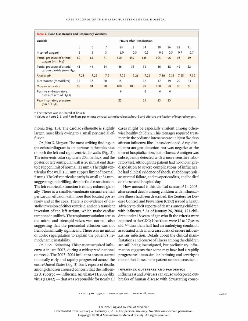
Laboratory values are shown in Tables 1, 2, and 3.An electrocardiogram showed a rate of 118 beatsper minute, a PR interval of 125 msec, QRS durationof 88 msec, QT of 292 msec, corrected QT of 409msec, and ST-segment changes consistent with ear-ly repolarization or pericarditis. A chest radiographwas normal (Fig. 1A), and a bedside echocardio-gram showed no pericardial fluid. Specimens ofblood, urine, and sputum were taken for bacterialand viral cultures and testing for viral antigens. Arapid influenza screening of a nasal swab was neg-ative. Intravenous vancomycin, ceftriaxone, and nor-mal saline fluid boluses totaling 3.5 liters, mor-phine, and ketorolac were administered. He wasadmitted to the pediatric intensive care unit.
In the intensive care unit he reported difficultybreathing and rated the muscle pain in his neck,back, and legs 9 out of 10. The axillary temperaturewas 34.5°C, blood pressure 155/80 mm Hg, andpulse 124 beats per minute, and the respirationsranged from 11 to 53 per minute. There were de-creased breath sounds in both lungs, withoutwheezes or stridor. The oxygen saturation was 98percent while he was breathing oxygen at 2 litersper minute by nasal cannula. Laboratory studiesare shown in Tables 1, 2, 3, and 4. Triplex sonogra-phy of the lower extremities showed no evidence ofdeep venous thromboses. A triple-lumen femoralcatheter and radial-artery catheter were placed.Droplet precautions were instituted. Calcium glu-conate, sodium bicarbonate, morphine sulfate, lac-tated Ringer’s solution, and normal saline wereadministered intravenously.
Six hours after presentation the blood pressurewas 123/86 mm Hg, the pulse 135 beats per minute,and the respirations 11 per minute, and urine output
had decreased. Treatment with dopamine (10 to20 µg per kilogram of body weight per minute) tomaintain a systolic blood pressure of 140 was start-ed, and aggressive fluid resuscitation was contin-ued. One hour later the blood pressure was 98/76mm Hg and the mean arterial pressure 65 mm Hg;epinephrine was added. The patient reported in-creasing difficulty breathing. Blood gas values areshown in Table 3. Eight hours after admission thetrachea was electively intubated after the patient wastreated with ketamine, vecuronium bromide, mid-azolam, and fentanyl; epinephrine and dopamine atincreasing doses and milrinone were administered.Adequate oxygenation was maintained thereafter,with an end-expiratory pressure of 6 cm of water, apeak inspiratory pressure of 25 cm of water, and afraction of inspired oxygen of 0.5 (Table 3). Echo-cardiography (Fig. 2 [a video clip is available withthe full text of this article at www.nejm.org]) re-vealed depressed biventricular function and diffuse-ly hypokinetic ventricles, with an ejection fractionof 40 percent. A small pericardial effusion was seenposteriorly and at the apex. There was no significantmitral or aortic regurgitation.
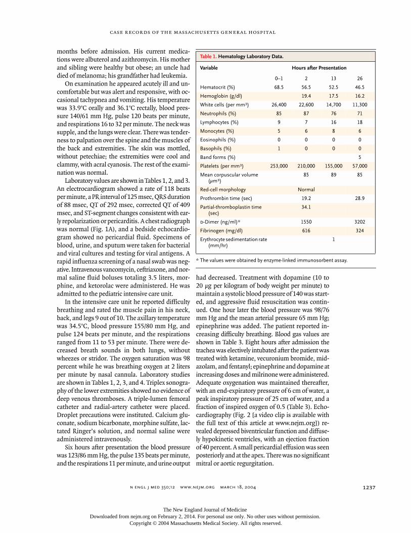
* The values were obtained by enzyme-linked immunosorbent assay.
Table 1. Hematology Laboratory Data.
Variable Hours after Presentation
0–1 2 13 26
Hematocrit (%) 68.5 56.5 52.5 46.5
Hemoglobin (g/dl) 19.4 17.5 16.2
White cells (per mm
3
) 26,400 22,600 14,700 11,300
Neutrophils (%) 85 87 76 71
Lymphocytes (%) 9 7 16 18
Monocytes (%) 5 6 8 6
Eosinophils (%) 0 0 0 0
Basophils (%) 1 0 0 0
Band forms (%) 5
Platelets (per mm
3
) 253,000 210,000 155,000 57,000
Mean corpuscular volume (µm
3
)85 89 85
Red-cell morphology Normal
Prothrombin time (sec) 19.2 28.9
Partial-thromboplastin time (sec)
34.1
d
-Dimer (ng/ml)* 1550 3202
Fibrinogen (mg/dl) 616 324
Erythrocyte sedimentation rate (mm/hr)
1
The New England Journal of Medicine Downloaded from nejm.org on February 2, 2014. For personal use only. No other uses without permission.
Copyright © 2004 Massachusetts Medical Society. All rights reserved.
n engl j med
350;12
www.nejm.org march
18
,
2004
The
new england journal
of
medicine
1238
Four hours later the blood pressure was 81/65mm Hg; drotrecogin alfa was given, and levofloxa-cin was added to broaden his antibiotic coverage.Dobutamine was administered, but was discontin-ued after two hours because the blood pressure de-creased to 79/55 mm Hg. Norepinephrine was givenby intravenous infusion. Cosyntropin was admin-istered, and the cortisol level rose from 27.1 µg perdeciliter (748 nmol per liter) to 41.5 µg per deciliter(1145 nmol per liter) one hour later. Hydrocortisonetreatment every eight hours was instituted. The fluid
intake was 10,115 ml and output 1500 ml on thefirst hospital day.
On the morning of the second hospital day, theblood pressure was 116/17 mm Hg, the pulse 177beats per minute, and the temperature 39.6°C. Ace-taminophen was given. A chest radiograph revealeddecreased lung volumes and the development ofperihilar indistinctness, which may have reflectedinterstitial edema or developing viral pneumonia,and slight enlargement of the cardiac silhouette(Fig. 1B). Toxicology screening of blood was nega-tive except for the presence of doxylamine (0.02 mgper liter). Twenty-nine hours after presentation theaxillary temperature was 40.4°C (104.8°F) and theblood pressure 82/55 mm Hg despite increasingdoses of dopamine, epinephrine, and norepineph-rine. A transthoracic echocardiogram revealed anestimated ejection fraction of 47 percent, diffuselyhypokinetic ventricles, and an underfilled left ven-tricle. There was a moderate apical and posteriorpericardial effusion. Dobutamine treatment was be-gun again, and fluid administration was increased.The lung sounds were coarse, with secretions rang-ing from scant and thin to moderately creamy. Theresults of laboratory tests are shown in Tables 1, 2,and 3.
Thirty-one hours after presentation, bradycardiadeveloped, followed rapidly by asystole, and cardio-pulmonary resuscitation was initiated. Epinephrineand atropine boluses, bicarbonate, calcium, isopro-terenol, insulin, and intravenous fluids were admin-istered. Defibrillation with electroshock and exter-nal and internal pacing were attempted, withoutevidence of capture. The patient was pronounceddead 32 hours after arrival in the emergency room.The microbiology laboratory reported the detectionof influenza A antigen in a nasal swab obtained onthe previous day. An autopsy was performed.
Dr. Todres
: May we review the chest radiograph andthe echocardiograms?
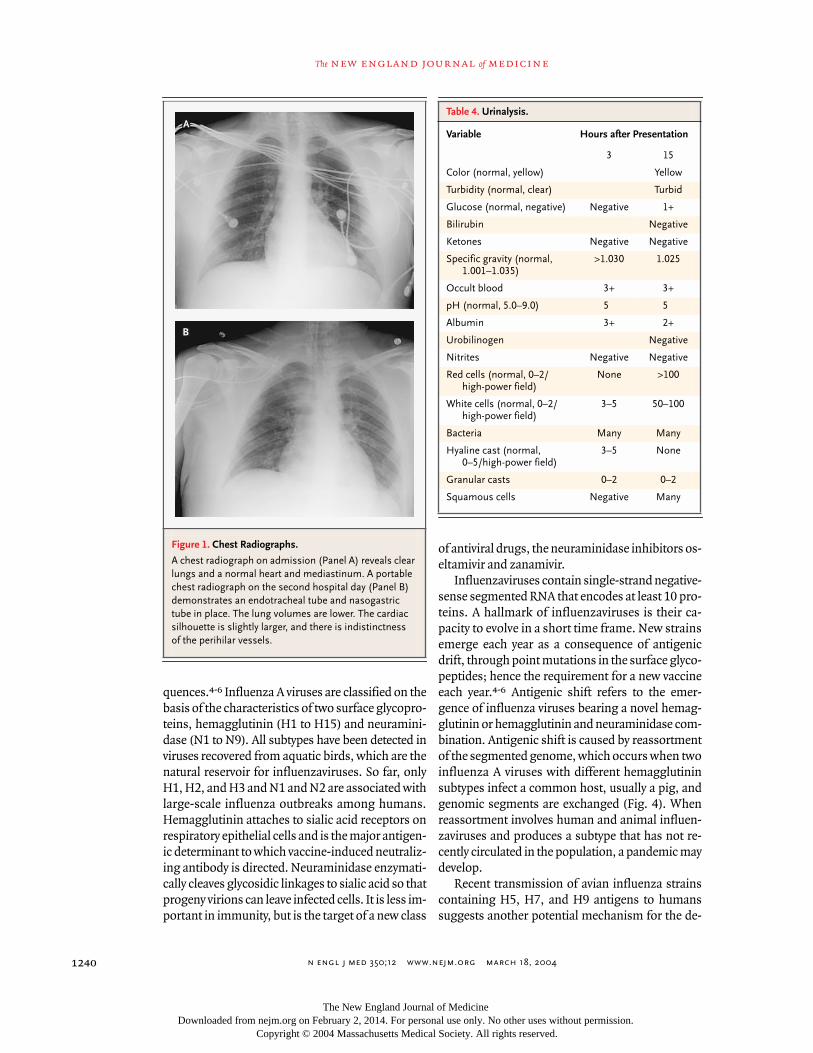
Dr. Jo-Anne O. Shepard
: The chest radiographobtained on admission reveals well-inflated, clearlungs and no evidence of a pleural effusion. Theheart is normal in size (Fig. 1A). A portable chestradiograph obtained on the second hospital day, af-ter intubation and the placement of a nasogastrictube, revealed lower lung volumes and the develop-ment of perihilar indistinctness that may have re-flected interstitial edema or developing viral pneu-
differential diagnosis
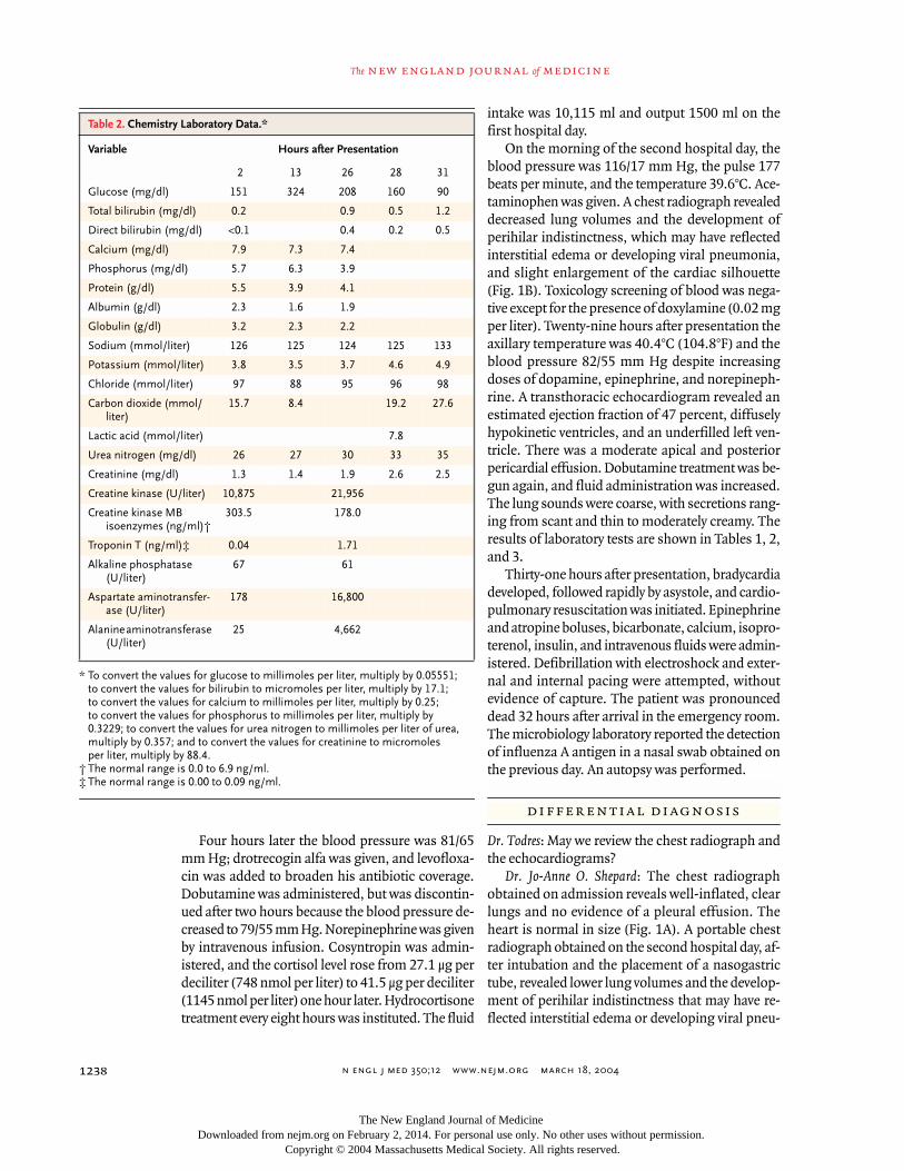
* To convert the values for glucose to millimoles per liter, multiply by 0.05551; to convert the values for bilirubin to micromoles per liter, multiply by 17.1; to convert the values for calcium to millimoles per liter, multiply by 0.25; to convert the values for phosphorus to millimoles per liter, multiply by 0.3229; to convert the values for urea nitrogen to millimoles per liter of urea, multiply by 0.357; and to convert the values for creatinine to micromoles per liter, multiply by 88.4.
† The normal range is 0.0 to 6.9 ng/ml.
‡ The normal range is 0.00 to 0.09 ng/ml.
Table 2. Chemistry Laboratory Data.*
Variable Hours after Presentation
2 13 26 28 31
Glucose (mg/dl) 151 324 208 160 90
Total bilirubin (mg/dl) 0.2 0.9 0.5 1.2
Direct bilirubin (mg/dl) <0.1 0.4 0.2 0.5
Calcium (mg/dl) 7.9 7.3 7.4
Phosphorus (mg/dl) 5.7 6.3 3.9
Protein (g/dl) 5.5 3.9 4.1
Albumin (g/dl) 2.3 1.6 1.9
Globulin (g/dl) 3.2 2.3 2.2
Sodium (mmol/liter) 126 125 124 125 133
Potassium (mmol/liter) 3.8 3.5 3.7 4.6 4.9
Chloride (mmol/liter) 97 88 95 96 98
Carbon dioxide (mmol/liter)
15.7 8.4 19.2 27.6
Lactic acid (mmol/liter) 7.8
Urea nitrogen (mg/dl) 26 27 30 33 35
Creatinine (mg/dl) 1.3 1.4 1.9 2.6 2.5
Creatine kinase (U/liter) 10,875 21,956
Creatine kinase MB isoenzymes (ng/ml)†
303.5 178.0
Troponin T (ng/ml)‡ 0.04 1.71
Alkaline phosphatase (U/liter)
67 61
Aspartate aminotransfer-ase (U/liter)
178 16,800
Alanine aminotransferase (U/liter)
25 4,662
The New England Journal of Medicine Downloaded from nejm.org on February 2, 2014. For personal use only. No other uses without permission.
Copyright © 2004 Massachusetts Medical Society. All rights reserved.
n engl j med
350;12
www.nejm.org march
18, 2004
case records of the massachusetts general hospital
1239
monia (Fig. 1B). The cardiac silhouette is slightlylarger, most likely owing to a small pericardial ef-fusion.
Dr. John G. Morgan
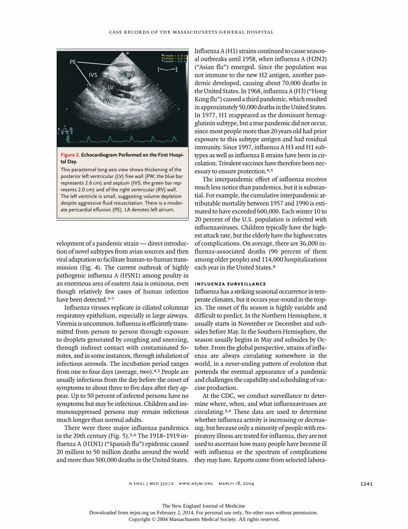
: The most striking finding onthe echocardiogram is an increase in the thicknessof both the left and right ventricular walls (Fig. 2).The interventricular septum is 20 mm thick, and theposterior left ventricular wall is 26 mm at end dias-tole (upper limit of normal, 11 mm). The right ven-tricular free wall is 11 mm (upper limit of normal,5 mm). The left ventricular cavity is small at 34 mm,suggesting underfilling, despite fluid resuscitation.The left ventricular function is mildly reduced glob-ally. There is a small-to-moderate circumferentialpericardial effusion with more fluid located poste-riorly and at the apex. There is no evidence of dia-stolic inversion of either ventricle, and only transientinversion of the left atrium, which make cardiactamponade unlikely. The respiratory variation acrossthe mitral and tricuspid valves was normal, alsosuggesting that the pericardial effusion was nothemodynamically significant. There was no mitralor aortic regurgitation to explain the patient’s he-modynamic instability.
Dr. Julie L. Gerberding
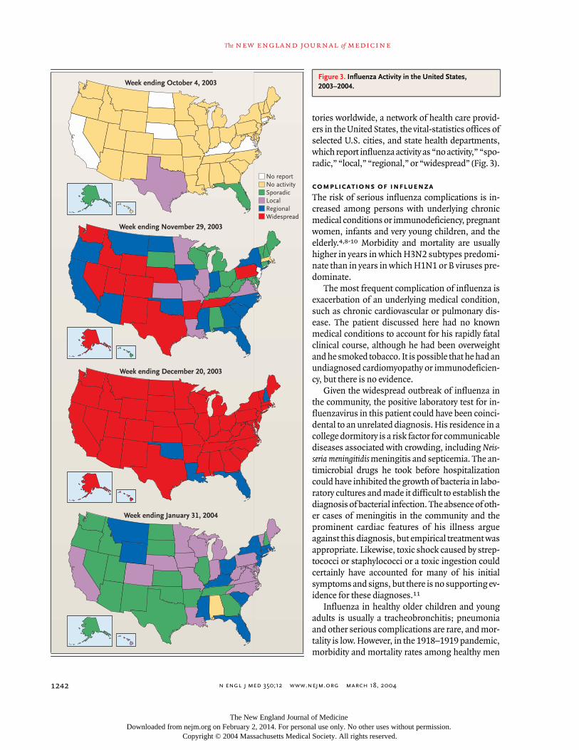
: This patient acquired influ-enza A in late 2003, during a widespread nationaloutbreak. The 2003–2004 influenza season startedunusually early and rapidly progressed across theentire United States (Fig. 3). Early reports of deathsamong children aroused concern that the influen-za A subtype — influenza A/Fujian/411/2002-likevirus (H3N2) — that was responsible for nearly all
cases might be especially virulent among other-wise healthy children. This teenager required treat-ment in the pediatric intensive care unit just five daysafter an influenza-like illness developed. A rapid in-fluenza-antigen detection test was negative at thetime of hospitalization, but influenza A antigen wassubsequently detected with a more sensitive labo-ratory test. Although the patient had no known pre-disposition to severe complications of influenza,he had clinical evidence of shock, rhabdomyolysis,acute renal failure, and myopericarditis, and he diedon the second hospital day.
How unusual is this clinical scenario? In 2003,after several deaths among children with influenza-like illness had been described, the Centers for Dis-ease Control and Prevention (CDC) issued a healthadvisory to elicit reports of deaths among childrenwith influenza.
1
As of January 26, 2004, 121 chil-dren under 18 years of age who fit the criteria werereported to the CDC; 19 of them were 12 to 17 yearsold.
2,3
Less than half had an underlying conditionassociated with an increased risk of severe influen-zavirus infection. Details about the clinical mani-festations and course of illness among the childrenare still being investigated, but preliminary infor-mation suggests that some may have had a rapidlyprogressive illness similar in timing and severity tothat of the illness in the patient under discussion.
influenza outbreaks and pandemics
Influenza A and B viruses can cause widespread out-breaks of human disease with devastating conse-
* The trachea was intubated at hour 8.
† Values at hours 5, 6, and 7 are liters per minute by nasal cannula; values at hour 8 and after are the fraction of inspired oxygen.
Table 3. Blood Gas Results and Respiratory Variables.
Variable Hours after Presentation
5 6 7 8* 11 14 18 26 28 31
Inspired oxygen† 2 5 5 1.0 0.5 0.5 0.5 0.5 0.7 0.7
Partial pressure of arterial oxygen (mm Hg)
80 61 71 350 152 145 105 96 98 59
Partial pressure of arterial carbon dioxide (mm Hg)
41 44 54 46 35 31 36 38 49 52
Arterial pH 7.23 7.22 7.2 7.12 7.26 7.21 7.30 7.33 7.25 7.39
Bicarbonate (mmol/liter) 17 18 20 15 12 17 19 20 31
Oxygen saturation 98 94 90 100 100 99 100 98 96 96
Positive end-expiratory pressure (cm of H
2
O)6 6 6 6
Peak inspiratory pressure (cm of H
2
O)22 25 25 25
The New England Journal of Medicine Downloaded from nejm.org on February 2, 2014. For personal use only. No other uses without permission.
Copyright © 2004 Massachusetts Medical Society. All rights reserved.
n engl j med
350;12
www.nejm.org march
18
,
2004
The
new england journal
of
medicine
1240
quences.
4-6
Influenza A viruses are classified on thebasis of the characteristics of two surface glycopro-teins, hemagglutinin (H1 to H15) and neuramini-dase (N1 to N9). All subtypes have been detected inviruses recovered from aquatic birds, which are thenatural reservoir for influenzaviruses. So far, onlyH1, H2, and H3 and N1 and N2 are associated withlarge-scale influenza outbreaks among humans.Hemagglutinin attaches to sialic acid receptors onrespiratory epithelial cells and is the major antigen-ic determinant to which vaccine-induced neutraliz-ing antibody is directed. Neuraminidase enzymati-cally cleaves glycosidic linkages to sialic acid so thatprogeny virions can leave infected cells. It is less im-portant in immunity, but is the target of a new class
of antiviral drugs, the neuraminidase inhibitors os-eltamivir and zanamivir.
Influenzaviruses contain single-strand negative-sense segmented RNA that encodes at least 10 pro-teins. A hallmark of influenzaviruses is their ca-pacity to evolve in a short time frame. New strainsemerge each year as a consequence of antigenicdrift, through point mutations in the surface glyco-peptides; hence the requirement for a new vaccineeach year.
4-6
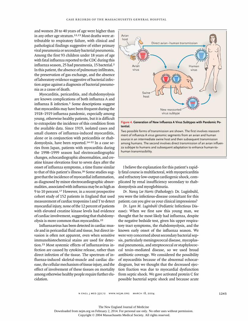
Antigenic shift refers to the emer-gence of influenza viruses bearing a novel hemag-glutinin or hemagglutinin and neuraminidase com-bination. Antigenic shift is caused by reassortmentof the segmented genome, which occurs when twoinfluenza A viruses with different hemagglutininsubtypes infect a common host, usually a pig, andgenomic segments are exchanged (Fig. 4). Whenreassortment involves human and animal influen-zaviruses and produces a subtype that has not re-cently circulated in the population, a pandemic maydevelop.
Recent transmission of avian influenza strainscontaining H5, H7, and H9 antigens to humanssuggests another potential mechanism for the de-
Figure 1. Chest Radiographs.
A chest radiograph on admission (Panel A) reveals clear lungs and a normal heart and mediastinum. A portable chest radiograph on the second hospital day (Panel B) demonstrates an endotracheal tube and nasogastric tube in place. The lung volumes are lower. The cardiac silhouette is slightly larger, and there is indistinctness of the perihilar vessels.
A
B
Table 4. Urinalysis.
Variable Hours after Presentation
3 15
Color (normal, yellow) Yellow
Turbidity (normal, clear) Turbid
Glucose (normal, negative) Negative 1+
Bilirubin Negative
Ketones Negative Negative
Specific gravity (normal, 1.001–1.035)
>1.030 1.025
Occult blood 3+ 3+
pH (normal, 5.0–9.0) 5 5
Albumin 3+ 2+
Urobilinogen Negative
Nitrites Negative Negative
Red cells (normal, 0–2/high-power field)
None >100
White cells (normal, 0–2/high-power field)
3–5 50–100
Bacteria Many Many
Hyaline cast (normal, 0–5/high-power field)
3–5 None
Granular casts 0–2 0–2
Squamous cells Negative Many
The New England Journal of Medicine Downloaded from nejm.org on February 2, 2014. For personal use only. No other uses without permission.
Copyright © 2004 Massachusetts Medical Society. All rights reserved.
n engl j med
350;12
www.nejm.org march
18, 2004
case records of the massachusetts general hospital
1241
velopment of a pandemic strain — direct introduc-tion of novel subtypes from avian sources and thenviral adaptation to facilitate human-to-human trans-mission (Fig. 4). The current outbreak of highlypathogenic influenza A (H5N1) among poultry inan enormous area of eastern Asia is ominous, eventhough relatively few cases of human infectionhave been detected.
5-7
Influenza viruses replicate in ciliated columnarrespiratory epithelium, especially in large airways.Viremia is uncommon. Influenza is efficiently trans-mitted from person to person through exposureto droplets generated by coughing and sneezing,through indirect contact with contaminated fo-mites, and in some instances, through inhalation ofinfectious aerosols. The incubation period rangesfrom one to four days (average, two).
4,5
People areusually infectious from the day before the onset ofsymptoms to about three to five days after they ap-pear. Up to 50 percent of infected persons have nosymptoms but may be infectious. Children and im-munosuppressed persons may remain infectiousmuch longer than normal adults.
There were three major influenza pandemicsin the 20th century (Fig. 5).
5,6
The 1918–1919 in-fluenza A (H1N1) (“Spanish flu”) epidemic caused20 million to 50 million deaths around the worldand more than 500,000 deaths in the United States.
Influenza A (H1) strains continued to cause season-al outbreaks until 1958, when influenza A (H2N2)(“Asian flu”) emerged. Since the population wasnot immune to the new H2 antigen, another pan-demic developed, causing about 70,000 deaths inthe United States. In 1968, influenza A (H3) (“HongKong flu”) caused a third pandemic, which resultedin approximately 50,000 deaths in the United States.In 1977, H1 reappeared as the dominant hemag-glutinin subtype, but a true pandemic did not occur,since most people more than 20 years old had priorexposure to this subtype antigen and had residualimmunity. Since 1997, influenza A H3 and H1 sub-types as well as influenza B strains have been in cir-culation. Trivalent vaccines have therefore been nec-essary to ensure protection.
4,5
The interpandemic effect of influenza receivesmuch less notice than pandemics, but it is substan-tial. For example, the cumulative interpandemic at-tributable mortality between 1957 and 1990 is esti-mated to have exceeded 600,000. Each winter 10 to20 percent of the U.S. population is infected withinfluenzaviruses. Children typically have the high-est attack rate, but the elderly have the highest ratesof complications. On average, there are 36,000 in-fluenza-associated deaths (90 percent of themamong older people) and 114,000 hospitalizationseach year in the United States.
8
influenza surveillance
Influenza has a striking seasonal occurrence in tem-perate climates, but it occurs year-round in the trop-ics. The onset of flu season is highly variable anddifficult to predict. In the Northern Hemisphere, itusually starts in November or December and sub-sides before May. In the Southern Hemisphere, theseason usually begins in May and subsides by Oc-tober. From the global perspective, strains of influ-enza are always circulating somewhere in theworld, in a never-ending pattern of evolution thatportends the eventual appearance of a pandemicand challenges the capability and scheduling of vac-cine production.
At the CDC, we conduct surveillance to deter-mine where, when, and what influenzaviruses arecirculating.
3,4
These data are used to determinewhether influenza activity is increasing or decreas-ing, but because only a minority of people with res-piratory illness are tested for influenza, they are notused to ascertain how many people have become illwith influenza or the spectrum of complicationsthey may have. Reports come from selected labora-
Figure 2. Echocardiogram Performed on the First Hospi-tal Day.
This parasternal long-axis view shows thickening of the posterior left ventricular (LV) free wall (PW; the blue bar represents 2.6 cm) and septum (IVS; the green bar rep-resents 2.0 cm) and of the right ventricular (RV) wall. The left ventricle is small, suggesting volume depletion despite aggressive fluid resuscitation. There is a moder-ate pericardial effusion (PE). LA denotes left atrium.
PE
IVS
LV
LAPW
RV
The New England Journal of Medicine Downloaded from nejm.org on February 2, 2014. For personal use only. No other uses without permission.
Copyright © 2004 Massachusetts Medical Society. All rights reserved.
n engl j med
350;12
www.nejm.org march
18
,
2004
The
new england journal
of
medicine
1242
tories worldwide, a network of health care provid-ers in the United States, the vital-statistics offices ofselected U.S. cities, and state health departments,which report influenza activity as “no activity,” “spo-radic,” “local,” “regional,” or “widespread” (Fig. 3).
complications of influenza
The risk of serious influenza complications is in-creased among persons with underlying chronicmedical conditions or immunodeficiency, pregnantwomen, infants and very young children, and theelderly.
4,8-10
Morbidity and mortality are usuallyhigher in years in which H3N2 subtypes predomi-nate than in years in which H1N1 or B viruses pre-dominate.
The most frequent complication of influenza isexacerbation of an underlying medical condition,such as chronic cardiovascular or pulmonary dis-ease. The patient discussed here had no knownmedical conditions to account for his rapidly fatalclinical course, although he had been overweightand he smoked tobacco. It is possible that he had anundiagnosed cardiomyopathy or immunodeficien-cy, but there is no evidence.
Given the widespread outbreak of influenza inthe community, the positive laboratory test for in-fluenzavirus in this patient could have been coinci-dental to an unrelated diagnosis. His residence in acollege dormitory is a risk factor for communicablediseases associated with crowding, including
Neis-seria meningitidis
meningitis and septicemia. The an-timicrobial drugs he took before hospitalizationcould have inhibited the growth of bacteria in labo-ratory cultures and made it difficult to establish thediagnosis of bacterial infection. The absence of oth-er cases of meningitis in the community and theprominent cardiac features of his illness argueagainst this diagnosis, but empirical treatment wasappropriate. Likewise, toxic shock caused by strep-tococci or staphylococci or a toxic ingestion couldcertainly have accounted for many of his initialsymptoms and signs, but there is no supporting ev-idence for these diagnoses.
11
Influenza in healthy older children and youngadults is usually a tracheobronchitis; pneumoniaand other serious complications are rare, and mor-tality is low. However, in the 1918–1919 pandemic,morbidity and mortality rates among healthy men
Week ending October 4, 2003
Week ending November 29, 2003
Week ending December 20, 2003
Week ending January 31, 2004
No reportNo activitySporadicLocalRegionalWidespread
Figure 3. Influenza Activity in the United States, 2003–2004.
The New England Journal of Medicine Downloaded from nejm.org on February 2, 2014. For personal use only. No other uses without permission.
Copyright © 2004 Massachusetts Medical Society. All rights reserved.
n engl j med
350;12
www.nejm.org march
18, 2004
case records of the massachusetts general hospital
1243
and women 20 to 40 years of age were higher thanin any other age stratum.
12,13
Most deaths were at-tributable to respiratory failure, with clinical andpathological findings suggestive of either primaryviral pneumonia or secondary bacterial pneumonia.Among the first 93 children under 18 years of agewith fatal influenza reported to the CDC during thisinfluenza season, 25 had pneumonia, 15 bacterial.
2
In this patient, the absence of pulmonary infiltrates,the preservation of gas exchange, and the absenceof laboratory evidence suggestive of bacterial infec-tion argue against a diagnosis of bacterial pneumo-nia as a cause of death.
Myocarditis, pericarditis, and rhabdomyolysisare known complications of both influenza A andinfluenza B infection.
5
Some descriptions suggestthat myocarditis may have been frequent during the1918–1919 influenza pandemic, especially amongyoung, otherwise healthy patients, but it is difficultto extrapolate the incidence of this condition fromthe available data. Since 1919, isolated cases andsmall clusters of influenza-induced myocarditis,alone or in conjunction with pericarditis or rhab-domyolysis, have been reported.
14-22
In a case se-ries from Japan, patients with myocarditis duringthe 1998–1999 season had electrocardiographicchanges, echocardiographic abnormalities, and cre-atine kinase elevations four to seven days after theonset of influenza symptoms, a time frame similarto that of this patient’s illness.
20
Some studies sug-gest that the incidence of myocardial inflammation,as diagnosed by minor electrocardiographic abnor-malities, associated with influenza may be as high as9 to 10 percent.
17
However, in a recent prospectivecohort study of 152 patients in England that usedmeasurement of cardiac troponins I and T to detectmyocardial injury, none of the 12 percent of patientswith elevated creatine kinase levels had evidenceof cardiac involvement, suggesting that rhabdomy-olysis is more common than myocarditis.
22
Influenzavirus has been detected in cardiac mus-cle and in pericardial fluid and tissue, but direct in-vasion is often not apparent, even when sensitiveimmunohistochemical stains are used for detec-tion.
23
Most systemic effects of influenzavirus in-fection are caused by cytokine release, rather thandirect infection of the tissue. The spectrum of in-fluenza-induced skeletal-muscle and cardiac dis-ease, the cellular mechanism of tissue injury, and theeffect of involvement of these tissues on mortalityamong otherwise healthy people require further elu-cidation.
I believe the explanation for this patient’s rapid-ly fatal course is multifactoral, with myopericarditisand refractory low-output cardiogenic shock, com-plicated by renal insufficiency secondary to rhab-domyolysis and myoglobinuria.
Dr. Nancy Lee Harris
(Pathology): Dr. Luginbuhl,you were the infectious-disease consultant for thispatient; can you give us your clinical impressions?
Dr. Lynn M. Luginbuhl
(Pediatric Infectious Dis-ease): When we first saw this young man, wethought that he most likely had influenza, despitethe negative bedside test, given his upper respira-tory tract symptoms, the rhabdomyolysis, and theknown early onset of the influenza season. Wewere very concerned about secondary bacterial sep-sis, particularly meningococcal disease, mycoplas-mal pneumonia, and streptococcal or staphylococ-cal toxin–mediated disease, so we used broadantibiotic coverage. We considered the possibilityof myocarditis because of the abnormal echocar-diogram, but we thought that the decreased ejec-tion fraction was due to myocardial dysfunctionfrom septic shock. We gave activated protein C forpossible bacterial septic shock and because acute
Figure 4. Generation of New Influenza A Virus Subtypes with Pandemic Po-tential.
Two possible forms of transmission are shown. The first involves reassort-ment of influenza A virus genomic segments from an avian and human source in an intermediate swine host and then subsequent transmission among humans. The second involves direct transmission of an avian influen-za subtype to humans and subsequent adaptation to enhance human-to-human transmissibility.
The New England Journal of Medicine Downloaded from nejm.org on February 2, 2014. For personal use only. No other uses without permission.
Copyright © 2004 Massachusetts Medical Society. All rights reserved.

n engl j med
350;12
www.nejm.org march
18
,
2004
The
new england journal
of
medicine
1244
respiratory distress syndrome seemed to be devel-oping. At the time of death the clinical picture re-mained one of irreversible shock, and we were stillconcerned that he had a secondary bacterial infec-tion. We then learned that he was influenza A–pos-itive, and we knew that no bacteria had grown at 24hours. Thus, we began to consider the possibilitythat his death might be due to influenza A alone.
Influenza A infection with shock, caused by eitherbacterial superinfection or possibly influenza, com-plicated by rhabdomyolysis, renal failure, and dis-seminated intravascular coagulation.
Influenza A infection with shock due to multiplefactors, including possible myopericarditis and se-vere rhabdomyolysis with myoglobinuria and renalfailure.
Dr. Harris
: Dr. Richard L. Kradin will present the au-topsy findings.
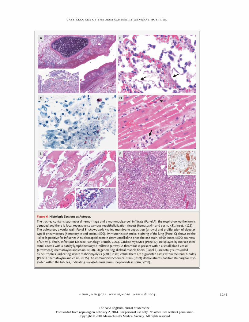
Dr. Richard L. Kradin
: At autopsy, the tracheobron-chial tree was diffusely erythematous and the respi-ratory epithelium was denuded. Microscopically,the trachea and large bronchi were congested andedematous and contained submucosal hemorrhageand a mononuclear-cell inflammatory infiltrate. Theepithelium was denuded, and there was patchy re-parative squamous reepithelialization (Fig. 6A). Thelungs weighed 2800 g together and were plum col-ored, congested, and edematous, but with minimalconsolidation. The alveoli were filled with macro-phages and desquamated epithelium. There wasearly hyaline membrane formation and prolifera-tion of alveolar type II pneumocytes, findings con-sistent with diffuse alveolar damage (Fig. 6B). Noviral inclusions were identified, and there was noevidence of bacterial infection. The influenza A vi-rus isolated from the sputum was subtyped asH3N2. Immunohistochemical staining performedat the CDC identified influenza A nucleocapsidprotein within pulmonary epithelial cells (Fig. 6C).All postmortem fluids, including sputum, blood,urine, and cerebrospinal fluid, were negative forbacterial growth.
The pericardium contained approximately 400ml of serosanguineous fluid. There were no pericar-dial adhesions, and there was no anatomical evi-dence of cardiac tamponade. The heart was en-
clinical diagnosis
dr. julie l. gerberding’s
diagnosis
pathological discussion
Figure 5. Emergence of New Influenza A Virus Subtypes in Humans.
H5
H7
H1
H3H2
H1
Avianflu
1998
1980 1996 2003
1997 2003
1957–8"Asian" influenza
H2N2
1918–9"Spanish" influenza
H1N1
1968–9"Hong Kong"
influenzaH3N2
1915 1925 1935 1945 1955 1965 1975 1985 1995 2005
H9
1999
2004
The New England Journal of Medicine Downloaded from nejm.org on February 2, 2014. For personal use only. No other uses without permission.
Copyright © 2004 Massachusetts Medical Society. All rights reserved.
n engl j med
350;12
www.nejm.org march
18, 2004
case records of the massachusetts general hospital
1245
Figure 6. Histologic Sections at Autopsy.
The trachea contains submucosal hemorrhage and a mononuclear-cell infiltrate (Panel A); the respiratory epithelium is denuded and there is focal reparative squamous reepithelialization (inset) (hematoxylin and eosin, ¬31; inset, ¬125). The pulmonary alveolar wall (Panel B) shows early hyaline membrane deposition (arrows) and proliferation of alveolar type II pneumocytes (hematoxylin and eosin, ¬500). Immunohistochemical staining of the lung (Panel C) shows epithe-lial cells positive for influenza A nucleocapsid protein (immunoalkaline phosphatase stain, ¬300; inset, ¬500; courtesy of Dr. W.-J. Shieh, Infectious Disease Pathology Branch, CDC). Cardiac myocytes (Panel D) are splayed by marked inter-stitial edema with a patchy lymphohistiocytic infiltrate (arrow). A thrombus is present within a small blood vessel (arrowhead) (hematoxylin and eosin, ¬300). Degenerating skeletal-muscle fibers (Panel E) are totally surrounded by neutrophils, indicating severe rhabdomyolysis (¬300; inset, ¬500).There are pigmented casts within the renal tubules (Panel F; hematoxylin and eosin, ¬125). An immunohistochemical stain (inset) demonstrates positive staining for myo-globin within the tubules, indicating myoglobinuria (immunoperoxidase stain, ¬250).
A B
C D
E F
The New England Journal of Medicine Downloaded from nejm.org on February 2, 2014. For personal use only. No other uses without permission.
Copyright © 2004 Massachusetts Medical Society. All rights reserved.
n engl j med
350;12
www.nejm.org march
18
,
2004
The
new england journal
of
medicine
1246
larged, at 590 g (normal for the patient’s weight,less than 450 g [0.45 percent of body weight]), withconcentric biventricular hypertrophy. Microscopi-cally, there was cardiac myocyte hypertrophy and apatchy lymphohistiocytic infiltrate in perivascularareas associated with interstitial edema (Fig. 6D).Although not meeting formal criteria for viral myo-carditis, the changes are consistent with “border-line myocarditis” and with the spectrum of findingsthat may be observed in influenza infection.
24
Therewas focal contraction-band myocyte necrosis andscattered intravascular fibrin thrombi. This type ofnecrosis can be produced by catecholamines,
25
andthe fibrin thrombi found in the heart and other or-gans are signs of disseminated intravascular coag-ulation.
Skeletal muscle showed severe rhabdomyolysiswith numerous degenerating and necrotic musclefibers, marked edema, and focal infiltration of neu-trophils (Fig. 6E). Immunostaining of cardiac andskeletal muscle at the CDC did not reveal evidenceof influenzavirus. The renal glomeruli were nor-mal, but the proximal tubules contained pigment-ed casts (Fig. 6F), which were shown by immuno-staining to be myoglobin (inset, Fig. 6F). Therewas severe ischemic hepatic injury with centrilobu-lar necrosis. The adrenal glands were normal.
Influenza produces no consistent cytopathicchanges. Uncomplicated infection causes tracheo-bronchitis characterized by necrosis, ulceration, anddenudation of the respiratory epithelium, followedby reparative squamous reepithelialization. Thepathological changes of influenza pneumonia
26
in-clude bronchiolocentric exudation of histiocytes,obliterative bronchiolitis with organizing pneumo-nia, and diffuse alveolar damage with necrosis andhemorrhage. Myocarditis is rare, but myocardialinflammatory-cell infiltrates were observed at au-topsy in approximately one third of 33 patients dy-ing from influenza.
27
Although myalgias are com-mon, severe rhabdomyolysis is unusual; it occursmore often in young patients and can be complicat-ed by myoglobinuria and renal failure, as in thiscase.
28-32
Skeletal-muscle biopsies generally donot reveal direct viral infection.
33
The mechanisms of viral pathogenesis are mostlikely complex. In addition to direct viral replicationin epithelial cells, proinflammatory cytokine re-lease
34
and abnormalities in the interferon system
35
may contribute to the morbidity and mortality. Inthis case, death is attributable to multisystem dis-ease complicating influenza A H3N2 infection, in-
cluding tracheobronchitis, pneumonia, possibleearly myopericarditis, severe rhabdomyolysis withmyoglobinuria and acute renal failure, disseminat-ed intravascular coagulation, and hepatic centri-lobular necrosis. The striking cardiac hypertrophy ,which may have been associated with the patient’shistory of obesity,
36
may have placed him at in-creased risk for complications.
A Physician
: Would early initiation of antiviraltherapy have altered the course?
Dr. Gerberding
: Antiviral drugs have been docu-mented to shorten the course of the illness by onlya day or two. One study of oseltamivir found thattreatment may reduce some complications,
37
butno studies have shown that treatment reduces fataloutcomes.
Dr. Harris
: I wonder whether severe rhabdomy-olysis could be the dominant cause of the shock-like symptoms in this patient’s clinical presenta-tion. Influenza A is the most common infectiouscause of rhabdomyolysis.
28-32
Severe rhabdomyol-ysis can lead to shock due to massive fluid redistri-bution into necrotic muscle, respiratory acidosis,disseminated intravascular coagulation, and myo-globinuria with renal failure, all of which were seenin this case.
31
He had unremitting hypovolemicshock, despite a net fluid gain of over 20 liters in 32hours. Although his weight was 100 kg four monthsearlier, the autopsy service recorded his weight as144 kg, suggesting that a remarkable amount of ex-travascular fluid had accumulated. In one reportedcase of a child with fatal rhabdomyolysis associatedwith influenza B infection, muscle biopsy showeda clinically unsuspected carnitine palmitoyl trans-ferase II deficiency.
38
It is possible that an unrecog-nized metabolic disorder may predispose patientsto rhabdomyolysis in influenza A infection.
Dr. Gerberding
: This tragic case reminds us thatinfluenzavirus is a serious pathogen and that weneed to do more to prevent this very preventable ill-ness through vaccination programs.
Influenza A infection with rhabdomyolysis, severe;myoglobinuria; viral tracheobronchitis and pneu-monia; virus-associated cardiac changes (“border-line myocarditis”) and catecholamine-induced my-onecrosis; pericardial effusion.
Disseminated intravascular coagulation.Hepatic centrilobular necrosis.Cardiac hypertrophy of unknown cause.
anatomical diagnoses
The New England Journal of Medicine Downloaded from nejm.org on February 2, 2014. For personal use only. No other uses without permission.
Copyright © 2004 Massachusetts Medical Society. All rights reserved.
n engl j med
350;12
www.nejm.org march
18, 2004
case records of the massachusetts general hospital
1247
references
1.
Update: influenza-associated deathsreported among children aged <18 years —United States, 2003-04 influenza season.MMWR Morb Mortal Wkly Rep 2003;52:1254-5.
2.
Update: influenza-associated deaths re-ported among children aged <18 years —United States, 2003–04 influenza season.MMWR Morb Mortal Wkly Rep 2004;52:1286-8.
3.
Update: influenza activity United States— January 18–24, 2004. MMWR Morb Mor-tal Wkly Rep 2004;53:63-5.
4.
Prevention and control of influenza: rec-ommendations of the Advisory Committeeon Immunization Practices (ACIP). MMWRMorb Mortal Wkly Rep 2003;52(RR-8):1-36.[Erratum, MMWR Morb Mortal Wkly Rep2003;52:526.]
5. Plotkin SA, Orenstein WA, eds. Vaccines.4th ed. Philadelphia: Saunders, 2004:339-46.6. Nicholson KG, Wood JM, Zambon M.Influenza. Lancet 2003;362:1733-45.7. Outbreaks of avian influenza A (H5N1)in Asia and interim recommendations forevaluation and reporting of suspect cases —United States, 2004. MMWR Morb MortalWkly Rep 2004;53:97-100.8. Thompson WW, Shay DK, Weintraub E,et al. Mortality associated with influenzaand respiratory syncytial virus in the UnitedStates. JAMA 2003;289:179-86.9. Izurieta HS, Thompson WW, KramarzP, et al. Influenza and the rates of hospital-ization for respiratory disease among in-fants and young children. N Engl J Med2000;342:232-9.10. Quach C, Piché-Walker L, Platt R, MooreD. Risk factors associated with severe influ-enza infections in childhood: implicationfor vaccine strategy. Pediatrics 2003;112:662-3. abstract.11. Tolan RW Jr. Toxic shock syndromecomplicating influenza A in a child: case re-port and review. Clin Infect Dis 1993;17:43-5.12. Stevens KM. Cardiac stroke volume as adeterminant of influenzal fatality. N Engl JMed 1976;295:1363-6.13. Lucke B, Wight T, Kime E. Pathologicanatomy and bacteriology of influenza: epi-demic of autumn, 1918. Arch Intern Med1919;24:154-237.
14. Adams CW. Postviral myopericarditisassociated with the influenza virus: report ofeight cases. Am J Cardiol 1959;4:56-67.15. Edelen JS, Bender TR, Chin TDY. En-cephalopathy and pericarditis during anoutbreak of influenza. Am J Epidemiol1974;100:79-84.16. Kessler HA, Trenholme GM, Harris AA,Levin S. Acute myopathy associated with in-fluenza A/Texas/1/77 infection: isolation ofvirus from a muscle biopsy specimen. JAMA1980;243:461-2.17. Karjalainen J, Nieminen MS, Heikkila J.Influenza A1 myocarditis in conscripts. ActaMed Scand 1980;207:27-30.18. Proby AM, Hackett D, Gupta S, Cox TM.Acute myopericarditis in influenza A infec-tion. Q J Med 1986;60:887-92.19. Berry L, Braude S. Influenza A infectionwith rhabdomyolysis and acute renal failure— a potentially fatal complication. PostgradMed J 1991;67:389-90.20. Onitsuka H, Imamura T, Miyamoto N, etal. Clinical manifestations of influenza Amyocarditis during the influenza epidemicof winter 1998-1999. J Cardiol 2001;37:315-23.21. Yoshino M, Suzuki S, Adachi K, Fukaya-ma M, Inamatsu T. High incidence of acutemyositis with type A influenza virus infec-tion in the elderly. Intern Med 2000;39:431-2.22. Greaves K, Oxford JS, Price CP, ClarkeGH, Crake T. The prevalence of myocarditisand skeletal muscle injury during acute viralinfection in adults: measurement of cardiactroponins I and T in 152 patients with acuteinfluenza infection. Arch Intern Med 2003;163:165-8.23. Guarner J, Shieh WJ, Dawson J, et al.Immunohistochemical and in situ hybridi-zation studies of influenza A virus infectionin human lungs. Am J Clin Pathol 2000;114:227-33.24. Aretz HT, Billingham ME, Edwards WD,et al. Myocarditis: a histopathologic defini-tion and classification. Am J CardiovascPathol 1987;1:3-14.25. Baroldi G, Mittleman RE, Parolini M,Silver MD, Fineschi V. Myocardial contrac-tion bands: definition, quantification andsignificance in forensic pathology. Int J Le-gal Med 2001;115:142-51.
26. Yeldandi A, Colby TV. Pathologic fea-tures of lung biopsy specimens from influ-enza pneumonia cases. Hum Pathol 1994;25:47-53.27. Oseasohn R, Adelson L, Kaji M. Clinico-pathologic study of thirty-three fatal cases ofAsian influenza. N Engl J Med 1959;260:509-18.28. DiBona FJ, Morens DM. Rhabdomyoly-sis associated with influenza A: report of acase with unusual fluid and electrolyte ab-normalities. J Pediatr 1977;91:943-5.29. Berlin BS, Simon NM, Bovner RN. Myo-globinuria precipitated by viral infection.JAMA 1974;227:1414-5.30. Dell KM, Schulman SL. Rhabdomyoly-sis and acute renal failure in a child with in-fluenza A infection. Pediatr Nephrol 1997;11:363-5.31. Warren JD, Blumbergs PC, ThompsonPD. Rhabdomyolysis: a review. MuscleNerve 2002;25:332-47.32. Singh U, Scheld WM. Infectious etiolo-gies of rhabdomyolysis: three case reportsand review. Clin Infect Dis 1996;22:642-9.33. Craighead JE. Pathology and pathogen-esis of human viral disease. San Diego, Cal-if.: Academic Press, 2000.34. Hayden FG, Fritz R, Lobo MC, AlvordW, Strober W, Straus SE. Local and systemiccytokine responses during experimental hu-man influenza A virus infection: relation tosymptom formation and host defense. J ClinInvest 1998;101:643-9.35. Katze MG, He Y, Gale M Jr. Viruses andinterferon: a fight for supremacy. Nat RevImmunol 2002;2:675-87.36. Duflou J, Virmani R, Rabin I, Burke A,Farb A, Smialek J. Sudden death as a resultof heart disease in morbid obesity. Am HeartJ 1995;130:306-13.37. Kaiser L, Wat C, Mills T, Mahoney P,Ward P, Hayden F. Impact of oseltamivirtreatment on influenza-related lower respi-ratory tract complications and hospitaliza-tions. Arch Intern Med 2003;163:1667-72.38. Kelly KJ, Garland JS, Tang TT, Shug AL,Chusid MJ. Fatal rhabdomyolysis followinginfluenza infection in a girl with familialcarnitine palmityl transferase deficiency. Pe-diatrics 1989;84:312-6.Copyright © 2004 Massachusetts Medical Society.
35-millimeter slides for the case recordsAny reader of the Journal who uses the Case Records of the Massachusetts General Hospital as a medical teaching exercise or reference material is eligible to receive 35-mm slides, with identifying legends, of the pertinent x-ray films, electrocardiograms, gross specimens, and photomicrographs of each case. The slides are 2 in. by 2 in., for use with a standard 35-mm projector. These slides, which illustrate the current cases in the Journal, are mailed from the Department of Pathology to correspond to the week of publication and may be retained by the subscriber. Each year approximately 250 slides from 40 cases are sent to each subscriber. The cost of the subscription is $450 per year. Application forms for the current subscription year, which began in January, may be obtained from Lantern Slides Service, Department of Pathology, Massachusetts General Hospital, Boston, MA 02114 (telephone 617-726-2974).
Slides from individual cases may be obtained at a cost of $35 per case.
The New England Journal of Medicine Downloaded from nejm.org on February 2, 2014. For personal use only. No other uses without permission.
Copyright © 2004 Massachusetts Medical Society. All rights reserved.