Embed Size (px)
Citation preview
Planta (Berl.) 110, 159- 164 (197:1) © by Spl'inger.Verlag 197:1
Natural Segregation of Nucleolar Components in the Course
of a Plant Cell Differentiation
Wernel' w. Franke, Michael F. Trendelenburg, and Ulrich Scheel'
Department of Cell Biology, Institute of Biology n, University of Freibut"g i.Br., D·7800 Freiburg, Schanzlestr. 9- 15,
Federal Republic of Germany
Received November 2, 1972
Summm·y. Segregation of the nu cleolar components is described in the differentiated nucleus of the generative cell in the growing Clivia a nd Lilium pollen tubes. This finding of a natural nucleolar segregation is discussed against the background of current v iews of the correlations of nllcleolar mor-phology a nd transcriptional activ ity.
Introduction
Changes in the distribution of the granular and fibrillar parts of the nucleolus are prominent when the nucleolar RNA synthetic activity is repressed by a series of specific drugs. Characteristically, these changes result in a distinct separation of the two structural components, usually as cap or hemisphere formation, referred to as " nucleolar segregation " (Bernhard et al., 1965; Bernhard and Granboulan, 1968 ; Goldblatt et al., 1970). A similar but naturally occurring nucleolar segregation has been observed in some amphibian cell types such as in embryogenetic stages of the O-nu mutant of the clawed toad, Xenopus laevis (for review see Hay, 1968), during later stages of erythropoiesis (Tooze and Davies, 1967) , and in hepatocytes of adult newts (Reddy and Svoboda, 1972). Other cellular situations which have been interpreted as representing nucJeoJar segregation are clearly different since they exhibit the pars granulosa merely as a uniform cortex around the fibrillar core [for examples see the micrographs of embryogenetic and oogenetic stages of some amphibian, ophiurid, and crustacean species as were discussed by Gimenez-Martin and Stockert (1970) and Reddy and Svoboda (1972); see further the article on the chloragogen cells of the oligochaete, Tubifex tubi fex (Djaczenko et al., 1969) and the study of Gimenez-Martin and Stockert (1970) on meiotic stages of the onion, Allium cepa]. In addition, we do not consider these cases to be examples of "true " nucleolar segregation since in some of them there is no decrease in nucleolar
1 I Planta (Be!'!. ), Bd. HO
160 W. W. Franke et al.: Segregation of Nucleolar Components
RNA synthesis. The present article shows that true nucleolar segregation OCCUTS dUTing the differentiation of the generative cell of a plant, in apparent concomitance with the progressive inactivation of the nuclear RNA synthesis (Jalouzot, 1969).
l\1aterial and Methods Pollen tubes of Olivia min'iata and Lilium longijlorum were grown on the
surface of a 10% sucrose solution with 10 ppm of boric acid and fixed at room temperature as described previously (Franke et al., 1972). Processing for electron microscopy was identical as in the earl ier articles. For sectioning the Epon blocks were longitudinally oriented. Micrographs were taken with a Siemens Elmiskop lA.
Results and Discussion
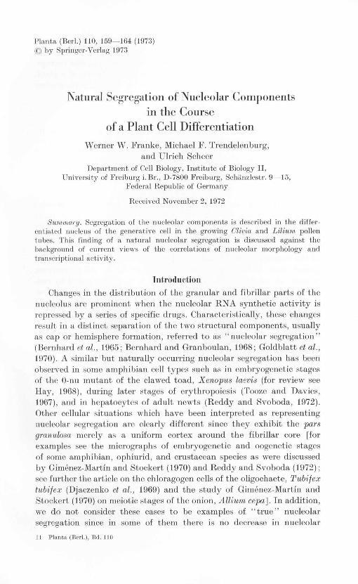
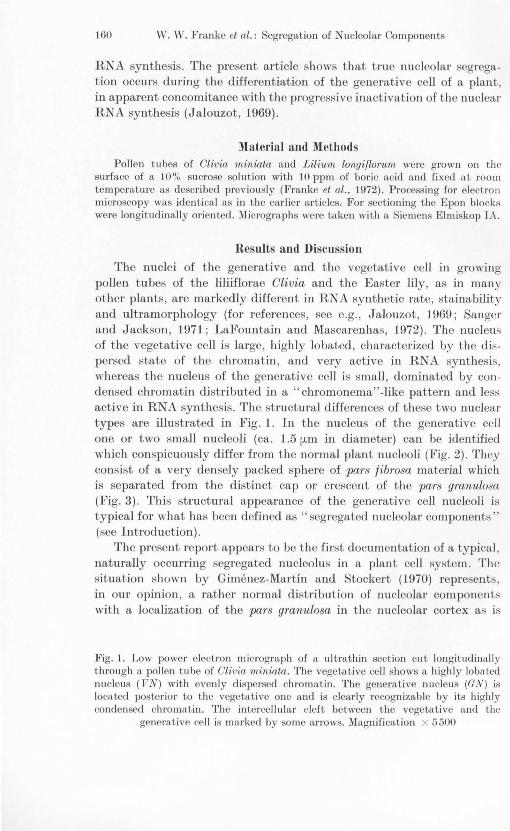
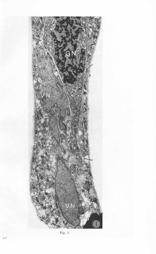
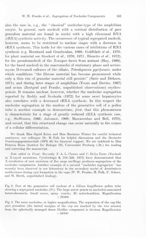
The nuclei of the generative and the vegetative cell in growing pollen tubes of the liliiflorae Clivia and the Easter lily, as in many other plants, are markedly different in RN A synthetic rate, stainability and ultramorphology (for references, see e.g., Jalouzot, 1969; Sanger and Jackson, 1971; LaFountain and Mascarenhas, 1972). The nucleus of the vegetative cell is large, highly lobated, characterized by the dispersed state of the chromatin, and very active in RN A synthesis, whereas the nucleus of the generative cell is small , dominated by condensed chromatin distributed in a "chromonema" -lilw pattern and less active in RNA synthesis. The structural differences of these two nuclear types are illustrated in Fig. 1. In the nucleus of the generative ccll one or two small nucleoli (ca. 1.5 [.Lm in diameter) can be identified which conspicuously differ from the normal plant nucleoli (Fig. 2). They consist of a very densely packed sphere of pars fibrosa material which is separated from the distinct cap or crescent of the pars granulosa (Fig. 3). This structural appearance of the generative cell nucleoli is typical for what has been defined as "segregated nucleolar components" (see Introduction).
The present report appears to be the first documentation of a typical, naturally occurring segregated nucleolus in a plant cell system. The 'ituation shown by Gimenez-Martin and Stockert (1970) represents, in our opinion, a rather normal distribution of nucleolar components with a localization of the pars granulosa in the nucleolar cortex as is
Fig. 1. Low power electron micrograph of a ultrathin section cut longitudinally t hrough a pollen tube of Ol'iv-ia min'iata. The vegetative ce ll shows a highly lobatcd nucleus (VN) with even ly dispersed chromatin. The generative nucleus (GN) is located posterior to the vegetative onc and is clearly recognizable by its highly condensed chromatin. The intercellular cleft between the vegetative and the
generative cell is marked by some arrows. Magnification X 5500
11 ·
W. W. Franke et al.: Segregation of Nueleolar Components 163
also the case in, e.g., the "claAsical" nucleolus- type of the amphibian oocyte. In general, such nucleoli with a cortical distribution of pars granulosa material are found in nuclei with a high ribosomal RNA (rRNA) synthetic activity. The occurrence of typical segregated nucleoli, however, seems to be restricted to nuclear stages with a decrease in rRNA synthesis. This holds for the various cases of inhibitions of RNA synthesis (e.g. Bernhard and Granboulan, 1968; Goldblatt et al., 1970 ; for plant material see Stockert et al., 1970, 1971 ; Risuefio et al., 1972) , for the pseudonucleoli of the X enopus laevis O-nu mutant (Hay, 1968) , for the fused nucleoli in the macronuclei of stationary phasc and actinomycin D-treated cultures of the ciliate, Tetrahymena pyriformis, under which conditions "the fibrous material has become pronounced while only a thin rim of granular material still persists" (Satir and Dirksen, 1971) , and during later stages of amphibian (Tooze and Davies, 1967) and avian (Zentgraf and Franke, unpublished observations) erythropoiesis. It remains unclear, however, whether the nucleolar segregation reportcd by Reddy and Svoboda (1972) for some newt hepatocytes also correlates with a decreased rRNA synthesis . In this respect the nucleolar segregation in the nucleus of the generative cell of a pollen tube is another example to dcmonstrate , first , that this phenomenon is characteristic for a stage of greatly reduced rRNA synthesis (see, e.g., Stcffensen , 1966 ; Jalouzot, 1969; Mascarenhas and Bell, 1970), and second, that this strnctural change can occur naturally in the course of a cellular diffcrentiation.
We thank Miss Sigrid Krien and Miss Marianne Winter for careful technical assistance, our co lleague Dr. H. Falk for helpful discussions and the Deutsche L~orschungsgemeinschaft (SFB 46) for financial suppor·t. We are indebted to Miss Patri cia Reau (Institut flIr Biologic 1H, Universitat Freiburg i.Br.) for reading and correcting the manuscript.
Note added in ProoJ: Rccently F . A. L. Clowes and C. DeLa Torre (Nucleoli in X-rayed meristell1s. Cytobiologie H, 318- 326, 1972) have demonstrated that X-irradiation of root meristem of Zea mays seerl lings produces segregation of the nucleola r components. Another exa mple of a natural" nu cleola r segregation " has recently bccn observcd in our laboratory in the seconda ry nuclei of Aeetabulal·i(£ 1I1eciiterranea during cyst formation in the caps (W. W. Franke, H. Falk, U. Scheer, and W. Herth, lU1published finding).
Fig. 2. Part of the generative ce ll nu cleus of a Lilium 10ngiJlorum pollen tube showing a segregated nu clcolu s (Ne). The large a rrow points to nucleolus·associated heterochromatin. Small arrow, spiny vesicle; Mi mitochondrion . Magnification
X 22000
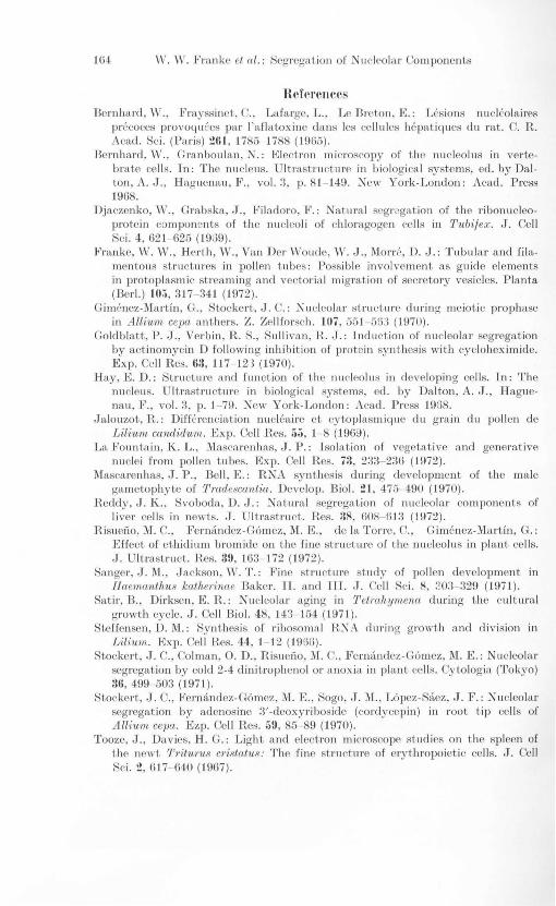
Fig. :3. The same nuclcolus, at highcr magnification. The separation of the cap-like 1Ja1·s gra,nulosa (the lateral margins of the cap are marked by the two arrows) from the spherically arranged dense fibrillar component is obviolls. Magnification
X 56000
164 W. W. I.'ra nke et al.: Segregat ion of Nucico la,t' Co mponents
Uefl'I'Cllces Bemha rd , W. , L"rayssinet. C. , La fa rge, L" Le B reton, K : Lesions nu cleola,ires
precoces provoqu ees par I'aflatox ine dans les cellules hepatiques du rat. C. R. Acacl. Sci. (Pari s) 21t1 , 1785- 1788 (1965).
Bemha rd , W. , Granboula n, N.: Elect ron mi croscopy of t he nu cleolus in vertebrate ce lls. T n: The nu cleus. U lt rastru cture in b iolog ical systems, cd. by Dalton, A. J., H ag uena u, I~ . , vol. :3, p.81- 149. New York-London : Acad . Press 1968.
Djaczenko, W. , Gra bska, J ., li'iladoro, L", : Natura l segrcgation of t he ribonu cleoprote in componcnts of the Iluc:lcoli of C'hloragogen cells in ,[,ubi/ex. J. Cell Sci , 4, 62 1- 625 (19G9).
li'ranke, W. W. , H er th, W. , Va n Del' Woude, W. J., MorI''', D. J.: Tubul a r a nd filamcntou s structures in pollen t ubes : Possible involve ment as guide elements in pT'Otoplasm ic stream ing and vecto ria l m igration of secretory vesicles. P lanta (Bed .) 10;;,317- 341 (1972).
Gimcnez-Ma rtin, G., Stockert, J, C.: Nu cleola r str'uC'\ure during meiotic prophase in Allium cepa a nthers. Z. Zellforsch. JUI , 551- 56:l (1970).
Goldblatt, P. J. , Verbin, R. S., Sulli van , H. J.: Lndu ction of nu cleola r segrcgation by act inomycin D following inhi bition of pT'Otcin sy nt hesis with cycloh eximide. Exp. Cell Res. 63, 117- 123 (1970).
H ay, E. D.: Structurc and fun ction of t he nu cleolus in developing ce lls. Ln: The nucleu '. Ult rastructLll'e in b iologica l systems, ed. by Daltoll, A. J. , H aguenau, 1"., vol. 3, p. 1 79, New York- London: Acad. Press 1958.
Ja louzot, H.: Differenciation nucleaire et cytoplasmique d u grain du pollen de Liliurn candidurn. I!:xp. Cell R.es. ';ii , 1- 8 (1969).
La Foun tain, K. L., lVLascarcnh as, J . P.: Lsolation of vegetative and generative nu clei from pollen t ubes. Exp. Cell R es. 73, 2:33- 2:3(j (1972) .
Mascarenhas, J. P. , Bell , E.: RNA synt hesis during development of t he male ga metophyte of 'l'mdeswntia. Develop. BioI. 21 , 471)- 490 (1970).
H,eddy, J. K. , Svoboda, D. J.: Natural segregation of nu cleola r co mponents of I ivc r cell s in newts. J. U lt rastruct. Res. 38, 608- 61:3 (1972) .
]~ i s u cno, M. C., Fermi ndez -Gomez, M. I]: " de la TOI'I'e, C., Gim6nez-Ma rtin , G. : I!:ffeet of ethidium bromide on t he fin e stru ct.ure of t he nu cleolus in plant cell s. J . Ultrastru ct. Hes. 3!1, 163- 172 (1972).
Sa nger, J. M., J ackson, \ V. 1'.: I~in e structure study of poll E'n developmen t in lIaemanthus kathel'ina.e Baker. Ll. a nd I] L. J. Cell Sc i. 8, :i O:3- :329 (1971 ).
Satir, B., Dirksen, E. R: N ucl eola r ag ing in T etrahymena, during the cul t ural growth cycle. J . Cell BioI. 48, 14'3- 154 (1971).
Steffensen, D. M.: Synthesis of ri bosomal R.NA d uring gT'Owth a. nd division in Lilium. Exp. Cell Res. 44, 1- 12 (1960).
Stockert, J. C., Colm a n, O. D. , Risuei'io, M. C., Ji'erIHi ndcz-Gomez, M. E.: N ucleolar segrcgation by cold 2-4 dinitrophenol 0 1' a noxia in plant cells. Cytologia. (Tokyo) ;jli, 499- 503 (197 t ).
Stockcrt, J. C., Fcrna nclez-Co mez, M. K, Sogo, J. M., Lopez-Saez, J. I~ .: Nu cleolar segregation by adenosinc 3'-deoxyribos ide (cordycepin ) in root t ip cell s of Allium ce1J[t. Ezp. Cell Hes. 59, 85- 89 (1970).
Tooze, J., Davies, H. G.: Light a nd electl'On mi croscope studics on t he splcen of t he newt rrl'itul'U8 c1'istatus: The fine structlll'e of erythropoietic cells. J . Cell Sci. !! , 617- 640 (1967 ).


