Embed Size (px)
Citation preview
REVIEW ARTICLE
Myositis autoantibodies and clinical phenotypes
Anna Ghirardello • Elisabetta Borella •
Marianna Beggio • Franco Franceschini •
Micaela Fredi • Andrea Doria
Received: 16 June 2014 / Accepted: 1 July 2014 / Published online: 23 August 2014
� Springer International Publishing Switzerland 2014
Abstract Autoantibodies are powerful diagnostic tools in
idiopathic inflammatory myopathies, especially for con-
firming the diagnosis and contributing to the definition of
disease subsets. They are present in over 80 % of patients
with immuno-mediated myositis and directed towards
ubiquitously expressed intracellular complexes. Most of
these autoantibodies are reported also in other autoimmune
diseases, while some are considered myositis-specific.
Myositis autoantibodies are traditionally categorized in two
groups, based on their diagnostic accuracy: myositis-spe-
cific antibodies (MSA) and myositis-associated antibodies
(MAA), the latter mostly occurring in myositis-overlap
syndromes. Besides the so-called traditional MSA,
including anti-synthetases, anti-SRP and anti-Mi-2 anti-
bodies, additional newly conceived immune targets have
been recently identified, mostly in patients with severe
forms of dermatomyositis or necrotizing myopathy. They
mainly encompass enzymatic proteins essentially involved
in the regulation of gene transcription or post-translational
modifications, i.e., TIF1-c, NXP-2, MDA5, SAE and
HMGCR. Among the MAA, anti-PM/Scl and anti-Ku
characterize an overlap polydermatomyositis/systemic
sclerosis syndrome with severe interstitial lung
involvement.
Keywords Autoimmune myositis � Autoantibodies �Myositis-specific antibodies � Overlap syndrome
Introduction
Serum autoantibodies towards ubiquitary intracellular
constituents are found in more than 80 % of patients with
polymyositis (PM) or dermatomyositis (DM). Some auto-
antibody specificities are shared with other connective
tissue diseases (CTD), others are almost exclusive of
patients with autoimmune myositis. As well as in other
CTD, the identification of autoantibodies in serum has
become an important milestone in the diagnosis of idio-
pathic inflammatory myopathies (IIM).
The clinically important autoantibodies are classically
categorized in two groups based on their diagnostic
accuracy: the myositis-specific antibodies (MSA) and the
myositis-associated antibodies (MAA) (Table 1). By
definition, MSA are specific of autoimmune myositis,
diagnostic specificity exceeding 90 %, and target cyto-
plasmic or nuclear ribonucleoproteins involved in key
processes of cell biology such as gene transcription,
protein synthesis and translocation, and innate antiviral
immune response (Table 2). They are mutually exclusive
and closely associated with distinct disease subsets dif-
fering in clinical involvement and prognosis [1]. On the
other hand MAA, even though present in up to 50 % of
myositis patients, are not disease-specific, frequently
associated with MSA, and mostly found in myositis-
overlap syndrome, primarily myositis-systemic sclerosis
(PDM/SSc) [2].
A. Ghirardello � E. Borella � M. Beggio � A. Doria (&)
Division of Rheumatology, Department of Medicine,
University of Padova, Via Giustiniani, 2, 35128 Padua, Italy
e-mail: [email protected]
F. Franceschini � M. Fredi
Rheumatology and Clinical Immunology Unit,
AO Spedali Civili di Brescia, Brescia, Italy
M. Fredi
Department of Clinical and Experimental Sciences,
University of Brescia, Brescia, Italy
123
Autoimmun Highlights (2014) 5:69–75
DOI 10.1007/s13317-014-0060-4
Myositis-specific autoantigens are largely heterogeneous
in functional and biochemical characteristics: the classic
ones are the aminoacyl-tRNA synthetases (ARS), the Mi-2
helicase/histone deacetylase protein complex, and the sig-
nal recognition particle (SRP) [3–5]. During the last dec-
ade, new putative myositis autoantigens have been
identified, including TIF1-c, NXP-2, MDA5, SAE, and
HMGCR [6–11]. The diagnostic accuracy of newly con-
ceived autoantibodies is far from being ascertained; how-
ever, they are promising and in the future they could
expand the MSA spectrum improving our ability in diag-
nosis and classification of myositis.
Myositis-specific antibodies (MSA) and disease subsets
Anti-ARS antibodies
ARS are cytoplasmic enzymes that catalyze the binding of
each amino acid to its cognate tRNA during protein syn-
thesis. As immunological targets, eight tRNA synthetases
have been identified so far: histidyl (Jo-1), threonyl (PL-7),
alanyl (PL-12), glycyl (EJ), isoleucyl (PL-12), asparaginyl
(KS), tyrosyl (Ha), and phenylalanyl (Zo) synthetases.
Antibodies to ARS overall occur in 25–35 % of IIM
patients [1]. Anti-Jo-1 is the most common, found in
20–30 % of patients with PM and 60–70 % of myositis
patients with interstitial lung disease (ILD). Anti-PL-7, PL-
12, are found in less than 5 %, and anti-KS, -OJ, -EJ, -Zo,
-Ha, in less than 2 % of PM or DM. Taken together, anti-
ARS are associated with a clinical syndrome featured by
myositis and high occurrence of ILD, arthritis, Raynaud’s
phenomenon and ‘‘mechanic’s hands’’, named anti-ARS
syndrome. Lungs and joints are the major organs involved,
and disease prognosis is strictly related to pulmonary
involvement. Even if anti-ARS syndrome was described as
a clinical variant of PM or DM, it is now considered a true
overlap syndrome [12].
Anti-Mi-2 antibody
Mi-2/nucleosome remodeling and histone-deacetylase
complex participates in the regulation of gene expression
via chromatin modifications. Anti-Mi-2 antibodies are
found in 10–30 % of patients with IIM, especially DM,
being associated with specific skin involvement, like Got-
tron’s sign or papules and heliotrope rash, lung sparing and
good response to steroids [4]. Moreover, anti-Mi-2 anti-
body seems to be associated with a lower risk of parane-
oplastic myositis, thus being considered a good prognostic
factor [4, 13].
Anti-SRP antibody
SRP is a highly conserved cytoplasmic multimeric ribo-
nucleoprotein, consisting of six polypeptides complexed
with one 7SL RNA, that is involved in secretory protein
recognition and translocation across the rough endoplasmic
reticulum. Anti-SRP antibodies are specific for IIM, being
found in 4–8 % of PM patients [1, 5]. They are strictly
associated with the anti-SRP syndrome, a severe necro-
tizing myopathy, histologically characterized by abundant
myofiber necrosis and regeneration, with scarce inflam-
mation. Rapidly progressive muscle weakness and poor
Table 1 Autoantibodies in
poly/dermatomyositis Myositis-Specific Antibodies Myositis-Associated Antibodies
• Anti-aminoacyl-tRNA synthetases • Anti-Ro
• Anti-Mi-2 • Anti-PM/Scl
• Anti-SRP • Anti-Ku
• Anti-TIF1-γ • Anti-U1RNP
• Anti-NXP-2
• Anti-MDA5
• Anti-SAE
• Anti-HMGCR
Novel autoantibodies
70 Autoimmun Highlights (2014) 5:69–75
123
response to standard treatment are the characteristic prog-
nostic features [5].
Anti-TIF1-c antibody
This novel putative MSA, firstly described few years ago,
is exclusively found in 20–30 % of adult as well as juvenile
DM. It targets nuclear transcription factors belonging to the
human transcriptional intermediatory factor (TIF-1) family,
primarily TIF1-c. Anti-TIF1-c antibody is significantly
associated with aggressive skin lesions both in adult and
juvenile DM, whereas the established association with
paraneoplastic DM is largely confined to adult patients,
especially the older ones ([50 years). Intriguingly, TIF-1
proteins are over-expressed in solid tumors, i.e., adeno-
carcinoma, and implicated in the regulation of p53 onco-
gene suppressor. The diagnostic value of anti-TIF1-cantibody testing has been recently assessed: high antibody
levels anti-TIF1-c increase the risk of cancer-associated
DM in adults, whereas negative antibody prompts to
exclude concurrent malignancy, due to its very high neg-
ative predictive value [6].
Anti-NXP-2 antibody
This autoantibody targets a 140-kDa nuclear protein,
named nuclear matrix protein 2 (NXP2), which plays a role
in the regulation of p53-induced apoptosis after oncogenic
stimuli. To date, anti-NXP-2 antibodies have been reported
in about 25 % of juvenile or adult DM, and rarely in PM.
Joung-onset DM, severe cutaneous lesions, including cal-
cinosis, and muscle contractures are the prominent features
[14]. An association with cancer has been observed in
adults, especially in males. Recently, Fiorentino et al. [7]
assessed the diagnostic value of anti-TIF1-c and anti-NXP-
2 antibody testing for cancer-associated DM, confirming
that they overall occur in more than 50 % of otherwise
‘‘antibody negative’’ patients, and are independently asso-
ciated with cancer. Moreover, the authors provided evi-
dence that accurate laboratory testing for such antibodies
should be included in the diagnostic workup in order to
identify the vast majority of patients with cancer-associated
DM.
Anti-MDA5 antibody
This novel MSA has been firstly described in 20–30 % of
Asian populations, as specific of DM and associated with a
clinical subset characterized by clinically amyopathic DM
and rapidly progressive ILD [8]. IFN-induced melanoma
differentiation-associated protein 5 (MDA5) is as the
identified autoantigen, which regulates the innate antiviral
immune response against cytoplasmic viral RNA. The
clinical relevance of anti-MDA5 antibodies seems to be
confirmed also in Caucasian IIM cohorts [9], but it is still a
matter of research.
Anti-SAE antibody
Strictly related to a specific HLA haplotype, anti-small
ubiquitin-like modifier activating enzyme (SAE) antibodies
are found in 8 % of adult DM patients, presenting with
severe skin disease, dysphagia and systemic features;
prognosis is favorable [10]. They target the SUMO-1
activating enzyme heterodimer, a small nuclear protein
structurally similar to ubiquitin, which leads to a post-
Table 2 Myositis-specific antibodies: target antigens and clinical
associations in adult myositis patients
Autoantibody Immune target Function of
autoantigen
Clinical
associations
Anti-ARS
(Jo-1, PL-7,
PL-12, EJ,
OJ, KS, Ha,
Zo)
tRNA
synthetases
Aminoacylation
of tRNAs
PM
Anti-
synthetase
syndrome
Anti-Mi-2 NuRD subunit Gene
transcription
Nucleosome
remodeling
‘‘Classic
DM’’
Mild disease
Anti-TIF1-c Transcriptional
intermediary
factor 1c
Ubiquitination
Gene
transcription
Severe DM
Cancer-
associated
DM
Anti-NXP-2 Nuclear matrix
protein 2
Gene
transcription
Severe DM
Cancer-
associated
DM
Anti-MDA5 Melanoma
differentiation-
associated
protein 5
Innate antiviral
response
Amyopathic
DM
ILD
Poor
prognosis
Anti-SAE SUMO-1
activating
enzyme
Protein
sumoylation
Gene
transcription
DM
Initially
amyopathic
DM
Anti-SRP Signal
recognition
particle
Protein
translocation
across the ER
Necrotizing
myopathy
Anti-
HMGCR
3-Hydroxy-3-
methylglutaryl-
CoA reductase
Cholesterol
biosynthesis
Necrotizing
myopathy
Prior statin
use
tRNAs transfer RNAs, PM polymyositis, NuRD nucleosome remod-
eling-histone deacetylase, DM dermatomyositis, ILD interstitial lung
disease, SUMO-1 small ubiquitin-like modifier 1, ER rough endo-
plasmic reticulum
Autoimmun Highlights (2014) 5:69–75 71
123
translational enzymatic aggregation/conjugation of pro-
teins, called ‘‘protein sumoylation’’.
Anti-HMGCR antibody
A putative MSA was discovered in 3–8 % of adult patients
with IIM, as directed against 3-hydroxy-3-methylglutaryl-
coenzyme A reductase (HMGCR), a key enzyme of the
cholesterol biosynthesis, specifically inhibited by statins.
Anti-HMGCR antibody has been proposed as a serological
marker of immune-mediated necrotizing myopathy, often
but not exclusively induced by statin exposure [11]. Clin-
ical phenotype of anti-HMGCR-positive patients resem-
bled that observed in other forms of IIM: proximal muscle
weakness, markedly elevated creatine kinase levels, myo-
pathic features on electromyography, and response to
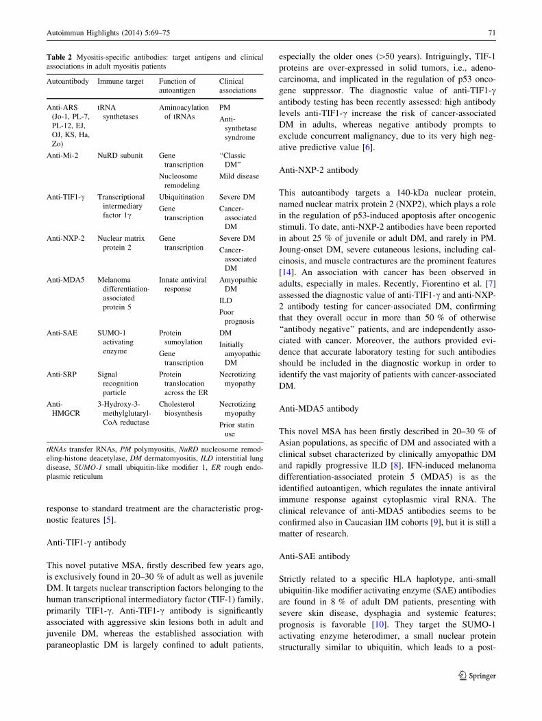
immunosuppressive therapy. Myofiber necrosis and
degeneration is the characteristic histopathologic feature,
occasionally accompanied by aspecific inflammatory cell
infiltration (Fig. 1).
Myositis-associated antibodies (MAA) and overlap
syndromes
Major MAA include anti-Ro/SSA, anti-PM/Scl, and anti-
Ku and anti-U1RNP antibodies. MAA are often found
when PM or DM occurs as a component of another
CTD, and thus contributing to the clinical spectrum of
an overlap syndrome, though MAA may also be present
in patients without overlap CTD. The overlap syndrome
between PM/DM and systemic sclerosis (PDM/SSc),
also called scleromyositis, is the most common and
according to a recent review of the literature it appears
to represent more than 44 % of all scleroderma overlap
syndromes [2].
Fig. 1 Muscle biopsy from a patient with polymyositis and anti-
HMGCR autoantibodies (hematoxylin–eosin stain, original magnifi-
cation 920). The biopsy shows scattered necrotic fibers, some of
which invaded by mononuclear cells, basophilic regenerating fibers
and an inflammatory infiltrate in the perimysium. (Courtesy of Dr.
Vattemi G.)
72 Autoimmun Highlights (2014) 5:69–75
123
Anti-Ro/SSA antibody, mostly directed against the
Ro52 subunit, is the most prevalent MAA in myositis,
found in more than 30 % of patients, frequently concom-
itant with anti-ARS antibodies or other MAA [15]. It has
been recently reported that anti-Ro52 antibody could have
a prognostic value in anti-Jo-1 positive patients, being
associated with a higher risk of severe ILD, myositis, joint
involvement, and cancer compared with anti-Jo1-positive
patients without anti-Ro52 antibodies [16]. In addition,
anti-Ro/SSA and/or anti-La/SSB antibodies are frequently
encountered in PDM/Sjogren (SS) overlap syndrome,
which is reported in 5.3 % of PDM patients, while myositis
in primary SS is variably reported in 1–14 % of cases [17].
Anti-PM/Scl and anti-Ku antibodies can be somehow
considered markers of PDM/SSc. Antibodies to PM/Scl are
generally found in PM, DM or SSc patients, with the
highest occurrence in PDM/SSc. A meta-analysis of the
studies on anti-PM/Scl antibodies showed that anti-PM/Scl
was found in 31 % of patients with PDM/SSc, compared to
8 % of PM, 11 % of DM and 2 % of SSc. On the other
hand, PDM/SSc is diagnosed in 59 % of patients with anti-
PM/Scl [18]. Apart from myositis, the most frequent clin-
ical manifestations reported in anti-PM/Scl-positive PDM/
SSc patients consist of Raynaud’s phenomenon, ILD,
arthritis and skin involvement, including ‘‘mechanic’s
hands’’. While muscle involvement is often subclinical in
PDM/SSc with PM/Scl antibodies, ILD has the same
clinical and pathological features and severity observed in
patients with anti-ARS syndrome [19].
Even though detected in a wide spectrum of CTD, anti-
Ku antibody should be considered as a marker of PDM/
SSc, either because it was first described in patients with
PDM/SSc, which was reported in about 25 % of anti-Ku
positive patients, or because the clinical manifestations are
the same as in anti-ARS and anti-PM/Scl overlap syndrome
[20]. Moreover, while the inflammatory myopathy is usu-
ally mild and responds to corticosteroid treatment, lung
involvement is severe and refractory to corticosteroids, as
in both anti-ARS and anti-PM-Scl overlap syndrome [21].
Anti-PM/Scl and anti-Ku positive PDM/SSc are preferen-
tially associated with the limited cutaneous variant of SSc
[20, 22], presenting less digital ulcers and much more
myositis, arthritis and ILD, compared with anti-centromere
antibody positive limited SSc [20]. Notably, both anti-PM/
Scl antibody and the associated PDM/SSc recognize an
immunogenetic background characterized by the haplotype
HLA-DQA1*0501, DQB1*02, DRB1*0301 [22].
Anti-U1RNP antibodies are significantly more frequent
in PDM/systemic lupus erythematosus (SLE) overlap
syndrome than in SLE and they are associated with anti-Jo-
1 antibodies in such condition. In addition to myositis,
erosive arthritis, alopecia, ILD and oral ulcers are prevalent
findings in patients with PDM/SLE overlap syndrome.
Conversely, renal disease is very rare. Nevertheless,
patients with SLE and myositis were likely to die at a
young age. The onset of myositis can antedate, be con-
comitant or follow the appearance of SLE manifestations.
DM is more common than PM [23].
By reviewing a cohort of 110 outpatients with PDM
attending Brescia Rheumatology and Clinical Immunology
Unit from 1990 to 2013, an overlap syndrome with CTD
was diagnosed in 23 patients (21 %): 14 with SSc (61 %),
3 with SLE (13 %), 4 with SS (17 %) and 2 with rheu-
matoid arthritis (9 %). A similar prevalence of overlap
syndrome in PDM was also reported in recent studies [17],
where myositis was classified according to the commonly
used Bohan and Peter’s criteria. Interestingly, a modified
classification of these criteria has been proposed [15],
taking into account the presence of extra muscular clinical
manifestations and/or autoantibodies other than anti-Mi-2,
considered highly specific for pure DM. With such an
approach, the majority of inflammatory myositis should be
diagnosed as overlap myositis syndrome. Furthermore,
combining autoantibody testing to the original classifica-
tion allows us to better understand the value of autoanti-
bodies in the diagnosis and prognosis definition, and in
predicting the response to corticosteroid treatment.
Laboratory detection of myositis autoantibodies
Testing for MSA and MAA is a useful aid for the diagnosis
and subset definition of autoimmune myositis. However,
many target antigens are poorly expressed in cell/tissue
extracts or highly sensitive to degradation/denaturation,
thus limiting the standardization of methods for diagnostic
purpose. Therefore, most MSA are still detected through
different time-consuming non-standardized techniques.
Recently, a single multianalytic line blot assay has been
validated as screening test for MSA/MAA in clinical
practice [24]. On the basis of our experience, we suggest to
include line blot as an initial screen in suspected IIM.
Patients seronegative by line blot testing should be evalu-
ated by in-house speculative testing.
At present, new antibody specificities are accurately
detected by immunoprecipitation of radiolabeled proteins
or RNA molecules; however, commercially available
ELISA or immunoblot assays will be available in the near
future.
Autoantibodies as clues in the pathogenesis of myositis
The aforementioned association of MSA with distinct
clinical phenotypes and disease prognosis, and the corre-
lation between serum antibody levels and disease activity,
Autoimmun Highlights (2014) 5:69–75 73
123
provide clues on the pathogenetic role of autoantibodies in
PM/DM. Evidence is increasing that myositis autoantigens
are able to drive a B cell antigen-specific immune response
in muscles. Intriguingly, after the pioneeristic study by
Casciola-Rosen et al. [25], several groups confirmed the
aberrant expression of Jo-1, Mi-2, HMGCR and other
myositis antigens in regenerating fibers from PM and DM
muscle biopsies compared with controls. In addition,
overexpression of both classic and novel myositis antigens
has been demonstrated in other target organs, such as Jo-1
in the lung, Mi-2 in the epidermal basal membrane, and
also TIF1-c and NXP-2 in adenocarcinoma tissues. In
target tissues, myositis antigens show strong adjuvant and
chemoattractant properties, acting per se as proinflamma-
tory and immunostimulating agents, either in the muscle or
far from it, i.e., in the lung, skin or tumors. Thus, regen-
erating muscle fibers and tumor cells seem to share similar
immunostimulating phenotypes, suggesting a pathogenetic
link between cancer and autoimmunity in myositis [26].
Finally, association of most myositis autoantigens with
nucleic acids makes them likely to stimulate the IFN type I
secretion pathways which in turn can increase antigen
availability and propagation of the immune response [27].
Conclusions
MSA are disease serological markers, mutually exclusive
and closely associated with distinct disease subsets.
Moreover, they seem to be directly involved in the
induction and perpetuation of muscle damage. Therefore,
their detection in the early phase of the disease might be
helpful in the prediction of clinical course and disease
prognosis.
Conflict of interest None.
Human and animal rights All procedures performed in studies
involving human participants were in accordance with the ethical
standards of the institutional and/or national research committee and
with the 1964 Helsinki declaration and its later amendments or
comparable ethical standards.
Informed consent The study was approved by the Local Ethics
Committee and informed consent was obtained from all patients, in
compliance with the Helsinki Declaration.
References
1. Ghirardello A, Bassi N, Palma L et al (2013) Autoantibodies in
polymyositis and dermatomyositis. Curr Rheumatol Rep 15:335.
doi:10.1007/s11926-013-0335-1
2. Iaccarino L, Gatto M, Bettio S et al (2012) Overlap connective
tissue disease syndromes. Autoimmun Rev 3:363–373
3. Zampieri S, Ghirardello A, Iaccarino L, Tarricone E, Gambari PF,
Doria A (2005) Anti-Jo-1 antibodies. Autoimmunity 38:73–78
4. Ghirardello A, Zampieri S, Iaccarino L et al (2005) Anti-Mi-2
autoantibodies. Autoimmunity 38:79–83
5. Hengstman GJ, ter Laak HJ, Vree Egberts WT et al (2006) Anti-
signal recognition particle autoantibodies: marker of a necrotising
myopathy. Ann Rheum Dis 65:1635–1638
6. Trallero-Araguas E, Rodrigo-Pendas JA, Selva-O’Challagan A
et al (2012) Usefulness of anti-p155 autoantibody for diagnosing
cancer-associated dermatomyositis. A systematic review and
meta-analysis. Arthritis Rheum 64:523–532
7. Fiorentino DF, Chung LS, Christopher-Stine L et al (2013) Most
patients with cancer-associated dermatomyositis have antibodies
to nuclear matrix protein NXP-2 or transcription intermediary
factor 1 c. Arthritis Rheum 65:2954–2962. doi:10.1002/art.38093
8. Cao H, Pan M, Kang Y et al (2012) Clinical manifestations of
dermatomyositis and clinically amyopathic dermatomyositis
patients with positive expression of anti-melanoma differentia-
tion-associated gene 5 antibody. Arthritis Care Res 64:1602–1610
9. Hall JC, Casciola-Rosen L, Samedy LA et al (2013) Anti-mela-
noma differentiation-associated protein 5-associated dermato-
myositis: expanding the clinical spectrum. Arthritis Care Res
65:1307–1315. doi:10.1002/acr.21992
10. Tarricone E, Ghirardello A, Rampudda M, Bassi N, Punzi L,
Doria A (2012) Anti-SAE antibodies in autoimmune myositis:
identification by unlabelled protein immunoprecipitation in an
Italian patient cohort. J Immunol Methods 384:128–134
11. Mohassel P, Mammen AL (2013) Statin-associated autoimmune
myopathy and anti-HMGCR autoantibodies. Muscle Nerve
48:477–483. doi:10.1002/mus.23854
12. Mahler M, Miller FW, Fritzler MJ (2014) Idiopathic inflamma-
tory myopathies and the anti-synthetase syndrome: a compre-
hensive review. Autoimmun Rev 13:367–371. doi:10.1016/j.
autrev.2014.01.022
13. Iaccarino L, Ghirardello A, Bettio S et al (2014) The clinical
features, diagnosis and classification of dermatomyositis. J Au-
toimmun 48–49:122–127. doi:10.1016/j.jaut.2013.11.005
14. Ceribelli A, Fredi M, Taraborelli M et al (2012) Anti-MJ/NXP-2
autoantibody specificity in a cohort of adult Italian patients with
polymyositis/dermatomyositis. Arthritis Res Ther 14:R97. doi:10.
1186/ar3822
15. Troyanov Y, Targoff IN, Tremblay JL, Goulet JR, Raymond Y,
Senecal JL (2005) Novel classification of idiopathic inflammatory
myopathies based on overlap syndrome features and autoanti-
bodies: analysis of 100 French Canadian patients. Medicine
(Baltimore). 84:231–249
16. Marie I, Hatron PY, Dominique S et al (2012) Short-term and
long-term outcome of anti-Jo1-positive patients with anti-Ro52
antibody. Semin Arthritis Rheum 41:890–899
17. Vancsa A, Gergely L, Ponyi A et al (2010) Myositis-specific and
myositis-associated antibodies in overlap myositis in comparison
to primary dermatopolymyositis: Relevance for clinical classifi-
cation: retrospective study of 169 patients. Jt Bone Spine
77:125–130
18. Mahler M, Raijmakers R (2007) Novel aspects of antibodies to
the PM/Scl complex: clinical, genetics and diagnostic insights.
Autoimmun Rev 6:432–437
19. Lega JC, Cottin V, Fabien N, Thivolet-Bejui F, Cordier JF (2010)
Interstitial lung disease associated with anti-PM/Scl or anti-
aminoacyl-tRNA synthetase autoantibodies: a similar condition?
J Rheumatol 37:1000–1009
20. Cavazzana I, Fredi M, Taraborelli M, Quinzanini M, Tincani A,
Franceschini F (2013) A subset of systemic sclerosis but not of
systemic lupus erythematosus is defined by isolated anti-Ku
autoantibodies. Clin Exp Rheumatol 31:118–121
21. Rigolet A, Musset L, Dubourg O et al (2012) Inflammatory
myopathies with anti-Ku antibodies: a prognosis dependent on
associated lung disease. Medicine (Baltimore) 91:95–102
74 Autoimmun Highlights (2014) 5:69–75
123

22. Ho KT, Reveille JD (2003) The clinical significance of autoan-
tibodies in scleroderma. Arthritis Res Ther 5:80–93
23. Dayal NA, Isenberg DA (2002) SLE/myositis overlap: are the
manifestations of SLE different in overlap disease? Lupus
11:293–298
24. Ghirardello A, Rampudda M, Ekholm L et al (2010) Diagnostic
performance and validation of autoantibody testing in myositis by a
commercial line blot assay. Rheumatology (Oxford) 49:2370–2374
25. Casciola-Rosen L, Nagaraij K, Plots P et al (2005) Enhanced
autoantigen expression in regenerating muscle cells in idiopathic
inflammatory myopathy. J Exp Med 201:591–601
26. Zampieri S, Doria A, Adami N et al (2010) Subclinical myopathy in
patients affected with early stage colorectal cancer at disease onset:
evidence from skeletal muscle biopsies. Neurol Res 32:20–25
27. Lundberg IE, Helmers SB (2010) The type I interferon system in
idiopathic inflammatory myopathies. Autoimmunity 43:239–243
Autoimmun Highlights (2014) 5:69–75 75
123

![Advances in inclusion body myositis: genetics ...progress in clinical trials [9]. Atypical phenotypes and clinical presentations have been reported in as many as 24% of cases in some](https://img.dokumen.tips/doc/110x75/5ebfa66afb0d015eb650a52e/advances-in-inclusion-body-myositis-genetics-progress-in-clinical-trials-9.jpg)


![Fatal myositis and spontaneous haematoma induced by ......myositis in patients receiving ipilimumab plus nivolumab was 0.24% [5]. ICI-related myositis mimics primary dermatomyositis](https://img.dokumen.tips/doc/110x75/60a56f20301b9a411c564b9f/fatal-myositis-and-spontaneous-haematoma-induced-by-myositis-in-patients.jpg)


