Embed Size (px)
Citation preview

Mutations
1

2
What is a gene?
---TTGACAT------TATAAT-------AT-/-AGGAGGT-/-ATG CCC CTT TTG TGA---AACTGTA------ATATTA-------TA-/-TCCTCCA-/-TAC GGG GAA AAC ATT
(-10)(-35)
PROMOTER
5’3’
3’ 5’
antisense
sense
RIBOSOMEBINDING
SITE
U-/-AGGAGGU-/-AUG CCC CUU UUG UGA
5’ 3’
Met Pro leu leu stp
Prokaryotic Genes
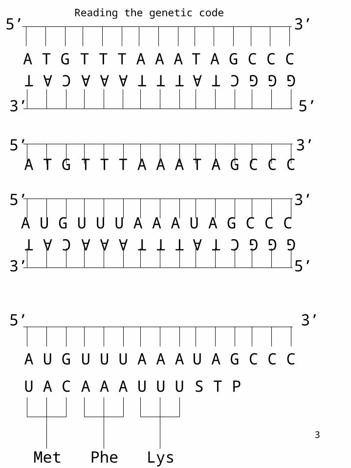
When all of these rules are satisfied then A segment of DNA will generate an RNA which will then be read by a ribosome and be translated into a protein.
3
Reading the genetic code
A T G T T T A A A T A G C C C
5’ 3’
C A T A A A T T T C T A G G G
5’3’
U A C
Met
A A A
Phe
U U U
Lys
S T P
A U G U U U A A A U A G C C C
5’ 3’
5’3’
C A T A A A T T T C T A G G GA U G U U U A A A U A G C C C
5’ 3’
A T G T T T A A A T A G C C C5’ 3’
4
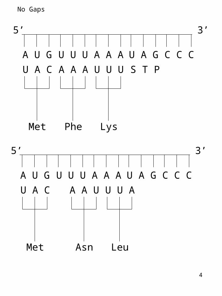
No Gaps
A U G U U U A A A U A G C C C
5’ 3’
U A C
Met
A A A
Phe
U U U
Lys
S T P
A U G U U U A A A U A G C C C
5’ 3’
U A C
Met
A A U
Asn
U U A
Leu
5
No overlaps
A U G A A A C C C U A G C C C
5’ 3’
U A C
Met
U U U
Lys
G G G
Pro
S T P
A U G A A A C C C U A G C C C
5’ 3’
U A C
Met
U U U
Lys
U G G
Trp

6
UUUUUCUUAUUG
CUUCUCCUACUG
AUUAUCAUAAUG
GUUGUCGUAGUG
UCUUCCUCAUCG
CCUCCCCCACCG
ACUACCACAACG
GCUGCCGCAGCG
UAUUACUAAUAG
CAUCACCAACAG
AAUAACAAAAAG
GAUGACGAAGAG
UGUUGCUGAUGG
CGUCGCCGACGG
AGUAGCAGAAGG
GGUGGCGGAGGG
Phe
Leu
Leu
Ile
Met
Val
Ser
Pro
Thr
Ala
Tyr
STOP
His
Gln
Asn
Lys
Asp
Glu
Cys
STOPTrp
Arg
Ser
Arg
Gly
U
C
A
G
U C A G
UCAG
UCAG
UCAG
UCAG
Fir
st
lett
er
Second letter
Th
ird le
tter
The GENETIC CODE
The code is a three letter code.

7
The code
3 amino acids are specified by 6 different codons5 amino acids are specified by 4 different codons1 amino acid is specified by 3 different codons9 amino acids are specified by 2 different codons2 amino acids are specified by 1 different codons
The degeneracy arises because
More than one tRNA specifies a given amino acidA single tRNA can base-pair with more than one codon
tRNAs do not normally pair with STOP codons
----UCC------UCA------AGCAGG
Ser
AGU
Ser
UCG
Ser
----UCC------UCA------AGG
Ser
AGG
Ser

8
The Genetic Code
Properties of the Genetic code:
1- The code is written in a linear form using the nucleotides that comprise the mRNA
2- The code is a triplet: THREE nucleotides specify ONE amino acid
3- The code is degenerate: more than one triplet specifies a given amino acid
4- The code is unambiguous: each triplet specifies only ONE amino acid
5- The code contains stop signs- There are three different stops
6- The code is comma less
7- The code is non-overlapping
xxxxxx
9
Mutations
10
Most mutations are harmful in their effects; only rarely are mutations beneficial.
A gene with one wild-type allele is monomorphic; a gene with two or more wild-type alleles is polymorphic.
The vast majority of traits are determined by alleles of more than one gene. This means that most traits are multifactorial traits.
A Heterogeneous Trait is one that may be caused by mutations in more than one gene.Human deafness is an example of a heterogeneous trait: Mutations in any of at least 50 genes lead to deafness.
An important class of mutations are conditional mutations- (Environment affects Phenotype). Conditional mutations are those that express their associated phenotype only under some conditions (restrictive conditions) and not others (permissive conditions).
Conditional lethal mutations are common.
Temperature-sensitive conditional mutations are invaluable in genetic research.
11
Generation of mutations
Spontaneous mutations
Replication induced mutations of DNAUsually base substitutions (Most errors are corrected)
Meiosis- segregation defects or defects during crossing over can induce mutationsSmall additions and deletions AND Large changes as well
Environment induced changesExposure to physical mutagens - Radioactivity or chemicals
Depurination (removal of A or G)Repair results in random substitution during
replication
Deamination (removal of amino group of base) (nitrous acid)
Cytosine--uracil--bp adenine--replication--
Oxidation (oxoG)guanine--oxoguanine--bp adenine--replication
--
Base analog incorporation during replication BU-T
Intercalating agents

Mutation rate
12
There are approximately 1013 cells in the human body
Each cell receives 1-10,000 DNA lesions per day (Lindahl and Barnes 2000). Almost all are repaired!!
Most pervasive mutagen is UV. 100,000 lesions per exposed cell per hour (Jackson and Bartek 2009).
Ionizing agents (X-rays/-rays) are most toxic because they generate double strand breaks (Ward 1988).
Chromosome instability (gain or loss of entire segments) is frequent - 40% of imbalances are entire arm imbalance while 45% are terminal segment imbalance (double strand break, nondysjunction etc)
Sequencing 179 humans as part of the 1000 genome project:On average, each person is found to carry approximately 250 to 300 loss-of-function variants in genes of which 50 are in genes previously implicated in genetic disorders.
Each individual on average has:1.3 million short indels (1-10,000 bp) and 20,000 large sequence variations (>10,000 bp).
Variation detected by the project is not evenly distributed across the genome: certain regions, containing repetitive sequences (sub-telomeres etc), show high rates of indels.

13
Sequencing the whole genomes of a family (2010 Science 328 636).
98 crossovers in maternal genome57 crossovers in paternal genome
Mutation rate is 1x10⁻⁸ per position per haploid genome(human genome is 3x10⁹ bp)
It was calculated that there are ~70 new mutations in each diploid human genome
Some sites such as CpG sites mutate at a rate 11 times higher than other sites
Exome sequencing of 2440 individuals (Science 2012 337 40)
Each person has ~100 loss of function mutations (~35 nonsense). 20 loss of function mutations are homozygous
Some alterations in sequence concentrate in specific geographic populations.Rare changes are population specific and their frequencies vary for each geographic population

14
Methods used to study mutations
Gross chromosomal changes- deletions, insertions, inversions, translocations
Cytology- microscopy- karyotype
Small mutationsSmall deletions, insertions and point mutations
Recombinant DNA technologies
15
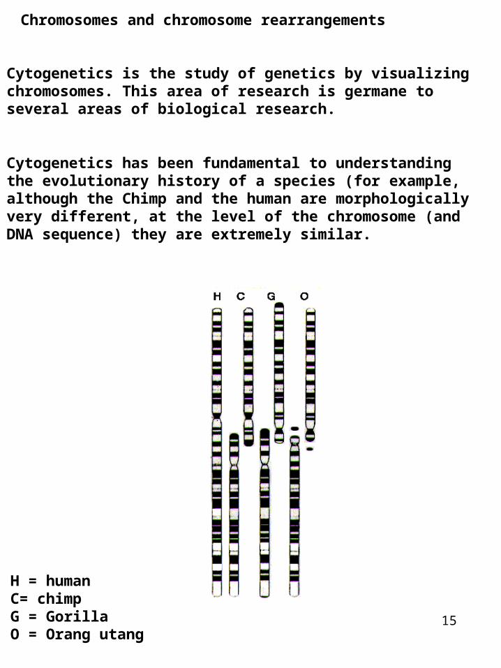
Chromosomes and chromosome rearrangements
Cytogenetics is the study of genetics by visualizing chromosomes. This area of research is germane to several areas of biological research.
Cytogenetics has been fundamental to understanding the evolutionary history of a species (for example, although the Chimp and the human are morphologically very different, at the level of the chromosome (and DNA sequence) they are extremely similar.
H = humanC= chimpG = GorillaO = Orang utang
16
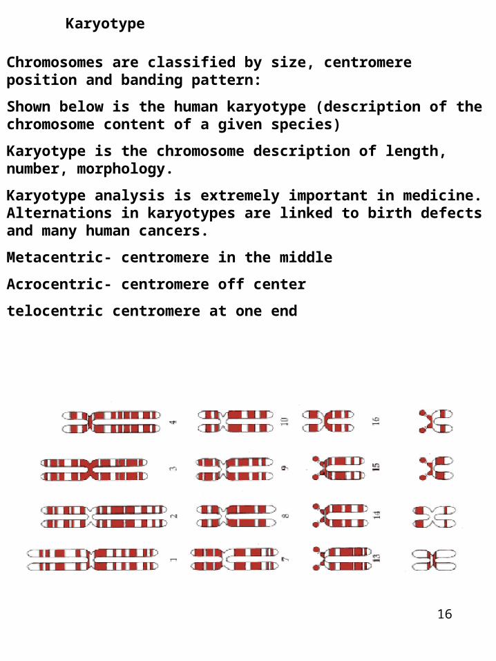
Chromosomes are classified by size, centromere position and banding pattern:
Shown below is the human karyotype (description of the chromosome content of a given species)
Karyotype is the chromosome description of length, number, morphology.
Karyotype analysis is extremely important in medicine. Alternations in karyotypes are linked to birth defects and many human cancers.
Metacentric- centromere in the middle
Acrocentric- centromere off center
telocentric centromere at one end
Karyotype
17
Banding patterns
Specialized stains produce unique banding patterns along each chromosome. Banding patterns are extremely useful for detecting abnormalities in chromosome structure.
For many of the chromosome stains- the molecular basis of the banding patterns is unclear. Nonetheless these techniques remain fundamental in many areas of genetic research
18
MU to bp
Genetic maps are based on recombination frequencies and describe the relative order and relative distance between linked genes.
Remember genes reside on chromosomes.
So what we would like to know is where are the genes located on the chromosomes
What does this mean in terms of chromosomes and DNA?
19
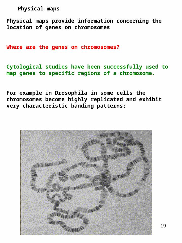
Physical maps
Physical maps provide information concerning the location of genes on chromosomes
Where are the genes on chromosomes?
Cytological studies have been successfully used to map genes to specific regions of a chromosome.
For example in Drosophila in some cells the chromosomes become highly replicated and exhibit very characteristic banding patterns:

20
In situ hybridization
Salivary glands
Squash on slide
Denature/Stain polytene chromosomes
label gene probe (you can only use this method if you have the gene cloned)
Hybridize probe to polytene chromosomes
Autoradiography
Chromosome loss/gain
21
Chromosome instability- Elevated gain or loss of complete chromosomes
Very Frequent in tumors
Frequent in in vitro fertilized embryos
Increase with age of parentMales have 2% rate of aneuploidy during gamete formationFemales have 15% rate of aneuploidy (that increases to 60% by age 50) (25% of all conceptions result in miscarriage due to aneuploidy usually at implantation stage)
Gross chromosomal rearrangements during in vitro fertilization. 40% of embryos carried entire chromosome imbalance
Entire chromosome aneuploidy in human adults: Only chromosome 13, 18, 21 and X/Y

22
Gross chromosomal changes
The Cri du chat syndrome in humans is a result of a deletion in the short arm of chromosome 5. This was determined by comparing banding patterns with normal and Cri du Chat individuals
Types of chromosome rearrangements that can be studied by karyotype analysis:
GROSS CHROMOSOMAL CHANGES
Deletions, Duplications, Inversions, Translocations
Copy Number Variation of chromosomes
23
CNV- copy number variation of entire chromosomes or of chromosome segments5% of an individuals genome displays CNV
Synuclein gene (involved in membrane stability of neurons). CNV is involved in Parkinsons
Many cancers- malignant cells most often gain additional copies of chromosome segments- genes in these segments are mis-expressed and this leads to mis-expression of other genes.
Tissues of identical twins show copy number differences at several loci (Bruder et al 2008)
Different tissues of single individual show copy number differences (Piotrowski et al., 2008)
Gross chromosomal rearrangements during in vitro fertilization55% of embryos carried terminal imbalance (sub-telomere loss) (Vanneste et al., 2009 -microarray based screen of IVF 35 embryo)
24
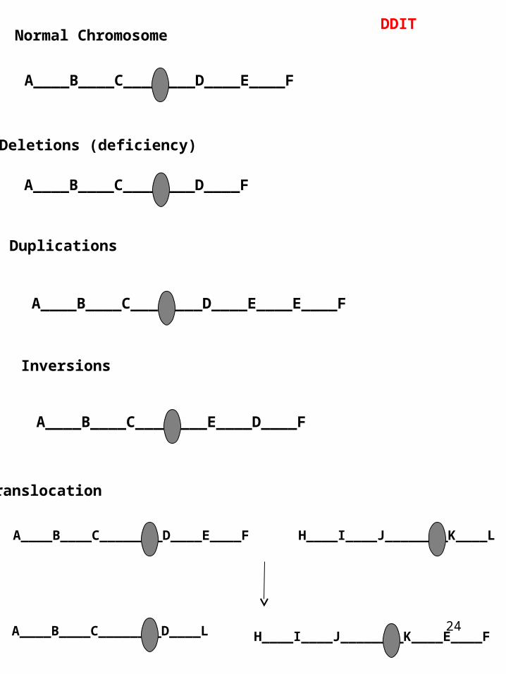
DDIT
Translocation
A____B____C________D____E____F
Normal Chromosome
Deletions (deficiency)
A____B____C________D____F
Duplications
A____B____C________D____E____E____F
Inversions
A____B____C________E____D____F
A____B____C________D____E____F
A____B____C________D____L
H____I____J________K____L
H____I____J________K____E____F
25
Insertion and deletions are frequent: Sequencing 179 humans as part of the 1000 genome project:On average, each person is found to carry approximately 250 to 300 loss-of-function variants in genes of which 50 are in genes previously implicated in inherited disorders.20,000 large structural variants were identified and 1.3 million short indels were identified.Variation detected by the project is not evenly distributed across the genome: certain regions, such as subtelomeric regions, show high rates of variation.
1 in 500 children have reciprocal translocation but such translocations are usually harmless. (However gametes produced by the children will have defects).
1 in 50 children have inversions (small and large). The heterozygous inversion carrier generally show no adverse phenotype (but produce abnormal meiotic products from crossing-over in the inversion loop).
26
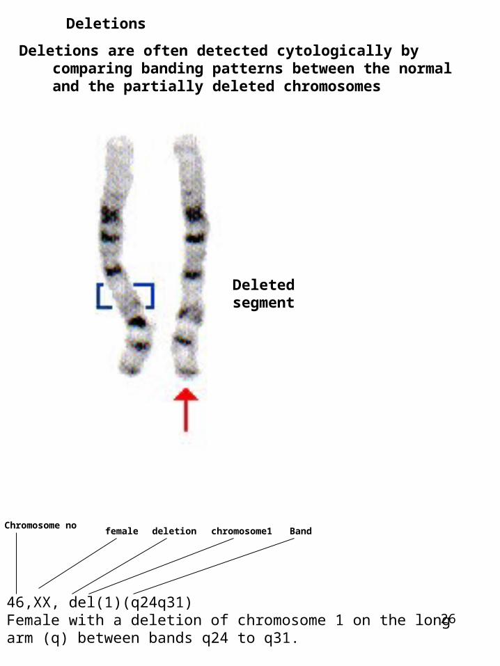
Deletions
Deletions are often detected cytologically by comparing banding patterns between the normal and the partially deleted chromosomes
Deletedsegment
46,XX, del(1)(q24q31)Female with a deletion of chromosome 1 on the long arm (q) between bands q24 to q31.
Chromosome nofemale deletion chromosome1 Band

27
In many instances deletions are too small to be detected cytologically. In these instances genetic/molecular techniques are used.
Since cytological deletions remove a contiguous set of genes, there is a high probability that an essential gene will be deleted. Therefore deletions will survive as heterozygotes and not homozygotes.
A____B________C____D
A____B________C____D
Normal
A____________C____D
A____________C____D
Homologous deletion(Lethal?)
A____________C____D
A____B________C____D
Heterologous deletion(NOT Lethal)
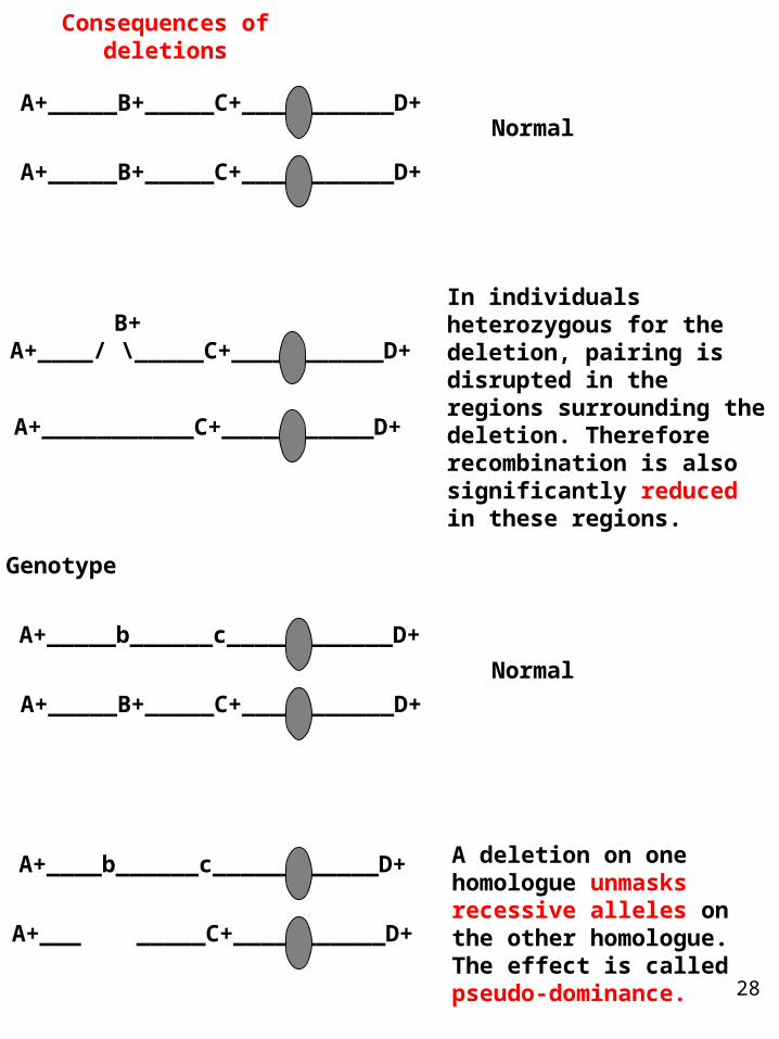
28
Consequences of deletions
B+A+____/ \_____C+___________D+
A+___________C+___________D+
In individuals heterozygous for the deletion, pairing is disrupted in the regions surrounding the deletion. Therefore recombination is also significantly reduced in these regions.
A deletion on one homologue unmasks recessive alleles on the other homologue. The effect is called pseudo-dominance.
A+____b______c____________D+
A+___ _____C+___________D+
Genotype
A+_____B+_____C+___________D+
A+_____B+_____C+___________D+
Normal
A+_____b______c____________D+
A+_____B+_____C+___________D+
Normal
29
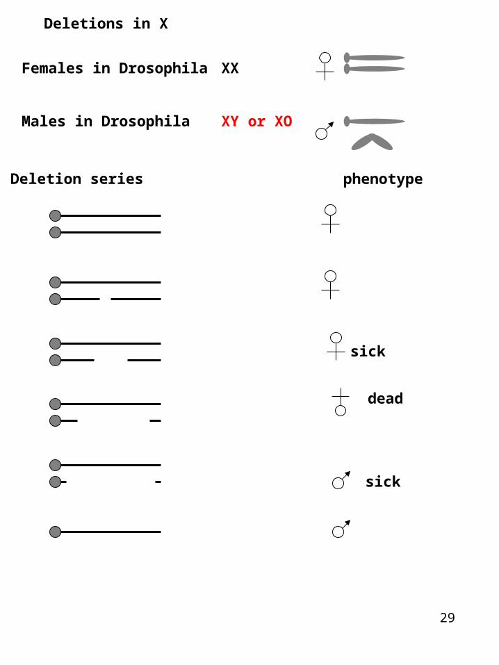
Deletions in X
Females in Drosophila XX
Males in Drosophila XY or XO
Deletion series phenotype
sick
dead
sick
30
Changes in chromosome structure
Deletions:
1. Hemizygosity from large deletions results in lethality- even the smallest cytologically defined deletions take out tens of 1,000's of bps and are likely to remove essential genes.
2. Organisms can tolerate hemizygosity from small but not large deletions. The reason for this is not entirely clear and is placed under the rubric of disrupting the overall ratio of gene products produced by the organism

xxxxxxxx
31
32
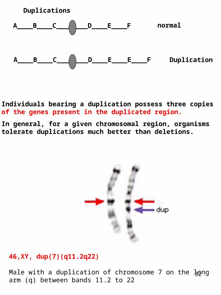
Duplications
Individuals bearing a duplication possess three copies of the genes present in the duplicated region.
In general, for a given chromosomal region, organisms tolerate duplications much better than deletions.
46,XY, dup(7)(q11.2q22)
Male with a duplication of chromosome 7 on the long arm (q) between bands 11.2 to 22
A____B____C________D____E____F
A____B____C________D____E____E____F
normal
Duplication
33
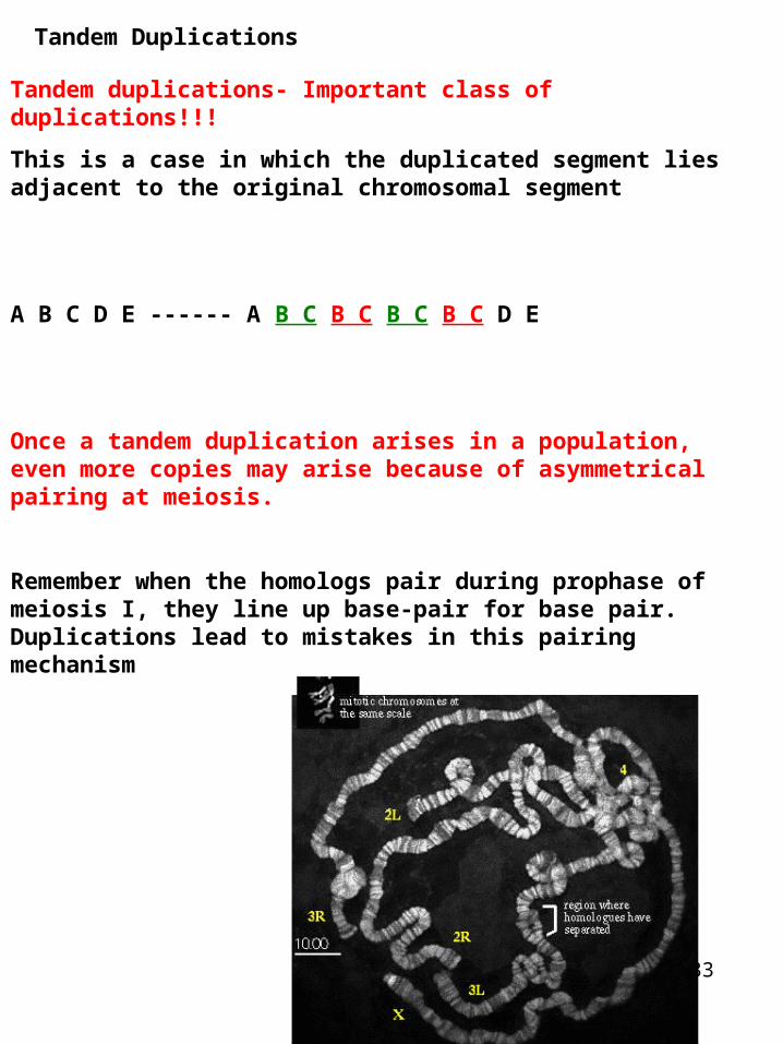
Tandem Duplications
Tandem duplications- Important class of duplications!!!
This is a case in which the duplicated segment lies adjacent to the original chromosomal segment
A B C D E ------ A B C B C B C B C D E
Once a tandem duplication arises in a population, even more copies may arise because of asymmetrical pairing at meiosis.
Remember when the homologs pair during prophase of meiosis I, they line up base-pair for base pair. Duplications lead to mistakes in this pairing mechanism
34
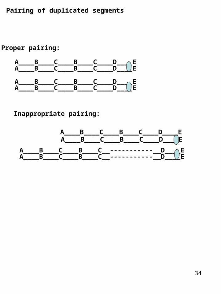
Pairing of duplicated segments
Proper pairing:
A____B____C____B____C____D____E
Inappropriate pairing:
A____B____C____B____C____D____E
A____B____C____B____C____D____EA____B____C____B____C____D____E
A____B____C____B____C____D____E
A____B____C____B____C__-----------__D____EA____B____C____B____C__-----------__D____E
A____B____C____B____C____D____E
35
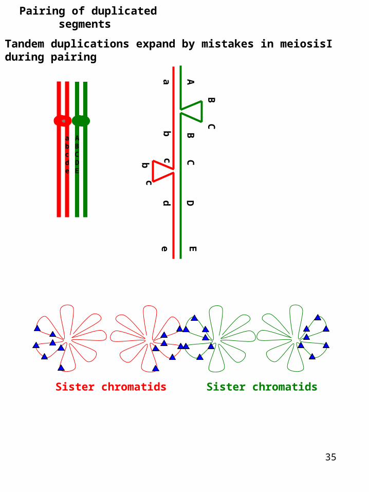
Pairing of duplicated segments
Tandem duplications expand by mistakes in meiosisI during pairing
A
BC
BC
DE
ab
cbc
de
Sister chromatids Sister chromatids
abcd
ABCD
e E
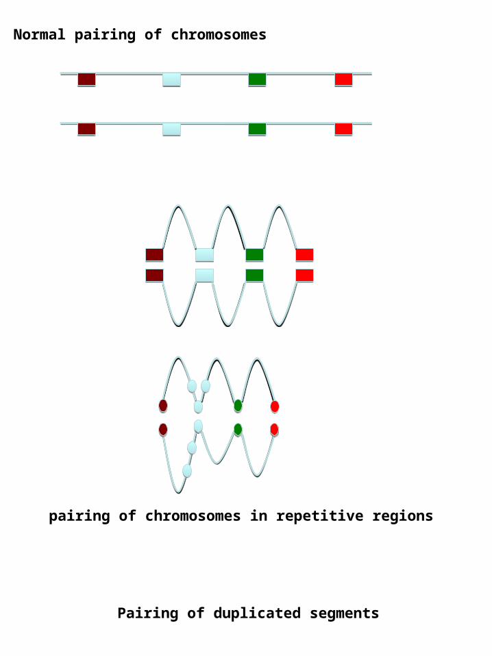
Normal pairing of chromosomes
pairing of chromosomes in repetitive regions
Pairing of duplicated segments
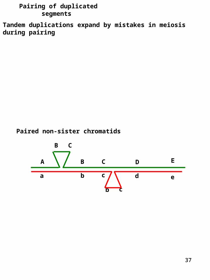
Pairing of duplicated segments
3737
Tandem duplications expand by mistakes in meiosis during pairing
A
B C
B C D E
a b c
b c
d e
Paired non-sister chromatids
38
Crossover in mispaired duplicated segments
A B C B C D
A B C B C D
a
b c bc
d
A B C B C D
A B C B C B C D
A B C D
A B C B C D
a
b cb
cd
What happens if you get a crossover after mis-pairing in meiosisI?
A
B C
B C D E
a b c
b c
d e
39
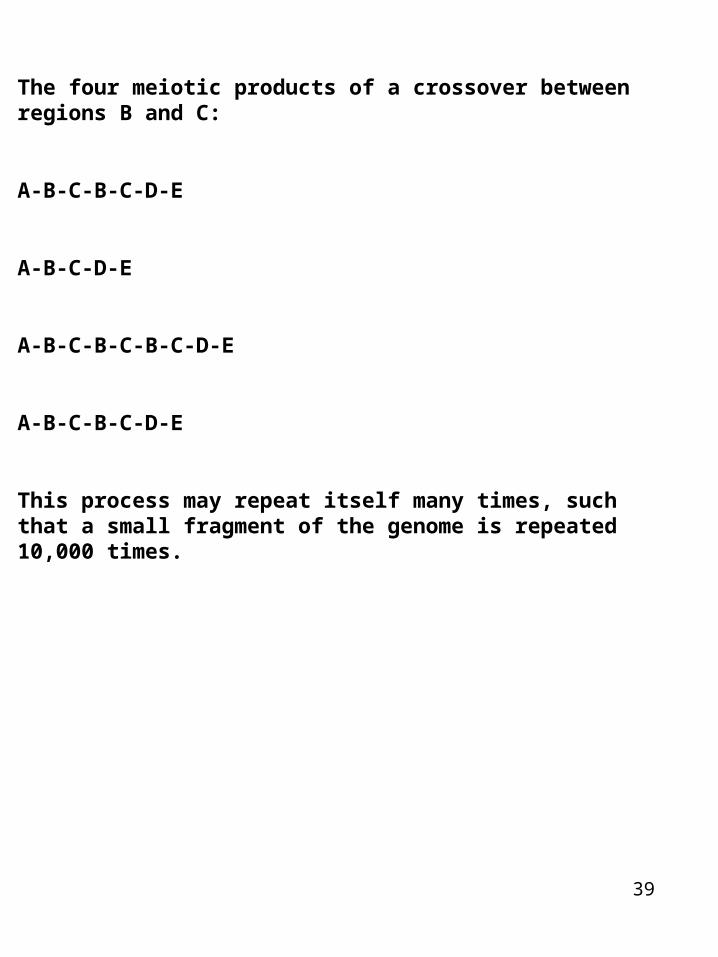
The four meiotic products of a crossover between regions B and C:
A-B-C-B-C-D-E
A-B-C-D-E
A-B-C-B-C-B-C-D-E
A-B-C-B-C-D-E
This process may repeat itself many times, such that a small fragment of the genome is repeated 10,000 times.
40
An example of this is near the centromeres of the Drosophila genome:
If you look at the DNA sequence in this region it consists of small 5-10 bp sequences (AATAC)n repeated 1,000s of times. It is believed to have arisen from unequal crossing over.
Repetitive DNA- cell does not like it- They try to reduce recombination of repetitive DNA by packaging the DNA with proteins to form heterochromatin- cold spots of recombination along the chromosome
41
Duplications provide additional genetic material capable of evolving new function. For example in the above situation if the duplication for the B and C genes becomes fixed in the population- the additional copies of B and C are free to evolve new or modified functions.
This is one explanation for the origin of the tandemly repeated globin genes in humans. Each of these has a unique developmental expression pattern and provides a specialized function.
The hemoglobin in fetus has a higher affinity for oxygen since it acquires its oxygen from maternal hemoglobin via competition

xxxxxx
42
43
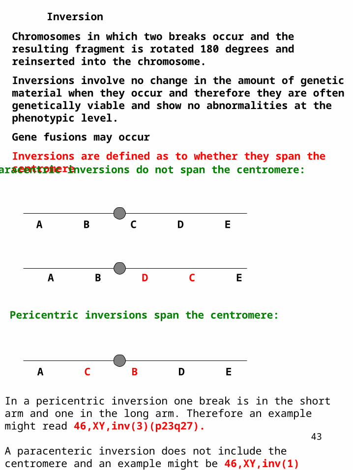
Inversion
Chromosomes in which two breaks occur and the resulting fragment is rotated 180 degrees and reinserted into the chromosome.
Inversions involve no change in the amount of genetic material when they occur and therefore they are often genetically viable and show no abnormalities at the phenotypic level.
Gene fusions may occur
Inversions are defined as to whether they span the centromere
Pericentric inversions span the centromere:
A B C D E
A B D C E
A C B D E
In a pericentric inversion one break is in the short arm and one in the long arm. Therefore an example might read 46,XY,inv(3)(p23q27).
A paracenteric inversion does not include the centromere and an example might be 46,XY,inv(1)(p12p31).
Paracentric inversions do not span the centromere:
44
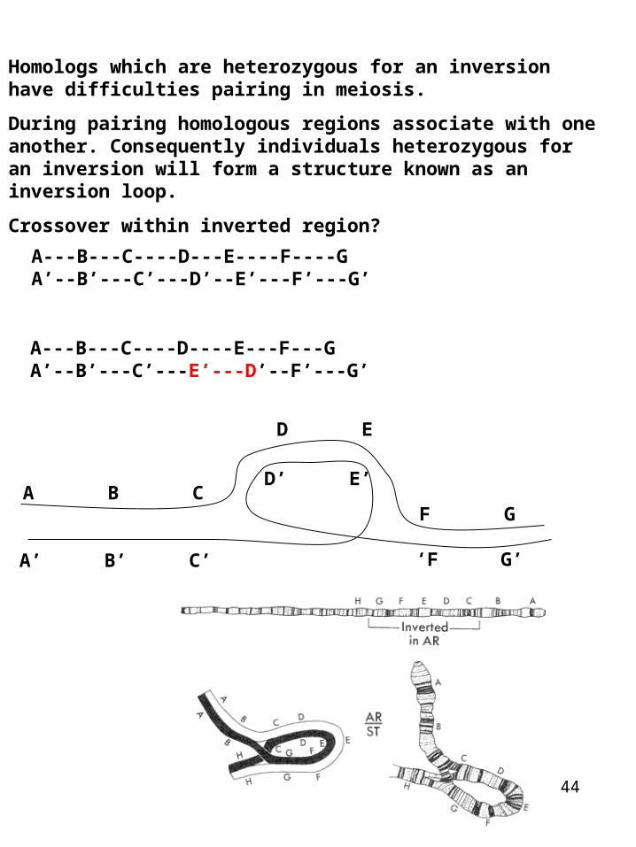
Homologs which are heterozygous for an inversion have difficulties pairing in meiosis.
During pairing homologous regions associate with one another. Consequently individuals heterozygous for an inversion will form a structure known as an inversion loop.
Crossover within inverted region?
A---B---C----D---E----F----GA ’--B’---C’---D’--E’---F’---G’
A B CF G
D E
A ’ B ’ C’ ‘F G ’
D ’ E ’
A---B---C----D----E---F---GA ’--B’---C’---E ’---D’--F’---G’
45
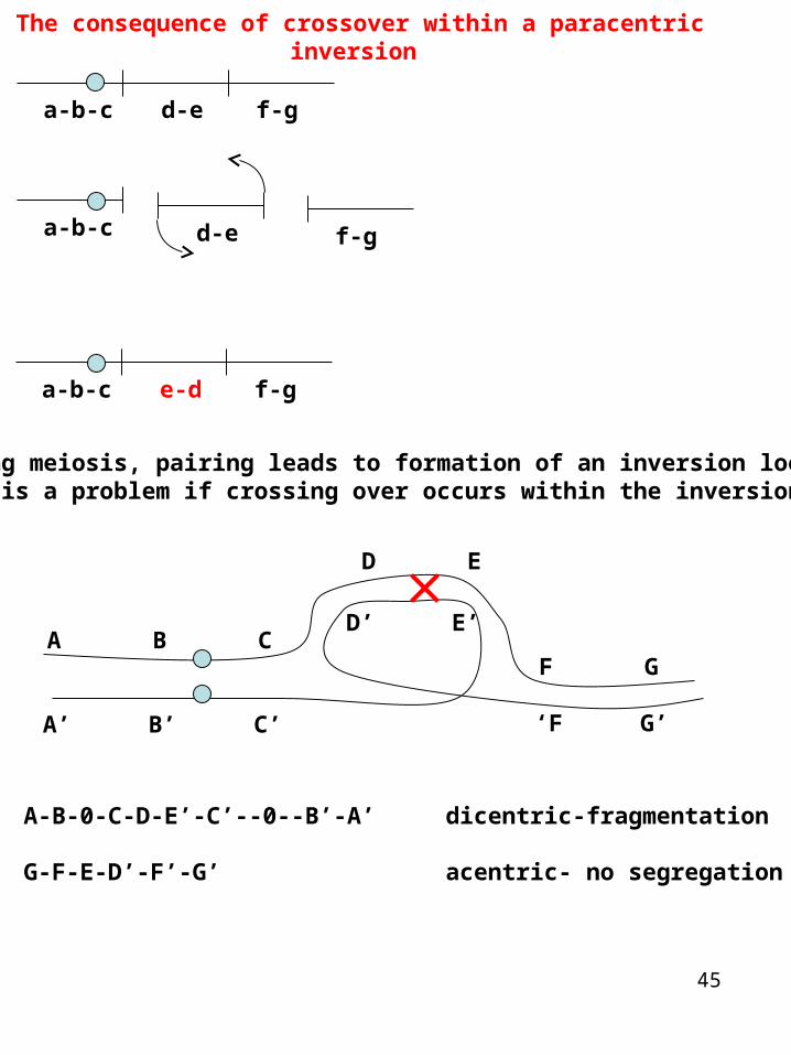
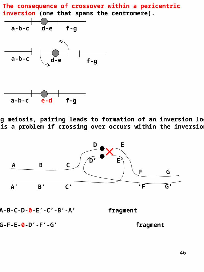
The consequence of crossover within a paracentric inversion
a-b-c d-e f-g
a-b-c e-d f-g
a-b-c f-gd-e
During meiosis, pairing leads to formation of an inversion loopThis is a problem if crossing over occurs within the inversion
A-B-0-C-D-E’-C’--0--B’-A’ dicentric-fragmentation
G-F-E-D’-F’-G’ acentric- no segregation
A B CF G
D E
A ’ B ’ C’ ‘F G ’
D ’ E ’
46
a-b-c f-g
During meiosis, pairing leads to formation of an inversion loopThis is a problem if crossing over occurs within the inversion
A-B-C-D-0-E’-C’-B’-A’ fragment
G-F-E-0-D’-F’-G’ fragment
The consequence of crossover within a pericentric inversion (one that spans the centromere).
a-b-c d-e f-g
d-e
a-b-c e-d f-g
A B CF G
D E
A ’ B ’ C’ ‘F G ’
D ’ E ’

47
Inversions •Paracentric inversion crosses over with a normal chromosome, the resulting chromosomes are an acentric, with no centromeres, and a dicentric, with 2 centromeres. •The acentric chromosome isn't attached to the spindle, so it gets lost during cell division, and the dicentric is usually pulled apart (broken) by the spindle pulling the two centromeres in opposite directions. These conditions are lethal.
•Pericentric inversion crosses over with a normal chromosome, the resulting chromosomes are duplicated for some genes and deleted for other genes. (They do have 1 centromere apiece though). •The gametes resulting from these do not produce viable progeny.• •Thus, either kind of inversion has lethal results when it crosses over with a normal chromosome. •The only offspring that survive are those that didn't have a crossover or crossed over in regions outside the inversion. •Thus when you count the offspring you only see the non-crossovers, so it appears that crossing over has been suppressed.
48
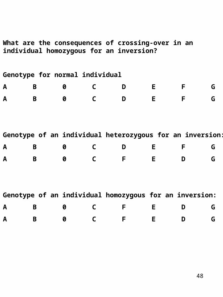
What are the consequences of crossing-over in an individual homozygous for an inversion?
Genotype for normal individual
A B 0 C D E F G
A B 0 C D E F G
Genotype of an individual heterozygous for an inversion:
A B 0 C D E F G
A B 0 C F E D G
Genotype of an individual homozygous for an inversion:
A B 0 C F E D G
A B 0 C F E D G

49
Translocations
A segment from one chromosome is exchanged with a segment from another chromosome.
Chromosome 1
A B C D E F----------------------0---------------------------------------------0-----------------------A B C D E F
Chromosome 2
O P Q R S T----------------------0---------------------------------------------0-----------------------O P Q R S T
Reciprocal translocation
A B C D S T----------------------0-----------------------
O P Q R E F----------------------0-----------------------
This is more specifically called a reciprocal translocation and like inversions (and unlike duplications and deficiencies) no genetic material is gained or lost in a reciprocal translocation.Non-reciprocal translocations may also occur
50
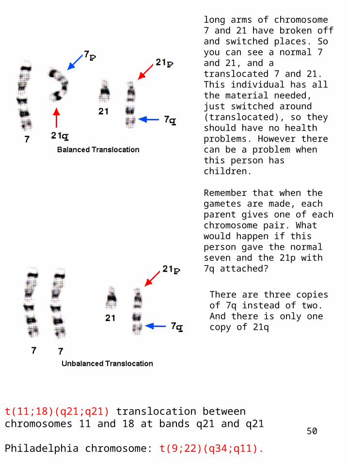
long arms of chromosome 7 and 21 have broken off and switched places. So you can see a normal 7 and 21, and a translocated 7 and 21.This individual has all the material needed, just switched around (translocated), so they should have no health problems. However there can be a problem when this person has children.
Remember that when the gametes are made, each parent gives one of each chromosome pair. What would happen if this person gave the normal seven and the 21p with 7q attached?
There are three copies of 7q instead of two. And there is only one copy of 21q
t(11;18)(q21;q21) translocation between chromosomes 11 and 18 at bands q21 and q21
Philadelphia chromosome: t(9;22)(q34;q11).
51
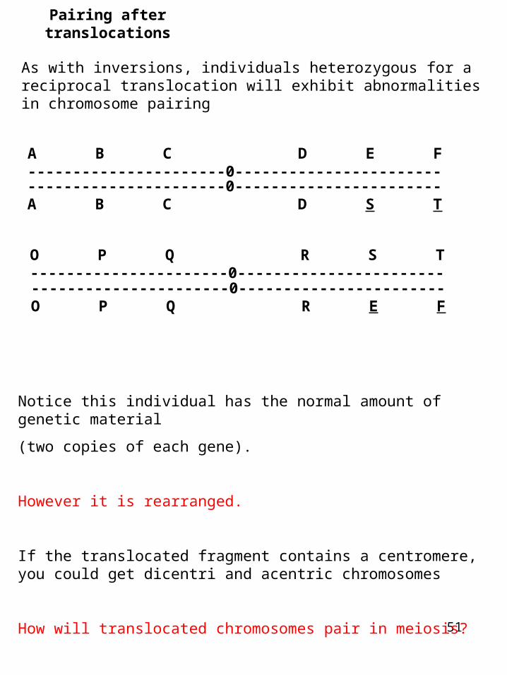
Pairing after translocations
As with inversions, individuals heterozygous for a reciprocal translocation will exhibit abnormalities in chromosome pairing
Notice this individual has the normal amount of genetic material
(two copies of each gene).
However it is rearranged.
If the translocated fragment contains a centromere, you could get dicentri and acentric chromosomes
How will translocated chromosomes pair in meiosis?
A B C D E F----------------------0-----------------------
O P Q R S T----------------------0-----------------------
----------------------0-----------------------A B C D S T
----------------------0-----------------------O P Q R E F
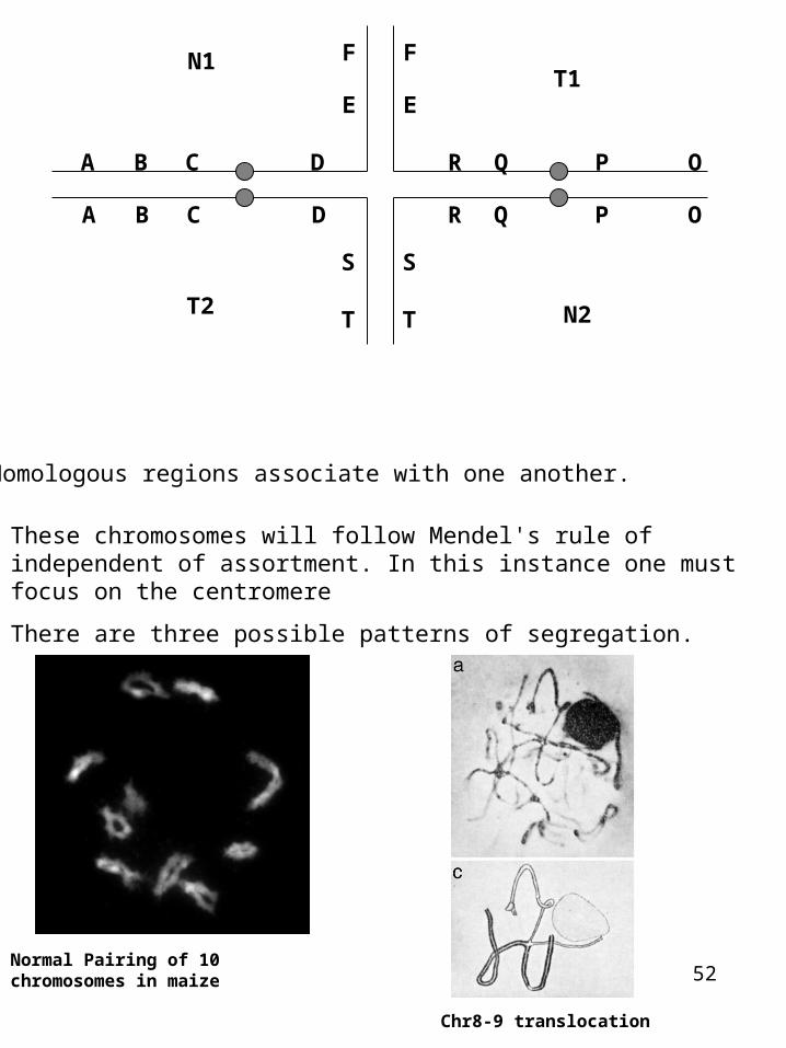
52
These chromosomes will follow Mendel's rule of independent of assortment. In this instance one must focus on the centromere
There are three possible patterns of segregation.
A B C D
E
F
A B C D
S
T
S
T
R Q P O
N1
E
F
R Q P O
T1
T2 N2
Homologous regions associate with one another.
Normal Pairing of 10 chromosomes in maize
Chr8-9 translocation
53
Segregation after translocations
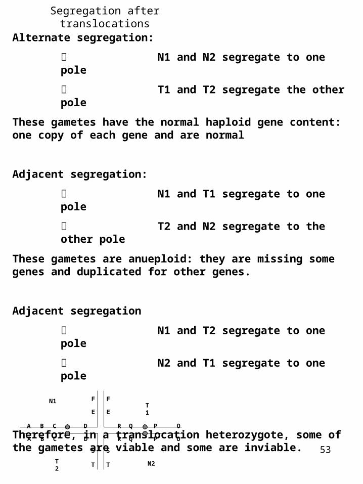
Alternate segregation:
キ N1 and N2 segregate to one pole
キ T1 and T2 segregate the other pole
These gametes have the normal haploid gene content: one copy of each gene and are normal
Adjacent segregation:
キ N1 and T1 segregate to one pole
キ T2 and N2 segregate to the other pole
These gametes are anueploid: they are missing some genes and duplicated for other genes.
Adjacent segregation
キ N1 and T2 segregate to one pole
キ N2 and T1 segregate to one pole
Therefore, in a translocation heterozygote, some of the gametes are viable and some are inviable.
A B C D
E
F
A B C D
S
T
S
T
R Q P O
N1
E
F
R Q P O
T1
T2
N2
54
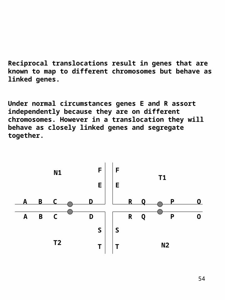
Reciprocal translocations result in genes that are known to map to different chromosomes but behave as linked genes.
Under normal circumstances genes E and R assort independently because they are on different chromosomes. However in a translocation they will behave as closely linked genes and segregate together.
A B C D
E
F
A B C D
S
T
S
T
R Q P O
N1
E
F
R Q P O
T1
T2 N2
55
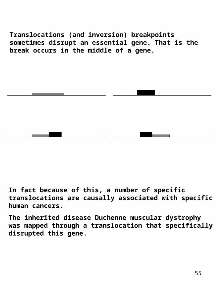
Translocations (and inversion) breakpoints sometimes disrupt an essential gene. That is the break occurs in the middle of a gene.
In fact because of this, a number of specific translocations are causally associated with specific human cancers.
The inherited disease Duchenne muscular dystrophy was mapped through a translocation that specifically disrupted this gene.

56
abl/bcr Fusion protein Chronic myelogenous and acute lymphotic leukemia
ALK/NPM Fusion Large cell lymphomas
HER2/neu Fusion Breast and cervical carcinomas
MYH11/CBFB Fusion Acute myeloid leukemia
ML/RAR Fusion Acute premyelocytic leukemia
ERG/TMPRSS2 Fusion prostate cancer
Gene fusion -prostate cancer -ERG merges with a prostate-specific gene called TMPRSS2. ERG is a transcription factors
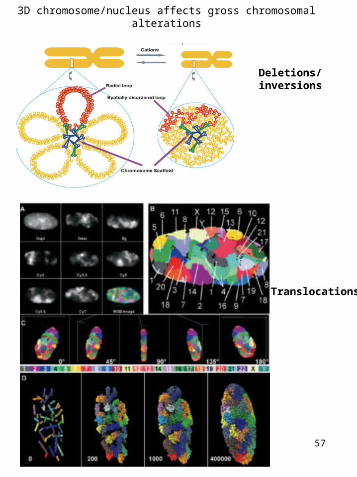
3D chromosome/nucleus affects gross chromosomal alterations
57
Deletions/inversions
Translocations
Xxxx
58


![PRIMARY RESEARCH Open Access Silencing SATB1 … [7-12]. RNA-interference ... 50- AGC TTT TCC AAA AAG GAT TTG GAA GAG AGT GTC TCT CTT GAA GAC ACT CTC TTC CAAATCCGGG-30; ... (HE…](https://img.dokumen.tips/doc/110x75/5aa8ab217f8b9a81188bd02d/primary-research-open-access-silencing-satb1-7-12-rna-interference-50-.jpg)