Embed Size (px)
Citation preview

MQP-BIO-DSA-1061
MUTATION SCREENING OF CANDIDATE BREAST CANCER
SUSCEPTIBILITY GENES IN NON-BRCA1/2 FAMILIES
A Major Qualifying Project Report
Submitted to the Faculty of the
WORCESTER POLYTECHNIC INSTITUTE
in partial fulfillment of the requirements for the
Degree of Bachelor of Science
in
Biology and Biotechnology
by
_________________________
Cara Schafer
January 14, 2009
APPROVED:
_________________________ _________________________
Csilla Szabo, Ph.D. David Adams, Ph.D.
Mayo Graduate School College of Medicine Biology and Biotechnology
Dept of Biochemistry and Molecular Biology WPI Project Advisor
Project Advisor

2
ABSTRACT
Although mutations in BRCA1 and 2 genes have previously been linked to breast
cancer, mutations in genes encoding other proteins in the BRCA DNA repair pathway
could also lead to this disease. This MQP used a candidate gene screening approach to
identify potential genetic changes in proteins previously shown to interact with BRCA1
and/or BRCA2 in repair pathways. PCR amplicons were analyzed by high resolution
melting analysis (HRMA) as a preliminary screen for mutations in six candidate genes
(Mre11, Rad50, MCPH1, NBS1, DSS1, and BCCIP) amplified from non-
BRCA1/BRCA2 breast cancer patient samples from BRCA-independent high-risk
families. Mutations in MCPH1 were further analyzed by DNA sequencing, which
showed frameshift/nonsense mutations, missense mutations, silent substitutions, and
intronic variants in 29 patients, 10 of which contained more than one mutation.
Mutations in exon-2 of DSS1 include protein truncating and missense mutations in highly
conserved domains.

3
TABLE OF CONTENTS
Signature Page 1
Abstract 2
Table of Contents 3
Acknowledgements 4
Background 5
Project Purpose 26
Methods 27
Results 34
Discussion 57
Bibliography 59
Appendix…………………………………………………………………………… 66

4
ACKNOWLEDGEMENTS
I would like to thank the following individuals for their kind assistance and support:
• Dr. Csilla Szabo, my advisor and mentor, for allowing me to work with her. I
wish to express my gratitude for her inspiration and guidance, and for her
enthusiasm and encouragement throughout the MQP.
• Dr. Fergus Couch for his support of this project.
• Bruce Eckloff and the Mayo DNA Sequencing Facility for providing the
sequencing data of the variants.
• Chris Hilker and the Genotyping Core for instruction and use of Pre-PCR
robotics.
• Dr. David Adams for his assistance initiating the project, for his constructive
comments and suggestions, and for his help editing the final MQP report.

5
BACKGROUND
Breast Cancer Description
Breast cancer is a malignancy that develops in tissues of the breast, usually in the
ducts and lobules. It occurs in a greater percentage in females, and rarely in males
(National Cancer Institute, 2007). Some of the first physically identifiable symptoms of
breast cancer include a lump or swelling, skin dimpling, nipple pain, discharge, or
retraction, redness, or scaliness of the nipple or breast skin (American Association for
Cancer Research, 2007).
Mammography is an effective and widely used diagnostic tool to detect changes
in breast tissue that may indicate cancer. Other diagnostic methods used are digital
mammography, magnetic resonance imaging, positron emission tomography, Sestamibi
scintimammography, and ductal lavage (National Cancer Institute, 2007). Family history
of breast cancer is the greatest predictor of risk for developing the disease. Other factors
include age, previous history of breast cancer, history of chest radiation therapy before
age 30, and existence of having dense breast tissue, particularly in older individuals.
Women with a history of early onset of menses or late age menopause, women who have
never had children or who have children after age 30, women who are obese after
menopause, or who use menopausal hormone therapy with estrogen plus progestin are
also considered to have increased risk for breast cancer (American Association for
Cancer Research, 2007; National Cancer Institute, 2007).

6
Breast Cancer Prevalence
Breast cancer is the second leading cause of cancer deaths in American women
(Friedenson, 2005). In 1997, breast cancer claimed the lives of approximately 44,910
Americans, with 43,900 women and 290 men losing their lives to the disease.
Approximately two million women living in the U.S. have been diagnosed with breast
cancer. The National Cancer Institute (2007) predicts that in the United States in 2007,
there will be 178,480 newly diagnosed cases of breast cancer in females, and 2,030 new
cases in males. Over 40, 000 people will die from breast cancer in 2007 (40,460 females
and 450 males) (National Cancer Institute, 2007). Mortality rates have, however,
decreased an estimated two percent per year in the past decade, with greater decreases
seen among young women (American Association for Cancer Research, 2007), possibly
due to mammography screening, early diagnosis, and improved management (adjuvant
tamoxifen therapy) and treatment of women with breast cancer (Hermon and Beral,
1996).
Risk Reduction Options in Familial Breast Cancer
Familial breast cancer is characterized by early onset diagnosis, an increased risk
of bilateral breast cancer, an increasing risk with increasing numbers of affected family
members, and increased risk for ovarian cancer. Women who have BRCA1 and BRCA2
gene mutations face this increased risk for breast and ovarian cancer. BRCA1 and
BRCA2 account for almost 80% of hereditary breast cancer, and 5 to 6% of all breast
cancers (Greene, 1997). At least eight candidate breast cancer susceptibility genes have
currently been identified (Greene, 1997) (discussed later). In a study conducted by the

7
Breast Cancer Linkage Consortium, 52% of families with breast cancer demonstrated
linkage to BRCA1, while 32% were linked to BRCA2, suggesting that 16% of breast
cancer families were linked to other predisposing genes (Ford et al., 1998). BRCA1 was
linked to 81% of breast-ovarian cancer families, while 14% were linked to BRCA2.
Linkage to BRCA2 was identified in76% of families with male and female breast cancer
(Ford et al., 1998). The cumulative risk of breast cancer was 27% by age 50 years, which
increased to 84% by age 70 years. Ovarian cancer risks were much smaller, at 0.4% until
age 50 years, but rose to 27% by age 70 years (Ford et al., 1998).
Some risk-reduction options are made available, as part of genetic counseling for
women who are BRCA1 or BRCA2 mutation carriers. Options offered include increased
surveillance, chemoprevention with tamoxifen, prophylactic oophorectomy (removal of
the ovaries), and prophylactic mastectomy (Uyei et al., 2006).
Uyei et al. (2006) reported a retrospective analysis of 554 women with BRCA1 and
BRCA2 gene mutations who were treated at The University of Texas M. D. Anderson
Cancer Center. Results obtained for data collected between 2000 and 2006 demonstrated
that women who had BRCA mutations, along with a history of breast cancer or ductal
carcinoma in situ, or a history of having had previous breast biopsies, were more likely to
select prophylactic surgery. Women with a family history of ovarian cancer opted for
prophylactic oophorectomy, while an individual’s personal history of ovarian cancer or
advanced breast cancer was more likely associated with a choice for surveillance only.
Breast cancer survivors with a history of treatment with total mastectomy chose
prophylactic mastectomy more often than breast cancer survivors with a history of
treatment with breast-conserving surgery or women with no history of breast cancer

8
(Uyei et al., 2006). Hartmann et al. (2001) reported that bilateral prophylactic
mastectomy decreased the risk of breast cancer in women with BRCA1 and BRCA2
mutations by approximately 90%.
Researchers at Lombardi Comprehensive Cancer Center in Washington, DC
reported that BRCA1 and BRCA2 carriers are recommended to undergo prophylactic
bilateral salpingo-oophorectomy (removal of an ovary with a fallopian tube) by age 35-40
years or when childbearing is complete, in an effort to significantly reduce the risk of
ovarian cancer (Nusbaum and Isaacs, 2007). This prophylactic surgery has been shown to
also reduce the risk of breast cancer when performed in premenopausal mutation carriers.
Finch et al. (2006) studied the incidence of ovarian, fallopian tube, and primary
peritoneal cancer in a large cohort of women with BRCA1 or BRCA2 mutations.
Prophylactic oophorectomy reduced the risk of ovarian
and fallopian tube cancer in the
BRCA1 and BRCA2 carriers by approximately 80%, although there was a post-
oophorectomy residual risk of approximately 4% to develop peritoneal cancer (Finch et
al. 2006). BRCA1 and BRCA2 carriers are also offered the option of increased
surveillance, with or without chemoprevention, or prophylactic surgery as part of a breast
cancer management protocol. Bilateral prophylactic oophorectomy is more commonly
chosen than bilateral prophylactic mastectomy in BRCA1/2 mutation carriers who are
unaffected (Freibel et al., 2007), as many women feel that bilateral prophylactic
mastectomy is too aggressive with increase risk for side effects (Uyei et al., 2006).
BRCA carrier status, to date, is not used as an independent factor to determine
prognosis for systemic treatment options (Nusbaum and Isaacs, 2007). Recently,
researchers have investigated the use PARP-1 [poly(ADP-ribose) polymerase-1]

9
inhibitors as a chemotherapeutic treatment for BRCA1/2 cancers. DeSoto and Deng
(2006) suggested that BRCA breast cancer cells were resistant to PARP-1 inhibitors
when used alone in the treatment. PARP-1 inhibitors did offer promise in the prevention
of BRCA related breast cancers, and may be successful when used in combination with
other chemotherapeutic agents in the treatment of BRCA related breast cancer (DeSoto
and Deng, 2006).
Genetic Causes of Breast Cancer
BRCA1 and BRCA2
Approximately 5-10% of all breast cancer results from the inheritance of highly
penetrant mutations in two susceptibility genes, BRCA1 (OMIM, 113705; GenBank,
U14680.1; Hall et al., 1990) and BRCA2 (OMIM, 600185; GenBank, U43746.1;
Wooster et al., 1994) which is consistent with an autosomal dominant transmission (Ford
et al., 1998; Pohlreich et al., 2005). These genes were first identified in 1994 (Miki et al.,
1994; Wooster et al., 1994; Breast and Ovarian Cancer, 2007), and are associated with
both breast and ovarian cancers (Troudi et al., 2007). Mutations in these two genes
account for 60% of all known mutation site-specific female breast cancers (Ford et al.,
1998). BRCA1 is found on the long arm of chromosome 17, mapped specifically to
chromosome 17q21 (Hall et al., 1990). The BRCA1 gene contains 24 exons, and
encodes a protein of approximately 220 kDa (1863 amino acids) (Cipollini et al., 2004).
BRCA2 is located on the long arm of chromosome 13. BRCA2 is also a large gene,
containing 27 exons that encode a protein of 380 kDa (3418 amino acids). Both BRCA1
and BRCA2 have an unusually large exon 11. The translational start site for both genes is

10
in exon 2. Both proteins are predominately nuclear, where phosphorylated versions of
both proteins are also located (Cipollini et al., 2004). While similar in some respects,
many differences exist between the two genes, which are not homologous. BRCA1 has
two protein motifs, while BRCA2 has BRC repeats and no relation to BRCA1 (Cipollini
et al., 2004) and is not highly conserved evolutionarily (Szabo et al., 1996).
BRCA1 and BRCA2 function as tumor suppressors, and are critical to the cellular
control of homologous recombination and double-strand break repair when DNAs are
damaged (Liu and West, 2002; Ford et al., 1994; Friedenson, 2005). Individuals with
mutations in these genes possess an increased lifetime risk for developing breast or
ovarian cancer. The cumulative risk of for developing breast cancer is approximately
28% by age 50 years, and 84% by age 70 years, with ovarian cancer risks determined to
be 0.4% by age 50 years and 27% by age 70 years (Ford et al., 1994). The lifetime risk
for developing breast cancer is similar in both BRCA1 and BRCA2 carriers, with a
possible lower risk in BRCA2 carriers <50 years of age (Ford et al., 1994).
Both BRCA1 and BRCA2, when functioning normally, play an active role in the
restoration of double-stranded breaks in DNA caused by radiation, which can occur
through exposure to DNA damaging agents(ionizing radiation) or through errors in
normal cellular replication (such as DNA synthesis, chromosomal segregation, metabolic
generation of oxygen radicals). Inactivating mutations occurring in BRCA1 or BRCA2
hinder the repair of DNA damage through homologous recombination. The accumulation
of mutations due to impaired DNA repair promotes the growth of cancer (Breast and
ovarian cancer, 2007). Researchers have reported that BRCA1 mutations also confer
modest risks for uterine, cervical, early-onset prostate and pancreatic cancers. BRCA2

11
mutations show a similar increased risk for prostatic, pancreatic, gallbladder, bile duct,
stomach cancers and melanoma (Easton et al., 1995; Friedenson, 2005).
Approximately 2000 distinct sequence variants in BRCA1 and BRCA2 have
already been recorded (Breast Cancer Mutation Database, 2007). Both BRCA1 and
BRCA2 genes have variants that are uniformly distributed along the entire coding
regions. Mutations have also been identified in intronic sequences flanking each exon in
both genes, some of which lead to altered splicing.
Breast cancer develops in a multistep process and is influenced by two types of
genes, oncogenes and suppressor genes (Osborne et al., 2004). Oncogenes refer to genes
that, when activated, can contribute to the development of cancer. Oncogenes produce
alterations that cause gain-of-function effects (Osborne et al., 2004). Tumor suppressor
genes function in slowing down cell growth, DNA repair, and apoptosis (American
Cancer Society, 2005). These genes refer to group of genes whose loss of function
promotes malignancy. Germline mutations in breast cancer occur in tumor suppressor
genes. Tumor suppressor genes can also contain sporadic acquired somatic mutations.
The tumor typically contains a mutation in one allele and a deletion of the remaining
allele. In 1971, Alfred Knudson proposed this "two-hit" hypothesis (in reference to
retinoblastoma) which stated that both gene alleles must be missing to unmask the
malignant phenotype. The activation of an oncogene and the mutation of a tumor
suppressor gene can produce changes that contribute to the
malignancy. The effects of
these alterations are complex due to the high number of changes in a typical case of
breast cancer and the interactions of the biological pathways involved (Osborne et al.,
2004).

12
Cancer risks increase from missing BRCA1 or BRCA2 protein sequences or non-
functional proteins, most likely caused by frameshift, nonsense, and splice site mutations
(Cipollini et al. 2004). Figure 1 shows how BRCA1and BRCA2 act as tumor suppressor
genes (deduced from an analysis of tumor specimen DNAs) (copied from Cipollini et al.
2004). An initial mutation in one allele (white circle in the figure) leads to a diminished
capacity for DNA repair. Eventually, the second non-mutated allele is entirely deleted
(figure right side).
Figure 1. Proposed Mechanism for Loss of Allele Function for BRCA1 and BRCA2 Tumor
Suppressor Genes. Initially one allele suffers a germline susceptibility point mutation or small deletion
(white circle), which leads to a diminished capacity for repairing DNA. Eventually the second unmutated
allele is deleted entirely, as found in tumor specimens (Copied from Cipollini et al. 2004).
The Role of Founder Mutations
The proportional contribution of BRCA1 and BRCA2 mutations has been shown
to differ in different populations around the world (Szabo and King, 1997). Vogel et al.
(2007) described eight different BRCA mutations and three variants within a small
sample in a Hispanic population. One of three mutations in the BRCA1 and BRCA2
genes was found in 2.0%-2.5% of Ashkenazi Jewish women (Struewing et al., 1997).

13
Studies have suggested that each founding mutation put these women at a high risk of
invasive breast cancer, which continues throughout life (Struewing et al., 1997; Warner
et al., 1999). In addition, an estimated 12% of the total number of breast cancers in the
Ashkenazi Jewish population is caused by mutations in the BRCA1 or BRCA2 gene
(Warner et al., 1999). Peto et al. (1999) reported that the mutation BRCA1-185delAG
has been identified in 20% of Ashkenazi Jewish women with early onset breast cancer,
while the mutation BRCA2- 6174delT is found in 8% of the Ashkenazi cases diagnosed
in women over 42 years of age. The authors speculated that, among Ashkenazi Jews,
BRCA1 mutations play a significant role in the risk for early-onset breast cancer, while
BRCA2 mutations affect the later onset of the disease. Ganguly et al. (1997) found that
that lower prevalence of mutations in both BRCA1 and BRCA2 genes was observed from
data collected from clinical families. Higher prevalence was found in linkage data
obtained from high-risk families collected in a research setting (Ganguly et al. 1997).
In Britain, BRCA1 mutation carriers were found in 3.1% of breast cancer
patients, and BRCA2 mutations in 3.0% of breast cancer patients under the age of 50
(Peto et al., 1999). For patients 50 years of age or older, the prevalence was 0.49% and
0.84%, respectively. Similarly, researchers in Australia found that an estimated 3.8% of
women before age 40 in that country carried a germline mutation in BRCA1 (Southey et
al., 1999). They reported seven rare BRCA variants, but argued that these did not have a
significant effect on the risk of breast cancer in the population studied.
In a recent study of 204 breast cancer patients in northern India, researchers found
a lower proportion of BRCA1 and BRCA2 mutations than seen in other populations,
although the proportion was still elevated for breast cancer patients versus the general

14
population (Saxena et al., 2006). Interestingly, 9 distinct BRCA1 and 9 distinct BRCA2
sequence variants were identified. 4 of the 9 BRCA1 mutations were unique to the
Indian population, accounting for 44% of the BRCA1 mutations found. Of the 9 BRCA2
mutations, 7 mutations (78%) were unique to the Indian population, accounting for 78%
of the BRCA2 mutations. In this group of patients studied from Northern India, these
unique mutations were distributed throughout the BRCA1 and BRCA2 gene exons
(Saxena et al., 2006).
Szabo and King (1997) analyzed the results of previous studies conducted by
researchers in seventeen countries, including Italy, Finland, Norway, Iceland, Israel,
Russia, Japan, Canada, Britain, and the United States. They explained that great
variability was noted among populations for proportions of high-risk families with
BRCA1 mutations. Russia displayed the largest proportion of BRCA1 mutations,
occurring in 79% of families with breast or ovarian cancer. Affected families had one of
two common alleles. The most common allele in Russia was BRCA1-5382insC, which is
also the most common allele found among the Europeans studied. The second most
common allele in Russia was BRCA1-4153delA (Szabo and King, 1997). This allele has
also been identified in affected families in Latvia (Csokay et al., 1999), Poland (Gorski et
al., 2000; Gorski et al., 2004), and Lithuania (Gronwald et al., 2005). Israel
demonstrated the next highest proportion of BRCA1 mutations in inherited breast and
ovarian cancer, occurring in 47% of high-risk families (Szabo and King, 1997). BRCA1
mutations were observed in 29% of Italian high-risk families, and in 20-25% of high-risk
families in Britain, France, Scandinavia, and Hungary. Less than 20% of high-risk
families in Holland, Belgium, Germany, Norway, and Japan had BRCA1 mutations.

15
Significantly fewer cases of BRCA2 mutations were noted for all countries except
Iceland, where a single BRCA2 mutation, specifically the 999del5 mutation, was
responsible for all the inherited breast and ovarian cancer in that country.
BRCA2 mutations are also more common than BRCA1 mutations in familial
male breast cancer, occurring in about 19% of familial male breast cancer in the United
States (Szabo and King, 1999). Overall, the authors proposed that “BRCA1 and BRCA2
have each undergone multiple mutations; the resultant alleles have migrated with the
peoples in which they occur; and disease-associated mutations have persisted, no doubt
because of late onset of disease and, hence, little or no deleterious impact of mutant
alleles on genetic fitness” (Szabo and King, 1997).
Non-BRCA Genes Associated with Breast Cancer
Recent studies suggest that the proportion of familial breast cancer cases due to
the BRCA1 and BRCA2 mutations may be smaller than initially believed (Kainu, et al.,
2000). Mutations in BRCA1 and BRCA2 account for only about 60% of mutation site-
specific female breast cancers (Ford et al., 1998), so additional susceptibility genes likely
exist (Walsh and King, 2007). But to date, gene identification efforts using linkage
analysis have not been successful at identifying non-BRCA genes, likely because that
approach identifies individual genes, each of which confers only a moderate risk
(Antoniou and Easton, 2006).
Kainu et al. (2000) used mathematical models to look for early somatic genetic
deletions in tumor tissues, and then applied targeted linkage analysis. The authors used
comparative genomic hybridization to investigate 61 breast tumors from
37 breast cancer

16
families, none of whom had BRCA1 or BRCA2 mutations. Mathematical models
predicted a loss of chromosome arm 13q as one of the first genetic events in these
familial cancers. This was demonstrated in a study of a Swedish family with five breast
cancer cases, where all patients evidenced clear 13q deletions at 13q21-q22 (Kainu, et al.,
2000). A subsequent study by Thompson et al., (2002) found no linkage to a
susceptibility locus at chromosome 13q21 and concluded that, if it did exist, its
contribution would be minimal in breast cancer families of European origin.
There exist multiple biologic functions for BRCA1 and BRCA2 proteins,
including “participating within a pathway that mediates error-free repair of DNA double
stranded breaks by homologous recombination (Friedenson, 2005).” BRCA1 and
BRCA2 gene products are placed within a biochemical sequence, which includes the
MRE11, Rad50 and NBS1 complex (MRN complex), ATM, CHEK2, BRCA1, BRCA2,
and Fanconi anemia proteins, often referred to as the BRCA pathway (Figure-2,
Friedenson, 2007). A breakdown in the critical protein function anywhere within this
DNA repair BRCA pathway may introduce mutations by repair of double strand breaks
by lower fidelity, error prone methods. Because some cancers are mediated by these
errors, this results in an increased risk for development of those cancers (Friedenson,
2005). More recently, Friedenson (2007) proposed that inactivation of any component
within the BRCA pathway would increase the risks for not only breast and ovarian
cancers, but also for lymphomas and leukemias. Where BRCA pathway mutations do not
exist, the functional encoded proteins provide protection from both breast and ovarian
cancer.

17
Figure 2. Schematic Diagram of the
BRCA Pathway for DNA Repair. Note
that BRCA1 and BRCA2 are only a part of
this key DNA repair pathway, which if any
key protein is rendered non-functional by
mutation, DNA mutations subsequently
increase. Thus this model predicts non-
BRCA mutations should also correlate
with breast cancer. (Figure from
Friedenson, 2007)
Gene mutations cause inactivation of BRCA1, BRCA2, and other critical proteins
within this "BRCA pathway" that inactivate this error-free repair process (Friedenson
2007). Liu and West (2002) described the pathway, which illustrates how, even though
BRCA1 and BRCA2 proteins interact together, only a minority of the BRCA1 protein is
actually found in association with BRCA2 at a given time. Recent identification of more
proteins that associate with either BRCA1 or BRCA2 further emphasizes that BRCA1
and BRCA2 each participate in different protein complexes, and these each have distinct
functions in DNA double strand breakage repair (See Figure 3 and Table I below).
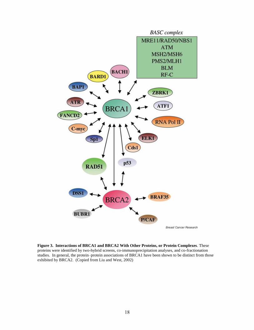
18
Figure 3. Interactions of BRCA1 and BRCA2 With Other Proteins, or Protein Complexes. These
proteins were identified by two-hybrid screens, co-immunoprecipitation analyses, and co-fractionation
studies. In general, the protein–protein associations of BRCA1 have been shown to be distinct from those
exhibited by BRCA2. (Copied from Liu and West, 2002)

19
Biological Functions BRCA-1-Interacting Proteins
DDR and repair MSH2, MSH6, MLH1, ATM, BLM and the RAD50-
MRE11-NBS1, DNA replication factor C, RAD51,
Fanconi anemia proteins, PCNA, H2AX, c-Abl, MDC1
Tumor suppressors ATM, ATR, p53, BRCA2, RB, BARD1, BACH1
Oncogenes c-Myc, casein kinase II, E2F1, E2F4, STK15, AKT
Transcription RNA polymerase II holoenzyme (RNA helicase A,
RPB2, RPB10α), CBP/p300, HDC and CtIP, estrogen
receptor α, androgen receptor, ZBRK1, ATF1, STAT1,
Smad3, BRCT-repeat inhibitor of hTERT expression
(BRIT1)
Cell cycle related Ayclin A, Cyclin D1, Cyclin D1, CDC2, Cdk2, Ckd4,
γ-tublin, p21, p27
Stress response, apoptosis MEKK3, IFI16, X-linked inhibitor of apoptosis protein
(XIAP)
Others BAP1, BIP1, BRAP2, importin α
Table 1. A list of BRCA1 Interacting Proteins. (Copied from Deng, 2006).
CHEK2 (OMIM 604373) (shown as purple in Figure-2, but not shown in Figure-3
or in Table-I) is a key checkpoint kinase of the BRCA pathway that serves as a tumor
suppressor in response to DNA double-strand breakage. The CHEK2*1100delC
mutation most likely accounts for familial risk of breast cancer in some non-BRCA1 and
non-BRCA2 patients, causing DNA damage and activation of cell-cycle checkpoints that
block cell proliferation and DNA repair. The impaired function of these checkpoints
results in instability in the genome and a subsequent increased risk for cancer (Weischer
et al., 2007). Investigators in Denmark reported the results of a 34 yearlong study of a
large sample in the Danish population (Weischer et al., 2007). The authors concluded
that CHEK2*1100delC heterozygosity was associated with a three-fold risk of breast
cancer in the Danish women studied. Conversely, researchers investigating a small
sample of hereditary breast and ovarian cancer families from the Slovak Republic did not

20
detect any 1100delC variant of the CHEK2 gene (Cierniková et al., 2005). Instead, the
investigators found a spectrum of eight mutations, one novel BRCA1 deletion, and one
recurrent BRCA2 mutation.
Other candidate genes have been found to act directly in double strand DNA
break repair. Walsh, et al. (2006) studied 300 US families with 4 or more cases of breast
or ovarian cancer who tested negative (wild type) for BRCA1 and BRCA2 mutations.
Patients were screened for genomic rearrangements in BRCA1 and BRCA2, and
germline mutations in CHEK2, TP53, and PTEN. Based on their findings, these
researchers estimated that, in a similar cancer population, one might expect that
approximately 12% would demonstrate a large genomic deletion or duplication in either
BRCA1 or BRCA2, and that 5% would carry a mutation in CHEK2 or TP53. Recently,
several mutations in genes in the BRCA-related pathways (Chek2, ATM, PALB2,
BRIP1) were shown to be associated with familial breast cancer (Walsh and King, 2007).
Besides BRCA1 and BRCA 2, other genetic syndromes are associated with
autosomal dominant inheritance of breast cancer risk. These include Li–Fraumeni
syndrome (a genetic disorder that causes breast cancer), as well as bone cancer
(osteosarcoma), muscle and soft tissue cancers, brain tumors, leukemias, and cancer of
the adrenal glands. These are caused by germline mutations in another key tumor
suppressor gene, p53, found in over 50% of families, with a reported penetrance of at
least 50% by age 50 years. Germline mutations in hCHK2 and TP53 genes have also
been associated with the Li-Fraumeni syndrome and related breast cancer (Cipollini et al.,
2004; Li-Fraumeni Syndrome, 2007). Cowden syndrome is caused by PTEN germline
mutations that promote an increased risk for not only developing breast cancer, but also

21
thyroid and endometrial cancer (lining of the uterus). In addition, patients with Cowden
syndrome are at risk for developing noncancerous breast and thyroid diseases, as well as
growths on the skin and mucous membranes called hamartomas (Cipollini et al., 2004;
Cowden Syndrome, 2007).
Approximately 1% of the general population may be heterozygote carriers of an
ATM gene mutation responsible for ataxia telangiectasia, a genetic autosomal recessive
disorder, with known risks for developing breast cancer (Cipollini et al., 2004). Another
identified autosomal recessive disorder, Peutz–Jeghers syndrome is characterized by
early onset of symptoms, which include hamartomatous polyps in the gastrointestinal
tract (Peutz-Jeghers Syndrome, 2007). Patients with Peutz–Jeghers syndrome face
lifetime risks for cancers of the gastrointestinal tract, pancreas, cervix, breast, and ovaries
(Cipollini et al., 2004; Peutz–Jeghers syndrome, 2007). Approximately half of the
patients with Peutz–Jeghers syndrome have mutations in STK11, which place them at
very high risk of developing breast cancer (Cipollini et al., 2004).
A recent study in Montreal found a high penetrance of PALB2 mutations in
probands (initial subjects tested) tested in 68 BRCA1/BRCA2-negative breast cancer
families of Ashkenazi Jewish, French Canadian, or mixed ethnic descent (Tischkowitz et
al., 2007). Seal et al. (2006) looked at truncating mutations in the Fanconi anemia J gene
BRIP1 in BRCA1/BRCA2 mutation-negative families. The authors reported that these
BRIP1 mutations could pose a risk as low-penetrance breast cancer susceptibility alleles.
However, other investigators were unable to substantiate an increase in risk of familial
breast cancer from BRIP1 variants (Lewis et al., 2005; Frank et al., 2007). Researchers
in Finland analyzed the Mre11 complex, composed of RAD50, NBS1 and MRE11 and

22
found that RAD50 and NBS1 haplo-insufficiency affected genomic integrity and
increased susceptibility to breast cancer (Heikkinen et al., 2006). The MRN complex is
comprised of MRE11, RAD50, and NBS1, which is the product of Nijmegan breakage
symdrome (Robert et al., 2006). Persons with Nijmegan breakage syndrome (NBS) are
susceptible to immunodeficiency and increased risk of malignancies (Tauchi et al., 2002).
The NBS1 gene product, nibrin, along with the rest of the MRN complex, is responsible
for detecting, signaling and repairing double strand breaks in DNA, and acts as a sensor
to recruit ATM to repair broken DNA molecules (Robert et al., 2006).
Li et al. (2006) recently investigated the protein DSS1 and described it as an
evolutionarily conserved acidic protein that binds to BRCA2. The authors explained that
DSS1 depletion causes hypersensitivity to DNA damage, similar to that seen with
BRCA2. They found that the presence of DSS1 was essential to the stability of the
BRCA2 protein in mammalian cells. Deletion, suppression, or mutation of DSS1 is
speculated to promote human breast and ovarian cancer, as well as sporadic and familiar
breast cancer where BRCA1 and BRCA2 mutations are not present (Li et al. 2006). Lu
et al. (2007) recently reported the results of their study, which showed that BCCIP
regulates homologous recombination and suppresses spontaneous DNA damage. They
proposed that BCCIP fragments that interact with BRCA2, or with the protein interacting
p21, inhibit DNA double-stranded break repair through homologous recombination.
When breaks in DNA structure occur, responses at the cellular level set off
numerous checkpoint and repair proteins. These responses synchronize a complex
signaling cascade that detects the DNA damage, with subsequent checkpoint activation,
DNA repair, cell cycle arrest and/or apoptosis (Chaplet et al. 2006). Lin et al. (2005)

23
studied the function of BRIT1 in DNA damage checkpoints. BRIT1 is identical to the
MCPH1 gene. Mutations in this gene are also found in patients with primary
microcephaly. BRIT1 is required for the expression of both BRCA1 and the checkpoint
kinase. Chk1 and phosphorylation of Nbs1 are dependant on BRIT1/MCPH1. The
authors speculated that since BRIT1/MCPH1 regulates Nbs1, BRCA1, and Chk1, defects
in BRIT1 will likely cause checkpoint defects. Rai et al. (2006) proposed that BRIT1
levels contribute to tumor progression by increasing the instability of the gene and
promoting metastasis. The authors emphasized that previous studies had shown that
BRIT1 expression was inversely correlated with the likelihood of breast cancer
metastasis and with the duration of relapse-free survival. The authors found decreases in
BRIT1 in the breast and ovarian cancer specimens studied.
The action of BRIT1 in signaling DNA damage is organized in a hierarchical
fashion as seen in Figure 4. The model proposed by Chaplet et al. (2006) suggests that
BRIT1 involvement begins early in the recruitment, and subsequent triggering of DNA
damage induces early mediators in the repair pathway.

24
Figure 4. A Model of BRIT1 Functions in DNA Damage Response, Genomic Integrity, and Tumor
Suppression. BRIT1 is an early regulator of the DNA damage response in both ATM and ATR pathways,
and is required for the expression of BRCA1 and Chk1. BRIT1 also functions as an adaptor downstream of
Chk1 in the ATR pathway, and is essential for maintenance of genomic integrity and tumor suppression.”
(Copied from Chaplet et al., 2006)
Chaplet et al. (2006) contend that BRIT1 may also be necessary to preserve an
intact chromatin structure, which is also essential to the DNA damage checkpoint and
repair machinery. BRIT1 depletion eliminates the DNA damage checkpoint and repair
response, causing both centrosomal defects as well as chromosomal aberrations. Human
carcinomas have been shown to have aberrantly reduced expression of BRIT, suggesting
that BRIT1 plays a role in the development and progression of cancer, further supporting
the role of BRIT1 as a tumor suppressor (Chaplet et al., 2006).
Although biochemical analyses have shown that multiple proteins participate in
the BRCA pathway for the repair of double stranded DNA breaks, and a loss of their

25
function should hypothetically correlate with an increased risk of breast or ovarian
cancer, to date, gene identification efforts using linkage analysis have not been successful
at identifying non-BRCA genes. This is likely because that approach identifies individual
genes, each of which confers only a moderate risk (Antoniou and Easton, 2006). Perhaps
a direct candidate gene sequencing effort which targets known BRCA pathway-
associated genes will succeed.

26
PROJECT PURPOSE
The overall objective of this project was to identify mutations within novel BRCA
pathway-associated genes that might contribute to the onset of breast cancer. The lab’s
main hypothesis is that the genetic loss of any gene related to DNA repair, and in
particular those genes known to participate in the BRCA-associated DNA repair pathway,
can lead to a loss of DNA repair, and the formation of cancer. This MQP study used a
candidate gene screening approach to identify novel susceptibility genes by directly
sequencing the coding regions of genes previously shown to interact with BRCA1 and/or
BRCA2 in the BRCA DNA repair pathway (Liu and West, 2002). PCR amplicons for
genes Mre11, Rad50, NBS1, DSS1, BCCIP, and MCPH1 were amplified from patient
DNAs from high-risk non-BRCA1/BRCA2 families. The amplicons were then analyzed
by High Resolution Melting Analysis (HRMA) to identify potential variants. Variants
for MCPH1 were further analyzed by DNA sequencing.

27
METHODS
Tissue Source
Germline DNA samples from blood were obtained from 288 subjects with a history of
breast cancer. All subjects were patients from high-risk non-BRCA1/BRCA2 families,
obtained from various collaborating centers in Europe and the United States.
DNA For PCR Optimization
Human placental DNA (Clontech) was used for PCR optimization.
PCR
Mutation analysis was performed on specific candidate genes known to
participate in the BRCA pathway for DNA repair using traditional PCR (not real time
PCR) followed by DNA sequencing.
PCR Primer Pair Optimizations
PCR primer pairs were designed using bioinformatics tools for specific gene
exons for the following genes: MCPH1, NBS1, DSS1, and BCCIP. Bioinformatics tools
included online applications such as Primer3 Input v. 0.4.0., UCSC’s ePCR and BLAT,
and NCBI’s GenBank. NCBI’s GenBank was accessed in order to obtain cDNA
information for the novel susceptibility genes.
Primer-3 was used to pick the primer pairs, which were then analyzed to confirm
that each forward primer melting temperature was similar to the reverse primer melting

28
temperature, resulting in a more stringent annealing temperature range for the PCR
product. By analyzing the Primer3 output, primer conditions could be created to avoid
primer dimer formation and check for stem-loop structures. The primer pairs were also
designed to include a length of approximately 50 nucleotides on either end of the exon to
ensure that the entire coding region would be included. These conditions were necessary
for designing primer pairs for mutation screening of all coding sequences and intron and
exon boundaries. PCR product size was limited to less than 500 bp for optimal HRMA
sensitivity. For large exons, overlapping PCR products were amplified to fit these
conditions. UCSC’s ePCR and BLAT applications determined each primer pair derived
from unique sequence within the human genome to produce a single PCR product.
The new primers were obtained from Integrated DNA Technologies.
Optimization of PCR conditions was performed using human placental DNA provided by
Clontech for each primer pair, and also for existing primer pairs for MRE11 and RAD50.
Optimization was defined as the specific conditions producing the maximum amplicon
band intensity without non-specific band amplification. Ideal PCR conditions were
determined using a gradient cycler sampling 10 Tm (annealing temperatures) per primer
pair. Each reaction contained 20 ng of human placental DNA, PCR master mix (1X
Buffer, 1mM MgCl2, 200 μM dNTPs, 0.5 U Qiagen Hotstar Taq Polymerase), and
individual primer pairs (250 uM each) in 96-well Bio-Rad polypropylene Multiplate
format. The PCR machine used was a Peltier Thermal Cycler DNAEngine Tetrad2
created by MJ Research. PCR amplification used the general protocol, shown in Table-
II, and any change from the general protocol is given in the figure legends.

29
Table-II: General PCR Conditions Used for Gene Amplification
Step Temperature Time Purpose
1 95ºC 15 Min Initial denaturation and activation of enzyme
2 95ºC 30 Sec Denaturation
3 50-72ºC 30 Sec Temperature gradient annealing
4 72ºC 30 Sec Amplification
5 2 Hrs Repeat steps 2-4, 39 more times
6 72ºC 5 Min Elongation
7 25ºC ∞ Incubation
PCR of Candidate Genes
Optimized PCR conditions for each gene were then implemented on germline
DNA from patient blood samples. PCR was performed on white 384-well Bio-Rad
Microseal Polypropylene Microplates, each reaction containing 10 ng of non-BRCA1/2
high risk patient DNA. High-throughput liquid handling robotics were used for sample
transfer (BIOMEK). White PCR plates were necessary for fluorescence analysis to
prevent fluorescence bleed from well to well. The brand of PCR machines used for PCR
of the candidate genes included Hybaid Satellite 384 Thermal Cyclers and Thermo
Hybaid Multiblock System Software which was programmed for optimal annealing
temperatures. The resulting amplicons were then analyzed by using High Resolution
Melting Analysis (HRMA) on the Light Scanner (see below).

30
DNA Amplicon Melting Curve Analysis
As a pre-screen to determine potential sequence mutations, analysis of melting
curves from individual amplicons was performed using IdahoTech Light Scanner Hi-Res
Melting technology and software. Visibly altered melting curves for patient samples
versus non-patient DNA indicated possible sequence variants or heteroduplexes that have
different melting temperatures than the normal (wild type or WT) melting temperatures.
A heteroduplex is made up of a mismatched, unstable pair of nucleotides (Figure-5). If a
dye is used to stain the DNA, its fluoresence decreases for heteroduplexes relative to
homoduplexes since less is intercalated.
Figure 5. dsDNA Saturated with LCGreen
Dye. LG Green Dye enables fluorescence of
dsDNA to be monitored during high resolution
melting analysis of PCR products (Copied from
Idaho Technology Inc., 2007)
Both homoduplexes and heteroduplexes are amplified by PCR during the reaction
(Figure-6). The melting temperature of a heteroduplex’s dsDNA is lower than the
melting temperature of a homoduplex, due to the base mismatches. LC green dye was
used to saturate dsDNA, and its fluorescence decreases for a heteroduplex. This
technique enabled the fluorescence of dsDNA to be monitored over a temperature range
of 45° C to 98° C (Figure-7), and this high resolution melting analysis of PCR products

31
depicted the various melting curves and peaks which were individually analyzed for
possible variants.
Figure 6. Theory For DNA Melting
Analysis. During the reannealing step
of PCR amplification, heteroduplexes
and homoduplexes of both wildtype
and mutant DNA become amplified.
(Copied from Cellular and Molecular
Biology - DNA Analysis, ncvs.org
2005)
Figure 7. Example of the
LightScanner Software Used for
the Identification of Mutations and
Polymorphisms. Software detects
the decrease in fluorescence
associated with heteroduplex
formation between mismatched
nucleotides through fluorescence
technology. (Mayo Clinic, 2007,
Example of BX-1-2-3_Mre11-6)

32
DNA Sequence Analysis
PCR amplicons identified from the melting curve analyses as potentially
containing mutations were subsequently verified by DNA sequencing. The amplicons of
potential variants were collected and transferred to a 96-well plate, on which the
ExoSAP-IT protocol was performed to remove excess nucleotides and primers prior to
robotic sequencing. 5 µL of molecular grade water and 2 µL of ExoSAP-IT were added
to each reaction amplicon. The samples were then placed on the PCR machine for PCR
clean up. The steps in the ExoSAP-IT protocol are as follows:
1. Incubate at 37° C for 20 minutes.
2. Incubate at 85° C for 20 minutes.
3. Incubate at 25° C forever.
5 µL of the reaction from the original plate was then transferred to a new 96 well plate.
Either the forward or reverse primer was then added to the corresponding amplicons and
then placed on the PCR machine for a 5 minute 95° C denature period. The plate was
removed and the samples were transferred to a clear 96 well plate required for
sequencing. The plate was sent to the Mayo Sequencing Core to be processed.
The ExoSAP-IT reagent, obtained from the USB Corporation, provided a one step
PCR clean up that removes excess primers and dNTPs, in preparation for robotic
sequencing. The plates were then sent to the core sequencing center at the Mayo Clinic
where sequence results were obtained. These results were analyzed for specific
mutations, including insertions, deletions, or frameshifts. The samples with mutations
were traced back to the initial DNA sample, so that the mutation could be identified along
with the individual’s family pedigree. Sequencing software used to detect these genetic
33
mutations included Applied Biosystems’ Sequence Scanner v1.0 and Gene Codes’
Sequencher 4.8. NCBI’s dbSNP was accessed to compare mutations found within the
novel susceptibility genes with those defined in the database as part of the HapMap
project.
Data Analysis
In order to determine if the identified mutations were disease specific mutations, a
comprehensive analysis was conducted on the conserved domains among various species.
These conserved domains have remained unchanged throughout evolution which
suggests a vital role in gene functionality. The genetic sequences obtained from the
NCBI (National Center for Biotechnology Information) database along with the online
bioinformatics tool ClustalW2 were used to align the different species’ MCPH1 protein
sequences. A percentage of cross species conservation was calculated for each identified
mutation.
Data Collection
A detailed list of the genetic mutations identified within the MCPH1 gene was
recorded to illustrate the family history, cross species conservation, type of mutation,
amino acid change, nucleotide change, location of the mutation, minor allele frequency
(MAF), observation frequency, and genotype of each sample analyzed.

34
RESULTS
The purpose of this project was to identify mutations in novel BRCA pathway-
associated genes that could contribute to the onset of breast cancer. The assumption was
that the genetic loss of any gene related to this important DNA repair pathway could
potentially lead to cancer, so breast cancer patients not showing the traditional BRCA1 or
2 mutations might show mutations in BRACA-related genes. Six BRCA-pathway genes
were identified as candidates: Mre11, Rad50, NBS1, DSS1, BCCIP, and MCPH1.
Mutations in these candidate genes were initially identified using High Resolution
Melting Analysis (HRMA) (LightScanner analysis) of PCR amplicons of individual
exons amplified from the DNA of non-BRCA1/BRCA2 breast cancer patient samples.
Positives for MCPH1 were further analyzed by DNA sequencing.
Primer Optimization on Human Placental DNA
The project was initiated by designing PCR primers for each exon of the six
candidate genes, then optimizing the PCR conditions for each primer pair using
commercially obtained human DNA. The optimum conditions determined for each
primer pair are shown in Table-III below. Figures 8-13 show example PCR gels for the
optimization of the primers for MCPH1. Tables IV and V show a typical 96-well format
for the PCR reactions with a primer annealing temperature gradient established across the
plate.

35
Table-III: Optimized PCR Conditions for Each Candidate Gene Tested.
Gene Candidate PCR Condition Changed From the General Protocol in Table-I
MCPH1
Exon-1
Exon-2
Exon-3
Exon-4
Exon-5
Exon-6
Exon-7
Exon-8a
Exon-8b
Exon-8c
Exon-8d
Exon-8e
Exon-8f
Exon-8g
Exon-8h
Exon-9
Exon-10
Exon-11
Exon-12
Exon-13
Exon-14a
Exon-14b
Strongest amplification at 66ºC
Strongest amplification between 51-63ºC (55ºC)
Strongest amplification between 52-63ºC (56ºC)
Strongest amplification between 56-63ºC (59ºC)
Strongest amplification between 56-66ºC (61ºC)
Strongest amplification between 54-66ºC (59ºC)
Strongest amplification between 54-66ºC (59ºC)
Strongest amplification at 63ºC (63ºC)
Strongest amplification between 52-66ºC (58ºC)
Strongest amplification between 52-66ºC (56ºC)
Strongest amplification between 59-69ºC (64ºC)
Strongest amplification between 56-63ºC (59ºC)
Strongest amplification between 54-66ºC (59ºC)
Strongest amplification between 56-66ºC (61ºC)
Strongest amplification between 60-63ºC (63ºC)
Strongest amplification between 54-66ºC (63ºC)
Strongest amplification between 51-63ºC (55ºC)
Strongest amplification between 56-63ºC (63ºC)
Strongest amplification between 56-63ºC (59ºC)
Strongest amplification between 63-69ºC (66ºC)
Strongest amplification between 59-66ºC (63ºC)
Strongest amplification between 56-66ºC (61ºC)
DSS1
Exon-1
Exon-2
Exon-3
Strongest amplification between 63-69ºC (65 ºC)
Strongest amplification at 63ºC (63 ºC)
Strongest amplification between 59-66ºC (65 ºC)
NBS1
Exon-1
Exon-2
Exon-3
In progress.
In progress.
In progress.
MRE11
Exon-1
In progress.

36
Exon-2
Exon-3
Exon-4
Exon-5
Exon-6
Exon-7
Exon-8
Exon-9
Exon-10
Exon-11
Exon-12
Exon-13
Exon-14
Exon-15
Exon-16
Exon-17
Exon-18
Exon-19
Exon-20
Strongest amplification between 59-66ºC (63ºC)
Strongest amplification between 54-59ºC (56ºC)
Strongest amplification between 54-63ºC (58ºC)
Strongest amplification between 54-63ºC (58ºC)
Strongest amplification between 56-66ºC (61ºC)
Strongest amplification between 50-52ºC (51ºC)
Strongest amplification between 54-63ºC (58ºC)
Failed, redesign primers
Strongest amplification between 51-56ºC (53ºC)
Strongest amplification between 54-63ºC (58ºC)
Strongest amplification between 56-63ºC (59ºC)
Strongest amplification between 59-66ºC (63ºC)
Strongest amplification between 51-63ºC (57ºC)
Strongest amplification between 56-59ºC (58ºC)
Strongest amplification between 52-63ºC (56ºC)
Strongest amplification between 54-59ºC (56ºC)
Strongest amplification between 52-59ºC (56ºC)
Strongest amplification between 52-59ºC (56ºC)
Strongest amplification between 54-63ºC (59ºC)
RAD50
Exon-1
Exon-2
Exon-3
Exon-4
Exon-5
Exon-6&7
Exon-8
Exon-9
Exon-10
Exon-11
Exon-12
Exon-13
Exon-14
Exon-15
Exon-16
Exon-17
Exon-18&19
Exon-20
Exon-21
Exon-22
Exon-23
Exon-24
Exon-25
(Using working stock primers previously designed)
Strongest amplification between 63-69ºC (66ºC)
Failed, redesign primers
Strongest amplification between 52-56ºC (54ºC at 50 cycles)
Strongest amplification between 52-56ºC (54ºC)
Failed, redesign primers
Failed, redesign primers
Failed, redesign primers; rerun - strongest 56-59ºC
Failed, redesign primers; rerun - redesign
Strongest amplification between 54-59ºC (56ºC)
Strongest amplification between 51-59ºC (54ºC)
Failed, redesign primers; rerun – strongest 54ºC
Failed, redesign primers; rerun – strongest 54-56ºC
Failed, redesign primers; rerun – strongest 52-56ºC
Strongest amplification 52-56ºC; rerun- strongest 51-54ºC (52ºC)
Strongest amplification between 51-56ºC (54ºC)
Failed, redesign primers; rerun – strongest 63ºC
Failed, redesign primers; rerun – strongest 56-63ºC
Failed, redesign primers; rerun – strongest 54-59ºC
Strongest amplification 56-59ºC (58ºC with 3 μL BX-1 DNA)
Strongest amplification 56-59ºC (58ºC with 3 μL BX-1 DNA)
Strongest amplification between 52-66ºC (56ºC)
Strongest amplification between 52-59ºC (56ºC)
Failed, redesign primers; rerun- 59-63ºC (weak); rerun - 56ºC
(strong) (60ºC)

37
BCCIP
Exon-1
Exon-2
Exon-3
Exon-4
Exon-5
Exon-6
Strongest amplification at 59ºC
Strongest amplification between 51-59ºC
Strongest amplification between 54-59ºC
Strongest amplification between 56-63ºC
Strongest amplification between 63-66ºC
Strongest amplification between 59-66ºC
Table-IV: Diagram of the 96-well Coordinates and Annealing Temperature
Gradient Used for Primer Optimization Tests.
This table shows the 96-well format used for primer optimization. A temperature
gradient ranging from 50-72°C (blue color) was distributed across the 96-well plate (50-
70°C for 10 samples of each primer pair).
Temperature Gradient (°C) 50 50.6 51.9 53.5 56 59.2 62.9 66 68.5 70.2 71.5 72
1 2 3 4 5 6 7 8 9 10 11 12
A A1 A2 A3 A4 A5 A6 A7 A8 A9 A10 N/A N/A
B B1 B2 B3 B4 B5 B6 B7 B8 B9 B10 N/A N/A
C C1 C2 C3 C4 C5 C6 C7 C8 C9 C10 N/A N/A
D D1 D2 D3 D4 D5 D6 D7 D8 D9 D10 N/A N/A
E E1 E2 E3 E4 E5 E6 E7 E8 E9 E10 N/A N/A
F F1 F2 F3 F4 F5 F6 F7 F8 F9 F10 N/A N/A
G G1 G2 G3 G4 G5 G6 G7 G8 G9 G10 N/A N/A
H H1 H2 H3 H4 H5 H6 H7 H8 H9 H10 N/A N/A

38
Table-V: Diagram of the Electrophoresis Lane Loadings.
The DNA marker and the PCR products were loaded into an agarose gel in
the order listed in this table, and electrophoresis was performed. The most
optimum primer annealing temperatures were determined by analyzing the
intensity of the amplicon bands present in the gel. The marker was loaded
into the bottom-most and top-most wells. Row A from the 96-well plate
was loaded into every other well on the gel. Row B was then loaded next
to the A wells. The same process was followed for rows C-H.
A/B C/D E/F G/H
marker marker marker marker
blank blank blank blank
blank blank blank blank
blank blank blank blank
blank blank blank blank
B10 D10 F10 H10
A10 C10 E10 G10
B9 D9 F9 H9
A9 C9 E9 G9
B8 D8 F8 H8
A8 C8 E8 G8
B7 D7 F7 H7
A7 C7 E7 G7
B6 D6 F6 H6
A6 C6 E6 G6
B5 D5 F5 H5
A5 C5 E5 G5
B4 D4 F4 H4
A4 C4 E4 G4
B3 D3 F3 H3
A3 C3 E3 G3
B2 D2 F2 H2
A2 C2 E2 G2
B1 D1 F1 H1
A1 C1 E1 G1
marker marker marker marker

39
Figure 8. Example of PCR Primer Optimization for MCPH1 Using Human Placental
DNA. Figure shows a variety of PCR signals. Primers 1 and 2 (first column), 3 and 4 (second
column), and 5 and 6 (third column) show strong amplification, while primers 7 and 8a (right
side) show weak amplification.
Figure 9. Example Assignment of Band Intensities. This figure shows the same gel in Figure
8 but with a list of temperatures tested and an assignment of band intensities. Each band was
assigned an intensity value for that amplicon ranging from – to +++, with +++ being the
strongest. The temperature gradient ran from 50-70°C.

40
Figure 10. PCR Band Intensities for MCPH1 Exons 8b-9. Exons 8h and 9
displayed very weak bands, indicating that the primers may need to be redesigned.
After running the PCR gradient on those two exons a second time, stronger bands
appeared on the gel that were later optimized.

41
Figure 11. PCR Band Intensities for MCPH1 Exons 10-
14b. The same method was used to determine the optimal
primer annealing temperatures for exons 10-14b. None of
these primers failed.
42
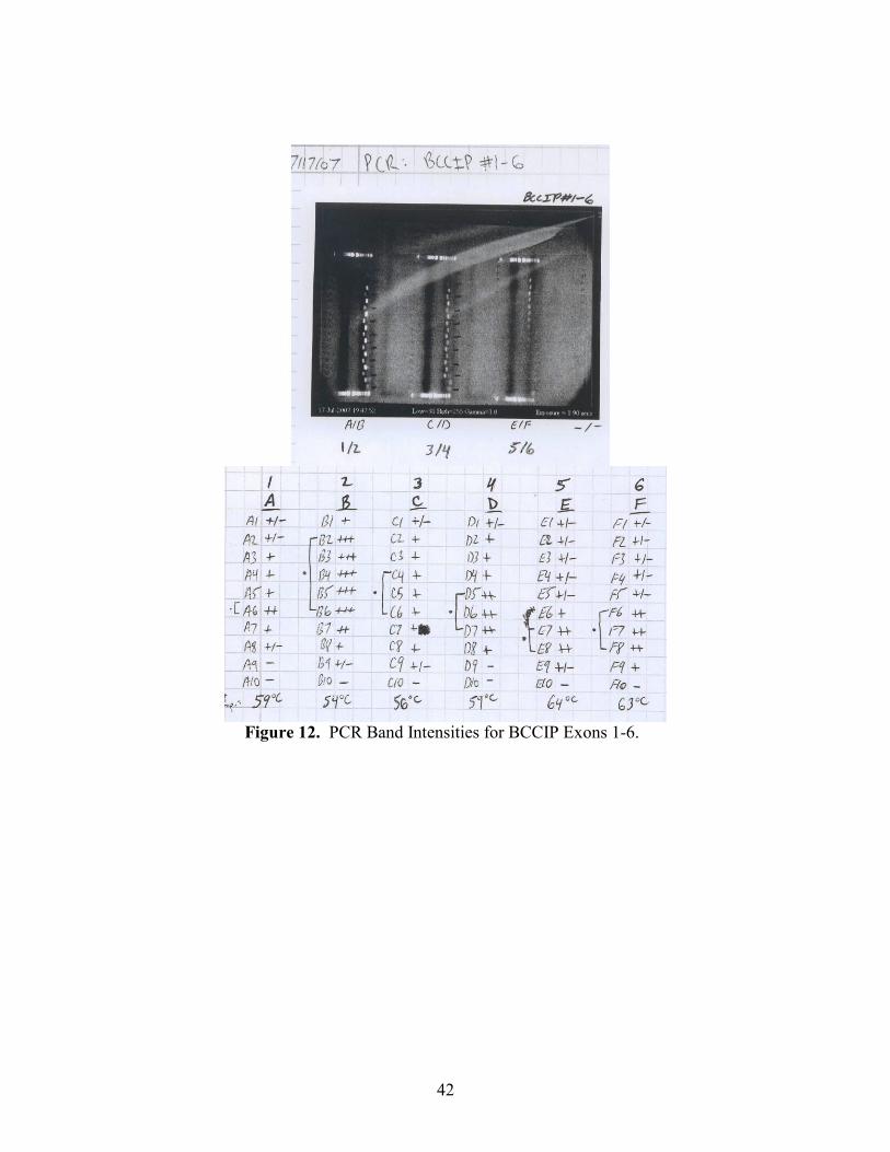
Figure 12. PCR Band Intensities for BCCIP Exons 1-6.

43
Figure 13. PCR Band Intensities for Mre11 Exons 10-17. None of the primers for
these exons failed in this gel.

44
PCR Amplification of Candidate Genes from Patient DNA
Following primer pair optimization, the optimized PCR conditions were then
applied to each of the six candidate genes from two hundred eighty-eight samples of
germline DNA obtained from various collaborating centers in Europe and the U.S. Each
patient had an extensive family history of breast cancer, but none had a BRCA1 or 2
mutation.
We began by individually amplifying each exon in the six target genes. Exons
larger than 500 bp were split into multiple amplicons for analyses. Table-VI shows a
summary of the number of amplicons successfully obtained for the six candidate genes.
All 14 exons were successfully obtained for MCPH1, all 3 exons for DSS1, 19 of 20
exons for Mre11, 23 of 25 exons for Rad50, all 16-17 exons for NBS1, and all 7 exons
for BCCIP were obtained.
Table-VI: Number of PCR Amplicons Obtained for Each Candidate Gene
and Number of Amplicons Analyzed by HRMA.
In this Table, column-1 shows the gene analyzed; column-2 shows the total known
number of exons for that gene; column-3 shows the number of amplicons analyzed for

45
that gene, and column-4 shows the number of amplicons analyzed by High Resolution
Melting Analysis (HRMA) for that gene.
High Resolution Melting Analysis (HRMA) and Variant Sequencing
PCR amplicons for MCPH1, DSS1, Mre11, and Rad50 were analyzed by High
Resolution Melting Analysis (HRMA) as a preliminary screen to identify potential
sequence variants. In this technique, heteroduplexes form during PCR between one DNA
strand from the WT DNA and the other strand from a patient’s variant gene. Mismatched
duplexes intercalate less fluorescent dye than pure duplex DNA, so a decreasing
fluorescent signal indicates the presence of potential sequence variants, which can
subsequently be further analyzed by DNA sequencing. HRMA Lightscanner Software
was used to determine whether melting curves varied from the wild type curves to
determine which of the 288 patient samples most likely contained mutations.
Table-VI (in the previous subsection) shows a summary of the number of
amplicons analyzed by HRMA. All 14 exons were analyzed for MCPH1, all 3 exons for
DSS1, 10 of 20 exons for MRE11, 13 of 25 exons for RAD50, and none for NBS1 or
BCCIP at this time. LightScanner software was used to display the HRMA data. Any
curves that visually differed from the normal (wild type) curve indicated a potential
sequence variant for that patient sample. Potential variants were then analyzed by
sequencing.
Of the four genes analyzed by PCR/HRMA to date, one MCPH1, was chosen for
sequence analysis. A preliminary analysis of DSS1 gene is shown in the Appendix, and
is nearly completed. DNAs were prepared for sequencing by using an ExoSAP IT

46
protocol to remove excess primers and nucleotides from the PCR reactions. The final
product samples were then placed on a sequencing plate and sent to the Mayo Clinic
sequencing core to be sequenced. The sequenced products were analyzed using
Sequencher software, which assisted in identifying the location of a possible mutation
within each amplicon.
Figure 14 shows the HRMA LightScanner output for MCPH1 Exon-6 for Patient H1.
The melting curve for exon-6 of patient H1 differs from the wild-type curve, indicating a
possible sequence variant. Figure 15 shows the Sequencher output for MCPH1 Exon-6 for
patient E4 and H1, both showing sequence variants relative to WT DNA. Figures 16-21 show
examples of other sequence variants identified.

47
Figure 14. HRMA LightScanner Output for MCPH1 Exon-6 for Patient H1 (BR-32-515-
d05-3184). The light colored double-peak curve for the patient DNA (lower right panel) differs
from the darker colored single peak for WT DNA indicating a potential sequence variant.
Figure 15. Sequencher Output for MCPH1 Exon-6 for Two Patients E4 (BR-32-127-d00-
2063) and H1 (BR-32-515-d05-3184). The upper sequence file for patient E4 (BR-32-127-d00-
2063) shows a homozygous S 171 R (TT) mutation relative to WT DNA (GG). Note: the WT
sequence is not shown in this figure. The lower sequence displays a heterozygous 581 G>T
mutation (GT).

48
Figure 16. HRMA LightScanner Output for MCPH1 Exon-7 for Patients K3 (FCP-119)
and P5 (COH-2394-469-1). This LightScanner output shows a double peak curve that is the
wild type curve and a multiple peak curve that corresponds to the frameshift mutation (K3).
Figure 17. Sequencher Output for MCPH1 Exon-7 for Two Patients E22 (COH-1228-863-
1) and K3 (FCP-119). This figure shows a frameshift mutation (Exon 7 c. 649 -7 ins T, IVS 6 -7
ins T) in the second sequence file relative to the WT sequence (top sequence).

49
Figure 18. HRMA LightScanner Output for MCPH1 Exon-8F for Patients J23 (FCP-10)
and P5 (COH-2394-469-1). This HRMA shows a pink curve wild type (P5) and an orange curve
(J23) that corresponds to the frameshift mutation.
Figure 19. Sequencher Output for MCPH1 Exon-8F for Two Patients J23 (FCP-10) and
A17. The first sequence file is an example of a frameshift mutation. In this sequence, there is an
apparent shoulder on the peaks, which is an indication of a frameshift mutation (1464 Δ del A, fs
(468) codon 466 stop 499). In this specific example, a stop codon is formed at codon 499 in exon
8f of the MCPH1 gene.

50
Figure 20. LightScanner Output for MCPH1 Exon-8F for Patients P5 (COH-2394-469-1),
G14 (COH-883-856-1), and J23 (FCP-10). The pink curve (P5) is the wild type curve. The
orange and dark blue curves (G14 and J23) are heterozygous mutations. The orange curve is also
a frameshift mutation.
Figure 21. Sequencher Output for MCPH1 Exon-8F for Patients J23 (FCP-10), A17, and
G14 (COH-883-856-1). This is another example of a frameshift mutation (1496 C>T, F 476 F).
The first sequence shows a mutation of C>T at position 85 in the frameshift/mutant sequence.
The mutation is homozygous. The second sequence displays the WT sequence, while the third
sequence shows an individual that is heterozygous for the SNP.

51
In order to determine how a particular amino acid would be altered by the
nucleotide mutation, the location of the nucleotide within the correct reading frame of the
coding sequence was identified. Altered codons will change the resultant protein
sequence, except for silent mutations where the protein is not altered or physically
changed. However, silent mutations may have some function in alternative splicing.
Once the locations of the amino acid changes had been recorded, the dbSNP
NCBI database was accessed to determine the frequency of the SNPs previously
identified in other population studies. Since the 288 patient samples consisted of
individuals from European countries, the Minor Allele Frequencies (MAFs) were
calculated from the data available on European populations. Novel SNPs, previously
unidentified, were not recorded in the dbSNP database. Frameshift mutations were also
not available in the dbSNP database, because frameshifts alter the sequence of multiple
nucleotides (rather than a single nucleotide). The sequence data analysis summary for
MCPH1 is shown in Table-VII below.

52
Table-VII: Summary of the Sequence Analysis Mutation Screening for MCPH1.
Coordinates exon nt change Genotype aa change dbSNP MAF Obs freq in X sequences
(mutants/#samples
sequenced)
Cross species
conservation
I.Frameshifts/nonsense
BX-1-2-3 J23 8f 1464 Δ del A fs (468) codon
466 stop 499
1 out of 23
BX-1-2-3 J23 8f 1464 Δ del A;
1496 C>T
CT fs (468) codon
466 stop 499;
F 476 F
rs2920676 0.0085 2 out of 23 2/8 del, 2/8 c, 4/8 nc
BX-1-2-3 K3 7 c. 649 -7 ins
T
IVS 6 -7 ins T 1 out of 16
BX-1-2-3 C5 13-1 2375 ins C (769) fs 772
Stop 777
3 out of 48
BX-1-2-3 E7 13-1 2375 ins C (769) fs 772
Stop 777
3 out of 48
BX-1-2-3 K16 13-1 2375 ins C (769) fs 772
Stop 777
3 out of 48
BX-1-2-3 J23 13-1 2375 Δ del C (769) fs
771Stop778
1 out of 48
BX-1-2-3 G4 13-1 del 244 fs 818 Stop
844
2 out of 48
BX-1-2-3 D5 13-1 2283del238
(ex.13 del)
fs 818 Stop
844
2 out of 48
II. Missense subst.
BX-1-2-3 H1 3 237 C>A CA Q 57 K No 7 out of 24 100% c
BX-1-2-3 M15 3 244 C>A CA T 59 N No 1 out of 24 100% c
BX-1-2-3 O20 3 250 A>G AG D 61 G No 1 out of 24 7/8 c, 1/8 nc
BX-1-2-3 I21 5 430 C>A CA P 121 Q No 14 out of 24 100% c
BX-1-2-3 O21 6 581 G>T TT S 171 R rs2442513 0.25 30 out of 31 3/8 c, 3/8 sc, 2/8 nc
BX-1-2-3 H1 6 581 G>T GT S 171 R rs2442513 0.25 1 out of 31 3/8 c, 3/8 sc, 2/8 nc
BX-1-2-3 G14 8b 931 C>A CA P 288 H rs35590577 ??? 1 out of 15 1/8 del, 1/8 nc, 6/8 c
BX-1-2-3 G14 8b 979 G>T GT R 304 I rs2083914 0.182 10 out of 15 5/8 c, 3/8 nc
BX-1-2-3 L5 8b 1008 G>C CC D 314 H rs930557 0.207 5 out of 15 1/8 del, 5/8 c, 2/8 nc
BX-1-2-3 O11 8b 1008 G>C GC D 314 H rs930557 0.207 9 out of 15 1/8 del, 5/8 c, 2/8 nc
BX-1-2-3 I15 8d-1 1243 A>G GG D 392 G rs2515569 1 46 out of 46 2/8 del, 1/8 c, 5/8 nc
(3G, 1R, 1Q)
BX-1-2-3 M4 13-1 2350 C>T CT A 761 V rs1057090 0.481 23 out of 48 5/8 c (4V,1M), 2/8 nc,
1/8 sc
BX-1-2-3 G9 13-1 2350 C>T TT A 761 V rs1057090 0.481 9 out of 48 5/8 c (4V,1M), 2/8 nc,
1/8 sc
BX-1-2-3 M4 13-1 2476 C>A CA A 806 A rs2912016 0.43 21 out of 48 5/8 c, 2/8 sc, 1/8 nc
BX-1-2-3 G9 13-1 2476 C>A AA A 806 A rs2912016 0.43 5 out of 48 5/8 c, 2/8 sc, 1/8 nc
BX-1-2-3 H3 13-1 2287 G>A AA C 740 Y No 1 out of 48 7/8 c, 1/8 nc
BX-1-2-3 B21 14 2540 C>T TT P 828 S No 6 out of 47 8/8 nc (4S, 2F, 1D, 1M
whch is sc to S)
BX-1-2-3 M8 14 2540 C>T TC P 828 S No 26 out of 47 8/8 nc (4S, 2F, 1D, 1M
whch is sc to S)
BX-1-2-3 D3 14 2562 C>A CA L 832 I No 47 out of 47? 6/8 c (5L, 1I), 2/8 sc
III. Silent substitutions
BX-1-2-3 A22 3 296 G>T TT V 76 V rs2305022 0.202 19 out of 24 100% c
BX-1-2-3 D23 3 296 G>T GT V 76 V rs2305022 0.202 4 out of 24 100% c
BX-1-2-3 I1 6 545 A>T TA S 159 S rs41313948 N/A 2 out of 31 100% c
BX-1-2-3 I15 8d-1 1280 T>A TA A 404 A No out of 46 2/8 del, 4/8 c, 2/8 sc
BX-1-2-3 M4 8d-1 1280 T>A TT? A 404 A No out of 46 2/8 del, 4/8 c, 2/8 sc
BX-1-2-3 G14 8f 1496 C>T CT F 476 F rs2920676 0.0085 2 out of 23 2/8 del, 2/8 c, 4/8 nc
BX-1-2-3 A21 8h 1850 G>A GA T 594 T rs2584 0.36 5 out of 8 2/8 del, 3/8 c, 3/8 nc
BX-1-2-3 M20 8h 1850 G>A GA T 594 T rs2584 0.36 8 out of 21 2/8 del, 3/8 c, 3/8 nc
BX-1-2-3 O20 8h 1850 G>A AA T 594 T rs2584 0.36 3 out of 21 2/8 del, 3/8 c, 3/8 nc
BX-1-2-3 M4 13-1 2294 C>T CT S 742 S rs2912010 0.475 22 out of 48 2/8 c, 4/8 sc, 2/8 nc
BX-1-2-3 G9 13-1 2294 C>T TT S 742 S rs2912010 0.475 9 out of 48 2/8 c, 4/8 sc, 2/8 nc
BX-1-2-3 M4 13-1 2476 C>A CA A 806 A rs2912016 0.43 21 out of 48 5/8 c, 2/8 sc, 1/8 nc
BX-1-2-3 G9 13-1 2476 C>A AA A 806 A rs2912016 0.43 5 out of 48 5/8 c, 2/8 sc, 1/8 nc
BX-1-2-3 P7 13-1 2411 G>A GA L 781 L No 1 out of 48 100% c
IV. Intronic variants
BX-1-2-3 H3 I-9 2003+9 G>A AG ….. No 1 out of 47
BX-1-2-3 F17 I-9 2003+69
A>G
AG ….. No 1 out of 47
BX-1-2-3 M20 I-10F 2041+15
T>A
TA ….. No 1 out of 45
BX-1-2-3 D3 I-14 2576+8 C>A CA ….. No 47 out of 47?
Table-VII: Summary of the Sequence Analysis Mutation Screening for Gene MCPH1. The columns
in this table indicate the exon number for MCPH1, the observed nucleotide number change, the genotype
for the mutation, the amino acid change designation, whether the mutation has been identified in the dbSNP
database, the minor allele frequency (MAF), the observation frequency of the corresponding mutant, and
the cross species conservation of the mutant amino acid.

53
The project to date has found MCPH1 mutations in 29 of the 288 patient samples
tested, 10 of which demonstrated more than one mutation. Nine likely deleterious,
frameshift/nonsense mutations, nineteen missense mutations, fourteen silent substitutions,
and three intronic variants were found in the MCPH1 amplicons. The mutations that are
most likely disease-associated are the missense and frameshift mutations. Frameshifts,
missense mutations, and silent substitutions within amplicon 13-1 (exon 13) occurred
most often within each of the three categories. Six of the nine frameshifts, five of the
nineteen missense mutations, and five of the fourteen silent substitutions occurred in
amplicon 13-1 (exon13).
A cross species alignment was also performed for MCPH1 (Figure 22) for nine
different species to determine the degree of conservation. The more highly conserved the
sequence domain, the more likely that domain is required for function, thus if a mutation
occurs in a conserved domain it likely is deleterious. The amino acids highlighted in red
indicate the locations where mutations were observed in patient samples in the present
study. The cross species conservation for each of these mutations is also listed in the
rightmost column of Table-VII. We conclude from the amino acid alignments that
several domains in MCPH1 are highly conserved and are likely required for function, and
that 14 patient mutations were identified in these potential functional domains.

54
Figure 22. Amino Acid Alignment for Nine Biological Species of MCPH1. This analysis was
performed to determine conserved domains to determine which patient mutations likely disrupt functional
domains. The alignment was performed using Clustal 2.0.8 software. The species name is listed on the left
side of the figure, and the corresponding amino acid sequence is given for each species. The letters in red
indicate the amino acid mutations found in patient samples.

55
Pedigrees of Families with MCPH1 Mutations
Figures-23, 24, 25, and 26 show the family pedigrees of patient samples found to
have a serious frameshift mutation in the MCPH1 gene. The males are indicated as
squares, while the females are represented as circles. Patients with breast cancer are
highlighted in black. The age of cancer diagnosis (in years) is indicated within the
symbol and, in some cases, both ages are given for bilateral breast cancer. Other cancers
noted were bladder (Bl), colon (Col), liver (Li), ovarian (Ov), pancreatic (Panc), prostate
(Pr), sarcoma (Sarc), thyroid (Thy), and uterine (Ut). The lines through symbols indicate
deceased family members.
Figure 23. Pedigree of a
Female Patient Identified with
Breast Cancer at 41 Years of
Age with an MCPH1
Frameshift Mutation.
Figure 24. Pedigree of a Female
Patient Identified With Breast
Cancer at 40 Years of Age With an
MCPH1 Frameshift Mutation.

56
Figure 25. Pedigree of female patient
identified with bilateral breast cancer
first at age 51 then again at 55 years with
an MCPH1 Frameshift Mutation.
Figure 26. Pedigree of A
Female Patient Identified
With Breast Cancer at Age
51 with an MCPH1
Frameshift Mutation.
From these pedigrees it can be concluded that deleterious MCPH1 frameshift mutations
occur in families with inherited early onset cancers. In three of the four pedigrees shown,
one or more of the patient’s family members died of early onset breast cancer. Further
studies may eventually show how widespread these MCPH1 mutations are. Since these
patients are from non-BRCA families, it can be inferred that frameshift mutations within
the MCPH1 gene may have a significant effect on the onset of breast cancer.

57
DISCUSSION
The data obtained from this project used a candidate gene screening approach to
identify potential genetic changes in proteins previously shown to interact with BRCA1
and/or BRCA2 in cellular pathways. The coding sequences of 6 candidate genes
MRE11, RAD50, MCPH1, NBS1, DSS1, and BCCIP were amplified by PCR from 288
non-BRCA1/BRCA2 breast cancer patient DNA samples. The amplicons were initially
screened for mutations by High Resolution Melting Analysis (HRMA), and selected
positives were further analyzed by sequence analysis. Most of the data obtained so far is
from gene MCPH1, which to date shows frameshift/nonsense mutations, missense
mutations, silent substitutions, and intronic variants in 29 patients of the 288 patients
analyzed so far. Ten patient samples demonstrated more than one mutation in MCPH1.
For gene DSS1 (see Appendix), all exons for that gene were obtained by PCR, analyzed
by HRMA, and sequenced, but only exon 2 has been analyzed to date. Within that
amplicon, seven frameshift mutations and one SNP were found. Thus in this study to
date, putative breast cancer-associated mutations have been identified in candidate genes
known to interact with BRCA1 and/or BRCA2 in the double-stranded DNA break repair
pathway. Importantly, the identified mutations include protein truncating and missense
mutations in highly conserved domains.
The data from this project support the findings of Friedenson (2005) who
previously speculated that a breakdown in the DNA repair BRCA pathway may increase
the risk for development of breast cancer, and who also proposed that inactivation of any
component within the BRCA pathway may also increase the risks for ovarian cancers,

58
lymphomas, and leukemias (Friedenson 2007). In addition, Chaplet et al. (2006)
suggested that MCPH1 (also known as BRIT1) plays a role as a tumor suppressor.
Depletion of BRIT1 (MCPH1) eliminates the DNA damage checkpoint and repair
response, increasing the risk for the development and progression of cancer (Chaplet et
al., 2006). The majority of mutations that we found in MCPH1 were missense mutations
in conserved domains, so these could disrupt function. In addition, the nine frameshift
mutations observed in MCPH1 are probably serious mutations that most likely could
cause disease in those patients. So perhaps a proportion of the cancer observed in non-
BRCA1/BRCA2 families can be explained by these observed mutations in the MCPH1
gene.
Problems encountered while working on this project included difficulty
amplifying certain exons by PCR, which led to a redesign and reoptimization of primers
in many instances. In addition, the HRMA assay used as a pre-screening method to
identify potential mutations was not very efficient or consistent at detecting the mutations
later identified by sequencing. It was also somewhat difficult to manage the large
amounts of data generated in this project, especially when analyzing so many patient
samples.
In the future, database software programs may be useful for organizing the data.
In addition, MCPH1 mutation segregation studies and penetrance estimation studies
could be done to determine disease-risk. Additional studies could also be completed on
other candidate genes to continue to identify genes that contribute to an unexplained
familial risk for breast cancer.

59
BIBLIOGRAPHY
Antoniou AC, Easton DF (2006) Models of Genetic Susceptibility to Breast Cancer.
Oncogene 25: 5898-5905.
Breast and Ovarian Cancer (2007) Genes and Disease. 1 Dec 2007.
http://www.ncbi.nlm.nih.gov/books/bv.fcgi?call=bv.View..ShowSection&rid=g
nd.section.99
Breast Cancer Mutation Database (2007) National Human Genome Research Institute
< http://research.nhgri.nih.gov/bic/>
Cellular and Molecular Biology - DNA Analysis (2005) National Center for Voice and
Speech 20 Aug 2008 www.ncvs.org/ncvs/groups/cmb/dna.html
Chaplet M, Rai R, Jackson-Bernitsas D, Li K, Lin SY (2006) BRIT1/MCPH1: a guardian
of genome and an enemy of tumors. Cell Cycle. 2006 Nov;5(22):2579-83. Epub
2006 Nov 15.
Cierniková S, Tomka M, Kovac M, Stevurkova V, Bella V, JNovotny J and Zajac V
(2005) Mutation screening of BRCA1, BRCA2 and CHEK2*1100delC in Slovak
HBOC families. Breast Cancer Research 7(Suppl 2): P1.02.
Cipollini G, Tommasi S, Paradiso A, Aretini P, Bonatti F, Brunetti I, Bruno M, Lombardi
G, Schittulli F, Sensi E, Tancredi M, Bevilacqua G, Caligo MA (2004) Genetic
alterations in hereditary breast cancer. Ann Oncol. 15 Suppl 1: I7-I13.
Cowden syndrome (2007) Genetics Home Reference. 8 Dec 2007.
http://ghr.nlm.nih.gov/condition=cowdensyndrome
Csokay B, Tihomirova L, Stengrevics A, Sinicka O, Olah E (1999) Strong founder
effects inBRCA1 mutation carrier breast cancer patients from Latvia. Mutation in
brief no. 258. Hum Mutat. 14(1): 92.
Deng, Chu-Xia (2006) BRCA1: cell cycle checkpoint, genetic instability, DNA damage
response and cancer evolution. Nucleic Acids Res. 34(5): 1416–1426.
De Soto JA and Deng, Chu-Xia (2006) PARP-1 inhibitors: are they the long-sought
genetically specific drugs for BRCA1/2-associated breast cancers? Int J Med Sci
3(4): 117–123.
Easton DF, Ford D, Bishop DT (1995) Breast and ovarian cancer incidence in BRCA1-
mutation carriers. Breast Cancer Linkage Consortium. Am J Hum Genet 56:265–
71.

60
Finch A, Beiner M, Lubinski J, Lynch HT, Moller P, Rosen B, Murphy J, Ghadirian P,
Friedman E, Foulkes WD, Kim-Sing C, Wagner T, Tung N, Couch F, Stoppa-
Lyonnet D, Ainsworth P, Daly M, Pasini B, Gershoni-Baruch R, Eng C,
Olopade OI, McLennan J, Karlan B, Weitzel J, Sun P, Narod SA; Hereditary
Ovarian Cancer Clinical Study Group. (2006) Salpingo-oophorectomy and the
risk of ovarian, fallopian tube, and peritoneal cancers in women with a BRCA1 or
BRCA2 Mutation. JAMA. Jul 296(2):185-92
Ford D, Easton DF, Stratton M, Narod S, Goldgar D, Devilee P, Bishop DT, Weber B,
Lenoir G, Chang-Claude J, Sobol H, Teare MD, Struewing J, Arason A,
Scherneck S, Peto J, Rebbeck TR, Tonin P, Neuhausen S, Barkardottir R, Eyfjord
J, Lynch H, Ponder BA, Gayther SA, Zelada-Hedman M, et al. (1998) Genetic
Heterogeneity and Penetrance Analysis of the BRCA1 and BRCA2 Genes in
Breast Cancer Families. The Breast Cancer Linkage Consortium. Am J Hum
Genet. 62: 676-689.
Friebel TM, Domchek SM, Neuhausen SL, Wagner T, Evans DG, Isaacs C, Garber JE,
Daly MB, Eeles R, Matloff E, Tomlinson G, Lynch HT, Tung N, Blum JL,
Weitzel J, Rubinstein WS, Ganz PA, Couch F, Rebbeck TR (2007) Bilateral
prophylactic oophorectomy and bilateral prophylactic mastectomy in a
prospective cohort of unaffected BRCA1 and BRCA2 mutation carriers. Clin
Breast Cancer. Dec; 7(11): 875-882.
Friedenson B (2005) BRCA1 and BRCA2 Pathways and the Risk of Cancers Other Than
Breast or Ovarian. Med Gen Med. 7: 60.
Friedenson B (2007) The BRCA1/2 pathway prevents hematologic cancers in addition to
breast and ovarian cancers. BMC Cancer 7: 152.
Frank B, Hemminki K, Meindl A, Wappenschmidt B, Sutter C, Kiechle M, Bugert P,
Schmutzler RK, Bartram CR, Burwinkel B (2007) BRIP1 (BACH1) variants and
familial breast cancer risk: a case-control study. BMC Cancer 7: 83.
Ganguly A, Leahy K, Marshall AM, Dhulipala R, Godmilow L, Ganguly T (1997)
Genetic testing for breast cancer susceptibility: frequency of BRCA1 and BRCA2
mutations. Genet Test. 1(2): 85-90.
Gorski B, Byrski T, Huzarski T, Jakubowska A, Menkiszak J, Gronwald J, Pluzańska A,
Bebenek M, Fischer-Maliszewska L, Grzybowska E, Narod SA, Lubiński (2000)
Founder mutations in the BRCA1 gene in Polish families with breast-ovarian
cancer. Am J Hum Genet. Jun; 66(6):1963-8. Epub 2000 Apr 28.
Górski B, Jakubowska A, Huzarski T, Byrski T, Gronwald J, Grzybowska E, Mackiewicz
A, Stawicka M, Bebenek M, Sorokin D, Fiszer-Maliszewska Ł, Haus O,
Janiszewska H, Niepsuj S, Góźdź S, Zaremba L, Posmyk M, Płuzańska M, Kilar

61
E, Czudowska D, Waśko B, Miturski R, Kowalczyk JR, Urbański K, Szwiec M,
Koc J, Debniak B, Rozmiarek A, Debniak T, Cybulski C, Kowalska E, Tołoczko-
Grabarek A, Zajaczek S, Menkiszak J, Medrek K, Masojć B, Mierzejewski M,
Narod SA, Lubiński J. (2004)A high proportion of founder BRCA1 mutations in
Polish breast cancer families. Int J Cancer. 110(5): 683-686.
Greene MH (1997) Genetics of breast cancer. Mayo Clin Proc. 72(1): 54-65.
Gronwald J, Elsakov P, Górski B, Lubiński J (2005) High incidence of 4153delA
BRCA1 gene mutations in Lithuanian breast- and breast-ovarian cancer families.
Breast Cancer Res Treat. 94(2): 111-113.
Hartmann LC, Sellers TA, Schaid DJ, Frank TS, Soderberg CL, Sitta DL, Frost MH,
Grant CS, Donohue JH, Woods JE, McDonnell SK, Vockley CW, Deffenbaugh
A, Couch FJ, Jenkins RB (2001) Efficacy of bilateral prophylactic mastectomy in
BRCA1 and BRCA2 gene mutation carriers. J Natl Cancer Inst. 93(21): 1633-
1637.
Hall JM, Lee MK, Newman B, Morrow JE, Anderson LA, Huey B, King MC (1990)
Linkage of early-onset familial breast cancer to chromosome 17q21.
Science. 250(4988): 1684-1689.
Heikkinen K, Rapakko K, Karppinen SM, Erkko H, Knuutila S, Lundán T, Mannermaa
A, Børresen-Dale AL, Borg A, Barkardottir RB, Petrini J, Winqvist R (2006)
RAD50 and NBS1 are breast cancer susceptibility genes associated with genomic
instability. Carcinogenesis. 27(8): 1593-1599.
Hermon C, Beral V (1996) Breast cancer mortality rates are levelling off or beginning to
decline in many western countries: analysis of time trends, age-cohort and age-
period models of breast cancer mortality in 20 countries. Br J Cancer. 73(7): 955-
960.
Kainu T, Juo SHH, Desper R, Schäffer A, Gillanders E, Rozenblum E, Freas-Lutz D,
Weaver D, Stephan D, Bailey-Wilson J, Kallioniemi O, Tirkkonen M, Syrjäkoski
K, Kuukasjärvi T, Koivisto P, Karhu R, Holli K, Arason A, Johannesdottir G,
Bergthorsson J, Johannsdottir H, Egilsson V, Barkardottir R, Johannsson O,
Haraldsson K, Sandberg T, Holmberg E, Grönberg
H, Olsson
H, Borg A,
Vehmanen P, Eerola H, PHeikkilä P, PyrhönenS, and Nevanlinna H (2000)
Somatic deletions in hereditary breast cancers implicate 13q21 as a putative novel
breast cancer susceptibility locus. Proc Natl Acad Sci USA 97: 9603-9608.
Knudson AG Jr. (1971) Mutation and cancer: statistical study of retinoblastoma. Proc
Natl Acad Sci USA 68: 820–823.
Lewis AG, Flanagan J, Marsh A, Pupo GM, Mann G, Spurdle AB, Lindeman GJ,
Visvader JE, Brown MA, Chenevix-Trench G; Kathleen Cuningham Foundation

62
Consortium for Research into Familial Breast Cancer (2005) Mutation analysis of
FANCD2, BRIP1/BACH1, LMO4 and SFN in familial breast cancer. Breast
Cancer Res.7(6): R1005-1016.
Li J, Zou C, Bai Y, Wazer DE, Band V, Gao Q (2006) DSS1 is required for the stability
of BRCA2. Oncogene 25(8): 1186-1194.
Li-Fraumeni syndrome (2007) Genetics Home Reference.8 Dec 2007.
http://ghr.nlm.nih.gov/condition=lifraumenisyndrome
Lin SY, Rai R, Li K, Xu ZX, Elledge SJ (2005) BRIT1/MCPH1 is a DNA damage
responsive protein that regulates the Brca1-Chk1 pathway, implicating
checkpoint dysfunction in microcephaly. Proc Natl Acad Sci USA. 18;102(42):
15105-15109.
Liu Y, West SC (2002) Distinct Functions of BRCA1 and BRCA2 in Double-Strand
Break Repair. Breast Cancer Res. 4: 9-13.
Lu H, Yue J, Meng X, Nickoloff JA, Shen Z (2007) BCCIP regulates homologous
recombination by distinct domains and suppresses spontaneous DNA damage.
Nucleic Acids Res. [Epub ahead of print]
Miki Y, Swensen J, Schattuck-Eidens D, Futreal PA,Harshman K, Tavtigian S, Liu QY,
et al (1994) Isolation of BRCA1, the 17q-linked breast and ovarian cancer
susceptibility gene. Science 266: 66–71.
National Cancer Institute (2007) Breast Cancer. 4 Dec 2007.
http://www.cancer.gov/cancertopics/types/breast
Nusbaum R, Isaacs C (2007) Management updates for women with a BRCA1 or BRCA2
mutation. Mol Diagn Ther. 11(3): 133-144.
Oncogenes and Tumor Suppressor Genes (2005) American Cancer Society. 24 Feb 2005
http://www.cancer.org/docroot/ETO/content/ETO_1_4x_oncogenes_and_tu
mor_suppressor_genes.asp 20 Aug 2008.
Osborne C, Wilson P, Tripathy D (2004) Oncogenes and Tumor Suppressor Genes in
Breast Cancer: Potential Diagnostic and Therapeutic Applications. The
Oncologist 9 (4) 361–377.
Peto J, Collins N, Barfoot R, Seal S, Warren W, Rahman N, et al. (1999) Prevalence of
BRCA1 and BRCA2 gene mutations in patients with early-onset breast cancer. J
Natl Cancer Inst 91: 943-949.
Peutz–Jeghers syndrome (2007) Genetics Home Reference. 8 Dec 2007.
http://ghr.nlm.nih.gov/condition=peutzjegherssyndrome

63
PC Green Melting Dyes (2007) Idaho Technology Inc. 21 Aug 2008
http://www.idahotech.com/LCGreen/index.html
Pohlreich P, Zikan M, Stribrna J, Kleibl Z, Janatova M, Kotlas J, Zidovska J, Novotny J,
Petruzelka L, Szabo C, Matous B (2005) High proportion of recurrent germline
mutations in the BRCA1 gene in breast and ovarian cancer patients from the
Prague area. Breast Cancer Res. 7(5): R728-736. Epub 2005 Jul 19.
Rai R, Dai H, Multani AS, Li K, Chin K, Gray J, Lahad JP, Liang J, Mills GB, Meric-
Bernstam F, Lin SY (2006) BRIT1 regulates early DNA damage response,
chromosomal integrity, and cancer. Cancer Cell. 10(2): 145-157.
Robert F, Hardy S, Nagy Z, Baldeyron C, Murr R, Dery U, Masson JY, Papadopoulo D,
Herceg Z, Tora L (2006) The Transcriptional Histone Acetyltransferase Cofactor
TRRAP Associates with the MRN Repair Complex and Plays a Role in DNA
Double-Strand Break Repair. Mol Cell Biol. Jan;26(2): 402-412.
Saxena S, Chakraborty A, Kaushal M, Kotwal S, Bhatanager D, Mohil RS, Chintamani
C, Aggarwal AK, Sharma VK, Sharma PC, Lenoir G, Goldgar DE, Szabo CI
(2006) Contribution of germline BRCA1 and BRCA2 sequence alterations to
breast cancer in Northern India. BMC Med Genet. 7: 75.
Schubert E, Lee M, Mefford H, Argonza R, Morrow J, Hull J, Dann J, King M (1997)
BRCA2 in American Families with Four or More Cases of Breast or Ovarian
Cancer: Recurrent and Novel Mutations, Variable Expression, Penetrance, and
the Possibility of Families Whose Cancer Is Not Attributable to BRCA1 or
BRCA2. Am J Hum Genet. 60: 1031-1040.
Seal S, Thompson D, Renwick A, Elliott A, Kelly P, Barfoot R, Chagtai T, Jayatilake H,
Ahmed M, Spanova K, North B, McGuffog L, Evans DG, Eccles D; Breast
Cancer Susceptibility Collaboration (UK), Easton DF, Stratton MR, Rahman N
(2006) Truncating mutations in the Fanconi anemia J gene BRIP1 are low-
penetrance breast cancer susceptibility alleles. Nat Genet. 38(11): 1239-1241.
Southey MC, Tesoriero AA, Andersen CR, Jennings KM, Brown SM, Dite GS, Jenkins
MA, Osborne RH, Maskiell JA, Porter L, Giles GG, McCredie MR, Hopper JL,
Venter DJ (1999) BRCA1 mutations and other sequence variants in a population-
based sample of Australian women with breast cancer. Br J Cancer 79(1): 34-39.
Struewing JP, Hartge P, Wacholder S, Baker SM, Berlin M, McAdams M, Timmerman
M, Brody L. Tucker M (1997) The risk of cancer associated with specific
mutations of BRCA1 and BRCA2 among Ashkenazi Jews. N Engl J Med
336: 1401-1408.

64
Szabo CI, King MC (1997) Population genetics of BRCA1 and BRCA2. Am J Hum
Genet. 60(5): 1013-1020.
Szabo CI, Wagner LA, Francisco LV, Roach JC, Argonza R, King MC, Ostrander EA
(1996) Human, canine and murine BRCA1 genes: sequence comparison among
species. Hum Mol Genet. 5(9): 1289-1298.
Thompson D, Szabo CI, Mangion J, Oldenburg RA, Odefrey F, Seal S, Barfoot R,
Kroeze-Jansema K, Teare D, Rahman N, Renard H, Mann G, Hopper JL, Buys
SS, Andrulis IL, Senie R, Daly MB, West D, Ostrander EA, Offit K, Peretz T,
Osorio A, Benitez J, Nathanson KL, Sinilnikova OM, Olàh E, Bignon YJ, Ruiz P,
Badzioch MD, Vasen HF, Futreal AP, Phelan CM, Narod SA, Lynch HT, Ponder
BA, Eeles RA, Meijers-Heijboer H, Stoppa-Lyonnet D, Couch FJ, Eccles DM,
Evans DG, Chang-Claude J, Lenoir G, Weber BL, Devilee P, Easton DF, Goldgar
DE, Stratton MR; KConFab Consortium.(2002) Evaluation of linkage of breast
cancer to the putative BRCA3 locus on chromosome 13q21 in 128 multiple case
families from the Breast Cancer Linkage Consortium. Proc Natl Acad Sci U S A.
Jan 22; 99(2): 827-831. Epub 2002 Jan 15.
Tischkowitz M, Xia B, Sabbaghian N, Reis-Filho JS, Hamel N, Li G, van Beers EH, Li
L, Khalil T, Quenneville LA, Omeroglu A, Poll A, Lepage P, Wong N, Nederlof
PM, Ashworth A, Tonin PN, Narod SA, Livingston DM, Foulkes WD (2007)
Analysis of PALB2/FANCN-associated breast cancer families. Proc Natl Acad
Sci USA. 104(16): 6788–6793.
Tauchi H, Matsuura S, Kobayashi J, Sakamoto S, Komatsu K (2002) Nijmegen breakage
syndrome gene, NBS1, and molecular links to factors for genome stability.
Oncogene. Dec 16;21(58): 8967-8980.
Troudi W, Uhrhammer N, Ben Romdhane K, Sibille C, Mahfoudh W, Chouchane L, Ben
Ayed F, Bignon YJ, Ben Ammar Elgaaied A (2007) Immunolocalization of
BRCA1 protein in tumor breast tissue: prescreening of BRCA1 mutation in
Tunisian patients with hereditary breast cancer? Eur J Histochem. 51: 219-226.
Uyei A, Peterson SK, Erlichman J, Broglio K, Yekell S, Schmeler K, Lu K, Meric-
Bernstam F, Amos C, Strong L, Arun B (2006) Association between clinical
characteristics and risk-reduction interventions in women who underwent BRCA1
and BRCA2 testing: a single-institution study. Cancer 107: 2745-2751.
Vogel K, Atchley D, Erlichman J, Broglio K, Ready K, V. Valero, Amo C, Hortobagyi
G, Lu K, and Arun B (2007) BRCA1 and BRCA2 Genetic Testing in Hispanic
Patients: Mutation Prevalence and Evaluation of the BRCAPRO Risk Assessment
Model J. Clin. Oncol. 25(29): 4635-4641.
Walsh T, Casadei S, Coats KH, Swisher E, Stray SM, Higgins J, Roach KC, Mandell J,
Lee MK, Ciernikova S, Foretova L, Soucek P, King MC (2006) Spectrum of

65
mutations in BRCA1, BRCA2, CHEK2, and TP53 in families at high risk of
breast cancer. JAMA 22: 1379-1388.
Walsh T, King M (2007) Ten Genes for Inherited Breast Cancer. Cancer Cell. 11: 103-
105.
Warner E, Foulkes W, Goodwin P, Meschino W, Blondal J, Paterson C, Ozcelik H, Goss
P, Allingham-Hawkins D, Hamel N, Di Prospero L, Contiga V, Serruya C, Klein
M, Moslehi R, Honeyford J, Liede A, Glendon G, Brunet JS, Narod S (1999)
Prevalence and penetrance of BRCA1 and BRCA2 gene mutations in unselected
Ashkenazi Jewish women with breast cancer. J Natl Cancer Inst. 21: 1241-1247.
Weischer M, Bojesen SE, Tybjaerg-Hansen A, Axelsson CK, Nordestgaard BG (2007)
Increased risk of breast cancer associated with CHEK2*1100delC. J Clin Oncol.
25(1): 57-63. Epub 2006 Jul 31.
Wooster R, Neuhausen S, Manigion J, Quirk Y, Ford D, CollinsN, Nguyen K, et al
(1994) Localisation of a breast cancer susceptibility gene (BRCA2) to
chromosome 13q by genetic linkage analysis. Science 265: 2088–2090.

66
Appendix
Sequence Analysis of DSS1
For gene DSS1, all exons for that gene were obtained by PCR, analyzed by
HRMA, and sequenced, but only the data for exon-2 has been analyzed to date. Within
exon-2 (Table-VIII, upper half), seven frameshift mutations and one SNP were found in 8
patient samples.
Table-VIII. Sequence Analysis of DSS1.
DSS1 Mutation Screening on BX-1-2-3
coord Seq_folder
sequencher
project exon seq file nt nt change
genoty
pe aa change dbSNP?
Obs freq
in X
sequences
(mutants/#
samples
sequence
d)
BX-1-2-3 M20 111307_be_2 2 2 fs until nt 90 FRAMESHIFT FRAMESHIFT 2 out of 11
BX-1-2-3 I4 111307_be_2 2 2 fs until nt 90 FRAMESHIFT FRAMESHIFT 2 out of 11
BX-1-2-3 C2 111307_be_2 2 2 fs until nt 156 FRAMESHIFT FRAMESHIFT 1 out of 11
BX-1-2-3 G4 111307_be_2 2 2 fs until nt 160 FRAMESHIFT FRAMESHIFT 1 out of 11
BX-1-2-3 K16 111307_be_2 2 2 fs until nt 158 FRAMESHIFT FRAMESHIFT 1 out of 11
BX-1-2-3 J5 111307_be_2 2 2 fs until nt 114 FRAMESHIFT FRAMESHIFT 1 out of 11
BX-1-2-3 L5 111307_be_2 2 2 fs until nt 104 FRAMESHIFT FRAMESHIFT 1 out of 11
BX-1-2-3 I4 111307_be_2 2 2 232 284 C>T CT F 52 F no 1 out of 11
This table contains the sequence analysis performed to date for DSS1. The data for exon-2 are
shown in this table. Identification of the variants is currently in progress. Frameshift mutations
have been positively identified within exon-2 for seven patient samples, and one patient sample
displayed a SNP within the same amplicon.
The DSS1 data obtained to date support Li et al.’s (2006) suggestion that DSS1
depletion causes hypersensitivity to DNA damage, similar to that shown with BRCA2
mutations. These authors found DSS1 to be essential to the stability of the BRCA2
protein in mammalian cells, and suggested that its deletion, suppression, or mutation,

67
would promote human breast and ovarian cancer, as well as sporadic and familiar breast
cancer where BRCA1 and BRCA2 mutations are absent (Li et al. 2006).