Embed Size (px)
Citation preview
Emojevwe’s Lecture note on physiology- Muscle Physiology Page 1 of 17
MUSCLE PHYSIOLOGY (PHS 213)
By Emojevwe V.
Course Objectives:
To familiarize students with the principles and basic facts of Human Physiology and
with some of the laboratory techniques and equipment used in the acquisition of
physiological data. The emphasis will be on muscles physiology. Since the course will
focus on muscle physiology, some cellular and molecular mechanisms will be
discussed in order to present a current view of physiological principles of muscle.
Where appropriate, basic chemical and physical laws will be reviewed in order to
enhance and to promote student understanding. The laboratory component of the course
is designed to reinforce the topics discussed in lecture, as well as to familiarize students
with some of the laboratory techniques and equipment used in muscle research
Student Learning Outcomes:
Upon successful completion of lecture portion of this course, the students will be able
to describe, identify, and/or explain:
• Structure and function of skeletal muscle, including excitation-contraction coupling, sliding filament mechanism, force generation, and isometric versus
isotonic contractions.
• Structure and functions of the cardiovascular system, including the mechanical
and electrical properties of cardiac muscle function.
• Structure and functions of smooth muscles including contraction of smooth muscle
Upon successful completion of the laboratory portion of this course, the students will be
able to describe, identify, explain, perform, and/or measure:
• Computer simulations of the membrane potential, action potential, and synaptic neurotransmission.
• Skeletal muscle mechanics, and the electromyogram (EMG).
Required Course Materials:
• Any textbook of physiology (human or medical) published within the last four years will be appropriate.
• Emojevwe’s Lecture Notes on Physiology. To obtain these notes as well as other supplementary materials, please visit the unimed portal
Emojevwe’s Lecture note on physiology- Muscle Physiology Page 2 of 17
MUSCLE PHYSIOLOGY
Muscle is an excitable tissue. The human body has over 600 muscles which perform many
useful functions and help us in doing everything in day-to-day life.
Classification of muscles: Muscles are classified by three different methods, based on
different factors:
I. Depending upon the presence or absence of striations; the muscles are divided into two
groups:
1. Striated Muscle: Striated muscle is the muscle which has a large number of
crossstriations (transverse lines). Skeletal muscle and cardiac muscle belong to this
category. 2. Non-striated Muscle: Muscle which does not have cross-striations is called
nonstriated muscle. It is also called plain muscle or smooth muscle. It is found in the wall
of the visceral organs.
II. Depending upon the control
1. Voluntary Muscle: Voluntary muscle is the muscle that is controlled by the will.
Skeletal muscles are the voluntary muscles. These muscles are innervated by somatic
nerves.
2. Involuntary Muscle: Muscle that cannot be controlled by the will is called
involuntary muscle. Cardiac muscle and smooth muscle are involuntary muscles. These
muscles are innervated by autonomic nerves.
III. Depending upon the situation.
1. Skeletal Muscle: Skeletal muscle is situated in association with bones forming the
skeletal system. The skeletal muscles form 40% to 50% of body mass and are voluntary
and striated. These muscles are supplied by somatic nerves. Fibers of the skeletal muscles
are arranged in parallel. In most of the skeletal muscles, muscle fibers are attached to
tendons on either end. Skeletal muscles are anchored to the bones by the tendons.
2. Cardiac Muscle: Cardiac muscle forms the musculature of the heart. These muscles
are striated and involuntary. Cardiac muscles are supplied by autonomic nerve fibers.
3. Smooth Muscle: Smooth muscle is situated in association with viscera. It is also
called visceral muscle. It is different from skeletal and cardiac muscles because of the
absence of cross striations, hence the name smooth muscle. Smooth muscle is supplied by
autonomic nerve fibers. Smooth muscles form the main contractile units of wall of the
various visceral organs.
In the preceding sections, I will be introducing you to the physiology of the skeletal, cardiac
and smooth muscles.
Emojevwe’s Lecture note on physiology- Muscle Physiology Page 3 of 17
SKELETAL MUSCLE
A skeletal muscle is a collection of muscle bundles or fascicles. Each fascicles is made up
of a number cells or (myofibrils). Each muscle is cell compose of hundreds to thousands
of myofibrils. Myofibrils are strings of the structural unit referred to as the sarcomere. The
sarcomere is the functional unit of skeletal and cardiac muscle. The sarcomere contains
the myofibrils; which are the proteins that are responsible for the contractile behavior of
the muscle cell. In addition, the sarcomere contains structural proteins (which maintain
the structural integrity of the sarcomere), as well as regulatory protein (which play an
important role in the regulation of muscle contractile activity).
A mature skeletal muscle cell is a long cell, which comes about through the fusion of many
embryonic muscle cells. Therefore, skeletal muscle cells are among the largest cells in the
body, whose length can be as long as tens of centimeters. The diameter of a typical skeletal
muscle cell is about 100 µm. Skeletal muscle cells are multinucleated, and they contain
numerous mitochondria. The plasma membrane of skeletal muscle cells (the sarcolemma)
is specialized in that it has invaginations that run deep into the muscle cell. These
invaginations are referred to as transverse tubules (or tubules). It is important to say that
the membrane of the t-tubule is continuous with the sarcolemma. Thus, t-tubules serve to
allow muscle action potentials to reach deep into the muscle cell. It is also important to
recognize that the lumen of the t-tubule is the extracellular fluid. Within the cytoplasm of
the skeletal muscle fiber (myoplasm or sarcoplasm), there are numerous specialized
structures, which we need to understand. A highly specialized endoplasmic reticulum
referred to as the sarcoplasmic reticulum is tightly wrapped around individual myofibrils
and functions to store a high concentration of Ca2+. The release of Ca2+ from the
sarcoplasmic reticulum is responsible for triggering muscular contraction. Another very
important structure is the sarcomere. As mentioned above, the sarcomere is the functional
unit of striated muscle, and it is discussed below.
Fine Details of the Sarcomere
The sarcomere is the functional unit of the muscle fiber. It is composed of overlapping
units of two different filamentous proteins; the thick and the thin filaments. Movement
of the thin filaments over the thick filaments brings about muscle shortening and force
generation. Thus, the sarcomere is responsible for the contractile ability of muscle cells.
In order to understand the function of the sarcomere, it is important that we first understand
the protein composition of the sarcomere. The proteins found in the sarcomere can be
placed into three proteins. As you read the following descriptions, be sure to examine the
attached figures in order to achieve a better understanding.
a. Contractile Proteins
i. Myosin (gives rise to the thick filaments) ii.
Actin (gives rise to the thin
filaments)
Emojevwe’s Lecture note on physiology- Muscle Physiology Page 4 of 17
b. Regulatory Proteins
i. Tropomyosin (in the absence of Ca2+ , it
covers the myosin –binding site of
actin)
ii. Troponin (Ca2+ sensor)
c. Structural Proteins
i. Titin (provide elasticity) ii. Nebulin
(run closely with actin)
iii. Dydrophine
iv. Desmin (it binds z line with sarcomere)
Now let us discuss these proteins
Contractile Proteins
As their name suggest, contractile proteins are in fact responsible for muscles shortening
(contraction). Myosin is the protein that makes up the thick filaments of the sarcomere.
Myosin has two structural domains; a head region and a tail region. The head and the tail
regions are connected through a flexible neck region. The tail region of myosin is used
to join many myosin molecules together in order to give rise to the thick filament. In
skeletal muscle, approximately 250 mysin molecules are intertwined to form a thick
filament. The myosin head has an actin-binding site to which ATP binds (see cross –
Bridge Cycle below). In a thick filament, the myosin molecules are arrange so that the
myosin heads are clustered at the ends (facing the Z disks), and the central region of the
thick filament is a bundle of myosin tails.
Actin is the protein that makes up the thin filaments of the sarcomere. An actin molecule
is a globular protein (globular actin or G-actin). Many G-actin molecules polymerize to
form filamentous actin (F-actin). In skeletal muscle, two F-actin polymers intertwine to
form a thin filament. Each G-actin molecule has a myosin binding site. Thus, the
interaction of actin and myosin take place via the interaction of myosin binding site of
Gactin with the actin binding site of the myosin of the myosin head group.
In order for the muscle to contract, the actin and the myosin molecules of the thin and thick
filaments have to interact with one another. When the actin and myosin interact, they are
said to be connected by cross-bridges. The cross-brides refer to the myosin head group
that interact with a myosin-binding site on G-actin.
Microscopic Details
Under the light or electron microscope, the arrangement of the thick and thin filaments in
a myofibril gives rise to a repeating pattern of alternating light and dark regions. One
repeat in the pattern roughly corresponds to what is known as the functional unit of striated

Emojevwe’s Lecture note on physiology- Muscle Physiology Page 5 of 17
muscle: the sarcomere. The special overlap of the thin and thick filaments within the
sarcomere gives rise to the contractile and shortening capability of the muscle cell. The
sarcomere has the following microscopic features:
Z Disks: Two adjacent Z disk along the myofibril mark the boundaries of a single
sarcomere. The Z disk are the attachment sites for the thin filaments. Therefore, from each
Z disk, thin filaments extend to two neighboring sarcomeres. When a muscle fiber
contracts, the Z disks of a sarcomere move closer together (see Sliding Filament model of
Contraction below). Thus, the sarcomere shortens as the muscle contracts.
A Band: The A Band is the entire length of the thick filament. At the outer edges of the A
Band, the thick and thin filaments overlap and, therefore, this region is the darkest region
of the sarcomere. The center of the A Band does not change as the muscle contracts.
H Zone: The H zone refer to the center of the A band where there is no overlap
between the thick and the thin filaments. Therefore, in the H zone, the filaments consist
only of the thick filament. The H zone becomes smaller as the muscle contracts because
the overlap between the thick and the thin filaments increases (i.e., region of naked or non-
overlap of the thick filament decreases).
I Band: This region is closest to the Z disk, and is the lightest region of the sarcomere.
The I Band is occupied by the thin filaments only. Each Z disk runs through the middle of
the I-band. Therefore, half of each I band belongs to one sarcomere, and the other half
belongs to the neighboring sarcomere. The I-band also shortens as the muscle contracts.
M Line: The M Line is the attachment site for the thick filaments. The M line is in the
middle of the A band and it is in the middle of the sarcomere.
In three dimensions, the sarcomere is a lattice of parallel and overlapping thick (myosin)
and thin (actin) filaments. If a cross-section view is obtained from the outer edges of the
A band where there is overlap between the thin and thick filaments, it can be seen that
every thin filament is surrounded by three thick filament, and every thick filament is
surrounded by six thin filaments.
Regulatory Proteins
Two regulatory proteins closely interact with actin. A regulatory protein called
tropomyosin spans over seven G-actin molecules and normally prevents the interaction of
the myosin head group with g-actin. When the myoplasmic Ca2+ concentration is low at
its resting level, tropomyosin covers the myosin binding site of G-actin. Tropomyosin also
closely interact with troponin, which is a Ca2+ sensor. Binding of Ca2+ to troponin moves
the troponin-tropomyosin complex out of the way exposing the myosin binding site. This
allows the myosin head to interact with G-actin.
Emojevwe’s Lecture note on physiology- Muscle Physiology Page 6 of 17
Structural Proteins
The structural integrity of the sarcomere is ensured by the presence of accessory structural
proteins titin and nebulin. Titin is a very elastic protein that stretches from one Z disk to
the M line. Its function is to bring the stretched muscle to its resting length. Nebulin is
positioned alongside the thin filaments and stabilizes them. Nebulin is inelastic.
SLIDING FILAMENT MODEL OF CONTRACTION
This theory proposed by A. F. Huxley in 1957 states that muscle contraction result from
the sliding of the thin filament over the thick filament. This explain how actin filament
slides over myosin and form acto-myosin complex during contraction. Muscle contraction
is an energy requiring event (ATP) that involves the movement of the thin filaments over
the thick filaments. As the thin filament are connected to the Z disks, and as muscle
contraction brings about movement of the thin filaments over the thick filaments and
towards the center of the sarcomere (the M line), a shortening of the sarcomere takes place.
This shortening brings the Z disks of a sarcomere closer together. During muscle
contraction, the size of the A band remains the same, but the I-band and H zone become
shorter as the thin filaments slide past the thick filaments.
A very important point to consider is that the tension generated in a muscle is directly
proportional to the overlap between the thick and thin filaments. This extent of the overlap,
of course, is a function of the number of myosin head groups that interact with thin
molecules. Thus, the greater the overlap, the larger the tension that is developed by the
muscle. However, it is possible for a muscle to generate tension without shortening. If one
pushes against a wall, tension is generated without much skeletal muscle shortening. Note
that this model is also called, Canoe and paddle, Walk along or ratchet theory of muscle
contraction
CROSS BIDGE CYCLE
The movement of the thin filaments over the thick filaments involves a series of
interactions between the myosin head groups of the thick filaments, and the actin molecules
of the thin filaments. The interaction can be summarized in a cycle referred to as the
crossbridge cycle. Cross-bridges refers to interaction site of the myosin head group and
actin molecules. Each cross-bridge has 3 components hinge, arm and head.
During muscle contraction, each myosin head group attaches to one actin molecule of the
thin filament. At a critical step in the cross-bridge cycle (power stroke; see below). At
the end of a power stroke, the myosin head releases it bound actin, swing back and binds
to a new actin molecule. As this process is repeated many times, the sarcomere shortens.
The cross-bridge cycle may be arbitrary divided into six steps:
Step 1: In this step, the myosin head interacts tightly with a G-actin of the thin filaments.
The part of the myosin head group that interact with actin is referred to as the actin-binding
Emojevwe’s Lecture note on physiology- Muscle Physiology Page 7 of 17
site. In this step, the myosin head makes a 45-degree angle with the thick filament. This is
referred to as the rigor state because if there is no ATP present (such as after death), the
thin and thick filaments maintain this tight interaction rendering the muscle very stiff (a
condition called rigor motis)
Step 2: In addition to the actin-binding site, the myosin head also has a nucleotide-binding
site. This is a site where ATP and ADP interact with myosin. The cytoplasmic ATP
concentration in skeletal muscle cell is 3-5 mM. In this step, an ATP molecule bind to the
nucleotide-binding site of the myosin head. Binding of ATP causes the release of the
myosin head from the G-actin molecule.
Step 3: In this step, the myosin head coverts the bound ATP to ADP and Pi both ADP and
Pi remain bound to the myosin head. Note that myosin is an ATPase (myosin ATPase) in
that it has the ability to hydrolyze ATP to ADP and inorganic phosphate (Pi).
Step 4: The energy released from the hydrolysis of ATP is used to change the conformation
of the myosin head, so that now it makes a 90-degree angle with the thick filament. This
change in conformation “energizes” the myosin head (i.e., it places it in a high-energy
state). At this point, if sufficient Ca2+ is present in the cytoplasm, the myosin head attaches
to a G-actin one or two positions away from the one bound in Step1. If there is not enough
Ca2+ present in the cytoplasm, the myosin head remains in this energized 90-degree angle.
A rise in the cytoplasmic Ca2+ concentration is essential and evokes series of events that
facilitates the binding of myosin head to G-actin again. Ironically, this step is referred to
as the “relaxed state”, meaning that the muscle is not contracting. Please note that the
relaxed state refers to the muscle cell and not to the conformation of the myosin molecule.
At rest, most skeletal muscle fibers are in this “relaxed state”.
Step 5: This is the power stroke step. Now, P1 is released from the myosin head. As P1
is released, the energized 90-degree angle myosin head begins to assume its
original45degree angle. However, as it is bound to a G-actin of the thin filament, the change
back to the 45-degree angle moves the actin filament toward the center of the sarcomere
(M line).
Step 6: At this point, ADP is released from the myosin head, and the myosin head remains
tightly bound to the G-actin. This brings us back to the beginning of the cycle at Step 1. If
there is ATP around (and if the cytoplasmic Ca2+ concentration is high; see below) the
Cross-bridge cycle repeats itself again and again resulting in the sliding of the thin
filaments over thick filaments, which will lead to muscle shortening.
It is important to emphasize that the cross-bridge cycle can take place only if the
cytoplasmic Ca2+ concentration is high. Thus, when skeletal muscles are at rest and the
cytoplasmic Ca2+ concentration is low, the cross-bridge cycle does not take place. Instead
the myosin head groups remains in an “energized” state (see Step 4 of the cross-bridge
cycle). When a motor neuron stimulates the skeletal muscle cell that it innervates, the

Emojevwe’s Lecture note on physiology- Muscle Physiology Page 8 of 17
ultimate result is a rise in the cytoplasmic Ca2+ concentration, which then allows the
crossbridge cycle to take place (see Excitation-Contraction Coupling below).
LENGTH TENSION RELATIONSHIP
The amount of tension developed by a muscle is directly proportional to the overlap
between the thick and thin filaments. The greater the number of myosin head groups that
interact with the actin filaments, the greater the amount of tension that the muscle cell can
produce. This overlap between the thick and thin filaments, in turn, is a function of the
length of the muscle. If the muscle is stretched so that at the level of the individual
sarcomere, very little overlap exist between the thin and the thick filaments, very little
tension can be developed. The muscle may also be forced to shorten to the point where the
thin filaments extending from opposite Z disks begin to collide. This causes a reduction in
the number of thin filaments that can effectively interact with the myosin heads. Therefore,
again, little tension can be developed. Thus, there is an optimum length for the muscle to
produce the maximum tension that it is capable of producing. At rest, most of our skeletal
muscles are at their optimum length, and on average the sarcomere has a resting length of
about 2.15µm. Tension developed by muscle tissue at rest is called passive tension, while
tension generated during isometric contraction is called total tension. The difference
between the total tension and the passive tension is called the active tension and it is the
real tension developed during contraction. In the laboratory, a curve can be obtained using
simple nerve preparation (gastrocnemius –sciatic preparation). This curve is called length
tension curve.
Excitation-Contraction Coupling (E-C Coupling) in skeletal Muscle
A muscle contraction begins when a signal is sent to the muscle from the central nervous
system. The activity of a somatic motor neuron leads to the release of acetycholine at the
neuromuscular junction. Acetycholine binds with nicotinic acetylcholine receptors,
causing them to open. Na+ entry into the muscle cell leads to depolarization of the
sarcolemma. The depolarization caused is always excitatory and is referred to as end-plate
potential. The end-plate potential is always above threshold and leads to the activation of
voltage-gated Na+ channels in the sarcolemma. Therefore, a new muscle action potential
is generated and travels along the sarcolemma. The action potential propagates deep into
the muscle fiber along the t-tubules. At special regions within the muscle fiber, the t-tubule
is flanked by the sarcoplasmic reticulum (also known as L-tubule). This arrangement is
referred to as the triad (composed of one t-tubule and the flanked sarcoplasmic reticulum
regions). Within the membrane of the t-tubule exits a voltage-gated molecule referred to
as the dihydropyridine receptor (DHP receptor). The DHP receptors are very closely
positioned to Ca2+ channel located in the membrane of the sarcoplasmic reticulum. These
Ca2+ channels are referred to as the ryanodine receptors. When the muscle is at rest, a
mechanical link between the DHP receptor and the ryanodine receptor keeps the ryanodine
Emojevwe’s Lecture note on physiology- Muscle Physiology Page 9 of 17
receptors closed. Therefore, there is literally a “mechanical plug” (provided by the DHP
receptor) that keeps the pore of the ryanodine receptor closed.
When an action potential propagates along the t-tubule, the depolarization caused by the
action potential changes the conformation of the DHP receptor leading to the removal of
the mechanical plug from the pore of the ryanodine receptor. Since Ca2+ is highly
concentrated in the sarcoplasmic reticulum, it diffuses down its concentration gradient
through the ryanodine receptor and into the cytoplasm. Usually, this leads to an increase
in the cytoplasmic Ca2+ concentration from resting value of 70 nM to about 1 µm (i.e 0.110
µmol). The available Ca2+ can now bind with troponin in order to trigger the power stroke
of the cross-bridge cycle. Recall that at low myoplasmic Ca2+ concentration, tropomyosin
fully covers the myosin binding sites of the thin filaments. When the cytoplasmic Ca2+
concentration rises, Ca2+ binds with troponin causing the troponintropomyosin Ca2+
complex to move, thereby exposing the myosin binding sites of the thin filament. When
this occurs, the myosin head groups can interact with the thin filaments and go through the
cross-bridge cycle.
Summary of Events That Lead to Skeletal Muscle Contraction and Relaxation
1. Activation of a somatic motor neuron is normally a voluntary decision that made in
the central nervous system.
2. Propagation of action potentials down the somatic motor neuron axon
3. Depolarization of the axon terminal of the somatic motor neuron, opening of
voltage-gated Ca2+ channels and entry of Ca2+ into the axon terminal.
4. Fusion of synaptic vesicles with the pre-synaptic plasma membrane, and subsequent
release of the neurotransmitter (acetylcholine) at the neuromuscular junction
5. Binding of acetylcholine to the nicotinic acetylcholine receptors located in the
plasma membrane of the skeletal muscle cell. Acetylcholine binding leads to the
opening of this ligand-gated ion channel.
6. Opening of the nicotinic acetylcholine receptor leads to the entry of Na+ into the
myoplasm which leads to depolarization of the plasma membrane (sarcolemma) of
the skeletal muscle cell. This depolarization is referred to as the end-plate potential.
7. At the neuromuscular junction, the end-plate potential is always excitatory and
therefore, leads to the activation of voltage-gated Na+ channels in the sarcolemma.
Activation of voltage-gated Na+ channel leads to the generation of muscle action
potentials that travels along the sarcolemma.
8. Propagation of the action potential deep into the muscle cell down the t-tubules.
9. The depolarization caused by the action potential in the t-tubule changes the
conformation of the dihydropyridine receptor. This change in conformation
releases the plug that normally keeps the sarcoplasmic ryanodine receptors closed.
10. Ryanodine receptors are Ca2+ channels and their opening releases Ca2+ into the
sarcoplasm.
11. Diffusion of calcium to the sarcomere units.
Emojevwe’s Lecture note on physiology- Muscle Physiology Page 10 of 17
12. Binding of Ca2+ to the troponin complex.
13. Displacement of the troponin-tropomyosin unit to expose the myosin cross-bride
binding site of G-actin.
14. Binding of myosin cross-bridges to the binding sites on G-actin molecules
15. Power stroke of the cross-bridges and movement of the thin filaments over the thick
filaments.
16. Continued cross-bridge cycling for as long as ATP is present and Ca2+ concentration
remain high in the myoplasm.
17. Muscle shortening and /or tension development
18. Muscle relaxation occurs when the train of motor neuron action potentials comes to
an end. In the absence of motor neuron action potentials, no additional muscle
action potentials are generated. The sarcolemma at the level of the t-tubules
repolarizes to its resting value of -90 mV. This repolarization once again places the
dihydropyridine plug over the ryanodine Ca2+ channels (closes it). Thus, no
additional Ca2+ enters the cytoplasm.
19. Calcium concentration in the myoplasm is brought back to normal due to the activity
of the sarcoplasmic Ca2+-Mg2+ ATPase which pumps Ca2+ back into the
sarcoplasmic reticulum.
20. Reduced Ca2+ in the myoplasm causes the troponin-tropomyosin complex to once
again cover the myosin binding site of actin.
21. The myosin head “waits “in its relaxed state (ADP and Pi bound) for another rise in
the Ca2+ concentration in the myoplasm.
Sources of Energy for muscle contraction: immediate source energy for muscle
contraction is the hydrolysis of ATP. ATP → ADP + Pi+ Energy This can only sustain
contraction for a fraction of time.
Other sources are from resynthesis of ATP from other sources which are:
Adenosine diphosphate, which is formed during ATP breakdown, is immediately utilized
for the resynthesis of ATP. But, for the resynthesis of ATP, the ADP cannot combine with
Pi. It should combine with a high energy phosphate radical such as creatine phosphate.
ADP + CP → ATP + Creatine. Energy produced in this reaction is sufficient to maintain
muscular contraction only for few seconds.
Emojevwe’s Lecture note on physiology- Muscle Physiology Page 11 of 17
Other sources ATP synthesis is anaerobic glycolysis which yield 4molecules of ATP and aerobic
glycolysis which yield 40ATP molecules.
Types of Muscle Contraction
a. Isotonic contraction: This is the type of muscular contraction in which the tension
remains the same and the length of the muscle fiber is altered (iso = same: tonic =
tension).Example: Simple flexion of arm, where shortening of muscle fibers occurs
but the tension does not change.
b. Isometric contraction: Isometric contraction is the type of muscular contraction in
which the length of muscle fibers remains the same and the tension is increased.
Example: Pulling any heavy object when muscles become stiff and strained with
increased tension but the length does not change.
Requirements for useful Muscular Contractions: A single action potential traveling down
a motor neuron results in a single twitch in the skeletal muscle cell that the axon innervates.
A single skeletal muscle cell twitch is not useful physiologically. Physiologically useful
skeletal muscle contractions are smooth and graded. Smooth contractions allow are needed
to avoid unwanted movements resulting from jerky twitches. Graded contractions allow an
individual to adjust the amount of tension that needs to be developed. The amount of tension
needed to lift a computer mouse is not as great as that needed to lift a computer. Two
mechanisms allow whole organisms to have smooth and graded contractions. These are
tetanic contractions (allow for smooth contractions) and recruitment of motor units (allow
for graded contractions). These two requirements are discussed below.
Tetanic Contractions: One action potential that travels down a somatic motor neuron axon
leads to the generation of one muscle fiber twitch. A twitch is a single contraction and
relaxation of a single muscle fiber. Just like action potentials, a single skeletal muscle cell
twitch is all-or-nothing. Depending on the skeletal muscle fiber type, a single twitch may
last 10-100 ms. three different phases can be seen in a single muscle twitch:
i. Latent period: the time from the initiation of the muscle action potential at the
neuromuscular junction to when the contraction of the muscle begins (about
1ms). This is the time requires for excitation-contraction coupling to take place
ii. Contraction period: this is the time during which the muscle fiber develop tension.
iii. Relaxation period: relaxation of muscle takes little more than the time it takes for
contraction
Remember that a single skeletal muscle cell twitch is not useful physiologically.
Physiologically useful skeletal muscle contractions are smooth and graded. In order to obtain
smooth contraction lots of rapid muscle twitch have to take place. Since somatic motor neuron
action potential leads to a single skeletal muscle twitch, multiple action potentials are then
needed to establish multiple twitches. Multiple twitches fused together are referred to as
tetanus. Tetanic contraction
Emojevwe’s Lecture note on physiology- Muscle Physiology Page 12 of 17
It is important to note that tetanic contraction is not permitted in cardiac muscle, this is to ensure
full relaxation of the heart before the next contraction.
Special note: Motor Unit:- group of muscle fibers that function together and the motor
neuron that controls their activities are collectively referred to as motor unit. It can also be
defined as the motor neuron and the muscle fibers it innervates.
CARDIAC MUSCLE Cardiac muscle cells within the myocardium are arranged in layers that completely encase the
chambers of the heart. The contraction of these cells pressurizes the blood inside the chambers
of the heart so that the blood can travel out of the heart to the rest of the body. Cardiac muscle
contains elements of both striated skeletal muscle and smooth muscle. The striated structure
of cardiac muscle is due to the arrangement of thick myosin and thin actin filaments which
are similar to the arrangement of skeletal muscle. Compared to skeletal muscle, cardiac
muscle is much shorter and has several branching processes. Intercalated disks join the ends
of cardiac muscle cells to each other. Gap junctions are situated adjacent to intercalated disks
which is similar to those found in many smooth muscle cells. The present of intercalated disc
causes the cells to behave like a functional Syncytium. One percent of cardiac cells do not
contract because the non-contracting cells have specialized features that aid in heart
excitation. The non-contracting cells form a network known as the conducting system and
contact other cardiac muscle cells at gap junctions. The conduction system begins the
heartbeat and assists in spreading the contraction impulse rapidly. The present of intercalated
disc causes the cells to behave like a functional Syncytium. The action potential of cardiac
muscle is prolong due to presence of a plateau. Excitation contraction coupling is same as in
skeletal muscles except for the fact that 20% of calcium needed for contraction enters the cell
from the ECF during action potential. For more comparism please see the table at the end of
this note
SMOOTH MUSCLE
Introduction to Smooth Muscle Function There is no conscious control over the activity of smooth muscle rather its activity is under
the control of the autonomic nervous system; therefore, its function is said to be
involuntary. Although, little attention is given to smooth muscle, this is not because of their
lack of importance, but mostly because of our lack of complete understanding of its function.
In fact, the function of the majority of internal organs depends on smooth muscle. For
example, the digestive system, the reproductive system, the respiratory system, the vascular
system (but not capillaries because their walls are composed of only a single layer of
endothelial cells), urinary bladder, as well as other organs all depend critically on smooth
muscle. However, the function of smooth muscle is more difficult to examine due to several
factors. First, due to lack of striation, smooth muscle ultrastructure is not as uniform and
amenable to study as that of skeletal and cardiac myocytes. Second, the relatively weaker
Emojevwe’s Lecture note on physiology- Muscle Physiology Page 13 of 17
forces of contraction generated by smooth muscle (primarily because these are smaller cells)
prevented a thorough characterization (at least in the early years when instrumentation was
not as readily available as today).
Smooth muscle cells are small fibers that lack sarcomeres and, therefore, the overlap of actin
and myosin is not able to generate as much force as the much larger striated skeletal or cardiac
muscle cells. In addition, twitches in smooth muscle cells can last much longer than those
of skeletal and cardiac cells. An important feature of smooth muscle cells is that they do not
fatigue easily. Because smooth muscle cells do not fatigue easily, they are able to maintain
tension for long periods of time. In fact, sustained contractions of smooth muscle cells serve
very important functions in the body. When smooth muscle cells undergo sustained
contractions, they are said to have tone. Thus, when speaking of smooth muscle function,
we must make distinction between tonic contractions and phasic contractions. Tonic
contractions refer to some constant level of tension that is developed by the smooth muscle
cell. An example would be in the walls of the arteries in which smooth muscle cells must
tonically contract in order to maintain the tone of the artery wall. If the smooth muscle cells
stimulated by the sympathetic nervous system (norepinephrine release), they will phasically
contract in order to bring about vasoconstriction. Here, the force of contraction is increased
transiently over its basal level.
Smooth muscle contractions are very slow. Skeletal muscle contractions and relaxations last
around 10–100 ms. Contractions of cardiac muscle cells can last 200–300 ms.
Smooth muscle contractions last 0.5–5.0 s, or longer.
After damage to skeletal or cardiac muscle cells, the existing healthy cells in the tissue do
not divide in order to replenish the lost cells. Unlike skeletal muscle and cardiac muscle,
however, recent studies suggest that some smooth muscle cells are able to give rise to new
cells. In fact, it is thought that it is this property of smooth muscle that gives rise to
pathological conditions such as arteriosclerosis.
Smooth Muscle Structure Smooth muscle fibers (cells) are spindle-shaped structures of 10–500 µm long and 5–10 µm
in diameter. Similar to cardiac muscle (but unlike skeletal muscle), smooth muscle fibers have
a single nucleus. Single smooth muscle cells do not extend the full length of the muscle.
Instead, smooth muscle cells are organized in groups which form sheets. Smooth muscle cells
are called “smooth” because they do not have the distinct banding pattern (striation) seen in
skeletal and cardiac muscle cells. Sarcomeres are absent in smooth muscle and, therefore,
the striation apparent in skeletal and cardiac muscle is absent in smooth muscle. Thin and
thick filaments, however, are present and function in a manner very similar to that described
for striated muscle. Therefore, overlap of the thin and thick filaments leads to shortening
(and rounding) as well as force generation in smooth muscle.
As mentioned, sarcomeres are absent in smooth muscle. The thin and thick filaments in the
cytoplasm are arranged in long bundles that tend to follow oblique lines (diagonal) with
Emojevwe’s Lecture note on physiology- Muscle Physiology Page 14 of 17
respect to the long axis of the cell. Therefore, contraction of smooth muscle leads not only
to shortening of the cell, but also leads to rounding of the cell (i.e., after contraction the cell
assumes a globular shape).
Both actin and myosin are present in smooth muscle. In addition, tropomyosin is present
and associates with actin. However, troponin is absent in smooth muscle. Therefore, Ca2+
is not directly involved in triggering the cross-bridge cycle. However, Ca2+
is indirectly
involved in this process. Within the cytoplasm, actin filaments are attached to dense bodies.
Long actin filaments ultimately attach to the plasma membrane in regions referred to as
protein attachment plaques. Smooth muscle myosin is arranged so that the myosin head
groups cover the entire length of the thick filament. This arrangement allows for maximum
overlap between the thin and thick filaments and, therefore, enables smooth muscle cells to
generate tension over a much wider range of cell length (from about half of the resting length
to about 2.5 times the resting length). This is not the case with striated muscle, as shortening
or stretching leads to sub- optimal overlap between the thin and thick filaments.
Transverse tubules (t-tubules) are absent in smooth muscle cells. Because smooth muscle
cells are much smaller than cardiac and skeletal muscle cells, t-tubules are not necessary to
spread the wave of depolarization deep within the cell.
The sarcoplasmic reticulum of smooth muscle cells is not as well-developed as that of
striated muscle. One reason for this is that Ca2+
is not the direct initiator of the crossbridge
cycle. Ca2+
however, serves as a second messenger signal in the cell and is still indirectly
required in order to activate a cascade of events that lead to cross-bridge cycling. The amount
of Ca2+
that is released from the sarcoplasmic reticulum of smooth muscle makes a small
contribution to the total amount of Ca2+
needed to bring about contraction. The majority of
Ca2+
enters from the extracellular space through plasma membrane Ca2+
channels.
Smooth Muscle Types
Single-Unit Smooth Muscle (or Unitary Smooth Muscle) 1. The cells of this smooth muscle type are electrically-coupled via gap junctions.
Electrical coupling allows the activity of a group of muscle fibers to become
coordinated (similar to the activity of the atria and ventricles of heart).
2. Because the cells are electrically-coupled, individual cells in the group do not need to
be innervated by the autonomic nervous system. Thus, innervation of only one or a few cells
in the group is sufficient to control the activity of the entire group. The entire group that is
connected by gap junctions is referred to as a functional syncytium.
3. The cells of this smooth muscle type exhibit pacemaker potentials. Note that
pacemaker potentials are unstable potentials that gradually depolarize to the threshold
Emojevwe’s Lecture note on physiology- Muscle Physiology Page 15 of 17
potential. Therefore, these cells exhibit spontaneous activity. Input of the nervous system
is not required to initiate the contractions of these smooth muscle cells. In fact, every single-
unit smooth muscle cell can act as a pacemaker to initiate an action potential (this is unlike
the heart in which cells of a specific region act as pacemakers; SA node). Once the action
potential gets under way in one cell, it sweeps across the sheet of muscle through the gap
junctions, so that the whole muscle sheet will contract at once. Thus, during relaxation,
the first cell to reach threshold becomes the pacemaker cell for the next contraction.
4. Although single-unit smooth muscle cells are capable of spontaneous contractions,
the level of the activity of the cells can be modified by the autonomic nervous system, i.e.,
they can be either inhibited or stimulated.
5. Unitary smooth muscle lines the viscera (e.g., lining of the gastrointestinal tract,
reproductive organs, urinary tract, and also some small blood vessels). Thus, it is also called
visceral smooth muscle.
Multi-Unit Smooth Muscle 1. These smooth muscle cells are not coupled via gap junctions.
2. Multi-unit smooth muscle is found in the lining of blood vessels. Thus, it is also Called
vascular smooth muscle. It is also found in the lining of large airways to the lungs,
muscles of the eyes used for accommodation, iris of the eye, sphincters and the base of
the hair follicles.
3. Muscular contraction is not spontaneous. Input from the autonomic nervous system is
required for contraction. Each muscle cell has to be individually innervated. Therefore,
contractile activity is said to be neurogenic. Both the parasympathetic and the
sympathetic divisions of the autonomic nervous system are involved. The nervous input
from these fibers can either inhibit or excite multi-unit smooth muscle cells.
(Remember that skeletal muscle cells have only excitatory synapses at the neuromuscular
junction).
Muscle Excitation-Contraction Coupling in Unitary Smooth Excitation-contraction coupling in smooth muscle can be summarized as the following:
1. Opening of Ca2+
channels in the plasma membrane leads to Ca2+
entry into the
myoplasm.
2. The majority (80%) of Ca2+
needed in smooth muscle contraction enters the cell from
the extracellular fluid while Some (20%) Ca2+
is released from the sarcoplasmic reticulum
through Ca2+
-induced Ca2+
release.
3. Ca2+
in the cytoplasm binds to the regulatory protein calmodulin to form Ca2+
-
calmodulin complex
Emojevwe’s Lecture note on physiology- Muscle Physiology Page 16 of 17
4. Calmodulin by itself cannot activate myosin light chain kinase, but the Ca2+
-
calmodulin complex can bind to the myosin light chain kinase to change its state from
inactive to active.
5. Activated myosin light chain kinase activates myosin ATPase by phosphorylating the
myosin head group. Therefore, myosin A T P a s e becomes active only when
hosphorylated by myosin light chain kinase.
6. Activated myosin ATPase can initiate the cross-bridge cycle. Once the myosin
ATPase is activated, the cross-bridge cycle is very similar to that seen in striated muscle
cells.
7. Muscle relaxation is a multi-step process. Cytoplasmic Ca2+
concentration is
reduced back to normal through the activity of sarcoplasmic and plasma membrane Ca2+
ATPase pumps. In addition, the plasma membrane Na+
/Ca2+
exchanger extrudes Ca2+
from the cytoplasm.
8. Reduced cytoplasmic Ca2+
concentration leads to the dissociation (i.e., release) of
Ca2+
from calmodulin. Therefore, inactivation of myosin light chain kinase ensues.
9. Myosin ATPase becomes inactive when the phosphate group (the phosphate added
by myosin light chain kinase) is removed by myosin light chain phosphatase. After
myosin ATPase is dephosphorylated, ATPase activity is again inhibited and the cross- bridge
cycle comes to a halt (i.e., contraction is stopped).
Contraction in Multi-Unit Smooth Muscle
Activities are triggered by nervous stimuli. Nerve secretes ACH or Noradrenaline which
depolarizes the membrane slightly leading to contraction. Action potential does not occur but
contraction is due to local depolarization or excitatory junction potential. Local depolarization
is developed because the multiunit smooth muscle fibers are too small to develop action
potential. This local depolarization travels throughout the entire smooth muscle fiber and
causes contraction
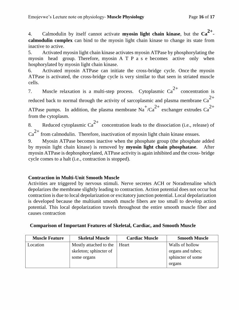
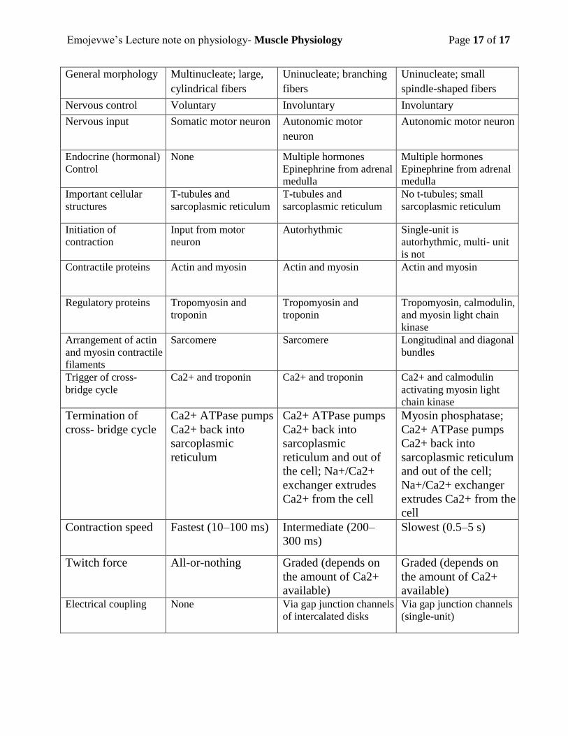
Comparison of Important Features of Skeletal, Cardiac, and Smooth Muscle
Muscle Feature Skeletal Muscle Cardiac Muscle Smooth Muscle
Location Mostly attached to the
skeleton; sphincter of
some organs
Heart Walls of hollow
organs and tubes;
sphincter of some
organs
Emojevwe’s Lecture note on physiology- Muscle Physiology Page 17 of 17
General morphology Multinucleate; large,
cylindrical fibers
Uninucleate; branching
fibers
Uninucleate; small
spindle-shaped fibers
Nervous control Voluntary Involuntary Involuntary
Nervous input Somatic motor neuron Autonomic motor
neuron
Autonomic motor neuron
Endocrine (hormonal)
Control
None Multiple hormones
Epinephrine from adrenal
medulla
Multiple hormones
Epinephrine from adrenal
medulla
Important cellular
structures
T-tubules and
sarcoplasmic reticulum
T-tubules and
sarcoplasmic reticulum
No t-tubules; small
sarcoplasmic reticulum
Initiation of
contraction
Input from motor
neuron
Autorhythmic Single-unit is
autorhythmic, multi- unit
is not
Contractile proteins Actin and myosin Actin and myosin Actin and myosin
Regulatory proteins Tropomyosin and
troponin
Tropomyosin and
troponin
Tropomyosin, calmodulin,
and myosin light chain
kinase
Arrangement of actin
and myosin contractile
filaments
Sarcomere Sarcomere Longitudinal and diagonal
bundles
Trigger of cross-
bridge cycle
Ca2+ and troponin Ca2+ and troponin Ca2+ and calmodulin
activating myosin light
chain kinase
Termination of
cross- bridge cycle
Ca2+ ATPase pumps
Ca2+ back into
sarcoplasmic
reticulum
Ca2+ ATPase pumps
Ca2+ back into
sarcoplasmic
reticulum and out of
the cell; Na+/Ca2+
exchanger extrudes
Ca2+ from the cell
Myosin phosphatase;
Ca2+ ATPase pumps
Ca2+ back into
sarcoplasmic reticulum
and out of the cell;
Na+/Ca2+ exchanger
extrudes Ca2+ from the
cell
Contraction speed Fastest (10–100 ms) Intermediate (200–
300 ms)
Slowest (0.5–5 s)
Twitch force All-or-nothing Graded (depends on
the amount of Ca2+
available)
Graded (depends on
the amount of Ca2+
available) Electrical coupling None Via gap junction channels
of intercalated disks
Via gap junction channels
(single-unit)