Embed Size (px)
Citation preview

Muscle
Physiology

3 Types of Muscles
1. skeletal- longest muscle cell,
striated, voluntary, elongated,
multi-nucleated
2. cardiac- heart muscle, striated,
involuntary, intercalated discs
3. smooth- non-striated, involuntary,
visceral muscle, slow and
sustained contractions, single
nucleus, less connective tissue
than skeletal muscle

3 Types of Muscles
Skeletal
Muscle
Smooth
Muscle
Cardiac
Muscle

Muscle Functions
• produces movement
• maintains posture
• stabilizes joints
• generates heat

Muscle Characteristics
• excitability- ability to receive and respond to a stimulus
• contractility- ability to shorten- special to muscle tissue
• extensibility- ability to stretch or lengthen beyond resting length
• elasticity- ability to recoil and resume its resting length

Organization of Skeletal Muscle
• muscle fibers are covered with
the endomysium and bundled into
groups or fasicles covered by
perimysium;
• fasicles are bundled and covered
by epimysium; the connective
tissue coverings are continuous
with the tendons connected to the
bones

Organization of Skeletal Muscle

Attachment
• insertion- the movable bone
• origin- immovable or less movable
bone (usually proximal)
• direct attachment- muscle
attaches directly to the bone
• indirect attachment- muscle
attaches through a tendon

Attachment

Muscle Cell Anatomy • sarcolemma- plasma membrane
• sarcoplasm- cytoplasm
• myoglobin- red pigment, stores oxygen in
muscles
• myofibrils- contractile elements of skeletal
muscles
• sarcomere- myofibril region between 2
successive Z discs
• A band- thick myofilaments – myosin
• I band- thin myofilaments – actin
• M line- center of the H zone
• Z disc- anchors the thin filaments and
connects the myofibrils
• H zone- area within the A band where thin and
thick filaments do not overlap

The Sarcomere

Myosin- thick filaments
– 2 interwoven protein chains
– 2 globular heads terminate the
chain AKA: cross bridges, myosin
heads
– bundles of myosin form a central
smooth section and its ends are
studded with myosin heads
– actin and ATP binding sites are
present

Actin- long, thin filaments
– helical structure with active
binding sites for myosin heads
– contains tropomyosin- a protein
to help stiffen the actin core; in
relaxed muscles they block
myosin binding sites
– contains troponin- 3 protein
complex
1. binds to actin
2. binds to tropomyosin and
helps position it
3. binds to Ca+2

Elastic Filaments
–composed of titin
–holds the thick filament in
place
–helps the muscle stretch and
recoil

Muscle Contraction
• Contraction is stimulated by the muscles
single motor nerve innervation which forms a
neuromuscular junction.
• Neuromuscular junction- axonal end of the
motor neuron and muscle fiber which are in
close contact, yet separated by a gel like
space, the synaptic cleft.
• Synaptic vesicles- membrane sacs filled with
acetylcholine (ACh)
• Motor end plate- highly folded area of
sarcolemma with ACh receptors
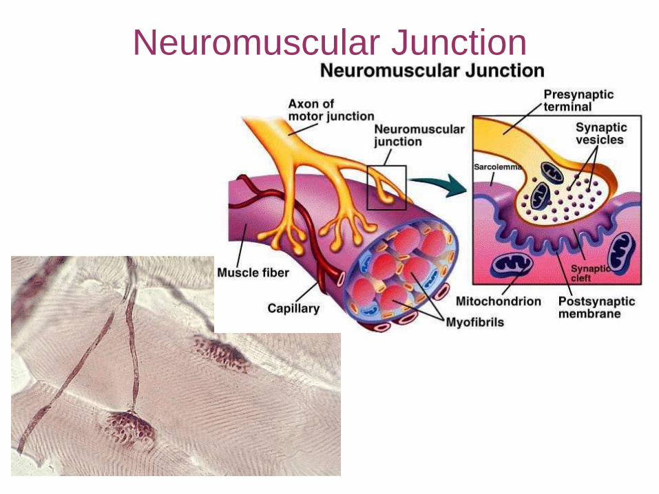
Neuromuscular Junction

Excitation-Contraction Coupling
• occurs during the latent period, the
time from action potential to
mechanical activity
• action potential continues down the
sarcolemma
• calcium ions are released and binds to
troponin, changing shape and removes
the blocking action of tropomyosin
• myosin cross bridges become
activated
– Sliding Filament Theory Occurs (To Come)
• calcium level falls and tropomyosin
blocks active sites

Muscle Contraction
Sliding Filament Theory of Contraction-
• Activated myosin heads are attracted
to exposed actin binding sites & cross
bridge attachment occurs
• The myosin head changes shape and
pulls on the actin sliding it to the
center of the sarcomere (ATP is used)
• A new ATP molecule binds to the
myosin head. The cross bridge
detaches from the actin.
• ATP causes the myosin head to change
shape and it reattaches to the actin to
repeat the process.

Sliding Filament Theory
View the Sliding Filament

Muscle Contraction
Stopping A Muscle Contraction
1. Nerve communication to contract
ends.
2. Lack of ATP will stop a contraction.
3. Low calcium ion concentration will
slow or stop contraction.
- calcium channel blockers – treat high blood
pressure – calcium is not released so
heart cannot beat as quickly

2 Categories of Contractions
1. isotonic contractions- muscles
change length, decreases the
angle at the joint
– concentric contractions- muscle
shortens
– eccentric contractions- muscle
contracts as it lengthens
2. isometric contractions- muscle
does not shorten or lengthen yet
tension continues to increase

Categories of Contractions

Classification of Muscle Fibers
• Speed of contraction- how fast or slow
they contract based upon how fast ATP
is split
• The major pathway for ATP production
– oxidative fibers- aerobic pathways
– glycolytic fibers- anaerobic path
(glycolysis)
• 3 types of skeletal muscle cells: slow
oxidative fibers, fast oxidative fibers,
fast glycolytic fibers

Myograms Drawings or tracings of the electrical current as it
travels through a muscle.
Twitch
Contraction-
one muscle cell
contracting,
one time

Myograms
4 Phases of a Twitch Contraction
1. Latent Phase
-AP has started
-Ca+2 is being released from sarcoplasmic
reticulum -Ca+2 is binding to troponin
-tropomyosin is exposing myosin binding
sites
2. Contraction Phase
-AP continues
-Na+1 moves into the cell -Muscle cell becomes depolarized

Myograms 4 Phases of a Twitch Contraction
3. Refractory Period
-cell has no membrane potential
-cell cannot respond to any neural stimulus
4. Relaxation Phase
-cell is repolarizing by pumping sodium out of
the cell
-will regain membrane potential

Strength of Contraction
Two ways to control the strength of a muscle contraction
1. By the size and number of motor units we recruit
2. Frequency of stimulation
3. Amount of Calcium ion released
4. Hydration
5. pH of sarcoplasm
6. Temperature of the muscle
7. Amount of stretch before contraction

Muscle Fatigue
• weakness and loss of contractility
Causes
• ATP synthesis declines
• ATP shortage slows Na+/K+ pump
• Motor nerves use their supply of Ach – junctional fatigue
• Lactic acid lowers pH of sarcoplasm
• K+ builds in extracellular fluid

Muscle Strength
Strength of muscle depends upon
• Muscle size
• Fasicle arrangement
• Size of active motor units
• Multiple motor unit summation
• Temporal summation
• Fatigue
• Length-tension relationships

Energy Requirements
Energy is provided by ATP & /CP
Night→Produce Extra ATP→ATP breakdown occurs to form ADP and Pi→Creatine Kinase attaches the Pi to creatine→Creatine Phosphate (CP)
First Energy Source –
Creatine Kinase reverses the reaction to remove Pi and then put the Pi with ADP to form ATP
Second Energy Source – Glycolysis
Third Energy Source – Cellular Respiration

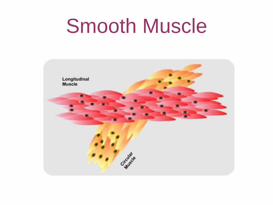
Smooth Muscle Types
• single unit or visceral muscle-
contracts as a unit and
contracts rhythmically
• multi-unit- independent cells,
graded contractions, lots of
nerve endings

Smooth Muscle - Smooth muscle has dense bodies with
intermediate filaments attached
- As the muscle contracts, it forms a spiral or corkscrew that pulls the dense bodies together
- The dense bodies are pulling at different angles

Smooth Muscle
– 2 layers of smooth muscle are present at
right angles
– longitudinal- dilate & shorten the
organ
– circular- constricts the cavity of the
organ and causes the organ to
elongate
• peristalsis- the alternating contraction and
relaxation of the opposing longitudinal and
circular layers - mixes substances and
squeezes them through the organ's internal
pathway
Smooth Muscle