Embed Size (px)
Citation preview
SECOND INTERNATIONAL WORKSHOP ON CYTOKINES / 111
229 232
CYmINE-INDUCED EXPRESSION OF HLA CLASS II ANTIGBN RY INSULIN (INS), EPIDERMAL GROWTH FACTOR (EGF), AND SOMATAMEDIN CULTURED HU!RN SYNOVIAL CELLS. Herbert 8. Lindsley and Donald D. Smith, Univ Kansas Medical Center, Kansas City, KS f35107. __-_-.
Adherent rheumatoid synovial cells (2nddth passage), cultured from patients undergoing joint replacement, were stimulated with human recombinant cytokines: interferon-gamms (IFN), interleukin-l-beta (IL-l), tumor necrosis factor-alpha (TNF) orz platelet-deeived growth factor (Pm), for 48-144 hr in culture medium (DNEM) containing 10% fetal calf serum. HLA Class II surface antigen was measured in a Cell ELISA microplate assay using monoclonal anti-class II Ab (L243) with an irrelevant Ab (P117) as a negative control. Class II Ag "as minimally present on non-cytokine-treated cells. IFN (10-1000 U/ml) yielded a time- and dose-dependent increase of 5500-9000% in Class II expression, which declined within 24 hr after removal of the cytokine. IL-1 (30 U/ml) "as less potent, showing an 800% increase: TNF (50 U/ml) showed a minimal increase, and PDGF (10 "g/ml) showed none. HLA Class II antigen is inducible in hum" synovial cells, and may allow these cells to participate in antigen pcesentation within the joint. (Supported in part by Pfizer, Inc.).
C (IGFl) AUGMENT IL-Z or IL-2 PLUS CONCANAVALIN A (CON A)- INDUCED RAT T CELL PROLIFERATION. A. Al Maghazachi, L. FitzCibbon and J. Mitchell. Biotechnology Research Institute, NRCC. 6100 Royalmount Avenue, MontrBs.1, QuBbec, Canada H4P 2R2
INS synergizes with a low dose of IL-2, or a low dose of IL-2 plus a suboptimal dose of CON A for T cell proliferation when added for 3 days with purified rat splenic T cells. Addition of CL INS receptor antibody (aIR-1) at days 0, 1 or 2 inhibits INS activity, suggesting that continuous contact of INS with T cells is necessary. EGF also synergizes with IL-2 or IL-Z plus CON A for T cell proliferation. Addition of IGFl does not augment EGF enhancing activity. However, addition of IGFl synergizes with CON A plus IL-2 when added at day 0, but not at days 1 or 2 of culture, indicating that ICFl must bind to T cells at the onset of the culture and that CON A must be present. Addition of aIR-1 inhibits IGFl activity when the antibody is added at day 1 but not at days 1 or 2 of culture suggesting that IGFl initially binds to INS
receptors, which may then induces the expression of the IGFI receptors. Such expression is probably potentiated by the presence of CON A. The effects of these growth factors are only observed after 3 days of culture with T cells, in the presence Of IL-2, and in serum-supplemented but not serum-free medium.
I" summary, these results provide evidence for novel effects of the endocrine system on the immune system.
230
PROSTAGLANDIN El BIOSYNTHESIS IN CULTURED CELLS SENSITIVE TO TNF-> (LYMF’HOTOXIN). K. J. Lonemuir. P. Se acuse. a d G. A. GranaeG Depts. of Physiology & Biophysics, and MAlecular biology & Biochemistry, Univ. of Calif., Irvine, CA 92717.
This study examined the time course of prostaglandin E2 for&nation
233
MULTIPLICATION STIMULATING FACTOR (IGF2) AUGMENTS THE PROLI- FERATION OF IL-2-PRIMED T CELLS. INHIBITION BY CYCLIC AMP ACTIVATORS. A. Al Maghazachi and L. FitzGibbon. Molecular Immunology Section, Biotechnology Research Institute, NRCC, 6100 Royalmount Ave., Montr&al, Quebec, Canada H4P 2R2.
IGF2 synergizes with a low dose of IL-2 for rat T cell induced by TNF-p (lymphotoxin). Mouse L929 cells (0.8-1.0 x 10 cells per 35 mm dish) were treated with 60,000 units per dish human recombinant TNF-8. Levels of PGE2 in the culture medium, O-33 h after addition of TNF, were measured by radioimmunoassay. PGE2 was undetectable at 0 h, rose during the first 6 h after TNF treatment, and remained essentially constant thereafter. After 6 h, PGE2 averaged 8.6 + 1.4 ng per dish (SE, n=S). All control dishes contamed less than 2 “g/dish PGE (0.93 2 0.18 “g/dish (n=S) after 6 h). In separate experiments, L 9% 9 cells were label d with 3H-arachido”ic
s acid prior to addition of TNF-p. Levels of H-labeled PGE2 in the lipid-extracted cudture medium were determined by thin layer chromatography. H-PGE rose during the initial 4 h after addition of TNF-@ to a level four-fol a lugher than control cultures, with no further increase thereafter. Indomethacin (50 pM) blocked PGE formation, and radiolabeled 5-hydroxyeicosatetraenoic acid (5- B ETE) was observed instead. This 4-6 h period of PGE2 biosynthesis represents a remarkably early event in the time course of TNF action on L929 cells. It occurs several hours before observable cell killing, and several hours before observable phospholipase activation as measured by loss of labeled arachidonic acid from glycerophospholipids. (Supported by ACS #IN-l66 and the Univ. Calif. Cancer Research Coordinating Committee.)
proliferation. Addition of CON A, EGF, INS, ICFl or INS plus IGFl did not enhance IGF2 activity. However, addition of EGF or Mannose-6-phosphate "M6P" which binds the same receptors as IGF2 (i.e. IGFII/M6P receptor) plus a suboptimal dose of CON A highly increased IGF2 activity. MlP was unable to enhance IGF2 activity. Also, M6P added to T cells plus IL-2, or T cells plus IL-2 plus CON A without IGF2 did not result in enhancement. These results suggest that primed T cells are increasingly triggered by IGF2, and this activity is potentiated by M6P or EGF.
231 234
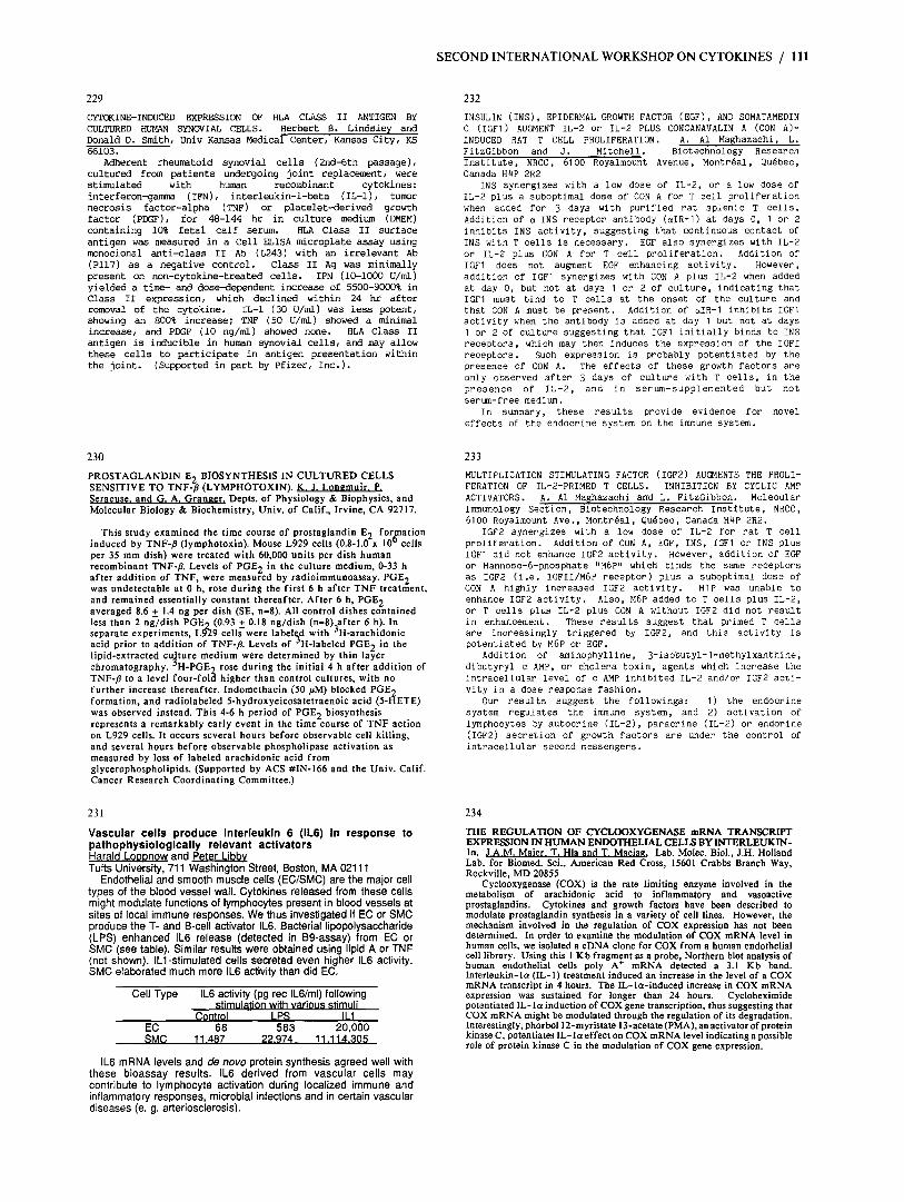
Vascular cells produce interleukln 6 (IL6) in response to oathoohvsloloalcallv relevant activators karald’ I &mow-and Peter I ibbv Tufts Universitv. 711 Washinaton Street. Boston. MA 02111
THE REGULATION OF CYCLOOXYGENASE mRNA TRANSCRIPT EXPRESSION IN HUMAN ENDOTHELIAL CELLS BY INTERLEUKIN- la. J.A.M. Maier. T. Hla and T. Maci8g. Lab. Molec. Biol., J.H. Holland Lab. for Biomed. Sci., American Red Cross, 15601 Crabbs Branch Way, Rockville, MD 20855 Endothelial and smooth m&.cle cells (EClSMk) are the major cell
types of the blood vessel wall. Cytokines released from these cells might modulate functions of lymphocytes present in blood vessels at sites of local immune responses. We thus investigated if EC or SMC produce the T- and B-cell activator IL6. Bacterial lipopolysaccharide (LPS) enhanced IL6 release (detected in BS-assay) from EC or SMC (see table). Similar results were obtained using lipid A or TNF (not shown). Ill-stimulated cells secreted even higher IL6 activity. SMC elaborated much more IL6 activity than did EC.
Cell Type IL6 act$iy i(pg ret IL6!ml) fol!;w;g st u at on with various stl u
Control LPS II 1 EC 68 503 20.000 SMC i 1.487 22.974 11 .114.305
IL6 mRNA levels and de novo protein synthesis agreed well with these bioassay results. IL6 derived from vascular cells may contribute to lymphocyte activation during localized immune and inflammatory responses, microbial infections and in certain vascular diseases (e. g. arteriosclerosis).
Addition of aminophylline, 3-isobutyl-1-methylxanthine, dibutyryl c AMP, or cholera toxin, agents which increase the intracellular level of c AMP inhibited IL-2 and/or IGF2 acti- vity in a dose response fashion.
Our results suggest the followings: 1) the endocrine system regulates the immune system, and 2) activation of lymphocytes by autocrine (IL-2), paracrine (IL-2) or endorine (IGF2) secretion of growth factors are under the control of intracellular second messengers.
Cyclooxygenase (COX) is the rate limiting enzyme involved in the metabolism of arachidonic acid to inflammatory and vasoactive prostaglandins. Cytokines and growth factors have been described to modulate prostaglandin synthesis in a variety of cell lines. However, the mechanism involved in the regulation of COX expression has not been determined. In order to examine the modulation of COX mRNA level in human cells, we isolated a cDNA clone for COX from a human endothelial cell library. Using this 1 Kb fragment as a probe, Northern blot analysis of human endothelial cells poly A+ mRNA detected a 3.1 Kb band. Interleukin-lo (IL-l) treatment induced an increase in the level of a COX mRNA transcript in 4 hours. The IL-la-induced increase in COX mRNA expression was sustained for longer than 24 hours. Cycloheximide potentiated IL-la induction of COX gene transcription, thus suggesting that COX mRNA might be modulated through the regulation of its degradation. InTerestingly, phorbol 12-myristate 13-acetate(PMA),a” activator of protein kinase C, potentiates IL- la effect on COX mRNA level indicating a possible role of protein kinase C in the modulation of COX gene expression.