Embed Size (px)
Citation preview
University of KentuckyUKnowledge
Physiology Faculty Publications Physiology
6-7-2016
Multiorgan Chronic Inflammatory HepatobiliaryPancreatic Murine Model Deficient in TumorNecrosis Factor Receptors 1 and 2Helieh S. OzUniversity of Kentucky, [email protected]
Right click to open a feedback form in a new tab to let us know how this document benefits you.
Follow this and additional works at: https://uknowledge.uky.edu/physiology_facpub
Part of the Gastroenterology Commons, Internal Medicine Commons, and the PhysiologyCommons
This Article is brought to you for free and open access by the Physiology at UKnowledge. It has been accepted for inclusion in Physiology FacultyPublications by an authorized administrator of UKnowledge. For more information, please contact [email protected].
Repository CitationOz, Helieh S., "Multiorgan Chronic Inflammatory Hepatobiliary Pancreatic Murine Model Deficient in Tumor Necrosis FactorReceptors 1 and 2" (2016). Physiology Faculty Publications. 84.https://uknowledge.uky.edu/physiology_facpub/84
Multiorgan Chronic Inflammatory Hepatobiliary Pancreatic Murine Model Deficient in Tumor Necrosis FactorReceptors 1 and 2
Notes/Citation InformationPublished in World Journal of Gastroenterology, v. 22, issue 21, p. 4988-4998.
© 2016 Baishideng Publishing Group Inc. All rights reserved.
This article is an open-access article which was selected by an in-house editor and fully peer-reviewed byexternal reviewers. It is distributed in accordance with the Creative Commons Attribution Non Commercial(CC BY-NC 4.0) license, which permits others to distribute, remix, adapt, build upon this work non-commercially, and license their derivative works on different terms, provided the original work is properlycited and the use is non-commercial. See: http://creativecommons.org/ licenses/by-nc/4.0/
Digital Object Identifier (DOI)https://doi.org/10.3748/wjg.v22.i21.4988
This article is available at UKnowledge: https://uknowledge.uky.edu/physiology_facpub/84

Helieh S Oz, Department of Physiology and Internal Medicine, College of Medicine, University of Kentucky Medical Center, Lexington, KY 40536, United States
Author contributions: Oz HS designed and executed the study, prepared the paper and provided funding; author has approved the final paper.
Supported by The NIH grant R03-DE019177.
Institutional animal care and use committee statement: All animal procedures were approved by the University of Kentucky Institutional animal care and use committee.
Conflict-of-interest statement: Author declares to have no commercial or associative interest that represents a conflict of interest in connection with the work submitted.
Data sharing statement: No additional data are available.
Open-Access: This article is an open-access article which was selected by an in-house editor and fully peer-reviewed by external reviewers. It is distributed in accordance with the Creative Commons Attribution Non Commercial (CC BY-NC 4.0) license, which permits others to distribute, remix, adapt, build upon this work non-commercially, and license their derivative works on different terms, provided the original work is properly cited and the use is non-commercial. See: http://creativecommons.org/licenses/by-nc/4.0/
Correspondence to: Helieh S Oz, DVM, PhD, AGAF, Department of Physiology and Internal Medicine, College of Medicine, University of Kentucky Medical Center, Lexington, KY 40536, United States. [email protected]: +1-859-2770988Fax: +1-859-3231000
Received: January 15, 2016Peer-review started: January 18, 2016First decision: February 18, 2016Revised: March 21, 2016
Accepted: April 7, 2016Article in press: April 7, 2016Published online: June 7, 2016
AbstractAIM: To provoke persistent/chronic multiorgan inflam-matory response and to contribute to stones formation followed by fibrosis in hepatobiliary and pancreatic tissues.
METHODS: Tumor necrosis factor receptors 1 and 2 (TNFR1/R2) deficient mice reared in-house were given dibutyltin dichloride (DBTC) twice within 10 d by oral gavage delivery. Sham control animals received vehicle treatment and naïve animals remained untreated throughout the study. Animals were monitored daily for symptoms of pain and discomfort. The abdominal and hindpaw hypersensitivity were assessed with von Frey microfilaments. Exploratory behaviors were recorded at the baseline, after initiation of treatment, and before study termination. Histopathological changes were examined postmortem in tissues. Collagen accumulation and fibrosis were confirmed with Sirius Red staining.
RESULTS: Animals lost weight after oral administration of DBTC and developed persistent inflammatory abdominal and hindpaw hypersensitivity compared to sham-treated controls (P < 0.0001). These pain related secondary mechanical hypersensitivity responses increased more than 2-fold in DBTC-treated animals. The drastically diminished rearing and grooming rates persisted after DBTC administration throughout the study. Gross as well as micropathology at one month confirmed that animals treated with DBTC developed chronic hepatobiliary injuries evidenced with activation of stellate cells, multifocal necrosis, fatty degeneration
Submit a Manuscript: http://www.wjgnet.com/esps/Help Desk: http://www.wjgnet.com/esps/helpdesk.aspxDOI: 10.3748/wjg.v22.i21.4988
4988 June 7, 2016|Volume 22|Issue 21|WJG|www.wjgnet.com
World J Gastroenterol 2016 June 7; 22(21): 4988-4998 ISSN 1007-9327 (print) ISSN 2219-2840 (online)
© 2016 Baishideng Publishing Group Inc. All rights reserved.
ORIGINAL ARTICLE
Multiorgan chronic inflammatory hepatobiliary pancreatic murine model deficient in tumor necrosis factor receptors 1 and 2
Basic Study
Helieh S Oz

of hepatocytes, periportal infiltration of inflammatory cells, and prominent biliary ductal dilation. The severity of hepatitis was scored 3.7 ± 0.2 (severe) in DBTC-treated animals vs score 0 (normal) in sham-treated animals. Fibrotic thickening was extensive around portal ducts, in hepatic parenchyma as well as in lobular pancreatic structures and confirmed with Sirius Red histopathology. In addition, pancreatic microarchitecture was presented with distortion of islets, and parenchyma, infiltration of inflammatory cells, degeneration, vacuolization, and necrosis of acinar cells and distention of pancreatic ducts. Extent of pancreatic damage and pancreatitis were scored 3.6 ± 0.4 (severe) for DBTC-treated in contrast to score 0 (normal) in sham-treated animals. The gall bladder became expanded with ductal distention, and occasional bile stones were detected along with microscopic hepatic lesions. DBTC-treated animals developed splenic hypertrophy with increased weight and length (P < 0.01) along with thymic atrophy (P < 0.001). Finally, colitic lesions and colitis were prominent in DBTC-treated animals and scored 3.4 ± 0.3 (moderately severe) vs 0 (normal) for the sham-treated animals.
CONCLUSION: This is the first report of chronic inflam-matory multiorgan hepatobiliary pancreatitis, along with fibrosis and calculi formation induced reliably utilizing oral DBTC administration in TNFR1/R2 deficient mice.
Key words: Inflammatory pain; Multiorgan; Hepatitis; Pancreatitis; Calculi formation; Gall bladder; Hepatobiliary inflammation
© The Author(s) 2016. Published by Baishideng Publishing Group Inc. All rights reserved.
Core tip: Currently there is no reliable model for chronic multiorgan inflammatory and fibrosis. Tumor necrosis factor (TNF)α initiates inflammation through TNFR1/R2. TNFR1/R2 deficient mice administered orally with dibutyltin dichloride (DBTC) developed significant persistent inflammatory and pain related secondary mechanical hypersensitivity. DBTC-animals showed severe chronic hepatobiliary injuries and prominent biliary ductal dilation. Extensive fibrotic thickening was evidenced around portal ducts, in hepatic and pancreatic structures. DBTC-animals had severe pancreatic damage and pancreatitis, hepatic lesions with expansion of gall bladder, bile stones and severe colitis. This is the first report of chronic inflammatory multiorgan hepatobiliary pancreatitis, fibrosis and calculi formation in TNFR1/R2 deficient mice.
Oz HS. Multiorgan chronic inflammatory hepatobiliary pancreatic murine model deficient in tumor necrosis factor receptors 1 and 2. World J Gastroenterol 2016; 22(21): 4988-4998 Available from: URL: http://www.wjgnet.com/1007-9327/full/v22/i21/4988.htm DOI: http://dx.doi.org/10.3748/wjg.v22.i21.4988
INTRODUCTIONFibrogenesis is a required process in wound healing, but persistent inflammatory and fibrotic reaction can lead to devastating symptoms and eventually organ failure[1,2]. Multiorgan fibrosis is typically the end product of various unresolved or repetitive tissue injuries from chronic inflammation, infection, radiation exposure, and abnormal repair outcome. Loss of function contributes to progression of morbidity and mortality. Multiorgan fibrosis is a common complication in cystic fibrosis[3], systemic sclerosis[4] and primary sclerosing cholangitis[5]. Chronic pancreatitis, initiated by idiopathic or recurrent inflammation, is manifested with irreversible destruction of exocrine parenchyma and pancreatic fibrosis. It is a potentially fatal progressive disease leading to diabetes mellitus and pancreatic cancer. Pancreatitis is associated with spontaneous visceral pain as a chief symptom in patients. Neural innervation of the pancreas is pivotal in the instigation and continuation of inflammation and pain response. Cellular destruction leads to activation of pancreatic sensory neurons causing release of neurotransmitters in the spinal cord and neurogenic signaling then back to the pancreas provoking plasma extravasation and neutrophil infiltration[6].
Multifactoral gallstones are one of the most prevalent gastrointestinal complications with serious outcomes such as gallstone pancreatitis and cancer. Gallstone disease is a chronic recurrent hepatobiliary complication which is characterized by formation of gallstones in the hepatic and bile duct, or gallbladder. It is manifested by impaired metabolism of cholesterol, bilirubin and bile acids[7].
Tumor necrosis factor α (TNFα) a proinflammatory cytokine, upregulates various cytokines/chemokines to initiate acute and chronic stages of inflammation. The biological action of TNFα is chiefly through two gene family receptors, TNFR1 and TNFR2. TNFα is released mainly by activated macrophages, in addition to astroglia, microglia, CD4+ lymphocytes, Natural killer cells (NK), and neurons[810]. The completelength membranecrossing TNFα (mTNFα) is sliced by the inducible TNF converting enzyme (TACE) to release soluble TNFα (sTNFα) and diffusible peptide[11]. TNFα release is associated with inflammatory response and pain related sensation in patients with pancreatitis, hepatitis and inflammatory bowel disease (IBD), as well as neuropathy[12]. TNFα contributes to development of neuropathic pain[13]. Soluble TNFR1 and R2 neutralize circulating TNFα to alleviate pain related responses to mechanical allodynia, thermal hyperalgesia or peripheral nerve injuries[1416]. TNFα plays an important function in the pathogenesis of acute pancreatitis. Recent investigations have demonstrated that TNFα inhibition drastically ameliorates the duration of experimental acute pancreatitis[17]. TNFα receptor 1 (TNFR1) gene deletion and etanercept application likewise ameliorated the duration of acute pancreatitis
4989 June 7, 2016|Volume 22|Issue 21|WJG|www.wjgnet.com
Oz HS. Chronic inflammatory multiorgan hepatobiliary pancreatitis model
in animal models, suggesting potential of etanercept and antiTNFα monoclonal antibodies as therapy in clinical pancreatitis[17]. Although, current clinical treatments with these biological agents may diminish inflammation and pain by reducing TNFα and other cytokines, the inflammatory response and pain is likely to reemerge in most patients with autoimmune disease including arthritis and IBD[10]. In addition, anti-TNFα monoclonal antibodies therapy has potential side effects such as provoking infections with JC virus, fungi and tuberculosis. Currently there is no cure or reliable mouse model for chronic pancreatitis.
Previously we have demonstrated that the baseline mechanical and thermal response to noxious stimulation is similar in TNFR1/R2 deficient mice vs wildtype background mice. However, TNFR1/R2 deficient mice develop more severe responses when similarly treated with various insults[10,16]. Animal models of acute and chronic pancreatitis have been utilized to examine mechanisms of pathogenesis, and to test possible therapeutic interventions. One of the most commonly used pancreatitis models is created by serial intraperitoneal administration of concentrations of caerulein, an ortholog of cholecystokinin[17]. Other chemically induced models have utilized dinbutyltin dichloride (DBTC). DBTC is a polyvinyl carbonate (PVC) plastic stabilizer/catalyzer additive, insecticide and biocide in agriculture, and antifouling agent in the paint and fabric industry that often contaminates food and water[18]. Tail vein slow injection of DBTC induces relatively unpredictable pancreatitis flares in rats[6]. However, DBTC injection is tedious and minor leakage results in tail necrosis, gangrene and animal loss. We hypothesized that oral administration of DBTC would provoke persistent and chronic pancreatitis in animals deficient in TNFreceptors. Similarly, TNFR1/R2 may accelerate inflammatory response in multiorgans and contribute to stones formation and fibrosis in hepato-biliary and pancreatic tissues. Here we report a chronic persistent DBTC-induced inflammatory model by oral gavage in TNFR1/R2 deficient mice persisting at least one month allowing more clinically relevant studies in this model. Pain related behaviors accompanying this model are characterized.
MATERIALS AND METHODSAnimalsAll animal procedures were approved by the University of Kentucky Institution Animal Care and Use Committee (IACUC). Mice were monitored daily for continued weight gain/loss and general health. Health status and procedures were documented daily on the UK IACUC Standard Operating Procedure (SOP-102) Post-Operative Evaluation form. Experiments were performed using dually deficient TNFR1/R2 mice (Jackson Laboratory) on a B6129SF2/J background inbred at the University of Kentucky animal facilities
and provided by Dr. Westlund. Mice were housed in individual cages with a 10 h/14 h dark/light reversed cycle to accommodate behavioral test during their active dark period. Mice were allowed free access to food and water ad libitum, except 2 h before and during behavioral testing.
Induction of persistent chronic pancreatitis Chronic persistent pancreatitis was induced in mice utilizing DBTC (Dibutyltin dichloride, Sigma-Aldrich, St Louis, MO). DBTC (10 mg/kg) was dissolved in 95% ethanol (two parts) and then mixed with glycerol (three parts) and given orally. Mice received DBTC by oral gavage (200 μL volume). Intragastric gavage administration was performed by Dr. Oz, an expert veterinarian scientist, in conscious animals, using appropriate bended gavage needles (22 gauge, 1 inch length, 1.25 mm ball diameter). Sham control mice were given the vehicle (95% ethanol + glycerol, 2:3) alone and Naïve control animals remained untreated. Animals were monitored until fully active. In order to induce chronic inflammation, mice received a 2nd
treatment by oral gavage within 10 d. Following induction of pancreatitis the animals were monitored daily for activity, appearance, and signs of abdominal discomfort. They were weighed regularly and tested for hypersensitivity on the hindpaw plantar foot pad and the shaved abdominal surface. After completion of the final behavioral testing, one month after induction, the animals were euthanatized with isoflurane overexposure, the thorax opened, blood samples collected by cardiac puncture, and tissue samples collected for histological evaluation.
Assessment of secondary mechanical allodynia by testing hindpaw withdrawal threshold Painrelated behavior was assessed throughout the study by the determining secondary mechanical threshold to assess hyperalgesia/allodynia. The von Frey test is a standard comparison used in the field of pain research. Day 0, baseline testing to determine footpad nociceptive responses was performed testing hindpaw withdrawal latency to mechanical stimuli with von Frey fibers. Reflex testing for secondary mechanical hyperalgesia/allodynia with von Frey fibers was developed by Max von Frey, who in 1896 identified “pain spots” on human skin. Mechanical nociceptive thresholds were analyzed as described previously[10,19]. Paw withdrawal response latencies were assessed weekly throughout the study. Mice were placed into clear cylindrical plastic enclosures (7 cm × 4 cm × 4 cm) on a smooth metal meshed (3 mm × 3 mm) platform (36 cm × 29 cm × 21.5 cm). Mechanical withdrawal threshold testing was done on the plantar surface of both hindpaws using a set of 8 von Frey monofilaments [(4.74) 6.0 g; (4.31) 2.0 g; (4.08) 1.0 g; (3.61) 0.4 g; (3.22) 0.16 g; (2.83) 0.07 g; (2.36) 0.02 g; (1.65) 0.008 g]. The von Frey
4990 June 7, 2016|Volume 22|Issue 21|WJG|www.wjgnet.com
Oz HS. Chronic inflammatory multiorgan hepatobiliary pancreatitis model
4991 June 7, 2016|Volume 22|Issue 21|WJG|www.wjgnet.com
against the floor, and abdomen retractions or arching the back. Recordings were masked and analyzed by the investigator.
Necropsy and sample collection Tissue collection: At the end of the one month ex-periment, animals were deeply anesthetized with isoflurane inhalation. Pancreatic, hepatic, gall bladder tissues were excised and a portion was fixed in cold 4% paraformaldehyde in 0.1 mol/L phosphate buffer saline (PBS). Thymus and splenic tissues were removed, weighed, and fixed in paraformaldehyde. Colonic tissues were removed and flushed with cold PBS, and portion of ascending and descending colon were fixed for histopathological examinations.
HistopathologyHepatic and pancreatic samples were collected and immerse fixed overnight in 4% paraformaldehyde in 0.1 mol/L PBS, then transferred into 70% ethanol and embedded in paraffin. Sections were cut (5 μm), rehydrated, stained with hematoxylin and eosin for histopathological changes. In order to detect collagen fiber deposits, sections were further stained with Sirius Red (Electron Microscopy Sciences, #2635702), using routine histological protocols[20].
Pancreatitis scoresPancreatic tissues and a portion of the small intestine along with spleen were removed and processed for histopathological evaluations of the pancreatitis. The severity of lesions was scored on a 04 grade on the basis of the histopathological changes as follows: 0 - normal pancreatic microstructure, no inflammatory mononuclear cell infiltration; 1 slight inflammatory mononuclear cell infiltration, with no detectable parenchymal destruction; 2 mild pancreatitis, edema, focal parenchymal destruction with mononuclear cell infiltration; 3 moderate pancreatitis, with diffuse parenchyma destruction, presence of necrosis, and reduced number of islets; and 4 severe pancreatitis, parenchyma mostly destroyed and replaced with adipose tissues, loss of pancreatic islets, presence of fibrosis and or calculi.
Hepatitis score A portion of the right lobe from liver tissues of each mouse was placed in an embedding cassette and fixed in paraformaldehyde as mentioned above. The specimens were dehydrated and embedded in paraffin, and tissue sections of 5 μm were stained with Hematoxylin Eosin. Each slide was evaluated under Ziess light microscopy. Hepatic lesions were graded on a scale of 0 to 4+ based on degeneration, inflammation, and necrosis as follow: Grade 0 - no detectable lesions, degeneration, infiltration of inflammatory cells, normal tissue appearance; Grade 1 - focal infiltration of inflammatory cells in the tissue and
filaments were applied perpendicularly to the plantar surface with sufficient force to bend the monofilament slightly and held for about 5 s, and 5 to 10 times with 15 s intervals. A positive response was defined as an abrupt withdrawal (flick response) of the foot during stimulation or immediately after the removal of stimulus. Whenever there was a negative or positive response, the next stronger or weaker filament was applied, respectively. Testing proceeded in this manner until four fibers had been applied after the first one caused a withdrawal response, allowing the estimation of the mechanical withdrawal threshold.
Pain-related behavioral evaluations for abdomen: Prior to induction of inflammation with DBTC admi-nistration, baseline testing of abdominal nociceptive responses to mechanical stimuli was performed with von Frey fibers applied to the upper left abdominal quadrant skin of mice as previously described[10,19]. Mechanical hypersensitivity in the abdominal area was quantified by measuring the number of withdrawal events (either abdominal withdraw from the von Frey filament or consequent licking of the abdominal area, or whole body withdrawal) in response to normally innocuous or subthreshold mechanical stimuli. Testing continued weekly throughout the study.
Evaluation of the pain-related posture: The abnormal posture of each animal with an affected hindlimb was given a single score using a subjective painrelated behavioral scale (spontaneous pain rating score 05) i.e. 0 normal; 1 curling of the toes, 2 aversion of the paw; 3 partial weight bearing; 4 nonweight bearing and guarding; and 5 avoidance of any contact with the hindlimb.
Pain-related gait disturbance: Gait disturbances (curling toes, limping, guarding, rearing and grooming were tallied by an observer blinded to treatment group as in our previous studies[10].
Spontaneous visceral pain assessment: The animals were placed individually in the observation chamber for a 25 min recording session. The observation chamber is a 28 cm × 17.5 cm × 12.5 cm seethrough plastic home cage with one mirrored side located in an isolated room with constant “white noise”. A digital camera located 0.5 meter from the chamber with an unobstructed view was used to record animals spontaneous visceral pain related behaviors. The camera was linked to a computer recording program for offline data analysis (Logitech Image Studio). The chamber was washed with a detergent disinfectant and dried after each use between animals. Postures defined as statistically significant increase in visceral painrelated behavior in this study included rearing, grooming and licking of the lower abdomen, stretching the abdomen or hindlimb, lowering the abdomen
Oz HS. Chronic inflammatory multiorgan hepatobiliary pancreatitis model
4992 June 7, 2016|Volume 22|Issue 21|WJG|www.wjgnet.com
hepatocytes degeneration; Grade 2 mild multifocal infiltration of inflammatory cells, and hepatocytes degeneration; Grade 3 - moderate multifocal infiltration of inflammatory cells and hepatocytes degeneration; and Grade 4 - severe diffuse infiltration of inflammatory cells, necrosis, or fibrosis.
Colitis evaluation scoresColonic tissues were flushed with PBS (pH 7.2) and a portion from proximal and distal colonic tissues were fixed for histological examinations. The fixed sections were processed and stained with Hematoxylin Eosin and slides evaluated by Ziess light microscopy. The severity of colitis was assessed with a histological semiquantitative grading score. The scores were based on histopathological features with a numeric value (0: normal to 4: severe) assigned according to the tissue involvement corresponding to the following criteria[21,22]. Grade 0: No detectable lesions, no inflammatory cells, and normal mucosal appearance; Grade 1: Focal inflammatory infiltrate in the mucosa; Grade 2: Mild multifocal inflammation with moderate expansion into the mucosa; Grade 3: Moderate multi-focal inflammation with moderate expansion of the mucosa; and Grade 4: Severe diffuse inflammation with crypt epithelium disruption and ulceration.
Statistical analysisAll results are expressed as mean and standard error of mean (± SEM) unless otherwise stated. Data were analyzed using paired ttest comparison of groups for histology or analysis of variance (ANOVA) followed by Bonferroni post hoc comparison using GraphPad Prism Software for behavioral testing over time (San Diego, CA, United States). Statistical significance was set at p ≤ 0.05.
RESULTSBody weight loss No major differences in body weight, behavioral analysis was detected between shamtreated and
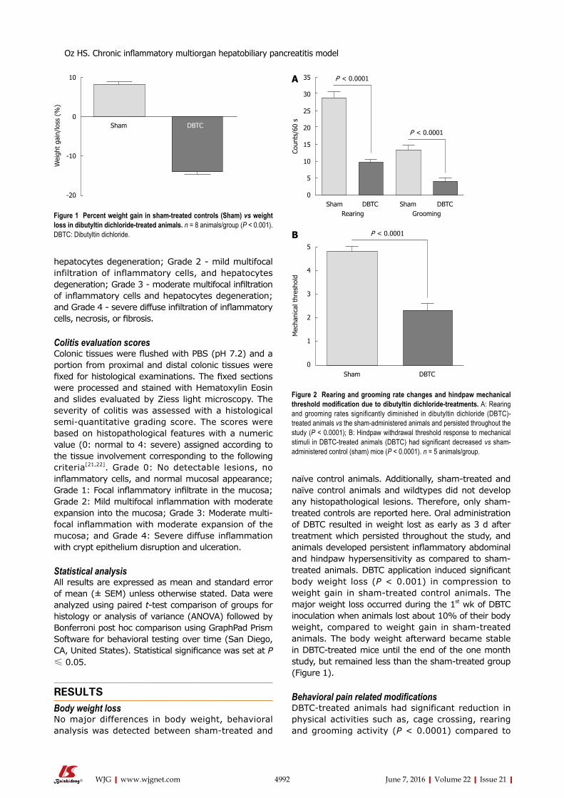
naïve control animals. Additionally, shamtreated and naïve control animals and wildtypes did not develop any histopathological lesions. Therefore, only shamtreated controls are reported here. Oral administration of DBTC resulted in weight lost as early as 3 d after treatment which persisted throughout the study, and animals developed persistent inflammatory abdominal and hindpaw hypersensitivity as compared to shamtreated animals. DBTC application induced significant body weight loss (p < 0.001) in compression to weight gain in shamtreated control animals. The major weight loss occurred during the 1st wk of DBTC inoculation when animals lost about 10% of their body weight, compared to weight gain in shamtreated animals. The body weight afterward became stable in DBTC-treated mice until the end of the one month study, but remained less than the shamtreated group (Figure 1).
Behavioral pain related modificationsDBTC-treated animals had significant reduction in physical activities such as, cage crossing, rearing and grooming activity (p < 0.0001) compared to
10
0
-10
-20
Wei
ght
gain
/loss
(%
)
Sham DBTC
Figure 1 Percent weight gain in sham-treated controls (Sham) vs weight loss in dibutyltin dichloride-treated animals. n = 8 animals/group (P < 0.001). DBTC: Dibutyltin dichloride.
35
30
25
20
15
10
5
0
Coun
ts/6
0 s
Sham DBTC Sham DBTC Rearing Grooming
P < 0.0001
P < 0.0001
5
4
3
2
1
0
Mec
hani
cal t
hres
hold
Sham DBTC
P < 0.0001
Figure 2 Rearing and grooming rate changes and hindpaw mechanical threshold modification due to dibutyltin dichloride-treatments. A: Rearing and grooming rates significantly diminished in dibutyltin dichloride (DBTC)-treated animals vs the sham-administered animals and persisted throughout the study (P < 0.0001); B: Hindpaw withdrawal threshold response to mechanical stimuli in DBTC-treated animals (DBTC) had significant decreased vs sham-administered control (sham) mice (P < 0.0001). n = 5 animals/group.
A
B
Oz HS. Chronic inflammatory multiorgan hepatobiliary pancreatitis model
4993 June 7, 2016|Volume 22|Issue 21|WJG|www.wjgnet.com
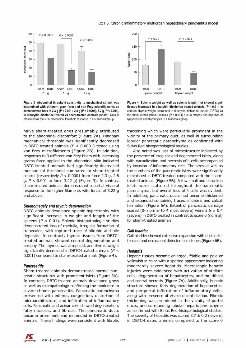
naïve shamtreated ones presumably attributed to the abdominal discomfort (Figure 2A). Hindpaw mechanical threshold was significantly decreased in DBTC-treated animals (p < 0.0001) tested using von Frey microfilaments (Figure 2B). In addition, responses to 3 different von Frey fibers with increasing grams force applied to the abdominal skin indicated DBTC-treated animals had significantly decreased mechanical threshold compared to shamtreated control (respectively p < 0.0001 from force 2.3 g, 2.8 g, p < 0.001 to force 3.22 g) (Figure 3). In contrast shamtreated animals demonstrated a partial visceral response to the higher filaments with forces of 3.22 g and above.
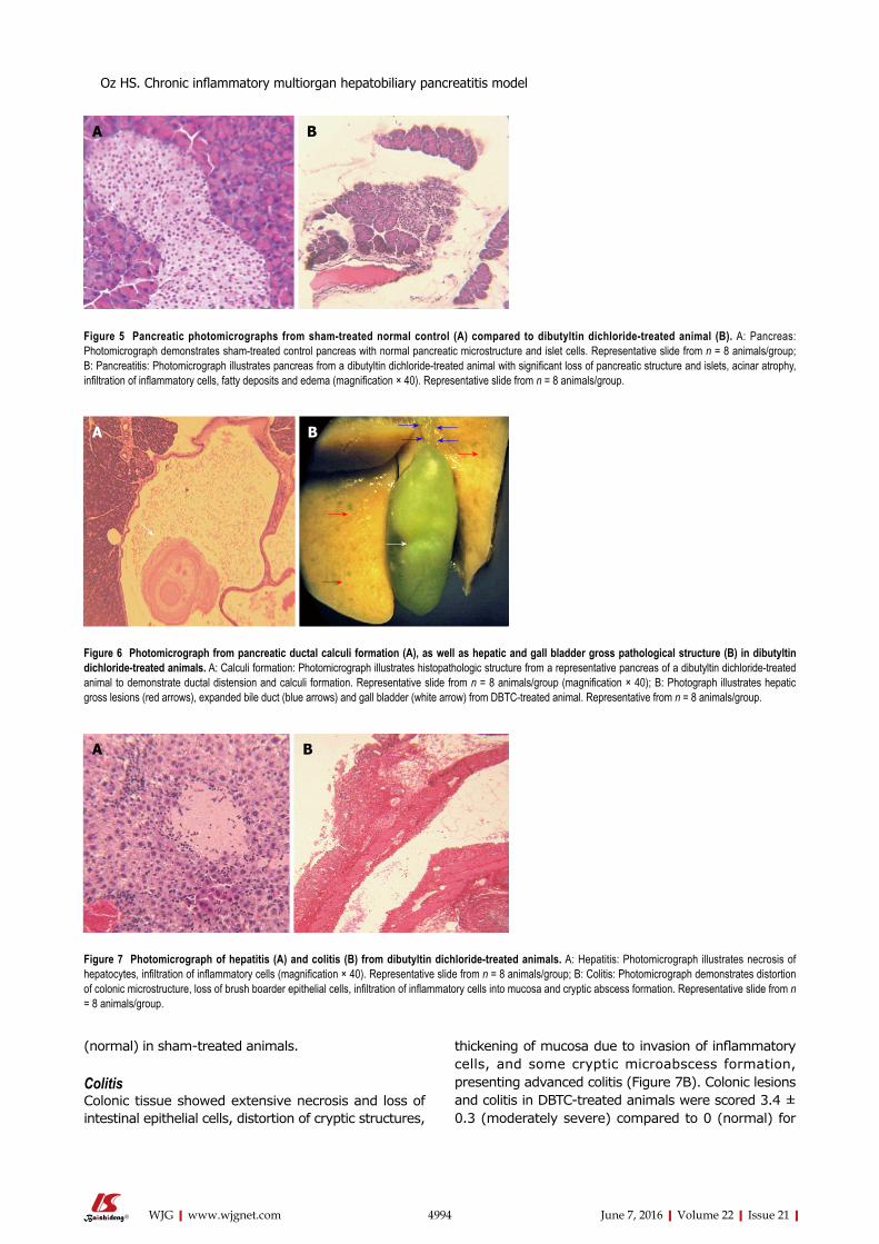
Splenomegaly and thymic degenerationDBTC animals developed splenic hypertrophy with significant increase in weight and length of the spleens (p < 0.01). Splenic histopathologic studies demonstrated loss of medulla, irregular formation of trabecules, with captured trace of blirubin and bile deposits. In contrast, thymic tissues from DBTC-treated animals showed central degeneration and atrophy. The thymus was atrophied, and thymic weight significantly decreased in DBTC-treated animals (p < 0.001) compared to shamtreated animals (Figure 4).
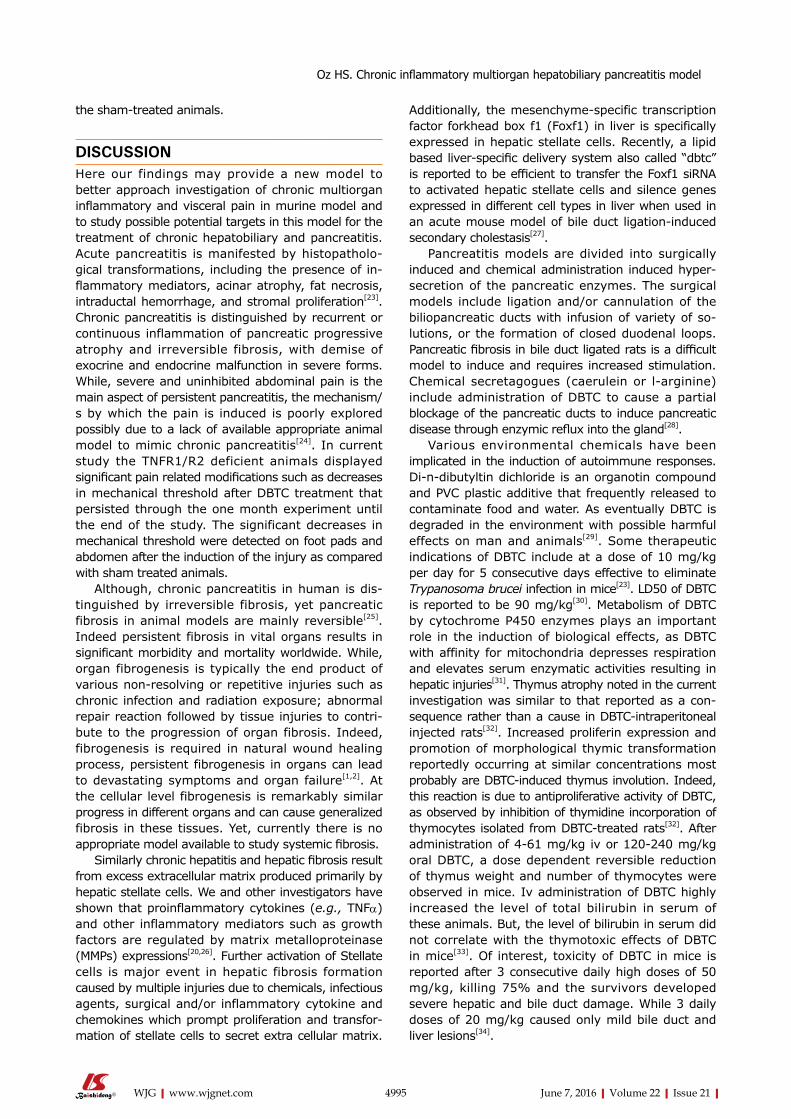
PancreatitisShamtreated animals demonstrated normal pancreatic structures with prominent islets (Figure 5A). In contrast, DBTC-treated animals developed gross as well as micropathology confirming the moderate to severe chronic pancreatitis. Pancreatic parenchyma presented with edema, congestion, distortion of microarchitecture, and infiltration of inflammatory cells. Pancreatic and acinar cells showed degeneration, fatty necrosis, and fibrosis. The pancreatic ducts became prominent and distended in DBTC-treated animals. These findings were consistent with fibrotic
thickening which were particularly prominent in the vicinity of the primary duct, as well in surrounding lobular pancreatic parenchyma as confirmed with Sirius Red histopathological studies.
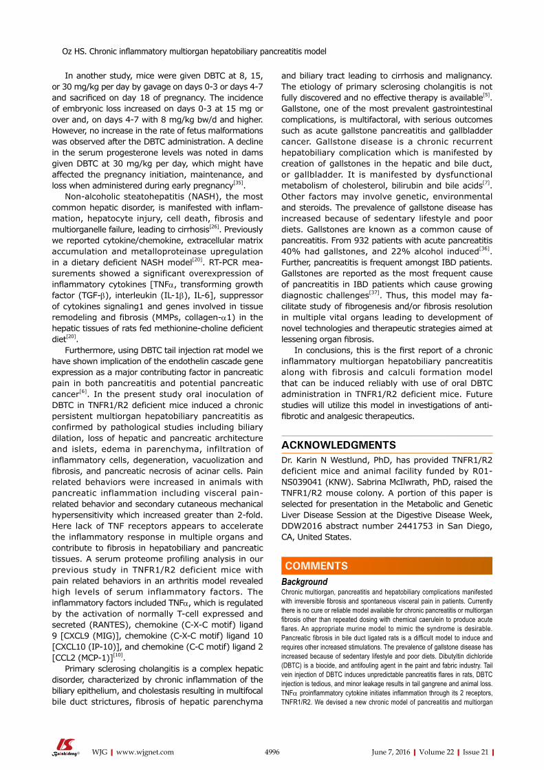
Also noted was loss of microstructure indicated by the presence of irregular and degenerated islets, along with vacuolization and necrosis of β cells accompanied by invasion of inflammatory cells. The sizes as well as the numbers of the pancreatic islets were significantly diminished in DBTC-treated compared with the sham-treated animals (Figure 5B). A few small and shrunken islets were scattered throughout the pancreatic parenchyma, but overall loss of β cells was evident. In addition, pancreatic ducts had become thickened and expanded containing traces of debris and calculi formation (Figure 6A). Extent of pancreatic damage scored (0 normal to 4 most severe) were 3.6 ± 0.4 (severe) in DBTC-treated in contrast to score 0 (normal) for shamtreated animals.
Gall bladder Gall bladder showed extensive expansion with ductal dis-tension and occasional detected bile stones (Figure 6B).
Hepatitis Hepatic tissues became enlarged, friable and pale or yellowish in color with a spotted appearance indicating moderately severe hepatitis. Macroscopic hepatic injuries were evidenced with activation of stellate cells, degeneration of hepatocytes, and multifocal and central necrosis (Figure 7A). Additionally, hepatic structure showed fatty degeneration of hepatocytes, and periportal infiltration of inflammatory cells, along with presence of visible ductal dilation. Fibrotic thickening was prominent in the vicinity of portal ducts, and surrounding lobular hepatic parenchyma as confirmed with Sirius Red histopathological studies. The severity of hepatitis was scored 3.7 ± 0.2 (severe) in DBTC-treated animals compared to the score 0
60
50
40
30
20
10
0
Mec
hani
cal t
hres
hold
Sham DBTC Sham DBTC Sham DBTC 2.3 g 2.8 g 3.2 g
P < 0.0001 P < 0.0001
P < 0.001
Figure 3 Abdominal threshold sensitivity to mechanical stimuli was determined with different gram forces of von Frey microfilaments as demonstrated here to 2.3 g (P < 0.001), 2.8 g (P < 0.0001), 3.2 g (P < 0.001), in dibutyltin dichloride-treated vs sham-treated controls (sham). Data is presented as the 50% mechanical threshold response. n = 5 animals/group.
200
150
100
50
0
Wei
ght
(mg)
Sham DBTC Sham DBTC Splenic weight Thymic weight
P < 0.01 P < 0.001
Figure 4 Splenic weight as well as splenic length (not shown) signi-ficantly increased in dibutyltin dichloride-treated animals (P < 0.01). In contrast thymic weight decreased in dibutyltin dichloride-treated (DBTC) vs the sham-treated (sham) animals (P < 0.001) due to atrophy and depletion of lymphocytes and thymocytes. n = 8 animals/group.
Oz HS. Chronic inflammatory multiorgan hepatobiliary pancreatitis model
4994 June 7, 2016|Volume 22|Issue 21|WJG|www.wjgnet.com
(normal) in shamtreated animals.
Colitis Colonic tissue showed extensive necrosis and loss of intestinal epithelial cells, distortion of cryptic structures,
thickening of mucosa due to invasion of inflammatory cells, and some cryptic microabscess formation, presenting advanced colitis (Figure 7B). Colonic lesions and colitis in DBTC-treated animals were scored 3.4 ± 0.3 (moderately severe) compared to 0 (normal) for
Figure 5 Pancreatic photomicrographs from sham-treated normal control (A) compared to dibutyltin dichloride-treated animal (B). A: Pancreas: Photomicrograph demonstrates sham-treated control pancreas with normal pancreatic microstructure and islet cells. Representative slide from n = 8 animals/group; B: Pancreatitis: Photomicrograph illustrates pancreas from a dibutyltin dichloride-treated animal with significant loss of pancreatic structure and islets, acinar atrophy, infiltration of inflammatory cells, fatty deposits and edema (magnification × 40). Representative slide from n = 8 animals/group.
A B
Figure 6 Photomicrograph from pancreatic ductal calculi formation (A), as well as hepatic and gall bladder gross pathological structure (B) in dibutyltin dichloride-treated animals. A: Calculi formation: Photomicrograph illustrates histopathologic structure from a representative pancreas of a dibutyltin dichloride-treated animal to demonstrate ductal distension and calculi formation. Representative slide from n = 8 animals/group (magnification × 40); B: Photograph illustrates hepatic gross lesions (red arrows), expanded bile duct (blue arrows) and gall bladder (white arrow) from DBTC-treated animal. Representative from n = 8 animals/group.
A B
Figure 7 Photomicrograph of hepatitis (A) and colitis (B) from dibutyltin dichloride-treated animals. A: Hepatitis: Photomicrograph illustrates necrosis of hepatocytes, infiltration of inflammatory cells (magnification × 40). Representative slide from n = 8 animals/group; B: Colitis: Photomicrograph demonstrates distortion of colonic microstructure, loss of brush boarder epithelial cells, infiltration of inflammatory cells into mucosa and cryptic abscess formation. Representative slide from n = 8 animals/group.
A B
Oz HS. Chronic inflammatory multiorgan hepatobiliary pancreatitis model
4995 June 7, 2016|Volume 22|Issue 21|WJG|www.wjgnet.com
the shamtreated animals.
DISCUSSION
Here our findings may provide a new model to better approach investigation of chronic multiorgan inflammatory and visceral pain in murine model and to study possible potential targets in this model for the treatment of chronic hepatobiliary and pancreatitis. Acute pancreatitis is manifested by histopathological transformations, including the presence of inflammatory mediators, acinar atrophy, fat necrosis, intraductal hemorrhage, and stromal proliferation[23]. Chronic pancreatitis is distinguished by recurrent or continuous inflammation of pancreatic progressive atrophy and irreversible fibrosis, with demise of exocrine and endocrine malfunction in severe forms. While, severe and uninhibited abdominal pain is the main aspect of persistent pancreatitis, the mechanism/s by which the pain is induced is poorly explored possibly due to a lack of available appropriate animal model to mimic chronic pancreatitis[24]. In current study the TNFR1/R2 deficient animals displayed significant pain related modifications such as decreases in mechanical threshold after DBTC treatment that persisted through the one month experiment until the end of the study. The significant decreases in mechanical threshold were detected on foot pads and abdomen after the induction of the injury as compared with sham treated animals.
Although, chronic pancreatitis in human is distinguished by irreversible fibrosis, yet pancreatic fibrosis in animal models are mainly reversible[25]. Indeed persistent fibrosis in vital organs results in significant morbidity and mortality worldwide. While, organ fibrogenesis is typically the end product of various nonresolving or repetitive injuries such as chronic infection and radiation exposure; abnormal repair reaction followed by tissue injuries to contribute to the progression of organ fibrosis. Indeed, fibrogenesis is required in natural wound healing process, persistent fibrogenesis in organs can lead to devastating symptoms and organ failure[1,2]. At the cellular level fibrogenesis is remarkably similar progress in different organs and can cause generalized fibrosis in these tissues. Yet, currently there is no appropriate model available to study systemic fibrosis.
Similarly chronic hepatitis and hepatic fibrosis result from excess extracellular matrix produced primarily by hepatic stellate cells. We and other investigators have shown that proinflammatory cytokines (e.g., TNFα) and other inflammatory mediators such as growth factors are regulated by matrix metalloproteinase (MMPs) expressions[20,26]. Further activation of Stellate cells is major event in hepatic fibrosis formation caused by multiple injuries due to chemicals, infectious agents, surgical and/or inflammatory cytokine and chemokines which prompt proliferation and transformation of stellate cells to secret extra cellular matrix.
Additionally, the mesenchymespecific transcription factor forkhead box f1 (Foxf1) in liver is specifically expressed in hepatic stellate cells. Recently, a lipid based liver-specific delivery system also called “dbtc” is reported to be efficient to transfer the Foxf1 siRNA to activated hepatic stellate cells and silence genes expressed in different cell types in liver when used in an acute mouse model of bile duct ligationinduced secondary cholestasis[27].
Pancreatitis models are divided into surgically induced and chemical administration induced hypersecretion of the pancreatic enzymes. The surgical models include ligation and/or cannulation of the biliopancreatic ducts with infusion of variety of solutions, or the formation of closed duodenal loops. Pancreatic fibrosis in bile duct ligated rats is a difficult model to induce and requires increased stimulation. Chemical secretagogues (caerulein or larginine) include administration of DBTC to cause a partial blockage of the pancreatic ducts to induce pancreatic disease through enzymic reflux into the gland[28].
Various environmental chemicals have been implicated in the induction of autoimmune responses. Dindibutyltin dichloride is an organotin compound and PVC plastic additive that frequently released to contaminate food and water. As eventually DBTC is degraded in the environment with possible harmful effects on man and animals[29]. Some therapeutic indications of DBTC include at a dose of 10 mg/kg per day for 5 consecutive days effective to eliminate Trypanosoma brucei infection in mice[23]. LD50 of DBTC is reported to be 90 mg/kg[30]. Metabolism of DBTC by cytochrome P450 enzymes plays an important role in the induction of biological effects, as DBTC with affinity for mitochondria depresses respiration and elevates serum enzymatic activities resulting in hepatic injuries[31]. Thymus atrophy noted in the current investigation was similar to that reported as a consequence rather than a cause in DBTC-intraperitoneal injected rats[32]. Increased proliferin expression and promotion of morphological thymic transformation reportedly occurring at similar concentrations most probably are DBTC-induced thymus involution. Indeed, this reaction is due to antiproliferative activity of DBTC, as observed by inhibition of thymidine incorporation of thymocytes isolated from DBTC-treated rats[32]. After administration of 461 mg/kg iv or 120240 mg/kg oral DBTC, a dose dependent reversible reduction of thymus weight and number of thymocytes were observed in mice. Iv administration of DBTC highly increased the level of total bilirubin in serum of these animals. But, the level of bilirubin in serum did not correlate with the thymotoxic effects of DBTC in mice[33]. Of interest, toxicity of DBTC in mice is reported after 3 consecutive daily high doses of 50 mg/kg, killing 75% and the survivors developed severe hepatic and bile duct damage. While 3 daily doses of 20 mg/kg caused only mild bile duct and liver lesions[34].
Oz HS. Chronic inflammatory multiorgan hepatobiliary pancreatitis model
4996 June 7, 2016|Volume 22|Issue 21|WJG|www.wjgnet.com
In another study, mice were given DBTC at 8, 15, or 30 mg/kg per day by gavage on days 03 or days 47 and sacrificed on day 18 of pregnancy. The incidence of embryonic loss increased on days 03 at 15 mg or over and, on days 47 with 8 mg/kg bw/d and higher. However, no increase in the rate of fetus malformations was observed after the DBTC administration. A decline in the serum progesterone levels was noted in dams given DBTC at 30 mg/kg per day, which might have affected the pregnancy initiation, maintenance, and loss when administered during early pregnancy[35].
Nonalcoholic steatohepatitis (NASH), the most common hepatic disorder, is manifested with inflammation, hepatocyte injury, cell death, fibrosis and multiorganelle failure, leading to cirrhosis[26]. Previously we reported cytokine/chemokine, extracellular matrix accumulation and metalloproteinase upregulation in a dietary deficient NASH model[20]. RTPCR measurements showed a significant overexpression of inflammatory cytokines [TNFα, transforming growth factor (TGFβ), interleukin (IL-1β), IL-6], suppressor of cytokines signaling1 and genes involved in tissue remodeling and fibrosis (MMPs, collagenα1) in the hepatic tissues of rats fed methionine-choline deficient diet[20].
Furthermore, using DBTC tail injection rat model we have shown implication of the endothelin cascade gene expression as a major contributing factor in pancreatic pain in both pancreatitis and potential pancreatic cancer[6]. In the present study oral inoculation of DBTC in TNFR1/R2 deficient mice induced a chronic persistent multiorgan hepatobiliary pancreatitis as confirmed by pathological studies including biliary dilation, loss of hepatic and pancreatic architecture and islets, edema in parenchyma, infiltration of inflammatory cells, degeneration, vacuolization and fibrosis, and pancreatic necrosis of acinar cells. Pain related behaviors were increased in animals with pancreatic inflammation including visceral painrelated behavior and secondary cutaneous mechanical hypersensitivity which increased greater than 2fold. Here lack of TNF receptors appears to accelerate the inflammatory response in multiple organs and contribute to fibrosis in hepatobiliary and pancreatic tissues. A serum proteome profiling analysis in our previous study in TNFR1/R2 deficient mice with pain related behaviors in an arthritis model revealed high levels of serum inflammatory factors. The inflammatory factors included TNFα, which is regulated by the activation of normally T-cell expressed and secreted (RANTES), chemokine (CXC motif) ligand 9 [CXCL9 (MIG)], chemokine (C-X-C motif) ligand 10 [CXCL10 (IP-10)], and chemokine (C-C motif) ligand 2 [CCL2 (MCP1)][10].
Primary sclerosing cholangitis is a complex hepatic disorder, characterized by chronic inflammation of the biliary epithelium, and cholestasis resulting in multifocal bile duct strictures, fibrosis of hepatic parenchyma
and biliary tract leading to cirrhosis and malignancy. The etiology of primary sclerosing cholangitis is not fully discovered and no effective therapy is available[5]. Gallstone, one of the most prevalent gastrointestinal complications, is multifactoral, with serious outcomes such as acute gallstone pancreatitis and gallbladder cancer. Gallstone disease is a chronic recurrent hepatobiliary complication which is manifested by creation of gallstones in the hepatic and bile duct, or gallbladder. It is manifested by dysfunctional metabolism of cholesterol, bilirubin and bile acids[7]. Other factors may involve genetic, environmental and steroids. The prevalence of gallstone disease has increased because of sedentary lifestyle and poor diets. Gallstones are known as a common cause of pancreatitis. From 932 patients with acute pancreatitis 40% had gallstones, and 22% alcohol induced[36]. Further, pancreatitis is frequent amongst IBD patients. Gallstones are reported as the most frequent cause of pancreatitis in IBD patients which cause growing diagnostic challenges[37]. Thus, this model may facilitate study of fibrogenesis and/or fibrosis resolution in multiple vital organs leading to development of novel technologies and therapeutic strategies aimed at lessening organ fibrosis.
In conclusions, this is the first report of a chronic inflammatory multiorgan hepatobiliary pancreatitis along with fibrosis and calculi formation model that can be induced reliably with use of oral DBTC administration in TNFR1/R2 deficient mice. Future studies will utilize this model in investigations of antifibrotic and analgesic therapeutics.
ACKNOWLEDGMENTSDr. Karin N Westlund, PhD, has provided TNFR1/R2 deficient mice and animal facility funded by R01NS039041 (KNW). Sabrina McIlwrath, PhD, raised the TNFR1/R2 mouse colony. A portion of this paper is selected for presentation in the Metabolic and Genetic Liver Disease Session at the Digestive Disease Week, DDW2016 abstract number 2441753 in San Diego, CA, United States.
COMMENTS BackgroundChronic multiorgan, pancreatitis and hepatobiliary complications manifested with irreversible fibrosis and spontaneous visceral pain in patients. Currently there is no cure or reliable model available for chronic pancreatitis or multiorgan fibrosis other than repeated dosing with chemical caerulein to produce acute flares. An appropriate murine model to mimic the syndrome is desirable. Pancreatic fibrosis in bile duct ligated rats is a difficult model to induce and requires other increased stimulations. The prevalence of gallstone disease has increased because of sedentary lifestyle and poor diets. Dibutyltin dichloride (DBTC) is a biocide, and antifouling agent in the paint and fabric industry. Tail vein injection of DBTC induces unpredictable pancreatitis flares in rats, DBTC injection is tedious, and minor leakage results in tail gangrene and animal loss. TNFα proinflammatory cytokine initiates inflammation through its 2 receptors, TNFR1/R2. We devised a new chronic model of pancreatitis and multiorgan
COMMENTS
Oz HS. Chronic inflammatory multiorgan hepatobiliary pancreatitis model
4997 June 7, 2016|Volume 22|Issue 21|WJG|www.wjgnet.com
inflammation in TNFR1/R2 deficient mice using oral DBTC.
Research frontiersCurrently, there is no cure or reliable model available for chronic pancreatitis and multiorgan fibrosis in mice. Persistent pancreatitis manifests with severe abdominal pain, but the mechanism/s by which induced is/are poorly explored possibly due to lack of appropriate models. Three daily doses of 20 mg/kg DBTC caused only mild bile duct and liver lesions, while 3 consecutive daily doses of 50 mg/kg DBTC were toxic and killed 75% of mice. TNFα proinflammatory cytokine initiates inflammation through its 2 receptors, TNFR1/R2. Proteome profiling analysis in our previous study in TNFR1/R2 deficient mice with persistent pain related behaviors in an arthritis model revealed high levels of serum inflammatory cytokines likely responsible for the multiorgan inflammatory response in this model.
Innovations and breakthroughsThis is the first report of a chronic inflammatory hepatobiliary pancreatitis, colitis and stone formation model that can be induced reliably with use of oral DBTC-administration in TNFR1/R2 deficient mice. These findings provide this new murine model to better approach investigation of chronic multiorgan inflammatory and visceral pain in murine mode. In addition, to facilitate study of fibrogenesis and/or fibrosis resolution in multiple vital organs leading to development of novel technologies and therapeutic strategies aimed at lessening organ fibrosis.
ApplicationsThis chronic inflammatory hepatobiliary pancreatitis, colitis/fibrosis and stone formation model can be induced reliably utilizing oral DBTC in TNFR1/R2 deficient mice. The model can be used to investigate chronic multiorgan inflammatory and visceral pain in mice to explore the mechanisms of injury and to study of fibrogenesis and/or fibrosis resolution in multiple vital organs leading to development of novel technologies and therapeutic strategies aimed at lessening organ fibrosis. This study grants ability for further investigation into the use of this model to explore mechanisms of multiorgan injury, the biochemical players and the therapeutic exploration for devastating chronic multiorgn inflammatory and fibrogenesis such as cystic fibrosis and systemic sclerosis as well as cholangitis.
TerminologyTNFR1/R2 deficient mice are transgenic animals lacking tumor necrosis factor receptor 1 and 2 with constant higher proinflammatory TNFα levels compared to wildtype background animals. Multiorgan damage, when 2 or more organs involved in the course of injury. Fibrogenesis is a process usually followed chronic inflammatory to form fibrous structures in and around tissues. Pain related mechanical hypersensitivity, is measured by von Frey microfilament (an accepted standard procedure), demonstrating decreased tolerance to a simple touch manifested with withdraw to protect against induced excess pressure while normal animals tolerate the mechanical touch with no withdrawal response to the fine microfilament touch.
Peer-reviewThis is an interesting and well-written paper. The author provided that TNFR1/R2 deficient mice treated with DBTC reveal the severe chronic injury of various internal organs which was proved with usage of histological methods.
REFERENCES1 De Langhe E, Lories R. Fibrogenesis, novel lessons from animal
models. Semin Immunopathol 2015; 37: 565-574 [PMID: 26141608 DOI: 10.1007/s00281-015-0510-8]
2 Medley JM, Kaplan E, Oz HS, Sundararaj SC, Puleo DA, Dziubla TD. Fibrin-targeted block copolymers for the prevention of postsurgical adhesions. J Biomed Mater Res B Appl Biomater 2011; 99: 102-110 [PMID: 21695779 DOI: 10.1002/jbm.b.31876]
3 Lavie M, Manovitz T, Vilozni D, Levy-Mendelovich S, Sarouk I, Weintraubv I, Shoseyov D, Cohen-Cymberknoh M, Rivlin J, Efrati O. Long-term follow-up of distal intestinal obstruction syndrome
in cystic fibrosis. World J Gastroenterol 2015; 21: 318-325 [PMID: 25574107 DOI: 10.3748/wjg.v21.i1.318]
4 Kavian N, Batteux F. Macro- and microvascular disease in systemic sclerosis. Vascul Pharmacol 2015; 71: 16-23 [PMID: 26044180 DOI: 10.1016/j.vph.2015.05.015]
5 Eaton JE, Talwalkar JA, Lazaridis KN, Gores GJ, Lindor KD. Pathogenesis of primary sclerosing cholangitis and advances in diagnosis and management. Gastroenterology 2013; 145: 521-536 [PMID: 23827861 DOI: 10.1053/j.gastro.2013.06.052]
6 Oz HS, Lu Y, Vera-Portocarrero LP, Ge P, Silos-Santiago A, Westlund KN. Gene expression profiling and endothelin in acute experimental pancreatitis. World J Gastroenterol 2012; 18: 4257-4269 [PMID: 22969188 DOI: 10.3748/wjg.v18.i32.4257]
7 Reshetnyak VI. Concept of the pathogenesis and treatment of cholelithiasis. World J Hepatol 2012; 4: 18-34 [PMID: 22400083 DOI: 10.4254/wjh.v4.i2.18]
8 Gregersen R, Lambertsen K, Finsen B. Microglia and macrophages are the major source of tumor necrosis factor in permanent middle cerebral artery occlusion in mice. J Cereb Blood Flow Metab 2000; 20: 53-65 [PMID: 10616793 DOI: 10.1097/00004647-200001000-00009]
9 Locksley RM, Killeen N, Lenardo MJ. The TNF and TNF receptor superfamilies: integrating mammalian biology. Cell 2001; 104: 487-501 [PMID: 11239407 DOI: 10.1016/S0092-8674(01)00237-9]
10 Westlund KN, Zhang L, Ma F, Oz HS. Chronic inflammation and pain in a tumor necrosis factor receptor (TNFR) (p55/p75-/-) dual deficient murine model. Transl Res 2012; 160: 84-94 [PMID: 22687964 DOI: 10.1016/j.trsl.2011.10.003]
11 Fang C, Shi B, Pei YY, Hong MH, Wu J, Chen HZ. In vivo tumor targeting of tumor necrosis factor-alpha-loaded stealth nanoparticles: effect of MePEG molecular weight and particle size. Eur J Pharm Sci 2006; 27: 27-36 [PMID: 16150582 DOI: 10.1016/j.ejps.2005.08.002]
12 Uçeyler N, Schäfers M, Sommer C. Mode of action of cytokines on nociceptive neurons. Exp Brain Res 2009; 196: 67-78 [PMID: 19290516 DOI: 10.1007/s00221-009-1755-z]
13 Marchand F, Perretti M, McMahon SB. Role of the immune system in chronic pain. Nat Rev Neurosci 2005; 6: 521-532 [PMID: 15995723 DOI: 10.1038/nrn1700]
14 Sommer C, Schmidt C, George A. Hyperalgesia in experimental neuropathy is dependent on the TNF receptor 1. Exp Neurol 1998; 151: 138-142 [PMID: 9582261 DOI: 10.1006/exnr.1998.6797]
15 Schäfers M, Svensson CI, Sommer C, Sorkin LS. Tumor necrosis factor-alpha induces mechanical allodynia after spinal nerve ligation by activation of p38 MAPK in primary sensory neurons. J Neurosci 2003; 23: 2517-2521 [PMID: 12684435]
16 Ma F, Zhang L, Oz HS, Mashni M, Westlund KN. Dysregulated TNFα promotes cytokine proteome profile increases and bilateral orofacial hypersensitivity. Neuroscience 2015; 300: 493-507 [PMID: 26033565 DOI: 10.1016/j.neuroscience.2015.05.046]
17 Malleo G, Mazzon E, Genovese T, Di Paola R, Muià C, Centorrino T, Siriwardena AK, Cuzzocrea S. Etanercept attenuates the development of cerulein-induced acute pancreatitis in mice: a comparison with TNF-alpha genetic deletion. Shock 2007; 27: 542-551 [PMID: 17438460 DOI: 10.1097/01.shk.0000246900.50445.1d]
18 DeWitt JC, Copeland CB, Luebke RW. An organotin mixture found in polyvinyl chloride (PVC) pipe is not immunotoxic to adult Sprague-Dawley rats. J Toxicol Environ Health A 2008; 71: 276-282 [PMID: 18253893 DOI: 10.1080/15287390701613025]
19 Oz HS. Toxoplasmosis complications and novel therapeutic synergism combination of diclazuril plus atovaquone. Front Microbiol 2014; 5: 484 [PMID: 25309522 DOI: 10.3389/fmicb.2014.00484]
20 Oz HS, Im HJ, Chen TS, de Villiers WJ, McClain CJ. Glutathione-enhancing agents protect against steatohepatitis in a dietary model. J Biochem Mol Toxicol 2006; 20: 39-47 [PMID: 16498637 DOI: 10.1002/jbt.20109]
21 Oz HS, Chen T, de Villiers WJ. Green Tea Polyphenols and Sulfasalazine have Parallel Anti-Inflammatory Properties in Colitis Models. Front Immunol 2013; 4: 132 [PMID: 23761791 DOI:
Oz HS. Chronic inflammatory multiorgan hepatobiliary pancreatitis model
4998 June 7, 2016|Volume 22|Issue 21|WJG|www.wjgnet.com
10.3389/fimmu.2013.00132]22 Oz HS, Chen TS, Nagasawa H. Comparative efficacies of 2 cysteine
prodrugs and a glutathione delivery agent in a colitis model. Transl Res 2007; 150: 122-129 [PMID: 17656332 DOI: 10.1016/j.trsl.2006.12.010]
23 Schmidt J, Hotz HG, Foitzik T, Ryschich E, Buhr HJ, Warshaw AL, Herfarth C, Klar E. Intravenous contrast medium aggravates the impairment of pancreatic microcirculation in necrotizing pancreatitis in the rat. Ann Surg 1995; 221: 257-264 [PMID: 7717779 DOI: 10.1097/00000658-199503000-00007]
24 Barreto SG, Saccone GT. Pancreatic nociception--revisiting the physiology and pathophysiology. Pancreatology 2012; 12: 104-112 [PMID: 22487519 DOI: 10.1016/j.pan.2012.02.010]
25 Miyauchi M, Suda K, Kuwayama C, Abe H, Kakinuma C. Role of fibrosis-related genes and pancreatic duct obstruction in rat pancreatitis models: implications for chronic pancreatitis. Histol Histopathol 2007; 22: 1119-1127 [PMID: 17616939]
26 Caldwell S. NASH (Nonalcoholic steatohepatitis): A case of multiorganelle failure. Free Radic Biol Med 2014; 75 Suppl 1: S6 [PMID: 26461413 DOI: 10.1016/j.freeradbiomed.2014.10.839]
27 Abshagen K, Brensel M, Genz B, Roth K, Thomas M, Fehring V, Schaeper U, Vollmar B. Foxf1 siRNA delivery to hepatic stellate cells by DBTC lipoplex formulations ameliorates fibrosis in livers of bile duct ligated mice. Curr Gene Ther 2015; 15: 215-227 [PMID: 25619889 DOI: 10.2174/1566523215666150126114634]
28 Foster JR. A review of animal models of nonneoplastic pancreatic diseases. Toxicol Pathol 2014; 42: 243-259 [PMID: 24178571 DOI: 10.1177/0192623313508479]
29 Kobayashi H, Suzuki T, Kasashima Y, Motegi A, Sato I, Matsusaka N, Ono N, Miura A, Saito F, Saito S. Effects of tri-, di- and mono-
butyltin on synaptic parameters of the cholinergic system in the cerebral cortex of mice. Jpn J Pharmacol 1996; 72: 317-324 [PMID: 9015740 DOI: 10.1254/jjp.72.317]
30 Shuaibu MN, Ameh DA, Bonire JJ, Adaudi AO, Ibrahim S, Nok AJ. Trypanocidal activity of organotin chlorides on Trypanosoma brucei-infected mice. Parasite 2000; 7: 43-45 [PMID: 10743647 DOI: 10.1051/parasite/2000071043]
31 Ueno S, Kashimoto T, Susa N, Shiota Y, Okuda M, Mutoh K, Hoshi F, Watanabe K, Tsuda S, Kawazoe S, Suzuki T, Sugiyama M. Effects of butyltin compounds on mitochondrial respiration and its relation to hepatotoxicity in mice and Guinea pigs. Toxicol Sci 2003; 75: 201-207 [PMID: 12805650 DOI: 10.1093/toxsci/kfg153]
32 Penninks A, Kuper F, Spit BJ, Seinen W. On the mechanism of dialkyltin-induced thymus involution. Immunopharmacology 1985; 10: 1-10 [PMID: 3877030 DOI: 10.1016/0162-3109(85)90053-0]
33 Hennighausen G, Lange P. Immunotoxic effects of dialkyltins used for stabilization of plastics. Pol J Pharmacol Pharm 1980; 32: 119-124 [PMID: 7454608]
34 Barnes JM, Stoner HB. Toxic properties of some dialkyl and trialkyl tin salts. Br J Ind Med 1958; 15: 15-22 [PMID: 13499843]
35 Ema M, Fujii S, Ikka T, Matsumoto M, Hirose A, Kamata E. Early pregnancy failure induced by dibutyltin dichloride in mice. Environ Toxicol 2007; 22: 44-52 [PMID: 17295259]
36 Nesvaderani M, Eslick GD, Vagg D, Faraj S, Cox MR. Epide-miology, aetiology and outcomes of acute pancreatitis: A retrospective cohort study. Int J Surg 2015; 23: 68-74 [PMID: 26384834 DOI: 10.1016/j.ijsu.2015.07.701]
37 Ramos LR, Sachar DB, DiMaio CJ, Colombel JF, Torres J. Inflam-matory Bowel Disease and Pancreatitis: A Review. J Crohns Colitis 2016; 10: 95-104 [PMID: 26351384 DOI: 10.1093/ecco-jcc/jjv153]
P- Reviewer: Khedmat H, Kurzepa J S- Editor: Gong ZM L- Editor: A E- Editor: Wang CH
Oz HS. Chronic inflammatory multiorgan hepatobiliary pancreatitis model

© 2016 Baishideng Publishing Group Inc. All rights reserved.
Published by Baishideng Publishing Group Inc8226 Regency Drive, Pleasanton, CA 94588, USA
Telephone: +1-925-223-8242Fax: +1-925-223-8243
E-mail: [email protected] Desk: http://www.wjgnet.com/esps/helpdesk.aspx
http://www.wjgnet.com
I S S N 1 0 0 7 - 9 3 2 7
9 7 7 1 0 07 9 3 2 0 45
2 1