Embed Size (px)
Citation preview
Page 1
Multimodal Preclinical Imaging for
Translational Research
Joshua McHattan
Product Line Manager – Asia Cluster

Page 2
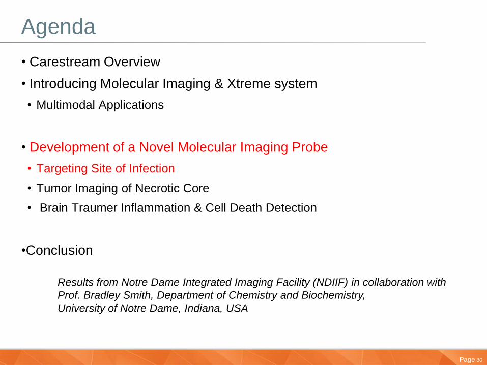
Agenda
• Carestream Overview
• Introducing Molecular Imaging & Xtreme system
• Multimodal Applications
• Development of a Novel Molecular Imaging Probe
• Targeting Site of Infection
• Tumor Imaging of Necrotic Core
• Brain Trauma Inflammation & Cell Death Detection
•Conclusion

Page 3
What We Do
A world leader in:
• Medical imaging
• Dental imaging and dental practice management software
• Molecular imaging
– The most extensive portfolio on the market
• Healthcare information solutions (RIS & PACS)
– More than 1,100 healthcare information management solutions currently installed worldwide (PACS, RIS, data management)
• Digital output solutions
– More than 50,000 KODAK DRYVIEW laser imagers on the market worldwide
• All categories of Kodak film
Page 4
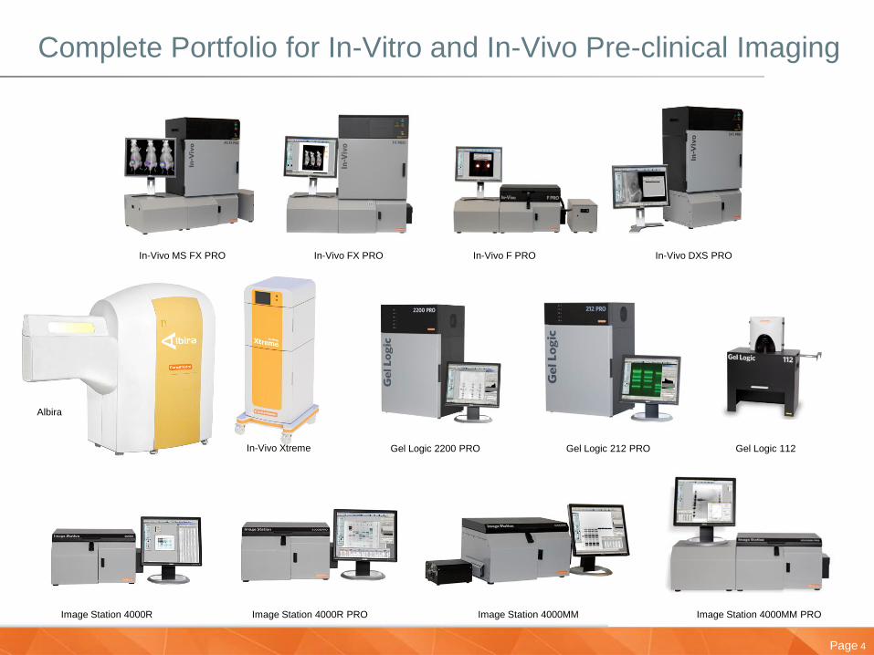
Complete Portfolio for In-Vitro and In-Vivo Pre-clinical Imaging
Image Station 4000MM PRO Image Station 4000R Image Station 4000R PRO Image Station 4000MM
In-Vivo MS FX PRO In-Vivo FX PRO In-Vivo F PRO In-Vivo DXS PRO
Gel Logic 2200 PRO Gel Logic 212 PRO Gel Logic 112
Albira
In-Vivo Xtreme

Page 5
How It All Ties Together
DNA, RNA, Protein Cells Animal Models Tissue & Systems Patient
Genetic Influences
•Developmental
•Aging
•Diet
•Environmental
•Pharmaceutical
Health endpoints
•Osteoporosis
•Diabetes
•Cancer
•Etc.
Genes Proteins Function
Information and Image Capture, Storage and Analysis (RIS & PACS)

Page 6
Why Whole Animal Molecular Imaging?
Detect & Monitor Disease Models over time
Monitor Drug Therapies Within the Same Model
Biodistribution
PK/PD
Probe Development
Fast, Efficient & Cost Reduction
Proof of Principle Within a True Hostile Environment

Page 7
Molecular Imaging in Disease Detection
Injection of probes
Red blood cell White blood cell
Labeled probes
Blood vessel
Circulation
Image capture
• Disease detection
and diagnosis
• Disease response to
therapy
• Guided surgery
• Accumulate at disease site
• Imaged
• Disease site identified
Page 8
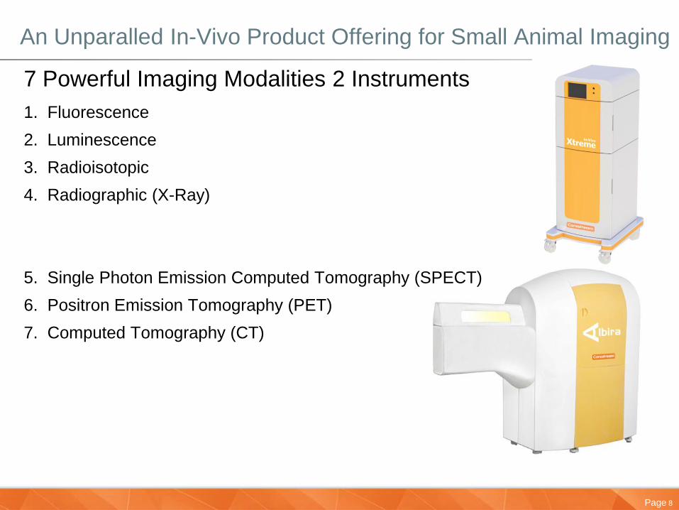
An Unparalled In-Vivo Product Offering for Small Animal Imaging
7 Powerful Imaging Modalities 2 Instruments
1. Fluorescence
2. Luminescence
3. Radioisotopic
4. Radiographic (X-Ray)
5. Single Photon Emission Computed Tomography (SPECT)
6. Positron Emission Tomography (PET)
7. Computed Tomography (CT)
Page 9
Page 10
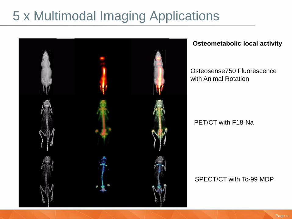
5 x Multimodal Imaging Applications
Osteosense750 Fluorescence
with Animal Rotation
PET/CT with F18-Na
SPECT/CT with Tc-99 MDP
Osteometabolic local activity
Page 11
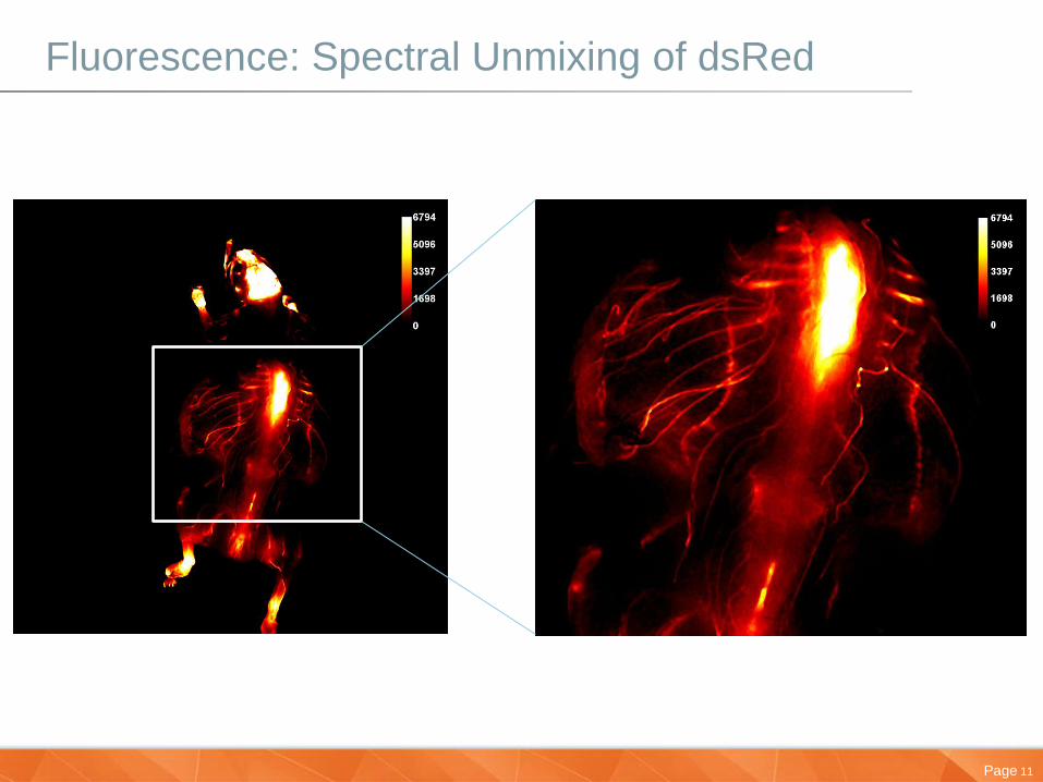
Fluorescence: Spectral Unmixing of dsRed
Page 12
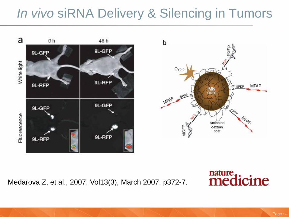
In vivo siRNA Delivery & Silencing in Tumors
Medarova Z, et al., 2007. Vol13(3), March 2007. p372-7.

Page 13
MARS OsteoSense 750: Bone Remodeling

Page 14
40min Dynamic Drug Delivery
Page 15
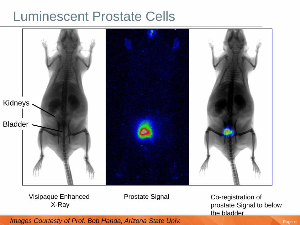
Luminescent Prostate Cells
Visipaque Enhanced
X-Ray
Prostate Signal Co-registration of
prostate Signal to below
the bladder
Bladder
Kidneys
Images Courtesty of Prof. Bob Handa, Arizona State Univ.
Page 16
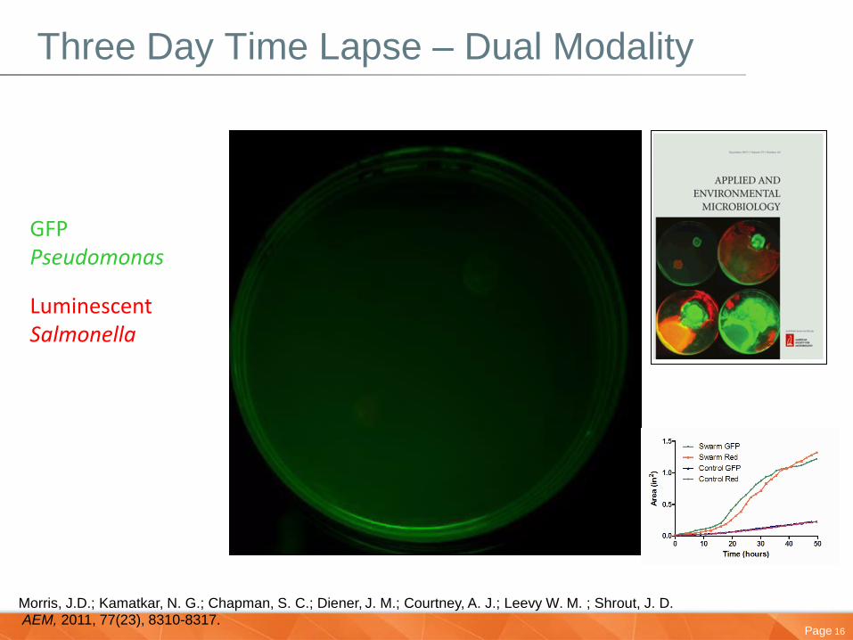
Three Day Time Lapse – Dual Modality
GFP Pseudomonas
Luminescent Salmonella
Morris, J.D.; Kamatkar, N. G.; Chapman, S. C.; Diener, J. M.; Courtney, A. J.; Leevy W. M. ; Shrout, J. D. AEM, 2011, 77(23), 8310-8317.
Page 17
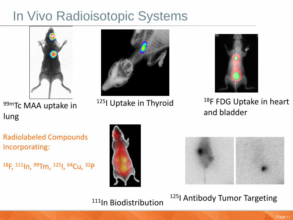
In Vivo Radioisotopic Systems
125I Uptake in Thyroid
18F FDG Uptake in heart and bladder
99mTc MAA uptake in lung
111In Biodistribution
125I Antibody Tumor Targeting
Radiolabeled Compounds Incorporating: 18F, 111In, 99Tm, 125I, 64Cu, 32P
Page 18
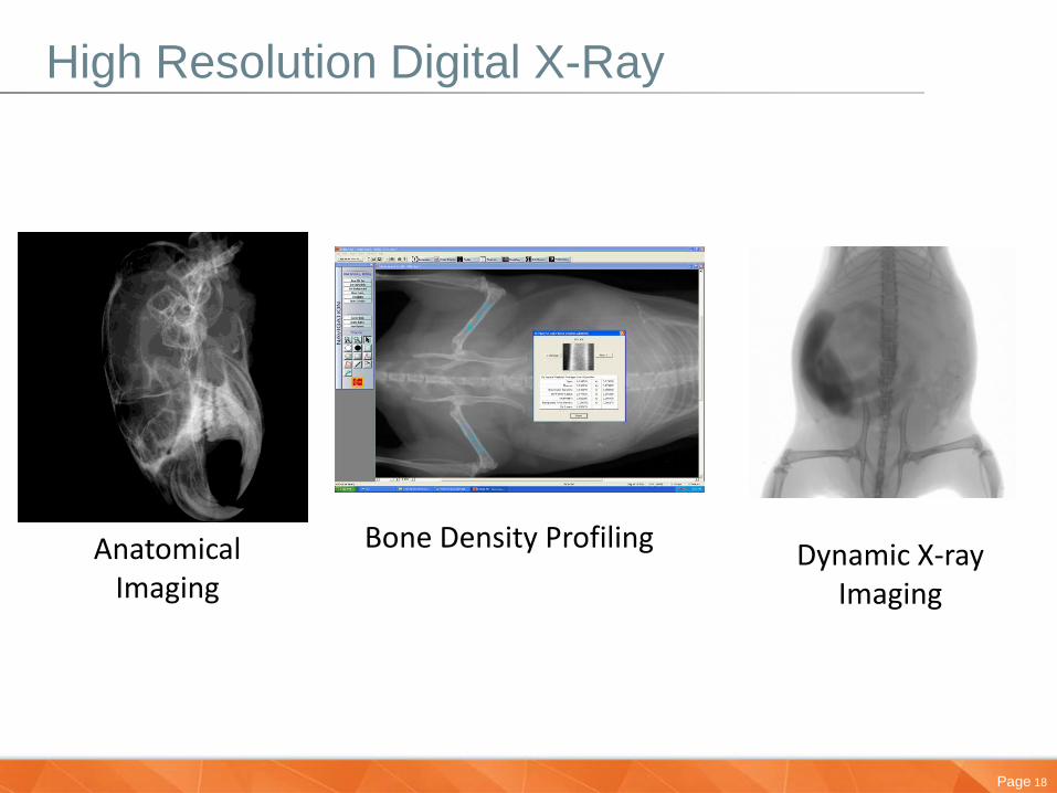
High Resolution Digital X-Ray
Bone Density Profiling
Anatomical Imaging
Dynamic X-ray Imaging

Page 19
X-Ray Vascular Imaging Using MARS
Page 20
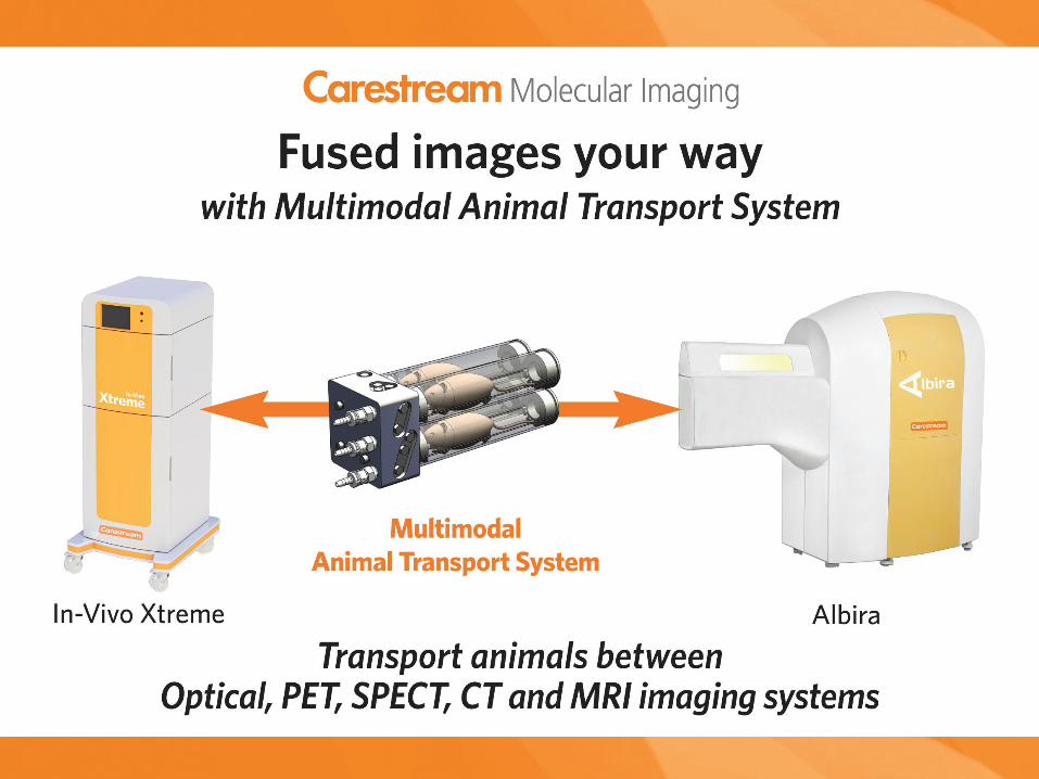
Xtreme Instrumentation
• How Does it Work?
Page 21
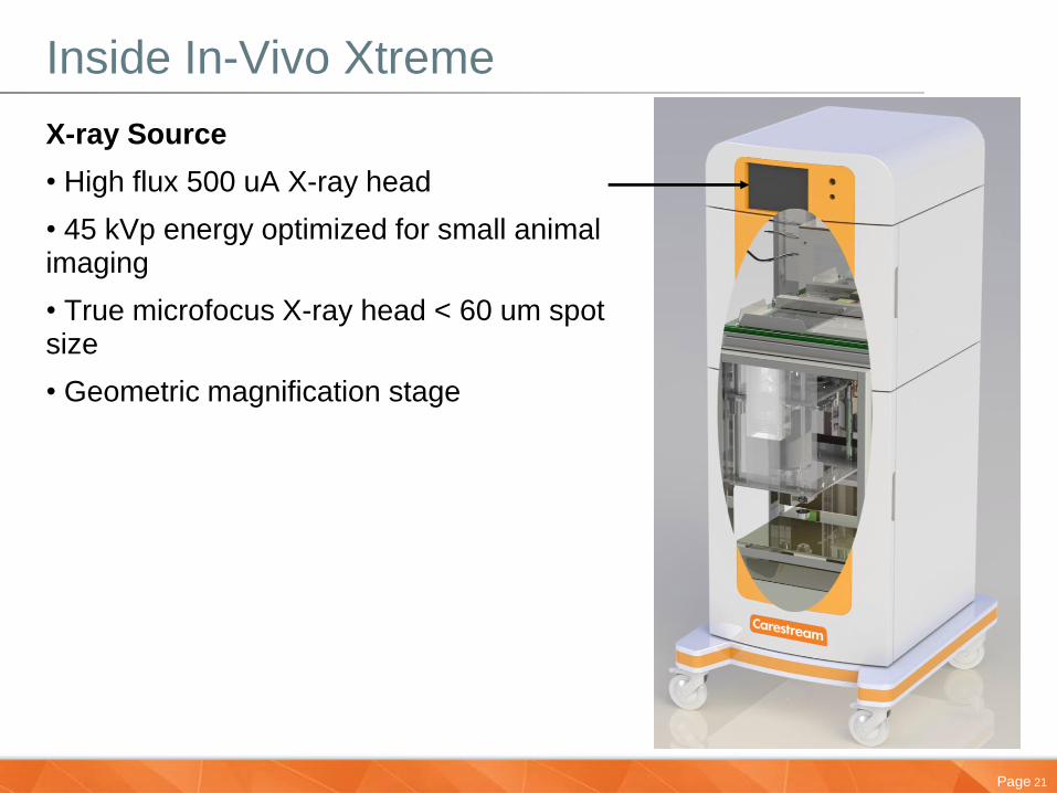
Inside In-Vivo Xtreme
X-ray Source
• High flux 500 uA X-ray head
• 45 kVp energy optimized for small animal imaging
• True microfocus X-ray head < 60 um spot size
• Geometric magnification stage
Page 22
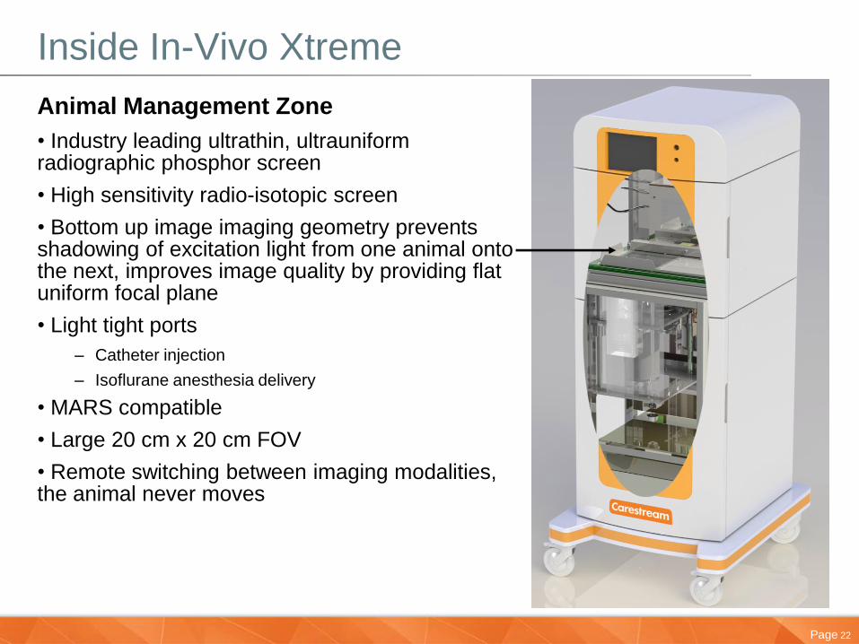
Inside In-Vivo Xtreme
Animal Management Zone
• Industry leading ultrathin, ultrauniform radiographic phosphor screen
• High sensitivity radio-isotopic screen
• Bottom up image imaging geometry prevents shadowing of excitation light from one animal onto the next, improves image quality by providing flat uniform focal plane
• Light tight ports
– Catheter injection
– Isoflurane anesthesia delivery
• MARS compatible
• Large 20 cm x 20 cm FOV
• Remote switching between imaging modalities, the animal never moves
Page 23
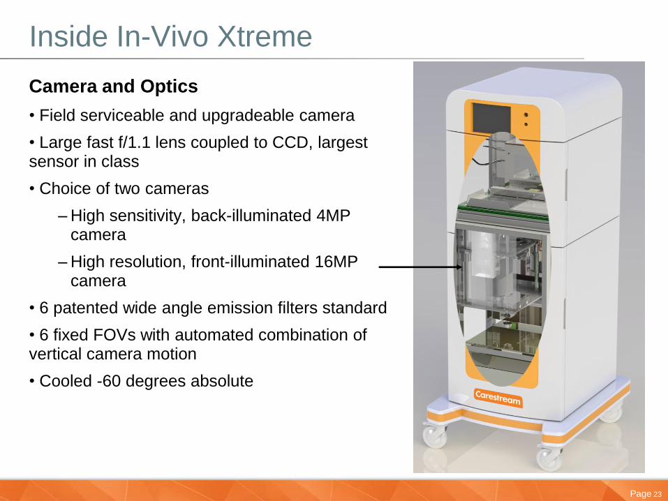
Inside In-Vivo Xtreme
Camera and Optics
• Field serviceable and upgradeable camera
• Large fast f/1.1 lens coupled to CCD, largest sensor in class
• Choice of two cameras
– High sensitivity, back-illuminated 4MP camera
– High resolution, front-illuminated 16MP camera
• 6 patented wide angle emission filters standard
• 6 fixed FOVs with automated combination of vertical camera motion
• Cooled -60 degrees absolute
Page 24
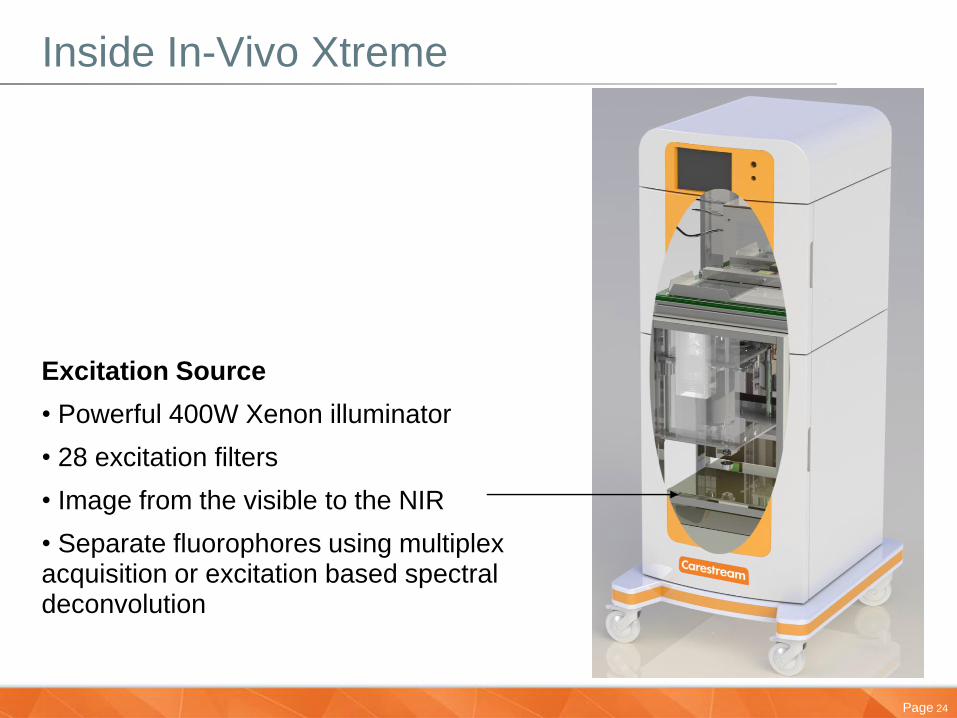
Inside In-Vivo Xtreme
Excitation Source
• Powerful 400W Xenon illuminator
• 28 excitation filters
• Image from the visible to the NIR
• Separate fluorophores using multiplex acquisition or excitation based spectral deconvolution
Page 26
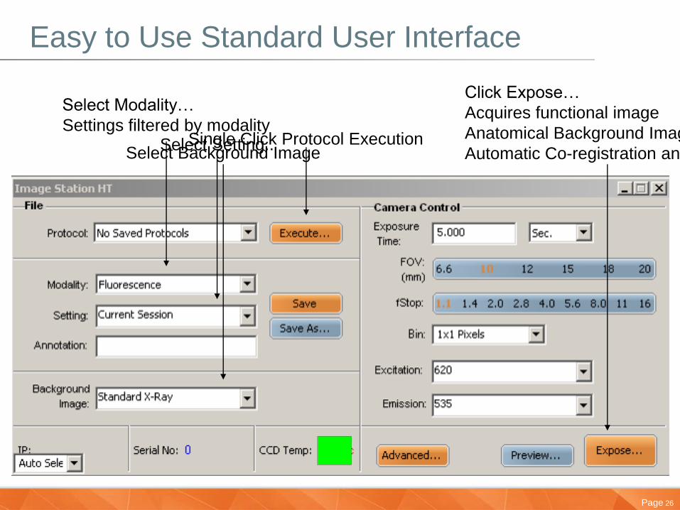
Easy to Use Standard User Interface
Single Click Protocol Execution
Select Modality…
Settings filtered by modality
Select Background Image
Click Expose…
Acquires functional image
Anatomical Background Image (X
Automatic Co-registration and Overlay
Select Setting..
Page 27
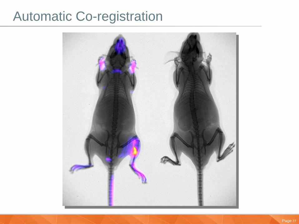
Automatic Co-registration
Page 28
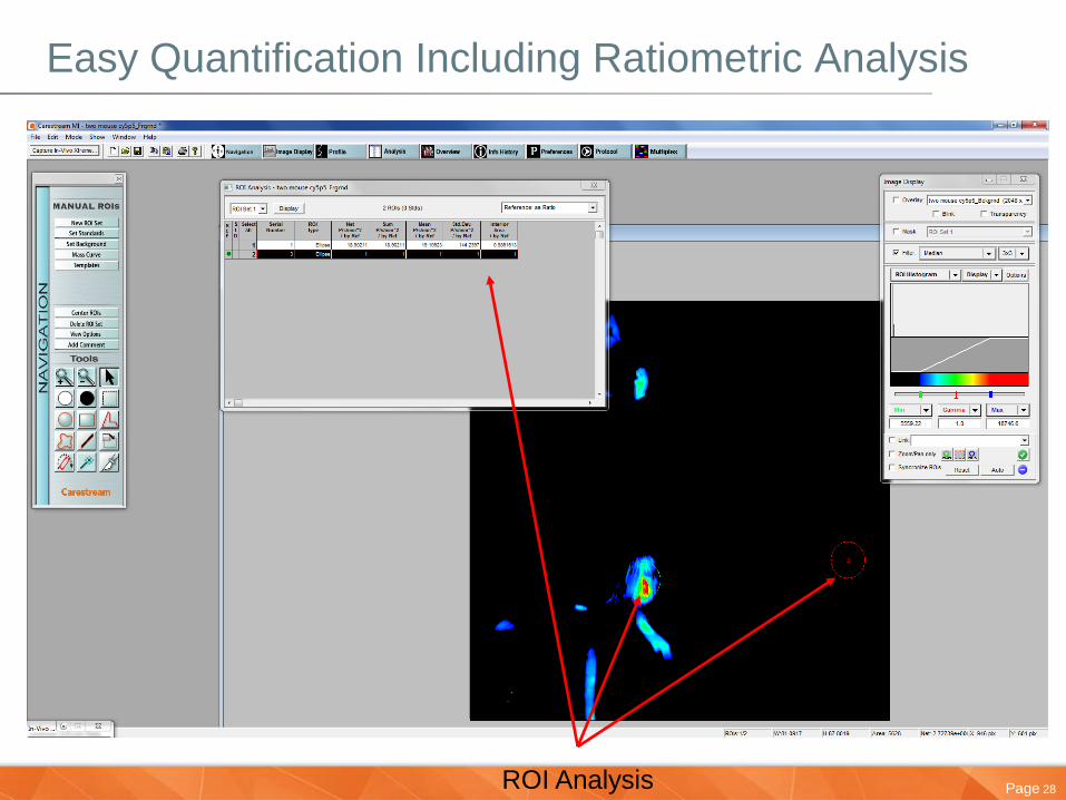
Easy Quantification Including Ratiometric Analysis
ROI Analysis
Page 29
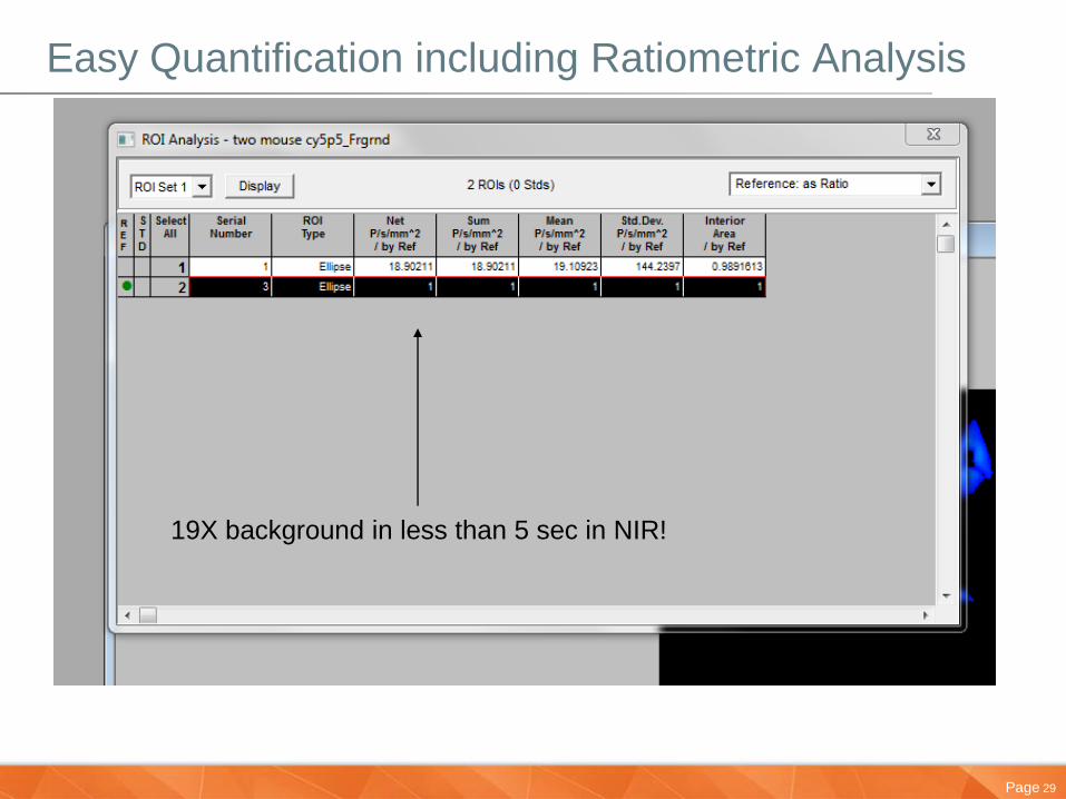
Easy Quantification including Ratiometric Analysis
19X background in less than 5 sec in NIR!
Page 30
Agenda
• Carestream Overview
• Introducing Molecular Imaging & Xtreme system
• Multimodal Applications
• Development of a Novel Molecular Imaging Probe
• Targeting Site of Infection
• Tumor Imaging of Necrotic Core
• Brain Traumer Inflammation & Cell Death Detection
•Conclusion
Results from Notre Dame Integrated Imaging Facility (NDIIF) in collaboration with
Prof. Bradley Smith, Department of Chemistry and Biochemistry,
University of Notre Dame, Indiana, USA
Page 31
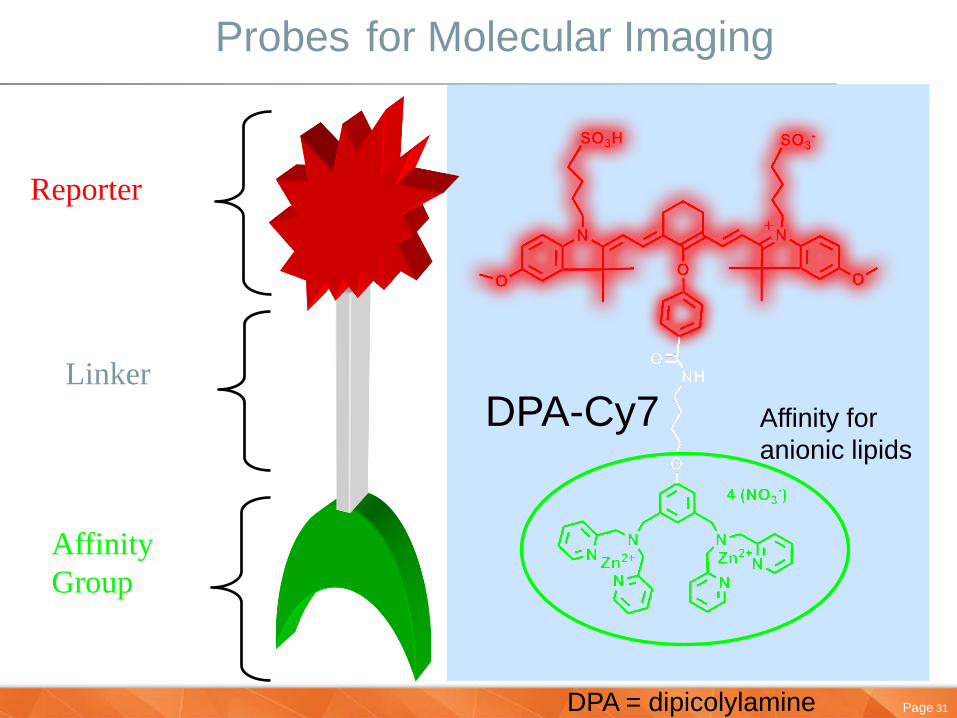
Probes for Molecular Imaging
Affinity
Group
Reporter
Linker
Affinity for
anionic lipids
DPA-Cy7
DPA = dipicolylamine
Page 32
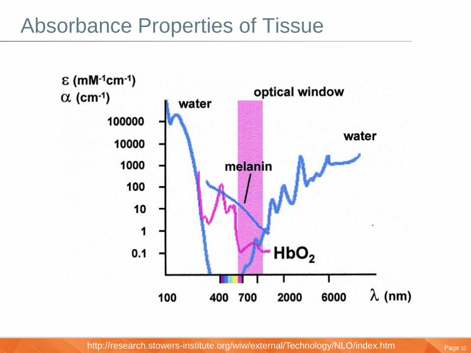
Absorbance Properties of Tissue
http://research.stowers-institute.org/wiw/external/Technology/NLO/index.htm
Page 33
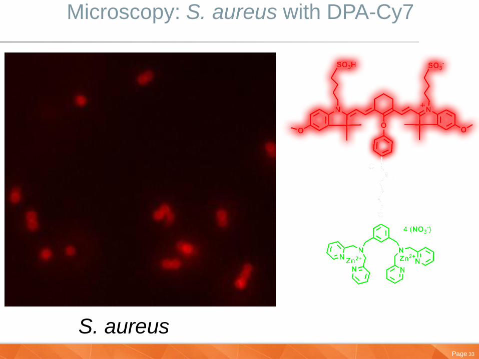
Microscopy: S. aureus with DPA-Cy7
DPA-Cy7 S. aureus
Page 34
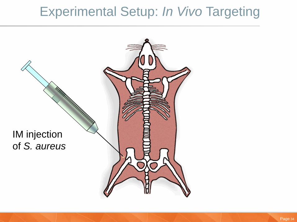
Experimental Setup: In Vivo Targeting
IM injection
of S. aureus
Page 35
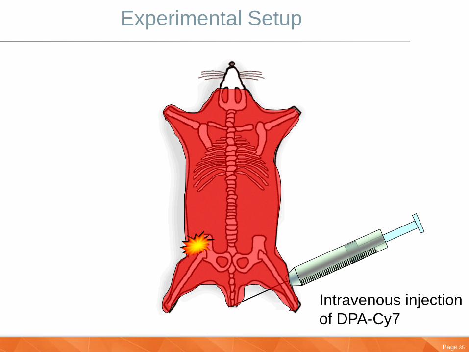
Experimental Setup
Intravenous injection
of DPA-Cy7

Page 36
In Vivo Imaging of Bacterial Infection
Pre-
injection
Post-
injection
6 h 12 h 18 h 21 h
This montage was prepared using ImageJ (free imaging software from NIH)
Leevy, W. M.; Gammon, S. T.; Jiang, H.; Johnson, J. R.; Marquez, M.; Piwnica-Worms, D.; Smith, B. D.
Bioconj. Chem. 2008, 686-692.
Excitation 755 nm, Emission 830 nm, No binning, 60 s acq, Fstop = 2.4
Page 37
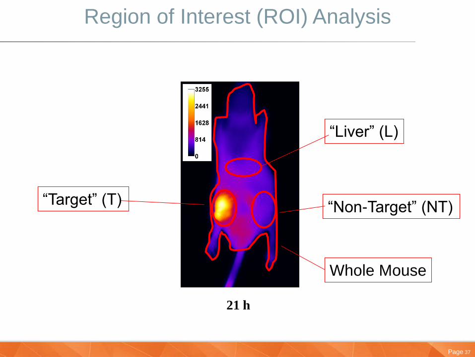
Region of Interest (ROI) Analysis
21 h
“Target” (T) “Non-Target” (NT)
“Liver” (L)
Whole Mouse
Page 38
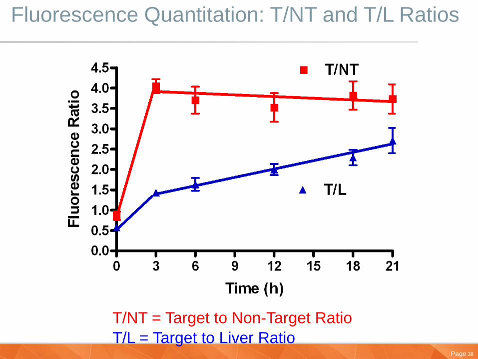
Fluorescence Quantitation: T/NT and T/L Ratios
T/NT = Target to Non-Target Ratio
T/L = Target to Liver Ratio
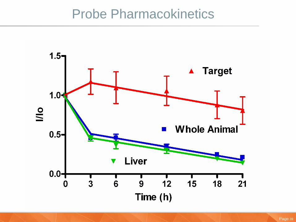
Page 39
Probe Pharmacokinetics
Page 40
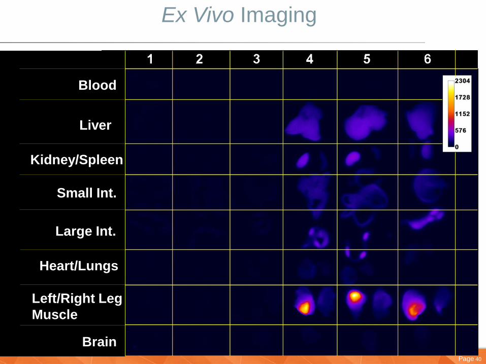
Ex Vivo Imaging
Blood
Liver
Kidney/Spleen
Small Int.
Large Int.
Heart/Lungs
Left/Right Leg
Muscle
Brain

Page 41
Ex Vivo Quantitation

Page 42
Multi-Modal Labeling
Non – Labeled Probe
Radiolabeled Probe Cu64 NIR Labeled Probe
Reflectance
Probe is now translatable into PET/SPECT imaging – Future goal to move into a clinical environment
Page 43
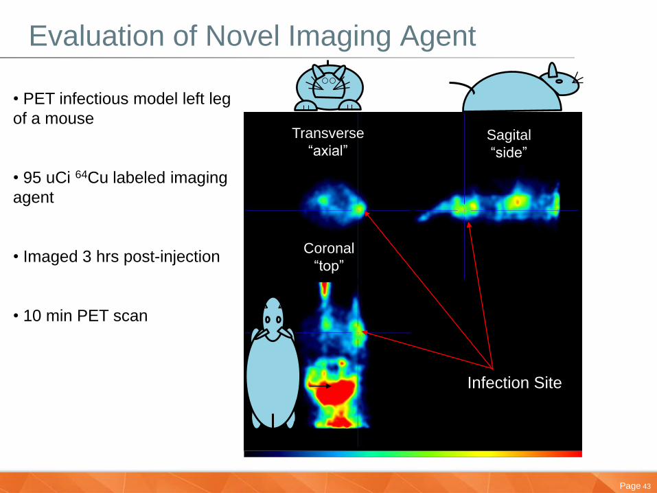
Evaluation of Novel Imaging Agent
• PET infectious model left leg
of a mouse
• 95 uCi 64Cu labeled imaging
agent
• Imaged 3 hrs post-injection
• 10 min PET scan
Infection Site
Transverse
“axial” Sagital
“side”
Coronal
“top”

Page 44
Agenda
• Carestream Overview
• Introducing Molecular Imaging & Xtreme system
• Multimodal Applications
• Development of a Novel Molecular Imaging Probe
• Targeting Site of Infection
• Tumor Imaging of Necrotic Core
• Brain Traumer Inflammation & Cell Death Detection
•Conclusion
Page 45
45
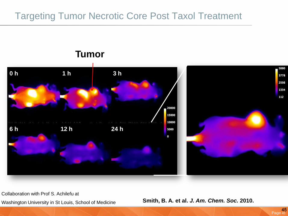
Tumor
0 h 3 h
6 h 12 h 24 h
1 h
Smith, B. A. et al. J. Am. Chem. Soc. 2010.
Targeting Tumor Necrotic Core Post Taxol Treatment
Collaboration with Prof S. Achilefu at
Washington University in St Louis, School of Medicine

Page 46
46
N
H3C
CH3 H3C
CH3
N
H3CO OCH3O
NO NN
N
NN
SO3 SO3H
O NH
Zn2+
Zn2+
4 NO3-
Using a PS Sensor to Detect Necrotic Tissue Within Tumors
PSS-794 is now commercially available from MTTI

Page 47
47 Smith, B. A. et al. J. Am. Chem. Soc. 2010.
Histological Analysis of Tumor Sections
Page 48
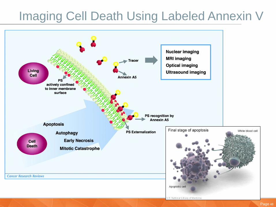
Imaging Cell Death Using Labeled Annexin V
Page 49
Agenda
• Carestream Overview
• Introducing Molecular Imaging & Xtreme system
• Multimodal Applications
• Development of a Novel Molecular Imaging Probe
• Targeting Site of Infection
• Tumor Imaging of Necrotic Core
• Brain Trauma Inflammation & Cell Death Detection
•Conclusion
Page 50
Brain (Frontal Cortex) Cryolesion Protocol
• Frontal Lobe cryolesion induced
• 1 hour later, PSVue 794 targeted probe injected IV
• 4 hours after injection, mice received 5 mg luminol injected IP
• X-ray, Near-infrared fluorescence, luminescence and RGB reflectance
(not shown) images taken
• Imaging repeated at 20 hours post cryolesion induction

Page 51
Brain (Frontal Cortex) Cryolesion - Xtreme
X-ray Luminescence
(Luminol)
Fluorescence
PSVue 794
Control Cryo 1 Cryo 2 Control Cryo 1 Cryo 2 Control Cryo 1 Cryo 2
5 hr post injury

Page 52
Brain (Frontal Cortex) Cryolesion – 5 Hour Time Point
Control Cryo 1 Cryo 2

Page 53
Brain (Frontal Cortex) Cryolesion
X-ray Luminescence
(Luminol)
Fluorescence
PSVue 794
Control Cryo 1 Cryo 2 Control Cryo 1 Cryo 2 Control Cryo 1 Cryo 2
20 hr post injury

Page 54
Brain Cryolesion/ Co-registration
Control Cryo 1 Cryo 2
Page 55
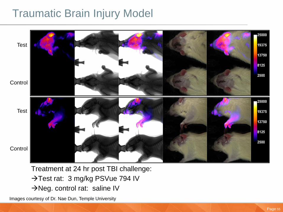
Traumatic Brain Injury Model
Treatment at 24 hr post TBI challenge:
Test rat: 3 mg/kg PSVue 794 IV
Neg. control rat: saline IV
Test
Control
Test
Control
Images courtesy of Dr. Nae Dun, Temple University
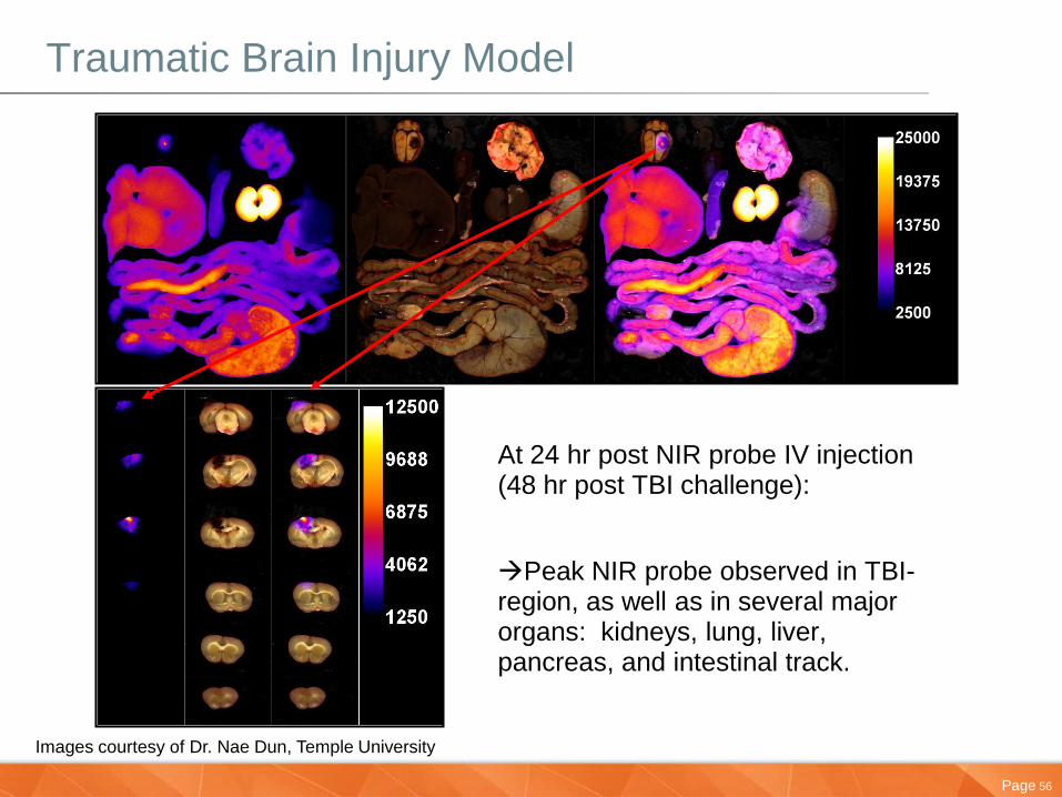
Page 56
Traumatic Brain Injury Model
At 24 hr post NIR probe IV injection (48 hr post TBI challenge):
Peak NIR probe observed in TBI-region, as well as in several major organs: kidneys, lung, liver, pancreas, and intestinal track.
Images courtesy of Dr. Nae Dun, Temple University

Page 57
Conclusion
• Whole animal imaging is a very useful tool for research development
• The In Vivo Xtreme system provides a powerful multimodal solution for molecular imaging
– Fluorescence
– Luminescence
– Radioisotopic
– High-resolution digital x-ray
• With up t 7 imaging modalities you have the freedom to combine many different molecular and anatomical imaging experiments
• Novel probes to detect disease can be easily assessed for effectiveness

Page 58
Thank you!
Contacts:
Joshua McHattan
Product Line Manager – Asia Cluster
Although the Carestream In-Vivo Xtreme can be used for in vivo and in vitro molecular
imaging of materials, researchers should be aware that the methods of preparing and
viewing the materials for molecular imaging may be subject to various patent rights.


![MSc in Translational (Neuroscience) · PDF fileMSc in Translational Pathology [Neuroscience] Why Translational Pathology? The MSc Translational Pathology (Neuroscience) course combines](https://img.dokumen.tips/doc/110x75/5a7454947f8b9a0d558bb440/msc-in-translational-neuroscience-a-msc-in-translational-pathology-neuroscience.jpg)