Embed Size (px)
Citation preview
IntroductionCancer cells overproducing cellular drug extruders maybecome resistant to a wide spectrum of drugs with dif-ferent structures or cellular targets. This phenomenonis called multidrug resistance (MDR) (1). Three ATP-dependent transmembrane drug transporters — themultidrug resistance gene product MDR1 P-glycopro-tein (Pgp) (1), the breast cancer resistance protein (2–4),and multidrug resistance protein 1 (MRP1) (5–9) — canrender cells multidrug resistant in vitro. MRP1 is a so-called glutathione-conjugate pump, or multispecificorganic anion transporter (10), and is a versatile pump.It can mediate the extrusion of glutathione-S conju-gates, glucuronide conjugates, and sulfate conjugates ofdrugs (10–13); it is the major transporter for endoge-nous LTC4, an important mediator of the inflammato-ry response (11, 14); and it can even extrude neutral andbasic organic compounds if the cell contains normallevels of glutathione (15–18).
To elucidate the physiological functions of theMDR-conferring transporters, mutant mice have been
generated lacking Mdr1a Pgp and Mdr1b Pgp (19, 20),or lacking Mrp1 (14, 21). Mdr1a Pgp plays an impor-tant role in the intestinal epithelium, where it active-ly excretes xenobiotics absorbed from the intestinallumen back into the lumen. It thereby limits the entryof Pgp substrates into the body. In addition, Mdr1aPgp plays an important role in the blood-brain barri-er (19, 20, 22–24). This barrier is primarily formed bythe endothelium of the blood capillaries in the brain.The presence of Mdr1a Pgp in the apical (luminal)plasma membranes of these capillary endothelial cells(25) is crucial for the protection of the brain againstseveral xenobiotics entering from the blood (19).
Mrp1-deficient mice lack the main high-affinity glu-tathione conjugate pump activity in erythrocytes, andcells derived from these mice show increased sensitivi-ty to anti-cancer drugs (14). Mrp1 has important pro-tective roles in the epithelium of the tongue and cheek,the urinary collecting duct epithelium, and the epithe-lial Sertoli cells in the testicular tubules; we have shownthat Mrp1 prevents drug-induced oral mucositis, dia-
The Journal of Clinical Investigation | February 2000 | Volume 105 | Number 3 279
Multidrug resistance protein 1 protects the choroid plexus epithelium and contributes to the blood-cerebrospinal fluid barrier
Jan Wijnholds,1 Elizabeth C.M. de Lange,2 George L. Scheffer,3 Dirk-Jan van den Berg,2
Carla A.A.M. Mol,1 Martin van der Valk,4 Alfred H. Schinkel,5 Rik J. Scheper,3
Douwe D. Breimer,2 and Piet Borst1
1Division of Molecular Biology and Center for Biomedical Genetics, The Netherlands Cancer Institute, 1066 CX Amsterdam, The Netherlands
2Division of Pharmacology, Leiden/Amsterdam Center for Drug Research, 2300 RA Leiden, The Netherlands3Department of Pathology, University Hospital Free University, 1081 HV Amsterdam, The Netherlands 4Department of Experimental Animal Pathology, and5Division of Experimental Therapy, The Netherlands Cancer Institute, 1066 CX Amsterdam, The Netherlands
Jan Wijnholds and Elizabeth C.M. de Lange contributed equally to this work.
Address correspondence to: Piet Borst, The Netherlands Cancer Institute, Plesmanlaan 121, 1066 CX Amsterdam, The Netherlands. Phone: 31-20-5122880; Fax: 31-20-6691383; E-mail: [email protected].
Received for publication August 26, 1999, and accepted in revised form December 21, 1999.
Multidrug resistance protein 1 (MRP1) is a transporter protein that helps to protect normal cells andtumor cells against the influx of certain xenobiotics. We previously showed that Mrp1 protectsagainst cytotoxic drugs at the testis-blood barrier, the oral epithelium, and the kidney urinary col-lecting duct tubules. Here, we generated Mrp1/Mdr1a/Mdr1b triple-knockout (TKO) mice, and usedthem together with Mdr1a/Mdr1b double-knockout (DKO) mice to study the contribution of Mrp1to the tissue distribution and pharmacokinetics of etoposide. We observed increased toxicity in theTKO mice, which accumulated etoposide in brown adipose tissue, colon, salivary gland, heart, andthe female urogenital system. Immunohistochemical staining revealed the presence of Mrp1 in theoviduct, uterus, salivary gland, and choroid plexus (CP) epithelium. To explore the transport func-tion of Mrp1 in the CP epithelium, we used TKO and DKO mice cannulated for cerebrospinal fluid(CSF). We show here that the lack of Mrp1 protein causes etoposide levels to increase about 10-foldin the CSF after intravenous administration of the drug. Our results indicate that Mrp1 helps to limittissue distribution of certain drugs and contributes to the blood-CSF drug-permeability barrier.
J. Clin. Invest. 105:279–285 (2000).

betes insipidus, and infertility (26). In addition, Mrp1-deficient mice have an impaired response to arachi-donic acid–stimulated inflammation, probably due tothe decreased cellular extrusion of LTC4 fromleukotriene-synthesizing cells (14).
Monoclonal antibodies against MRP1 (27, 28) detectthis transport protein in several tissues. Its presencehas been demonstrated in the basal plasma membraneof the Sertoli cells (26), in the basolateral membranesof the lung epithelium (26, 29), in the liver (30), in therat choroid plexus (CP) epithelium (31), and in trans-fected kidney cell lines overproducing human MRP1(32). Here, we show that Mrp1 is also present in themouse CP epithelium.
The brain is protected against drugs by 2 drug-per-meability barriers: the blood-brain barrier and theblood-cerebrospinal fluid (CSF) barrier. The blood-brain barrier is formed by the endothelium of the bloodcapillaries; diffusion of some hydrophobic drugsthrough this barrier is counteracted by transporterssuch as Mdr1a Pgp (19, 33). The blood-CSF barrier (34)is formed by the epithelium of the CP (35). Recently,some drug transporters (e.g., organic anion transporterprotein 1, MDR1 Pgp, and MRP1) have been postulat-ed to play a drug-transporting role in the CP (31, 36,37). The polarized choroidal epithelium uses activetransport mechanisms to extract micronutrients fromthe blood, and produces and secretes CSF (38). The CPalso has an important function in the clearance ofmetabolic waste products and substrates such as LTC4
(39), estradiol 17-β-D-glucuronide (37), prostaglandins(40), iodide (41), benzylpenicillin (42), cephalosporin(43), zidovudine (44–46), and 2,4,5-trichlorophenoxy-acetic acid (47) from the CSF toward the blood.
Using wild-type and Mrp1-deficient mice on an Mdr1aPgp/Mdr1b Pgp–null background (Mrp1+/+/Mdr1a–/–/Mdr1b–/– and Mrp1–/–/Mdr1a–/–/Mdr1b–/–, referred to asDKO and TKO for double knockout and triple knock-out, respectively) we demonstrate here that Mrp1 helpsto limit the accumulation of etoposide in brown adiposetissue, colon, salivary gland, heart, and the female uro-genital tract. Most importantly, we show that Mrp1 inthe CP protects against high etoposide levels in the CSF,indicating that Mrp1 is a critical component of theblood-CSF drug-permeability barrier. This implies thatspecific inhibitors of MRP1 and MDR1 Pgp may be usedto increase the permeability of the blood-CSF and blood-brain barriers for anti-cancer or other drugs.
MethodsAnimals. The animals used were Mdr1a- and Mdr1b-deficient (Mdr1a–/–/Mdr1b–/–) DKO mice and Mrp1-deficient (Mrp1–/–) mice, generated by gene targetingin embryonic stem cells as described by Schinkel et al.(19) and Wijnholds et al. (14). Using these mice,Mdr1a–/–/Mdr1b–/–/Mrp1–/– triple-knockout (TKO)mice were obtained by crossing. All mice used were ona 50% 129/Ola, 50% FVB genetic background. All ani-mals were housed in constant-temperature rooms with
a 12-hour-light/12-hour-dark cycle, and had access toa pelleted chow diet (Hope Farms BV, Woerden, TheNetherlands) and acidified water ad libitum. Mousehandling and experimental procedures were conduct-ed in accordance with the Netherlands Cancer Insti-tute guidelines for animal care and use.
Drug sensitivity and [3H]etoposide distribution in femalemice. Etoposide phosphate (Etopophos, 100 mg effec-tive etoposide; Bristol-Myers Squibb Pharmaceutical,Princeton, New Jersey, USA) was dissolved in sterile0.9% NaCl to obtain a stock solution of 4–20 mg/mL.Concentrations were adjusted to inject 5–6.5 µL/g bodyweight (20–130 mg/kg) intravenously in the tail vein offemale or male mice (11–16 weeks old) that were light-ly anesthetized with diethyl ether. Mice were weighedand were observed daily for a period of 2 weeks. Lethaltoxicity occurred between days 2 and 13 after injection.
For tissue distribution experiments, tracer [3H]etopo-side (475 Ci/mol) was diluted with carrier etoposide(Vepesid; Bristol-Myers Squibb Pharmaceutical) in 5%(wt/vol) glucose, and was injected into the tail vein at adose of 1 mg/kg. Tissues from female mice were col-lected at 4 hours and were processed as described (19).
Histological and immunohistochemical analyses. Histolog-ical and immunohistochemical analyses were per-formed as described (26, 27).
Cannulation of lateral brain ventricles for CSF sampling.Female DKO mice and TKO mice 11–16 weeks of age(25–27 g) were anesthetized by intraperitoneal injectionof 6 µL Hypnorm/Dormicum/water (1:1:2, vol/vol/vol)per gram body weight (Hypnorm, Janssen Pharmaceuti-ca, Beerse, Belgium; Dormicum; Roche Pharmaceuticals,Basel, Switzerland). A tail cannula consisting of 15 cm ofpolyethylene tubing (outer diameter 0.61 cm, innerdiameter 0.28 cm) and a 7-cm piece of Zeus tubing(outer diameter 0.48 mm, inner diameter 0.24 mm; ZeusIndustrial Products, Orangeburg, South Carolina, USA)was inserted into the tail artery to reach the aorta. The
280 The Journal of Clinical Investigation | February 2000 | Volume 105 | Number 3
Figure 1Loss of body weight in mice lacking Mrp1 on a genetic backgroundlacking Mdr1a Pgp and Mdr1b Pgp (TKO mice). DKO mice, TKOmice, and Mrp1–/– mice received a single intravenous dose of 60mg/kg Etopophos , and the loss of body weight (%) was measuredover 7 days. *P < 0.001.
anesthetized mouse was fixed in a stereotactic appara-tus. The skin covering the skull was shaved and disin-fected with 70% ethanol, and then cut crosswise andfolded aside. The exposed tissue was locally anesthetizedusing lidocaine (1% in 0.9% NaCl) and was removed byscraping to expose the skull. The skull was thoroughlycleaned. Two holes were drilled 1.1 mm apart (laterally)at 0.6 mm rostral to bregma. A homemade double can-nula made of 19-gauge and 23-gauge needles with 4-mmextending tips of fused silica (outer diameter 0.29 mm,inner diameter 0.10 mm; Composite Metal Services Ltd.,Worcester, UK) was fixed in a micromanipulator on thestereotactic apparatus. This double cannula was subse-quently lowered through the brain tissue into the later-al ventricles to a ventral coordinate at 2.5 mm to bregma,and fixed first with Cyanolit glue and then with dentalcement. The micromanipulator was carefully removed.The mouse was put on a heating pad at 37–39°C andwas not allowed to awaken. A solution of 145 mM NaCl,0.6 mM KCl, 1.0 mM MgCl2, 1.2 mM CaCl2, and 0.2 mMascorbic acid in 2 mM phosphate buffer (pH 7.4) wasperfused into the right lateral ventricle at a flow rate of0.1 µL/min at a constant rate with a syringe pump. Oneof the intracerebroventricular cannula lanes was used forinfusing the perfusion solution (Baby Bee Hive pump;Bioanalytical Systems Inc., West Lafayette, Indiana,USA), while the other was used to withdraw CSF at thesame flow rate into the outlet tubing (home-adjustedHarvard ‘33’ pump; Harvard Apparatus Inc., Holliston,Massachusetts, USA). Once the perfusion and collectionhad been running constantly for 10 minutes,Etopophos (60 mg/kg) was infused through a tail can-nula with the aid of a syringe pump (100 µL in 2 min-utes). During the 1 hour after the start of Etopophos
administration, CSF was collected through the outlettubing. The mouse was decapitated and blood and brainwere collected. Blood was centrifuged for 10 minutes at2,000 g to obtain plasma. CSF, brain tissue, and plasmawere stored at –30°C pending analysis.
Analysis of etoposide in plasma, CSF, and brain tissue. Aftergently thawing, the CSF sample was introduced into an
HPLC system that consisted of a Spherisorb C18 col-umn (100 mm × 0.8 mm inner diameter, 5 µm particlediameter; Capital HPLC Ltd., Broxburn, UK) using asolvent system of 50 mM sodium acetate buffer with 50µM EDTA (pH 3.8) and acetonitril (3.8:1 vol/vol) at aflow rate of 40 µL/min (48).
Plasma was diluted with phosphate buffer (50 mM atpH 6.0), and brain tissue was homogenized with a Poly-tron (0.2 g tissue/mL phosphate buffer). Etoposide indiluted plasma and homogenized brain tissue wasextracted with diethyl ether and dichloromethane (2:1,vol/vol), and then separated on an Econosil C18 column(250 mm × 4.6 mm inner diameter, 10 µm particle diam-eter; Alltech Associates Inc., Deerfield, Illinois, USA)using a solvent system of 50 mM sodium acetate with 50µM EDTA (pH 3.8) and acetonitril (2:1, vol/vol) at a flowrate of 1.1 mL/min (49). Detection was performed elec-trochemically (∆V = 900 mV) using a glassy carbon work-ing electrode (Decade; Antec Leyden BV, Zoeterwoude,The Netherlands). The system was set at 40°C.
Statistical evaluation. The results are presented as mean± SE. The difference between groups was evaluatedwith the unpaired, 2-tailed Student’s t test.
ResultsAltered drug sensitivity in mice lacking Mrp1 on anMdr1a/Mdr1b–null genetic background. We have previous-ly shown that Mrp1 deficiency renders mice hypersen-sitive to etoposide or its water-soluble derivative, etopo-side phosphate (Etopophos ) (14, 26). Becauseetoposide is also transported by Mdr1-type Pgps, wecrossed Mrp1–/– mice with DKO Mdr1a–/–/Mdr1b–/– miceto obtain TKO Mrp1–/–/Mdr1a–/–/Mdr1b–/– mice. No dif-ferences in health, life span, or fertility were observedamong TKO, DKO, Mrp1–/–, or wild-type mice in ouranimal facility. The TKO mice are more sensitive toetoposide than are DKO and Mrp1–/– mice; all 3 of thosegroups are more sensitive than are wild-type mice(Tables 1 and 2). Seven days after a single intravenous60 mg/kg dose of Etopophos , female Mrp1–/– mice (n =5) retained 92.3 ± 3.8% of their body weight (24.7 ± 1.1g at day 0) and DKO mice (n = 5) retained 99.5 ± 1.2% oftheir body weight (27.8 ± 0.9 g at day 0), whereas female
The Journal of Clinical Investigation | February 2000 | Volume 105 | Number 3 281
Table 1Etopophos toxicity in male wild-type, Mrp1–/–, DKO, and TKO mice
Survival of mice
Etopophos dose Wild type Mrp1–/– DKO TKO
(mg/kg)
20 – – – 3/3A
40 – – – 3/3A
60 – 3/3A – 1/3A, 3/4B
70 – – – 0/4A
80 – 3/3A, 5/5B 4/5B 0/3A
100 – – 3/5B –120 5/5B – 0/2B –
The number of surviving animals per group at each intravenous Etopophos doseis shown. The data are from 2 independent experiments (A and B), using mice11–16 weeks old. For additional toxicity experiments using wild-type and Mrp1–/–
mice, see ref. 26. TKO mice show increased sensitivity to Etopophos (P < 0.01).
Table 2Etopophos toxicity in female wild-type, Mrp1–/–, DKO, and TKO mice
Survival of mice
Etopophos dose Wild type Mrp1–/– DKO TKO
(mg/kg)30 – – – 3/350 – – – 3/370 – 3/3 3/3 0/390 – 3/3 1/3 –110 3/3 0/3 0/3 –130 3/3 – – –
The number of surviving animals per group at each intravenous Etopophos
dose is shown. Mice used were 11–13 weeks old. TKO mice show increasedsensitivity to Etopophos (P < 0.01).
TKO mice (n = 5) retained 74.1 ± 2.6% (P < 0.01) of theirbody weight (26.5 ± 0.7 g at day 0) (Figure 1).
Except for their weight loss, the drug-treated TKO miceshowed no clear gross abnormalities. Microscopic analy-sis at day 7 of TKO, DKO, and Mrp1–/– tissues from micetreated with 60 mg/kg Etopophos showed a disruptionof the epithelial layer of the tongue and cheek only in theTKO mouse tissue (all TKO mice were thus affected).Inflammation of the tongue epithelium was detected in 4out of 5 TKO animals (results not shown). The defectswere similar to those observed in Mrp1-deficient miceafter higher drug doses (26). Analysis of the colon of micetreated with 50–60 mg/kg Etopophos showed cryptdegeneration (9 of 12 TKO mice; 3 of 11 DKO mice) andcolitis (5 of 12 TKO mice; 1 of 11 DKO mice) (data notshown). The increase in gut damage to TKO mice suggeststhat Mrp1 contributes to the protection of the colon.Except for sporadic signs of inflammation in the stomach(in 3 of 5 mice), and a degeneration of the oocytes in theovary (in 2 of 5 mice), an extensive histopathologicalexamination revealed no further abnormalities in othertissues (data not shown) of the TKO mice.
We previously found that Mrp1-deficient mice, whentreated with a dose of 100 mg Etopophos per kg bodyweight, developed diabetes insipidus, demonstrated bythe overproduction of dilute urine (26). At close to themaximal drug dose tolerated by the TKO mice in meta-bolic cages (50 mg/kg), water intake, urine production(DKO, 1.7 ± 0.5 mL per 48 hours; TKO, 1.3 ± 0.3 mL per48 hours), food consumption, and feces production didnot differ between DKO and TKO mice. No differenceswere detected 7 days after drug administration in plas-ma sodium, urea, creatinine, or protein levels. Thenumber of leukocytes was reduced in TKO mice (DKO,[13.6 ± 1.5] × 109/L; TKO, [3.0 ± 1.2] × 109/L; P < 0.01),whereas hemoglobin, hematocrit, and the number ofthrombocytes did not differ between DKO mice andTKO mice (data not shown).
Altered drug tissue distribution in mice lacking Mrp1.Although Mrp1 is a ubiquitous protein present in sever-al epithelia (26, 27, 29, 31), our previous etoposide tissuedistribution experiments did not show a significantincrease in the accumulation of etoposide in Mrp1-defi-cient tissues (14). This negative result could be explained
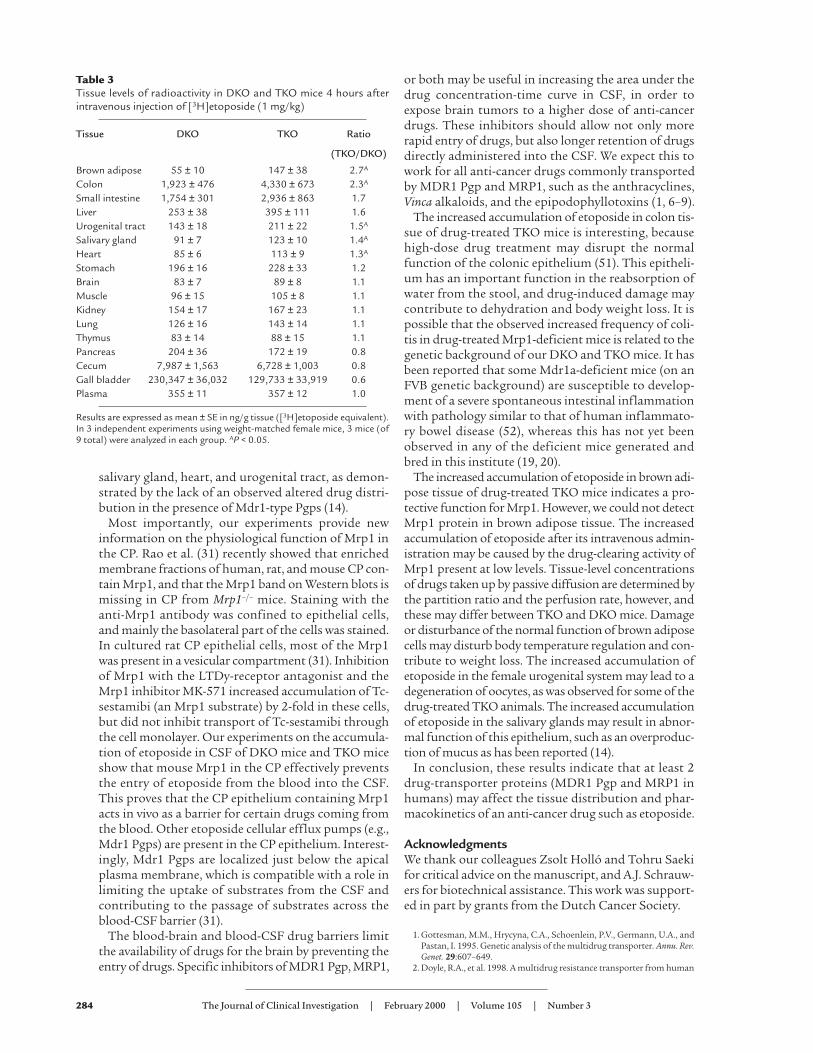
by redundancy of etoposide trans-porters; obvious candidates for theredundant transporters are the Mdr1-type Pgps. To test whether Mrp1 cancontribute to etoposide cellular clear-ing in vivo, we administered 1 mg/kgof radiolabeled etoposide intravenous-ly to DKO mice and TKO mice, anddetermined the tissue distribution oftotal radioactivity 4 hours after injec-tion. Table 3 shows that the etoposideplasma levels of DKO and TKO micewere comparable, indicating thatMrp1 does not significantly contributeto the renal, liver, or intestinal clear-ance of etoposide as was observed forMdr1-type Pgps (19, 20). In the TKOmice, however, we observed increasedetoposide accumulation in brown adi-pose (2.7-fold), colon (2.3-fold), uro-genital tract (1.5-fold), salivary gland(1.4-fold), and heart (1.3-fold) tissues(P < 0.05); no significant accumulationwas detected in brain, liver, kidney, or
282 The Journal of Clinical Investigation | February 2000 | Volume 105 | Number 3
Figure 2Mrp1 staining of frozen sections of wild-type andMrp1-deficient tissues using monoclonal anti-body MRPr1. Sections were counterstained withhematoxylin. Wild-type (a, c, e, g) and Mrp1-deficient mice (b, d, f, h). (a and b) Sections ofthe CP showing Mrp1 staining in the CP epithe-lium (a). (c and d) Sections of the salivary gland.(e and f) Sections of the oviduct. (g and h) Sec-tions of the uterus. Bar in a represents 100 µmand applies to a–d, g and h. Bar in e represents200 µm and applies to e and f.
other tissues. The observed altered distribution of etopo-side in some tissues (with no differences in plasma lev-els) indicates that Mrp1 contributes to the protectionagainst drugs of a limited set of tissues or cells. Theabsence of Mrp1 in the basolateral epithelia lining thelumina of some of these tissues (e.g., salivary gland,oviduct, or uterus) could lead to reduced active clearanceof drugs from such epithelia to the interstitial fluid andtoward the blood as proposed previously (26).
Mrp1 in salivary gland, uterus, oviduct, and CP. To corre-late the altered drug distribution with Mrp1 proteinlocation, we stained tissue sections from wild-type micewith the monoclonal antibody MRPr1 (27). As controls,we used mice lacking Mrp1, and isotype-matched con-trol antibodies. We found Mrp1 in the intercalated ductsof the salivary gland, and in the oviduct and uterus (Fig-ure 2, c, e, and g). Sections through the brain showed nostaining for Mrp1 in the endothelium of the blood cap-
illaries, but immunostaining was abundant in theepithelium of the CP (Figure 2a). In rat CP epithelium,staining for Mrp1 protein (31, 37) has been reported aswell. Mrp1 protein levels were below the detection levelin brown adipose tissue, colon, and heart (data notshown). The unexpected lack of immunostaining forMrp1 in these tissues may be due to the homogeneousdistribution of low levels of Mrp1. In previous experi-ments we showed that Mrp1 is present in heart (14)using Western blots. Using the MRPr1 monoclonal anti-body, Peng et al. detected mouse Mrp1 in all colon cellslining the crypt-villus axis, mainly in their basolateralmembranes (50). Immunostaining has shown MRP1 tobe present in the myocardium of the human heart as wellas in the epithelium of the human colon (27).
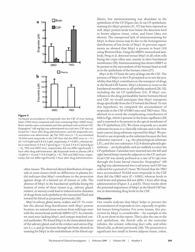
Mrp1 in the CP limits the entry of drugs into the CSF. Thepresence of Mrp1 in the CP prompted us to test the pos-sibility that Mrp1 contributes to the transport of drugsat the blood-CSF barrier. Mrp1 is known to route to thebasolateral membranes in all epithelia analyzed (26, 32),including the rat CP epithelium (31). If Mrp1 con-tributes to the drug-permeability barrier between bloodand CSF, we would anticipate that Mrp1 transportsdrugs specifically from the CP toward the blood. To testthis hypothesis, we compared the accumulation ofetoposide in the CSF of DKO mice and TKO mice. Thisallowed us to avoid the complicating contribution ofMdr1a Pgp, which is present in the brain capillaries (20)and is reported to be present in the apical membrane ofthe CP epithelium (31). We chose etoposide as a modelsubstrate because it is clinically relevant and is the best(anti-cancer) drug substrate reported for Mrp1. We pre-ferred to use etoposide as the substrate because the bestsubstrates reported — e.g., the physiological substrateLTC4 and the test substance S-(2,4-dinitrophenyl)-glu-tathione — are hydrophilic and are unlikely to enter theCP epithelium. Cannulas were inserted into the left andright lateral brain ventricles adjacent to the CP, and arti-ficial CSF was slowly perfused at a rate of 0.1 µL/minthrough the brain lateral ventricles. Etopophos (60mg/kg) was administered into a tail artery, and CSF wascollected for a period of 1 hour. We found that the TKOmice accumulated 10-fold more etoposide in the CSFthan did the DKO mice (P < 0.001), whereas levels intotal brain and plasma did not differ 1 hour after drugadministration (Figure 3, a and b). These results showthe potential importance of Mrp1 at the blood-CSF bar-rier in determining drug levels in the CSF.
DiscussionOur results indicate that Mrp1 helps to prevent theaccumulation of etoposide in vivo, especially in epithe-lial tissues lining lumina. For some tissues, this pro-tection by Mrp1 is considerable — for example in theCP, as we show in this report. This is also the case in theoral epithelium, the Sertoli cells in the testiculartubules, the urinary collecting duct cells, and whiteblood cells, as shown previously (26). The protection issignificant but small in brown adipose tissue, colon,
The Journal of Clinical Investigation | February 2000 | Volume 105 | Number 3 283
Figure 3Increased accumulation of etoposide into the CSF of mice lackingMrp1 (TKO mice) compared with mice containing Mrp1 (DKO mice).Brain vesicles were cannulated and then perfused with artificial CSF.Etopophos (60 mg/kg) was administered via a tail vein; CSF was col-lected for 1 hour after drug administration, and the etoposide con-centration was determined. (a) The TKO mice (n = 5) accumulated10-fold more etoposide in the CSF than did the DKO mice (n = 4 )(8.1 ± 0.9 µM and 0.8 ± 0.3 µM, respectively; P < 0.001), whereas lev-els in total brain (3.9 ± 0.7 µmol/kg [n = 5] and 2.9 ± 0.7 µmol/kg [n= 4], TKO and DKO mice, respectively) did not differ significantly 1hour after drug administration. (b) Etoposide levels in plasma (98 ±12 µM [n = 5] and 114 ± 24 µM [n = 4], TKO and DKO mice, respec-tively) did not differ significantly 1 hour after drug administration.
salivary gland, heart, and urogenital tract, as demon-strated by the lack of an observed altered drug distri-bution in the presence of Mdr1-type Pgps (14).
Most importantly, our experiments provide newinformation on the physiological function of Mrp1 inthe CP. Rao et al. (31) recently showed that enrichedmembrane fractions of human, rat, and mouse CP con-tain Mrp1, and that the Mrp1 band on Western blots ismissing in CP from Mrp1–/– mice. Staining with theanti-Mrp1 antibody was confined to epithelial cells,and mainly the basolateral part of the cells was stained.In cultured rat CP epithelial cells, most of the Mrp1was present in a vesicular compartment (31). Inhibitionof Mrp1 with the LTDy-receptor antagonist and theMrp1 inhibitor MK-571 increased accumulation of Tc-sestamibi (an Mrp1 substrate) by 2-fold in these cells,but did not inhibit transport of Tc-sestamibi throughthe cell monolayer. Our experiments on the accumula-tion of etoposide in CSF of DKO mice and TKO miceshow that mouse Mrp1 in the CP effectively preventsthe entry of etoposide from the blood into the CSF.This proves that the CP epithelium containing Mrp1acts in vivo as a barrier for certain drugs coming fromthe blood. Other etoposide cellular efflux pumps (e.g.,Mdr1 Pgps) are present in the CP epithelium. Interest-ingly, Mdr1 Pgps are localized just below the apicalplasma membrane, which is compatible with a role inlimiting the uptake of substrates from the CSF andcontributing to the passage of substrates across theblood-CSF barrier (31).
The blood-brain and blood-CSF drug barriers limitthe availability of drugs for the brain by preventing theentry of drugs. Specific inhibitors of MDR1 Pgp, MRP1,
or both may be useful in increasing the area under thedrug concentration-time curve in CSF, in order toexpose brain tumors to a higher dose of anti-cancerdrugs. These inhibitors should allow not only morerapid entry of drugs, but also longer retention of drugsdirectly administered into the CSF. We expect this towork for all anti-cancer drugs commonly transportedby MDR1 Pgp and MRP1, such as the anthracyclines,Vinca alkaloids, and the epipodophyllotoxins (1, 6–9).
The increased accumulation of etoposide in colon tis-sue of drug-treated TKO mice is interesting, becausehigh-dose drug treatment may disrupt the normalfunction of the colonic epithelium (51). This epitheli-um has an important function in the reabsorption ofwater from the stool, and drug-induced damage maycontribute to dehydration and body weight loss. It ispossible that the observed increased frequency of coli-tis in drug-treated Mrp1-deficient mice is related to thegenetic background of our DKO and TKO mice. It hasbeen reported that some Mdr1a-deficient mice (on anFVB genetic background) are susceptible to develop-ment of a severe spontaneous intestinal inflammationwith pathology similar to that of human inflammato-ry bowel disease (52), whereas this has not yet beenobserved in any of the deficient mice generated andbred in this institute (19, 20).
The increased accumulation of etoposide in brown adi-pose tissue of drug-treated TKO mice indicates a pro-tective function for Mrp1. However, we could not detectMrp1 protein in brown adipose tissue. The increasedaccumulation of etoposide after its intravenous admin-istration may be caused by the drug-clearing activity ofMrp1 present at low levels. Tissue-level concentrationsof drugs taken up by passive diffusion are determined bythe partition ratio and the perfusion rate, however, andthese may differ between TKO and DKO mice. Damageor disturbance of the normal function of brown adiposecells may disturb body temperature regulation and con-tribute to weight loss. The increased accumulation ofetoposide in the female urogenital system may lead to adegeneration of oocytes, as was observed for some of thedrug-treated TKO animals. The increased accumulationof etoposide in the salivary glands may result in abnor-mal function of this epithelium, such as an overproduc-tion of mucus as has been reported (14).
In conclusion, these results indicate that at least 2drug-transporter proteins (MDR1 Pgp and MRP1 inhumans) may affect the tissue distribution and phar-macokinetics of an anti-cancer drug such as etoposide.
AcknowledgmentsWe thank our colleagues Zsolt Holló and Tohru Saekifor critical advice on the manuscript, and A.J. Schrauw-ers for biotechnical assistance. This work was support-ed in part by grants from the Dutch Cancer Society.
1. Gottesman, M.M., Hrycyna, C.A., Schoenlein, P.V., Germann, U.A., andPastan, I. 1995. Genetic analysis of the multidrug transporter. Annu. Rev.Genet. 29:607–649.
2. Doyle, R.A., et al. 1998. A multidrug resistance transporter from human
284 The Journal of Clinical Investigation | February 2000 | Volume 105 | Number 3
Table 3Tissue levels of radioactivity in DKO and TKO mice 4 hours afterintravenous injection of [3H]etoposide (1 mg/kg)
Tissue DKO TKO Ratio
(TKO/DKO)
Brown adipose 55 ± 10 147 ± 38 2.7A
Colon 1,923 ± 476 4,330 ± 673 2.3A
Small intestine 1,754 ± 301 2,936 ± 863 1.7Liver 253 ± 38 395 ± 111 1.6Urogenital tract 143 ± 18 211 ± 22 1.5A
Salivary gland 91 ± 7 123 ± 10 1.4A
Heart 85 ± 6 113 ± 9 1.3A
Stomach 196 ± 16 228 ± 33 1.2Brain 83 ± 7 89 ± 8 1.1Muscle 96 ± 15 105 ± 8 1.1Kidney 154 ± 17 167 ± 23 1.1Lung 126 ± 16 143 ± 14 1.1Thymus 83 ± 14 88 ± 15 1.1Pancreas 204 ± 36 172 ± 19 0.8Cecum 7,987 ± 1,563 6,728 ± 1,003 0.8Gall bladder 230,347 ± 36,032 129,733 ± 33,919 0.6Plasma 355 ± 11 357 ± 12 1.0
Results are expressed as mean ± SE in ng/g tissue ([3H]etoposide equivalent).In 3 independent experiments using weight-matched female mice, 3 mice (of9 total) were analyzed in each group. AP < 0.05.
MCF-7 breast cancer cells. Proc. Natl. Acad. Sci. USA. 95:15665–15670.3. Allikmets, R., Schriml, L.M., Hutchinson, A., Romano-Spica, V., and
Dean, M. 1998. A human placenta-specific ATP-binding cassette gene(ABCP) on chromosome 4q22 that is involved in multidrug resistance.Cancer Res. 58:5337–5339.
4. Miyake, K., et al. 1999. Molecular cloning of cDNAs which are highlyoverexpressed in mitoxantrone-resistant cells: demonstration of homol-ogy to ABC transport genes. Cancer Res. 59:8–13.
5. Keppler, D., Leier, I., Jedlitschky, G., and König, J. 1998. ATP-dependenttransport of glutathione S-conjugates by the multidrug resistance pro-tein MRP1 and its apical isoform MRP2. Chem. Biol. Interact.111–112:153–161.
6. Cole, S.P., and Deeley, R.G. 1998. Multidrug resistance mediated by theATP-binding cassette transporter protein MRP. Bioessays. 20:931–940.
7. Cole, S.P.C., et al. 1992. Overexpression of a transporter gene in a mul-tidrug-resistant human lung cancer cell line. Science. 258:1650–1654.
8. Zaman, G.J.R., et al. 1994. The human multidrug resistance-associatedprotein MRP is a plasma membrane drug-efflux pump. Proc. Natl. Acad.Sci. USA. 91:8822–8826.
9. Cole, S.P.C., et al. 1994. Pharmacological characterization of multidrugresistant MRP-transfected human tumor cells. Cancer Res. 54:5902–5910.
10. Ishikawa, T., Li, Z.S., Lu, Y.P., and Rea, P.A. 1997. The GS-X pump inplant, yeast, and animal cells: structure, function, and gene expression.Biosci. Rep. 17:189–207.
11. Leier, I., et al. 1994. The MRP gene encodes and ATP-dependent exportpump for leukotriene C4 and structurally related conjugates. J. Biol. Chem.269:27807–27810.
12. Jedlitschky, G., et al. 1996. Transport of glutathione, glucuronate, andsulfate conjugates by the MRP gene-encoded conjugate export pump.Cancer Res. 56:988–994.
13. Müller, M., et al. 1994. Overexpression of the gene encoding the multidrugresistance associated protein results in increased ATP-dependent glu-tathione S-conjugate transport. Proc. Natl. Acad. Sci. USA. 91:13033–13037.
14. Wijnholds, J., et al. 1997. Increased sensitivity to anticancer drugs anddecreased inflammatory response in mice lacking the multidrug resist-ance-associated protein. Nat. Med. 3:1275–1279.
15. Zaman, G.J.R., et al. 1995. Role of glutathione in the export of com-pounds from cells by the multidrug-resistance-associated protein. Proc.Natl. Acad. Sci. USA. 92:7690–7694.
16. Loe, D.W., Deeley, R.G., and Cole, S.P. 1998. Characterization of vin-cristine transport by the M(r) 190,000 multidrug resistance protein(MRP): evidence for cotransport with reduced glutathione. Cancer Res.58:5130–5136.
17. Renes, J., de Vries, E.G., Nienhuis, E.F., Jansen, P.L., and Müller, M. 1999.ATP- and glutathione-dependent transport of chemotherapeutic drugsby the multidrug resistance protein MRP1. Br. J. Pharmacol. 126:681–688.
18. Rappa, G., Lorico, A., Flavell, R.A., and Sartorelli, A.C. 1997. Evidence thatthe multidrug resistance protein (MRP) functions as a co-transporter ofglutathione and natural product toxins. Cancer Res. 57:5232–5237.
19. Schinkel, A.H., et al. 1997. Normal viability and altered pharmacokinet-ics in mice lacking Mdr1-type (drug-transporting) P-glycoproteins. Proc.Natl. Acad. Sci. USA. 94:4028–4033.
20. Schinkel, A.H., et al. 1994. Disruption of the mouse Mdr1a P-glycopro-tein gene leads to a deficiency in the blood-brain barrier and to increasedsensitivity to drugs. Cell. 77:491–502.
21. Lorico, A., et al. 1997. Disruption of the murine MRP (Multidrug Resis-tance Protein) gene leads to increased sensitivity to etoposide (VP-16)and increased levels of glutathione. Cancer Res. 57:5238–5242.
22. Sparreboom, A., et al. 1997. Limited oral bioavailability and active epithe-lial excretion of paclitaxel (taxol) caused by P-glycoprotein in the intes-tine. Proc. Natl. Acad. Sci. USA. 94:2031–2035.
23. de Lange, E.C., de Bock, G., Schinkel, A.H., de Boer, A.G., and Breimer,D.D. 1998. BBB transport and P-glycoprotein functionality usingMDR1A (–/–) and wild-type mice. Total brain versus microdialysis con-centration profiles of rhodamine-123. Pharm. Res. 15:1657–1665.
24. Mayer, U., et al. 1997. Full blockade of intestinal P-glycoprotein andextensive inhibition of blood-brain barrier P-glycoprotein by oral treat-ment of mice with PSC833. J. Clin. Invest. 100:2430–2436.
25. Cordon-Cardo, C., et al. 1989. Multidrug-resistance gene (P-glycopro-tein) is expressed by endothelial cells at blood-brain barrier sites. Proc.Natl. Acad. Sci. USA. 86:695–698.
26. Wijnholds, J., et al. 1998. Multidrug resistance protein 1 protects theoropharyngeal mucosal layer and the testicular tubules against drug-induced damage. J. Exp. Med. 188:797–808.
27. Flens, M.J., et al. 1996. Distribution of the multidrug resistance-associ-ated protein (MRP) in normal and malignant human tissues. Am. J.
Pathol. 148:1237–1247.28. Hipfner, D.R., Gauldie, S.D., Deeley, R.G., and Cole, S.P. 1994. Detection
of the M(r) 190,000 multidrug resistance protein, MRP, with mono-clonal antibodies. Cancer Res. 54:5788–5792.
29. Wright, S.R., et al. 1998. Immunohistochemical detection of multidrugresistance protein in human lung cancer and normal lung. Clin. CancerRes. 4:2279–2289.
30. Mayer, R., et al. 1995. Expression of the MRP gene-encoded conjugateexport pump in liver and its selective absence from the canalicular mem-brane in transport-deficient mutant hepatocytes. J. Cell Biol. 131:137–150.
31. Rao, V.V., et al. 1999. Choroid plexus epithelial expression of MDR1 Pglycoprotein and multidrug resistance-associated protein contribute tothe blood-cerebrospinal-fluid drug-permeability barrier. Proc. Natl. Acad.Sci. USA. 96:3900–3905.
32. Evers, R., et al. 1996. Basolateral localization and export activity of thehuman multidrug resistance-associated protein in polarized pig kidneycells. J. Clin. Invest. 97:1211–1218.
33. Rubin, L.L., and Staddon, J.M. 1999. The cell biology of the blood-brainbarrier. Annu. Rev. Neurosci. 22:11–28.
34. Spector, R., and Johanson, C.E. 1989. The mammalian choroid plexus.Sci. Am. 261:68–74.
35. Mihorat, T.H. 1976. Structure and function of the choroid plexus andother sites of cerebrospinal fluid formation. Int. Rev. Cytol. 47:225–289.
36. Angeletti, R.H., et al. 1997. The choroid plexus epithelium is the site ofthe organic anion transport protein in the brain. Proc. Natl. Acad. Sci. USA.94:283–286.
37. Nishino, J., et al. 1999. Transepithelial transport of organic anions acrossthe choroid plexus: possible involvement of organic anion transporterand multidrug resistance-associated protein. J. Pharmacol. Exp. Ther.290:289–294.
38. Spector, R. 1989. Micronutrient homeostasis in mammalian brain andcerebrospinal fluid. J. Neurochem. 53:1667–1674.
39. Spector, R., and Goetzl, E.J. 1986. Leukotriene C4 transport and metab-olism in the central nervous system. J. Neurochem. 46:1308–1312.
40. DiBenedetto, F.E., and Bito, L.Z. 1986. Transport of prostaglandins andother eicosanoids by the choroid plexus: its characterization and physi-ological significance. J. Neurochem. 46:1725–1731.
41. Spector, R., and Lorenzo, A.V. 1974. The effects of salicylate andprobenecid on the cerebrospinal fluid transport of penicillin, aminosal-icyclic acid and iodide. J. Pharmacol. Exp. Ther. 188:55–65.
42. Ogawa, M., Suzuki, H., Sawada, Y., Hanano, M., and Sugiyama, Y. 1994.Kinetics of active efflux via choroid plexus of beta-lactam antibioticsfrom the CSF into the circulation. Am. J. Physiol. 266:R392–R399.
43. Nohjoh, T., et al. 1989. Transport of cefodizime, a novel third generationcephalosporin antibiotic, in isolated rat choroid plexus. J. Pharmacol. Exp.Ther. 250:324–328.
44. Sawchuk, R.J., and Hedaya, M.A. 1990. Modeling the enhanced uptakeof zidovudine (AZT) into cerebrospinal fluid. I. Effect of probenecid.Pharm. Res. 7:332–338.
45. Takasawa, K., Terasaki, T., Suzuki, H., and Sugiyama, Y. 1997. In vivo evi-dence for carrier-mediated efflux transport of 3′-azido-3′-deoxythymidineand 2′,3′-dideoxyinosine across the blood-brain barrier via a probenecid-sensitive transport system. J. Pharmacol. Exp. Ther. 281:369–375.
46. Masereeuw, R., Jaehde, U., Langemeijer, M.W.E., de Boer, A.G., andBreimer, D.D. 1994. In vitro and in vivo transport of zidovudine (AZT)across the blood brain barrier and the effect of transport inhibitors.Pharm. Res. 11:324–330.
47. Kim, C.S., and Pritchard, J.B. 1993. Transport of 2,4,5-trichlorophen-oxyacetic acid across the blood–cerebrospinal fluid barrier of the rabbit.J. Pharmacol. Exp. Ther. 267:751–757.
48. Burgio, D.E., Gosland, M.P., and McNamara, P.J. 1996. Modulation effectsof cyclosporine on etoposide pharmacokinetics and CNS distribution inthe rat utilizing microdialysis. Biochem. Pharmacol. 51:987–992.
49. Pérez-Urizar, J., Picazo, Y.F., Navarro-González, B., Flores-Murrieta, F.J.,and Castañeda-Hernandez, G. 1996. A new, rapid, and economical high-performance liquid chromatographic assay with electrochemical detec-tion for the determination of etoposide (VP-16) in human plasma sam-ples. J. Liq. Chrom. & Rel. Technol. 19:939–947.
50. Peng, K.-C., et al. 1999. Tissue and cell distribution of the multidrugresistance-associated protein (MRP) in mouse intestine and kidney. J.Histochem. Cytochem. 47:757–767.
51. Faucheron, J.L. 1999. Toxicity of non-steroidal anti-inflammatory drugsin the large bowel. Eur. J. Gastroenterol. Hepatol. 11:389–392.
52. Panwala, C.M., Jones, J.C., and Viney, J.L. 1998. A novel model of inflam-matory bowel disease: mice deficient for the multiple drug resistancegene, Mdr1a, spontaneously develop colitis. J. Immunol. 161:5733–5744.
The Journal of Clinical Investigation | February 2000 | Volume 105 | Number 3 285