Embed Size (px)
Citation preview
www.elsevier.com/locate/tvjl
The Veterinary Journal 171 (2006) 566–569
TheVeterinary Journal
Short communication
Muco-cutaneous candidiasis in two pigs with postweaningmultisystemic wasting syndrome
Priscila Zlotowski a, Daniela B. Rozza a, Caroline A. Pescador a, David E. Barcellos b,Laerte Ferreiro c, Edna M.C. Sanches c, David Driemeier a,*
a Laboratory of Veterinary Pathology, College of Veterinary Medicine, Federal University of Rio Grande do Sul (UFRGS),
Av. Bento Goncalves 9090, 91540-000 Porto Alegre, RS, Brazilb Department of Swine Medicine, College of Veterinary Medicine, Federal University of Rio Grande do Sul (UFRGS),
Av. Bento Goncalves 9090, 91540-000 Porto Alegre, RS, Brazilc Laboratory of Veterinary Mycology, College of Veterinary Medicine, Federal University of Rio Grande do Sul (UFRGS),
Av. Bento Goncalves 9090, 91540-000 Porto Alegre, RS, Brazil
Accepted 6 December 2004
Abstract
In two distinct commercial swine herds, poor weight gain and an increased number of animals showing wasting were observed
among nursery and growing pigs. Cases of postweaning multisystemic wasting syndrome (PMWS) and infection with Haemophilus
parasuis had been previously diagnosed in these herds. One growing wasted pig from each herd was necropsied and showed enlarged
lymph nodes. Pseudomembranous material adhered to the dorsum of the tongue, soft and hard palate in case 1, and in case 2, fibr-
inous material was seen as whitish plaques on the oesophageal surface with hyperkeratosis of the non-glandular stomach.
The main histological lesions in both cases were found in lymphoid tissues with a multifocal accentuated lymphohistiocytic infil-
trate, areas of lymphoid depletion and intracytoplasmic inclusions in histiocytic cells in lymph nodes and Payer�s patches. Focally,
extensive ulceration was found in the stratified pavement epithelium of the tongue with necrosis and necrosuppurative infiltrate in
case 1; in case 2, there was ulceration in the stomach with lymphohistiocytic infiltrate in the submucosa and ulceration in the mucosa
of the oesophagus associated with yeast cells and pseudo-hyphae. Candida albicans was isolated from the oral cavity lesions. Immu-
nohistochemistry of the lymph nodes was positive for porcine circovirus 2 (PCV2). The association between PMWS and mucocu-
taneous candidiasis reported here supports the potential immunosuppressive state of PMWS infected pigs.
� 2004 Elsevier Ltd. All rights reserved.
Keywords: Swine; PMWS; Immunosuppression; Mucocutaneous candidiasis
Postweaning multisystemic wasting syndrome
(PMWS) was first recognized in 1996 in Canada as
a new emerging disease causing wasting in post-weaning pigs. Since then, PMWS has been described
in pigs in Asia, North and South America and Europe
(Chae, 2004). The causative agent is porcine circovirus
2 (PCV2), a small non-enveloped DNA virus contain-
1090-0233/$ - see front matter � 2004 Elsevier Ltd. All rights reserved.
doi:10.1016/j.tvjl.2004.12.010
* Corresponding author. Tel.: +555133166107; fax: +555133166116.
E-mail address: [email protected] (D. Driemeier).
ing a unique single-strand circular genome (Ellis and
Allan, 2000). Many studies have indicated that dis-
eases or syndromes associated with PCV2 affect pigherds worldwide (Ellis and Allan, 2000) and the most
frequent clinical signs are wasting or a failure to
thrive, enlarged lymph nodes, dyspnoea, pallor, jaun-
dice and diarrhoea (Harding, 2004). Enlargement of
lymph nodes (mainly inguinal, mesenteric and medias-
tinal) is the main macroscopic finding at necropsy.
Other gross lesions have included non-collapsed,
tan–mottled lungs, kidneys with multiple pale foci of
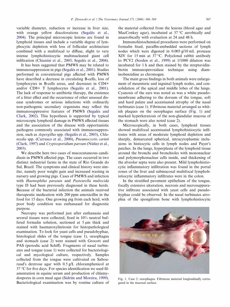
Fig. 1. Case 2: oesophagus. Fibrinous material longitudinally corru-
gated in the mucosal surface.
P. Zlotowski et al. / The Veterinary Journal 171 (2006) 566–569 567
variable diameter, reduction or increase in liver size,
with orange yellow discolorations (Segales et al.,
2004). The principal microscopic lesions are found in
lymphoid tissues and include a variable degree of lym-
phocytic depletion with loss of follicular architecture
combined with a multifocal to diffuse, slight to veryintense lymphohistiocytic multinucleated giant cell
infiltration (Chianini et al., 2003; Segales et al., 2004).
It has been suggested that PMWS may be related to
immunosuppression in pigs (Segales et al., 2001). Studies
performed in conventional pigs affected with PMWS
have described a decrease in circulating B-cells, loss of
lymphocytes in B-cells areas, and decreases in CD4+
and/or CD8+ T lymphocytes (Segales et al., 2001).The lack of response to antibiotic therapy, the existence
of a litter effect and the occurrence of other unusual dis-
ease syndromes or serious infections with ordinarily
non-pathogenic secondary organisms may reflect the
immunosuppressive features of PMWS (Segales and
Clark, 2002). This hypothesis is supported by typical
microscopic lymphoid damage in PMWS affected tissues
and the association of the disease with opportunisticpathogens commonly associated with immunosuppres-
sion, such as Aspergillus spp. (Segales et al., 2003), Chla-
mydia spp. (Carrasco et al., 2000), Pneumocystis carinii
(Clark, 1997) and Cryptosporidium parvum (Nunez et al.,
2003).
We describe here two cases of mucocutaneous candi-
diasis in PMWS affected pigs. The cases occurred in two
distinct industrial farms in the state of Rio Grande doSul, Brazil. The symptoms and clinical history were sim-
ilar, namely poor weight gain and increased wasting in
nursery and growing pigs. Cases of PMWS and infection
with Haemophilus parasuis and Pasteurella multocida
type D had been previously diagnosed in these herds.
Because of the bacterial infection the animals received
therapeutic medication with 200 ppm amoxicillin in the
food for 15 days. One growing pig from each herd, withpoor body condition was euthanased for diagnostic
purpose.
Necropsy was performed just after euthanasia and
several tissues were collected, fixed in 10% neutral buf-
fered formalin solution, sectioned at 5 lm thick and
stained with haematoxylin/eosin for histopathological
examination. To look for yeast cells and pseudohyphae,
histological slides of the tongue (case 1), oesophagusand stomach (case 2) were stained with Grocott and
PAS (periodic acid Schiff). Fragments of nasal turbin-
ates and tongue (case 1) were collected for bacteriologi-
cal and mycological culture, respectively. Samples
collected from the tongue were cultivated on Sabou-
raud�s dextrose agar with 0.5 g/L chloramphenicol at
37 �C for five days. For species identification we used fil-
amentation in equine serum and production of chlamy-dospores in corn meal agar (Sidrim and Moreira, 1999).
Bacteriological examination was by routine culture of
the material collected from the lesions (blood agar and
MacConkey agar), incubated at 37 �C aerobically and
anaerobically with evaluation at 24 and 48 h.
Immunohistochemical procedures were performed on
formalin fixed, paraffin-embedded sections of lymph
nodes which were digested in 0.005 g/10 mL proteaseXIV for 15 min at 37 �C. Polyclonal rabbit antibody
to PCV2 (Sorden et al., 1999) at 1/1000 dilution was
incubated for 1 h and then stained by the streptavidin-
biotin immunoperoxidase technique using diam-
inobenzidine as chromogen.
The main gross findings in both animals were enlarge-
ment of mesenteric and inguinal lymph nodes, and con-
solidation of the apical and middle lobes of the lungs.Cyanosis of the ears was noted as was a white pseudo-
membrane adhering to the dorsum of the tongue, soft
and hard palate and accentuated atrophy of the nasal
turbinates (case 1). Fibrinous material arranged as whit-
ish plaques on the oesophagus surface (Fig. 1) and
marked hyperkeratosis of the non-glandular mucosa of
the stomach were also noted (case 2).
Microscopically, in both cases, lymphoid tissuesshowed multifocal accentuated lymphohistiocytic infil-
trates with areas of moderate lymphoid depletion and
sharply, demarcated spherical, intracytoplasmic inclu-
sions in histiocytic cells in lymph nodes and Payer�spatches. In the lungs, hyperplasia of the lymphoid tissue
around the bronchi and bronchioles with mononuclear
and polymorphonuclear cells inside, and thickening of
the alveolar septa were also present. Mild lymphohistio-cytic inflammatory infiltration was found in the portal
zones of the liver and submucosal multifocal lymphoh-
istiocytic inflammatory infiltrates were in the colon.
In the stratified pavement epithelium of the tongue,
focally extensive ulceration, necrosis and necrosuppura-
tive infiltrate associated with yeast cells and pseudo-
hyphae could be observed. In the nasal turbinates atro-
phia of the spongiform bone with lymphohistiocytic
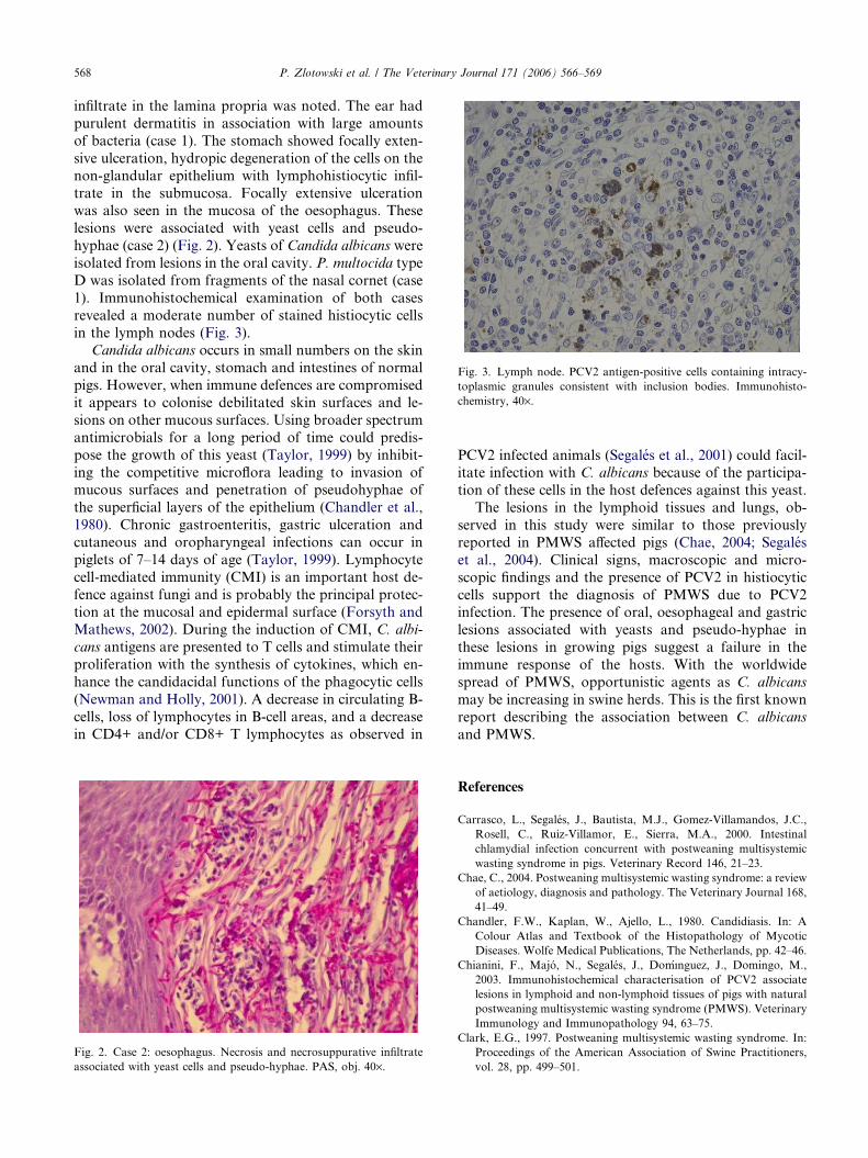
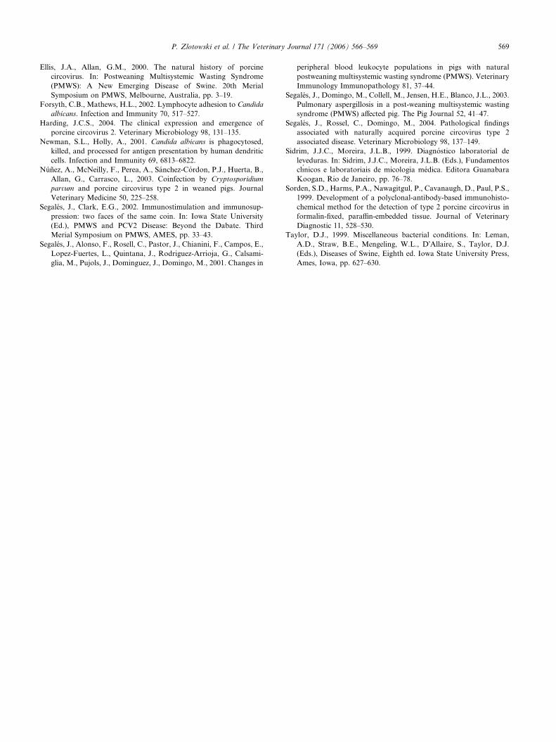
Fig. 3. Lymph node. PCV2 antigen-positive cells containing intracy-
toplasmic granules consistent with inclusion bodies. Immunohisto-
chemistry, 40·.
568 P. Zlotowski et al. / The Veterinary Journal 171 (2006) 566–569
infiltrate in the lamina propria was noted. The ear had
purulent dermatitis in association with large amounts
of bacteria (case 1). The stomach showed focally exten-
sive ulceration, hydropic degeneration of the cells on the
non-glandular epithelium with lymphohistiocytic infil-
trate in the submucosa. Focally extensive ulcerationwas also seen in the mucosa of the oesophagus. These
lesions were associated with yeast cells and pseudo-
hyphae (case 2) (Fig. 2). Yeasts of Candida albicans were
isolated from lesions in the oral cavity. P. multocida type
D was isolated from fragments of the nasal cornet (case
1). Immunohistochemical examination of both cases
revealed a moderate number of stained histiocytic cells
in the lymph nodes (Fig. 3).Candida albicans occurs in small numbers on the skin
and in the oral cavity, stomach and intestines of normal
pigs. However, when immune defences are compromised
it appears to colonise debilitated skin surfaces and le-
sions on other mucous surfaces. Using broader spectrum
antimicrobials for a long period of time could predis-
pose the growth of this yeast (Taylor, 1999) by inhibit-
ing the competitive microflora leading to invasion ofmucous surfaces and penetration of pseudohyphae of
the superficial layers of the epithelium (Chandler et al.,
1980). Chronic gastroenteritis, gastric ulceration and
cutaneous and oropharyngeal infections can occur in
piglets of 7–14 days of age (Taylor, 1999). Lymphocyte
cell-mediated immunity (CMI) is an important host de-
fence against fungi and is probably the principal protec-
tion at the mucosal and epidermal surface (Forsyth andMathews, 2002). During the induction of CMI, C. albi-
cans antigens are presented to T cells and stimulate their
proliferation with the synthesis of cytokines, which en-
hance the candidacidal functions of the phagocytic cells
(Newman and Holly, 2001). A decrease in circulating B-
cells, loss of lymphocytes in B-cell areas, and a decrease
in CD4+ and/or CD8+ T lymphocytes as observed in
Fig. 2. Case 2: oesophagus. Necrosis and necrosuppurative infiltrate
associated with yeast cells and pseudo-hyphae. PAS, obj. 40·.
PCV2 infected animals (Segales et al., 2001) could facil-itate infection with C. albicans because of the participa-
tion of these cells in the host defences against this yeast.
The lesions in the lymphoid tissues and lungs, ob-
served in this study were similar to those previously
reported in PMWS affected pigs (Chae, 2004; Segales
et al., 2004). Clinical signs, macroscopic and micro-
scopic findings and the presence of PCV2 in histiocytic
cells support the diagnosis of PMWS due to PCV2infection. The presence of oral, oesophageal and gastric
lesions associated with yeasts and pseudo-hyphae in
these lesions in growing pigs suggest a failure in the
immune response of the hosts. With the worldwide
spread of PMWS, opportunistic agents as C. albicans
may be increasing in swine herds. This is the first known
report describing the association between C. albicans
and PMWS.
References
Carrasco, L., Segales, J., Bautista, M.J., Gomez-Villamandos, J.C.,
Rosell, C., Ruiz-Villamor, E., Sierra, M.A., 2000. Intestinal
chlamydial infection concurrent with postweaning multisystemic
wasting syndrome in pigs. Veterinary Record 146, 21–23.
Chae, C., 2004. Postweaning multisystemic wasting syndrome: a review
of aetiology, diagnosis and pathology. The Veterinary Journal 168,
41–49.
Chandler, F.W., Kaplan, W., Ajello, L., 1980. Candidiasis. In: A
Colour Atlas and Textbook of the Histopathology of Mycotic
Diseases. Wolfe Medical Publications, The Netherlands, pp. 42–46.
Chianini, F., Majo, N., Segales, J., Domı´ nguez, J., Domingo, M.,
2003. Immunohistochemical characterisation of PCV2 associate
lesions in lymphoid and non-lymphoid tissues of pigs with natural
postweaning multisystemic wasting syndrome (PMWS). Veterinary
Immunology and Immunopathology 94, 63–75.
Clark, E.G., 1997. Postweaning multisystemic wasting syndrome. In:
Proceedings of the American Association of Swine Practitioners,
vol. 28, pp. 499–501.
P. Zlotowski et al. / The Veterinary Journal 171 (2006) 566–569 569
Ellis, J.A., Allan, G.M., 2000. The natural history of porcine
circovirus. In: Postweaning Multisystemic Wasting Syndrome
(PMWS): A New Emerging Disease of Swine. 20th Merial
Symposium on PMWS, Melbourne, Australia, pp. 3–19.
Forsyth, C.B., Mathews, H.L., 2002. Lymphocyte adhesion to Candida
albicans. Infection and Immunity 70, 517–527.
Harding, J.C.S., 2004. The clinical expression and emergence of
porcine circovirus 2. Veterinary Microbiology 98, 131–135.
Newman, S.L., Holly, A., 2001. Candida albicans is phagocytosed,
killed, and processed for antigen presentation by human dendritic
cells. Infection and Immunity 69, 6813–6822.
Nunez, A., McNeilly, F., Perea, A., Sanchez-Cordon, P.J., Huerta, B.,
Allan, G., Carrasco, L., 2003. Coinfection by Cryptosporidium
parvum and porcine circovirus type 2 in weaned pigs. Journal
Veterinary Medicine 50, 225–258.
Segales, J., Clark, E.G., 2002. Immunostimulation and immunosup-
pression: two faces of the same coin. In: Iowa State University
(Ed.), PMWS and PCV2 Disease: Beyond the Dabate. Third
Merial Symposium on PMWS, AMES, pp. 33–43.
Segales, J., Alonso, F., Rosell, C., Pastor, J., Chianini, F., Campos, E.,
Lopez-Fuertes, L., Quintana, J., Rodriguez-Arrioja, G., Calsami-
glia, M., Pujols, J., Dominguez, J., Domingo, M., 2001. Changes in
peripheral blood leukocyte populations in pigs with natural
postweaning multisystemic wasting syndrome (PMWS). Veterinary
Immunology Immunopathology 81, 37–44.
Segales, J., Domingo, M., Collell, M., Jensen, H.E., Blanco, J.L., 2003.
Pulmonary aspergillosis in a post-weaning multisystemic wasting
syndrome (PMWS) affected pig. The Pig Journal 52, 41–47.
Segales, J., Rossel, C., Domingo, M., 2004. Pathological findings
associated with naturally acquired porcine circovirus type 2
associated disease. Veterinary Microbiology 98, 137–149.
Sidrim, J.J.C., Moreira, J.L.B., 1999. Diagnostico laboratorial de
leveduras. In: Sidrim, J.J.C., Moreira, J.L.B. (Eds.), Fundamentos
clınicos e laboratoriais de micologia medica. Editora Guanabara
Koogan, Rio de Janeiro, pp. 76–78.
Sorden, S.D., Harms, P.A., Nawagitgul, P., Cavanaugh, D., Paul, P.S.,
1999. Development of a polyclonal-antibody-based immunohisto-
chemical method for the detection of type 2 porcine circovirus in
formalin-fixed, paraffin-embedded tissue. Journal of Veterinary
Diagnostic 11, 528–530.
Taylor, D.J., 1999. Miscellaneous bacterial conditions. In: Leman,
A.D., Straw, B.E., Mengeling, W.L., D�Allaire, S., Taylor, D.J.
(Eds.), Diseases of Swine, Eighth ed. Iowa State University Press,
Ames, Iowa, pp. 627–630.