Embed Size (px)
Citation preview

MR of the
Pelvis and Hip• Technique
• AVN
• Transient bone marrow edema
• Pelvic trauma– Bony
• Stress
• Occult
• Arthritis– Labrum
• Bursa
• Tendons
• Muscles
• Pediatric hip disorders
• Tumors
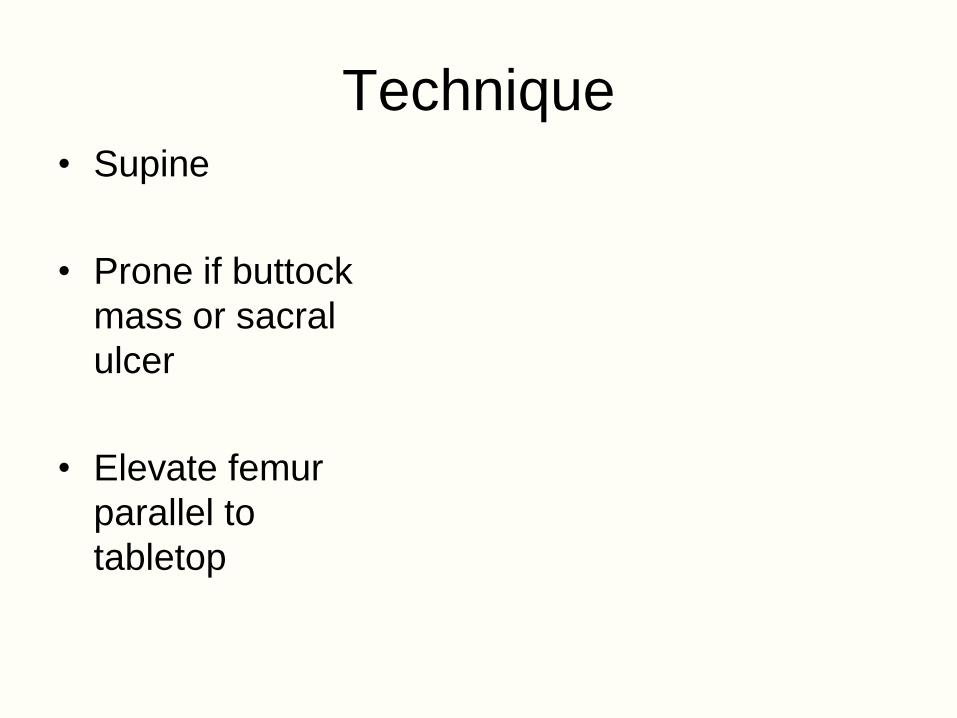
Technique• Supine
• Prone if buttock
mass or sacral
ulcer
• Elevate femur
parallel to
tabletop

Pelvis/Hip MR protocolPlane Seq TR/TE FOV Slice Matrix NEX Time
Localizer FMPIR 3500/17/160 40 5 128 2 2:05
Coronal T1 SE 600/14 36 5 192 1 4:50
Sagittal T1 SE 600/20 20 4 256 1 6:24
AxialT2
FSE-fs3000/90 36 5 192 2 5:30
CoronalT2
FSE-fs3000/90 36 5 192 2 5:12

Hip MR Arthrography
Indications
• Labral tear
• Paralabral ganglion
• Preoperative assessment of DDH
• Intraarticular bodies

Hip MRA technique
• Local anesthesia
• Anterolateral approach to
femoral head-neck junction
• Confirm needle position with
<1 cc contrast
• Inject 12 cc of diluted Gd-
DTPA

MR arthrography
• Anterolateral approach
• Inject 12-15 cc of 1:200
Gd-DTPA
• 3 planes of imaging with
T1 fat-sat
• Coronal IR or T2-w FSE

Normal variants
• Epiphyseal scar
• Synovial herniation pit
• Fovea
• Trabecular bands
• Intertrochanteric marrow
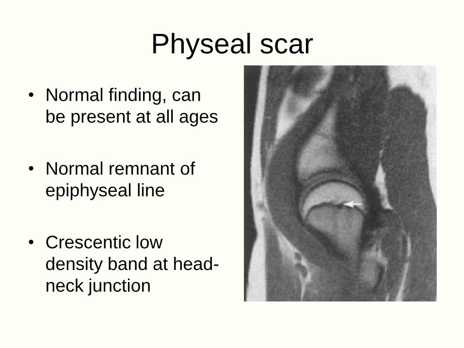
Physeal scar
• Normal finding, can
be present at all ages
• Normal remnant of
epiphyseal line
• Crescentic low
density band at head-
neck junction
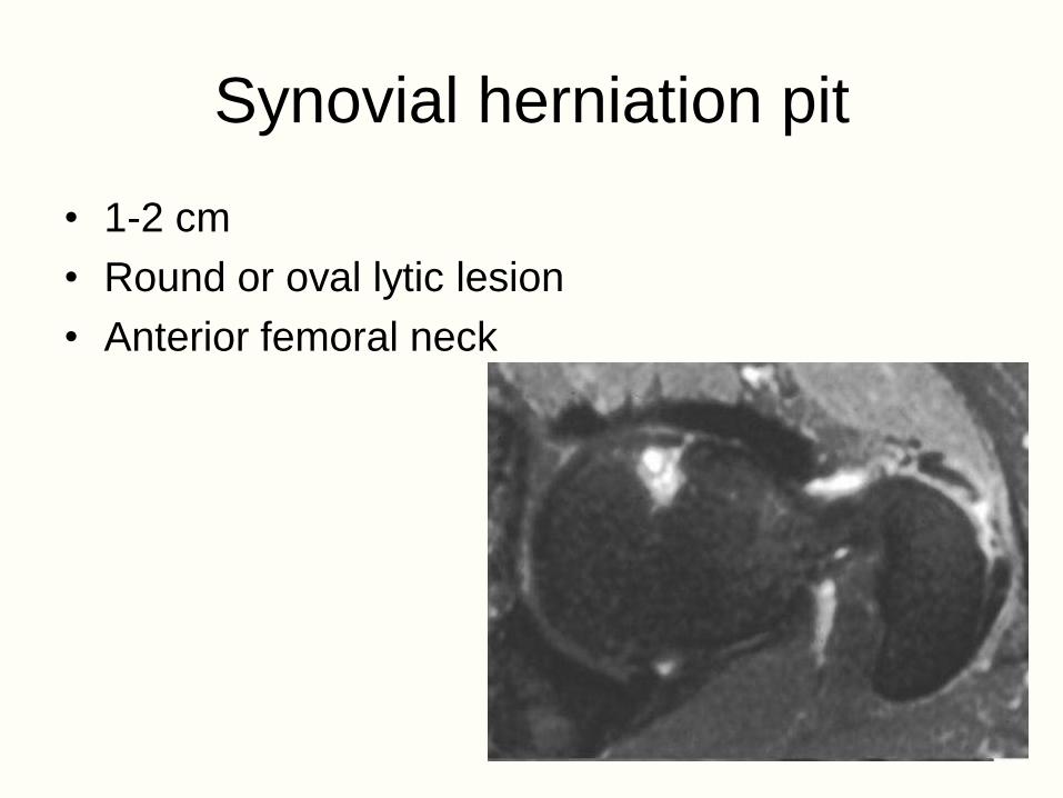
Synovial herniation pit
• 1-2 cm
• Round or oval lytic lesion
• Anterior femoral neck

Fovea
• Depression on central
surface of the femoral
head
• Devoid of cartilage
• Ligamentum teres femoris
arises from this
depression

Avascular necrosis (AVN)
• Avascular necrosis
• Aseptic necrosis
• Osteonecrosis
• Ischemic necrosis
• Bone infarction

Stages of AVN• Cell death
– Hematopoietic<Fat
– <Mesenchymal
• Cell proliferation
adjacent to necrosis
• Revascularization and
resorption of necrotic
segment
• Reossification by
creeping substitution
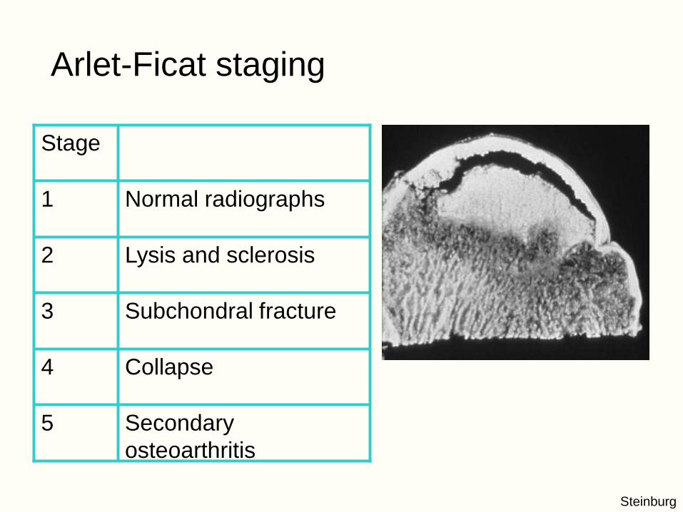
Arlet-Ficat staging
Secondary
osteoarthritis
5
Collapse4
Subchondral fracture3
Lysis and sclerosis2
Normal radiographs1
Stage
Steinburg

MG 1916259
AVN• MR is the most
sensitive method for noninvasive early detection
• Controversy about need for early detection
• Early therapeutic options limited
Cor T1

Avascular necrosis
• Line, band or ring of low signal
• Double-line sign
• Deformity
• Marrow edema

AVN
• Extent of disease
used to predict
likelihood of collapse
• Coronal image
• Extent of weight-
bearing surface
involved

Idiopathic bone marrow edema
• Middle-aged, male predominance
• Last trimester of pregnancy
• Left hip involved in 2/3 of cases
• Unknown etiology
• Self-limited

JP
Idiopathic marrow edema• Geographic signal loss
on T1w in femoral head and neck
• Signal loss extends to intertrochanteric line
• High signal on T2w and STIR
• No arcuate or linear bands of low signal
• No deformity
Cor T1
Cor T2

Edema: differential diagnosis
• Very early avascular
necrosis
• Osteomyelitis
• Osteoarthritis
• Stress fracture

Pelvic trauma
• Stress fracture
– Fatigue
– Insufficiency
• Occult fracture
• Soft tissue injury

Insufficiency fracturessacrum
superomedial
ileum
pubic rami
supracetabular
iliac wing
symphysis

MR of occult fracture
• Normal or
equivocal
radiographs
• High clinical
suspicion
• Major weight-
bearing region

Occult fracture
• Additional radiographs
• Fluoroscopy
• Scintigraphy
• Conventional tomography
• Computed tomography
• Magnetic resonance imaging

MR of occult fractures
• Coronal STIR of
entire pelvis
• Smaller FOV images
of injured area
– T1 (or PD FSE)
• Entire study less than
15 minutes

Labrum
• Attached directly to osseous rim
• Triangular, thickest posterosuperiorly
• Blends with transverse ligament at acetabular notch

Labral tears
• Traumatic or degenerative
• Pain, locking or premature osteoarthritis
• Higher incidence in patients with hip dysplasia
• Associated with supracetabular ganglion

Labral tear
• T1 images only show
swelling of labrum
• T2 images show high signal
in labrum
• MR arthrography most
accurate
Cor T2
CorT1
GM
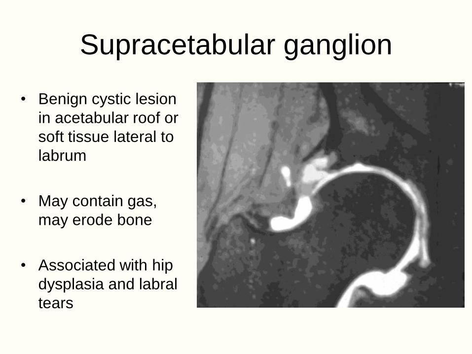
Supracetabular ganglion
• Benign cystic lesion
in acetabular roof or
soft tissue lateral to
labrum
• May contain gas,
may erode bone
• Associated with hip
dysplasia and labral
tears

Bursal anatomy• Communicating
bursae
– Iliopsoas
• Noncommunicating
bursae
– Trochanteric
– Subgluteal
http://www.aafp.org/afp/20000401/2109_f1.jpg

Soft tissue injury• Muscle strain
• Hematoma
• Tendon tear

Myotendinous avulsions
• Greater trochanter
– Gluteus medius
– Gluteus minimus
• Ischium
– Hamstrings

Gluteal attachments
Anterior Lateral Posterior
Pfirrmann C. et al

Gluteal tendon tear• Elderly
• Tear of gluteus
medius or minimus
tendon
• “Rotator cuff tear of
the hip”

Hamstring avulsion
• Young athlete
• Elderly patient

Pediatric hip disorders
• Developmental dysplasia
• Proximal focal femoral deficiency
• Transient synovitis
• Legg-Calve-Perthe's disease
• Slipped capital femoral epiphysis
Cor T1 FSGd

DG
Legg-Calvé-Perthes disease• Idiopathic necrosis
of femoral head in
children
• Ages 4-7


![[Chapter 12] Amputations of the Hip and Pelvis](https://img.dokumen.tips/doc/110x75/54654bcdaf795979338b4d41/chapter-12-amputations-of-the-hip-and-pelvis.jpg)
