Embed Size (px)
Citation preview
ORIGINAL RESEARCH
Mouse thymidylate synthase does not show the inactiveconformation, observed for the human enzyme
Anna Dowierciał1• Adam Jarmuła1
• Piotr Wilk1• Wojciech Rypniewski2 •
Monika Kowalska3• Tomasz Fraczyk1
• Joanna Ciesla1• Wojciech Rode1
Received: 2 July 2016 / Accepted: 29 August 2016 / Published online: 9 September 2016
� The Author(s) 2016. This article is published with open access at Springerlink.com
Abstract Crystal structures of mouse thymidylate syn-
thase (mTS) in complexes with (1) sulfate anion, (2) 20-deoxyuridine 50-monophosphate (dUMP) and (3) 5-fluoro-
dUMP (FdUMP) and N5,10-methylenetetrahydrofolate
(meTHF) have been determined and deposited in Protein
Data Bank under the accession codes 3IHI, 4E5O and
5FCT, respectively. The structures show a strong overall
similarity to the corresponding structures of rat and human
thymidylate synthases (rTS and hTS, respectively). Unlike
with hTS, whose unliganded and liganded forms assume
different conformations (‘‘inactive’’ and ‘‘active,’’ respec-
tively) in the loop 181–197, in each of the three mTS
structures, the loop 175–191, homologous to hTS loop
181–197, populates the active conformer, with catalytic
Cys 189 buried in the active site and directed toward C(6)
of the pyrimidine ring of dUMP/FdUMP, pointing to pro-
tein’s inability to adopt the inactive conformation. The
binary structures of either dUMP- or sulfate-bound mTS,
showing the enzyme with open active site and extended
C-terminus, differ from the structure of the mTS–5-
FdUMP–meTHF ternary complex, with the active site
closed and C-terminus folded inward, thus covering the
active site cleft. Another difference pertains to the con-
formation of the Arg44 side chain in the active site-flank-
ing loop 41–47, forming strong hydrogen bonds with the
dUMP/FdUMP phosphate moiety in each of the two
liganded mTS structures, but turning away from the active
site entrance and loosing the possibility of H-bonding with
sulfate in the sulfate-bound mTS structure.
Keywords Thymidylate synthase � Mouse � Crystal
structure � Active conformation
Introduction
Thymidylate synthase (TS; EC 2.1.1.45), a target in
chemotherapy, catalyzes the conversion of deoxyuridine
monophosphate (dUMP) and R-N5,10-methylenetetrahy-
drofolate (meTHF) to deoxythymidine monophosphate and
dihydrofolate, via reductive methylation, in which meTHF
serves as both methylene group donor and reducing agent
[1, 2]. As the sole de novo source of thymidylate synthesis
in eukaryotic cells, for over 50 years, the enzyme has been
a target in chemotherapy [3], with active forms of drugs
being dUMP or N5,10-meTHF analogues that inhibit the
enzyme [4, 5]. Between dUMP analogues active in
chemotherapy, the most prominent is 5-fluoro-dUMP
(FdUMP), a strong thymidylate synthase inhibitor being
the active form of drugs used in chemotherapy, such as
5-fluorouracil, 5-fluoro-20-deoxyuridine and 5-fluorocy-
tosine [2].
The first step of the reaction catalyzed by TS is the
nucleophilic addition of the active site cysteine (residue 189
in mouse TS) to the pyrimidine C(6) of the nucleotide. Of
essential role at this stage of catalysis are the proper binding
position and orientation of dUMP. The first is secured by
Anna Dowierciał and Adam Jarmuła have contributed equally to the
present work.
& Wojciech Rode
1 Laboratory of Comparative Enzymology, Department of
Biochemistry, Nencki Institute of Experimental Biology,
Polish Academy of Sciences, Warsaw, Poland
2 Institute of Bioorganic Chemistry, Polish Academy of
Sciences, Poznan, Poland
3 International Institute of Molecular and Cell Biology, Polish
Academy of Sciences, Warsaw, Poland
123
Struct Chem (2017) 28:667–674
DOI 10.1007/s11224-016-0840-8

anchoring the phosphate of dUMP via several conservative
hydrogen contacts from both TS subunits, while the latter by
the way of hydrogen bonding between the conserved
asparagine (Asn 220 in mTS) and the O4 and N3–H moieties
of the pyrimidine ring. Properly bound dUMP provides the
binding surface for the cofactor, resulting in the formation of
the ternary complex TS–dUMP–meTHF [2]. Following
activation of dUMP by the nucleophilic attack at the
pyrimidine C(6), the negative charge of cysteine residue is
delocalized, probably toward the C(4)=O of dUMP, and the
corresponding enolate anion is formed (Fig. 1) with its C(5)
position believed to become strongly nucleophilic. The latter
facilitates a consecutive attack of the methylene residue, in
the iminium ion form, resulting from opening of the imida-
zolidine ring of the cofactor. This leads to formation of the
covalent intermediate, ternary complex of the enzyme with
substrate and cofactor. Next, the C(5) hydrogen dissociates,
as proton, from dUMP pyrimidine ring, with concomitant b-
elimination of tetrahydrofolate, still remaining bound in the
active center. The latter enables hydride transfer from the
pteridine C(6), resulting in reduction of the pyrimidine C(5)
methylene group to methyl one. The reaction is completed by
regeneration of the pyrimidine 5,6 double bond, with con-
comitant elimination to form dTMP and enzyme [1, 2].
Structure determination of human TS complexes
revealed a unique feature. In contrast to structures from
other species, human TS loop 181–197 was observed to
populate two conformers, one of them, apparently inactive,
stabilized by a pair of hydrogen bonds from Arg163 to the
carbonyls of Ala191 and Leu192. As mouse TS protein
contains lysine residue at the position corresponding to
arginine residue in human TS (Lys157 in mTS vs. Arg163
in hTS), it has been hypothesized unable to populate the
inactive conformer [6]. In order to test that hypothesis,
learning the crystal structure of the mouse enzyme was of
interest.
An initial account of a part of the present results has
been published in a special issue of Pteridines [7], covering
materials of the 14th International Symposium on Pteridi-
nes and Folates (June 7–12, 2009, Jeju, Korea).
Materials and methods
Reagents
K/Na tartrate, PEG 3350, PEG 4000, Li2SO4, dUMP were
purchased from Sigma, dithiothreitol (DTT) was from Carl
Roth GmbH.
Recombinant mouse thymidylate synthase
This was overexpressed and purified as described previ-
ously [8, 9]. The enzyme was judged to be near homoge-
neous by denaturing polyacrylamide slab gel
electrophoresis using a 40, 20 and 10 lg sample. Enzyme
activity was measured spectrophotometrically [10], but at
the last purification step, the assay measuring tritium
release from [5-3H]dUMP was used as previously descri-
bed [11]. The specific activity of purified enzyme was
found to be 1.75 lmol min-1 mg -1 for conversion of
dUMP to dTMP at 37 �C.
Separation of non-phosphorylated fraction
of purified recombinant thymidylate synthase
preparation
The enzyme preparation was separated into phosphorylated
and non-phosphorylated fractions as previously described
[12]. Only the non-phosphorylated fraction was used for
crystallization.
Fig. 1 Thymidylate synthase-catalyzed reaction
668 Struct Chem (2017) 28:667–674
123
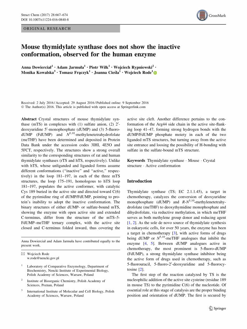
Crystallization and data collection
Purified protein was dialyzed against 5 mM Tris HCl, pH
7.5 buffer containing 5 mM DTT and then concentrated
using Amicon Centricon centrifugal filter. Crystals were
grown by the vapor diffusion method in the hanging drops
at 4 �C under following conditions. For TS, equal volumes
of protein solution at 15 mg/ml concentration and well
solution were mixed and allowed to equilibrate with 0.5 ml
of well solution, containing 0.1 M Tris HCl, pH 8.5, 0.2 M
Li2SO4 and 25 % (w/v) PEG 4000. For TS–dUMP com-
plex, a drop of protein solution at 25 mg/ml concentration,
containing 5.9 mM dUMP, was mixed as before 1:1 with
well solution and allowed to equilibrate with 0.5 ml of well
solution containing 0.2 M K Na Tartrate and 20 % PEG
3350. For TS–dUMP–meTHF complex, equal volumes of
mTS solution (25 mg/ml), containing 5 mM FdUMP and
10 mM meTHF and well solution (0.1 M Tris HCl, pH 8.5,
0.1 M MgCl2 and 19.5 % (w/v) PEG 4000) were mixed
and allowed to equilibrate with 0.5 ml of well solution.
Crystallization conditions are summarized in Table 1.
The crystals (Figs. 3, 4) were transferred for a few
seconds to cryoprotectant solution containing mother
liquor and 25 % butanodiol and 30 % glicerol for mTS–
dUMP and mTS, respectively, and flash-cooled in N2
vapors.
X-ray diffraction data were collected from three single
flash-frozen crystals either at the Max-Lab Lund University
Synchrotron (mTS and mTS–dUMP), using X-ray wave-
lengths of 1.038 and 0.908 A, or at the BESSY Syn-
chrotron (mTS–FdUMP–meTHF), using X-ray wavelength
of 0.918 A.
Data processing, structure determination
and refinement
Data were processed with Denzo and Scalepack [13]. Both
structures were determined by molecular replacement
carried out with the CCP4 package [14], using the rat TS
ternary complex without ligands as the search model. The
crystal structures of mTS, mTS–dUMP and mTS–FdUMP–
meTHF were determined at resolutions of 1.94, 1.70 and
1.55 A, respectively. The correctness of the structures was
evaluated using Sfcheck and Procheck from the CCP4
suite. Some X-ray data and model refinement parameters
are summarized in Table 2.
Results and discussion
The structures, consisting of either one dimer (mTS and
mTS–FdUMP–meTHF) or three dimers (mTS–dUMP) per
an asymmetric part of the unit cell, showed an overall
similarity to the corresponding structures of human and rat
TSs (hTS and rTS, respectively). The Ca RMSD values
between the liganded and unliganded mTS structures pre-
sented here, as well as between these structures and the
crystal structures of other ternary complexes with dUMP
and Tomudex, including those with hTS [15, 16], rTS [17]
and mTS [18], are shown in Table 3. The superimposition
of the mTS, mTS–dUMP and mTS–FdUMP–meTHF
structures is shown in Fig. 2. The similarity of the corre-
sponding TS structures is hardly unexpected in view of
highly conserved primary structures of different TS pro-
teins, as reflected by mTS amino acid sequence being 90 %
identical with those of hTS [19] and rTS [20], and about
40–55 % identical with prokaryotic and phage TS
sequences [19]. With respect to the latter, a recent com-
parison of amino acid sequences between TS of the para-
sitic nematode Trichinella spiralis and 33 other TS
proteins, both eukaryotic and prokaryotic, showed
43–68 % identities and 56–79 % similarities [21].
The mTS–dUMP structure showed the enzyme in the
active, open-state conformation. The dUMP pyrimidine
ring C(6) and the catalytic Cys189 were separated by dis-
tances of 3.19, 3.22, 3.19, 3.17, 3.28 and 2.94 A (subunits
A, B, C, D, E and F, respectively), precluding covalent
bondings. The substrate molecule was bound in a manner
similar to that observed in the corresponding complexes of
other mammalian and bacterial TSs, with the ligand
Table 1 Crystallization conditions used
System Concentrations of TS and ligand(s) in the protein solution Composition of the well solution
mTS mTS (15 mg/ml) 0.1 M Tris HCl, pH 8.5 0.2 M Li2SO4
No ligands 25 % (w/v) PEG 4000
mTS–dUMP mTS (25 mg/ml) 0.2 MK Na Tartrate
5.9 mM dUMP 20 % PEG 3350
mTS–FdUMP–meTHF mTS (25 mg/ml) 0.1 M Tris–HCl, pH 8.5
FdUMP (5 mM) 0.1 M MgCl2
meTHF (10 mM) 19.5 % PEG 4000
Struct Chem (2017) 28:667–674 669
123

anchored in the active site by several H-bonds to its
phosphate moiety from four arginines from both subunits
of the enzyme dimer (Arg44 and Arg209 from the first
subunit and Arg1690 and Arg1700 from the other subunit)
and single serine (Ser210). The orientation of dUMP was
secured by H-bonds between the conserved Asn 220 and
the O4 and N(3)-H moieties of the pyrimidine ring.
In the mTS–FdUMP–meTHF structure, the enzyme was
found in the active, closed state conformation, with the
catalytic Cys189 located in the active site and extended
toward FdUMP. The distances between mTS Cys189 cS
and FdUMP pyrimidine C(6) in the two subunits (A and B)
were equal, each 1.82 A, pointing to covalent bonding.
However, the distances between the FdUMP pyrimidine
C(5) and meTHF C(11) atoms were 2.28 A (subunit A) and
1.55 A (subunit B), indicating covalent bond between the
two ligands to have been formed only in the subunit B.
FdUMP was bound slightly shifted (*0.9 A), compared to
the position of dUMP in the mTS–dUMP structure, but
preserved all the non-covalent interactions of the latter
molecule. The conformation of the meTHF molecule was
stabilized by favorable contacts with Ile102 and Asn106
from a long loop–helix–loop segment (100–127), flanking
the entrance to the active site cleft, Ala305 and Met306
Table 2 Data collection and refinement statistics for mTS, mTS–dUMP and mTS–FdUMP–meTHF structures
Lattice type mTS (3IHI) mTS–dUMP (4E5O) mTS–FdUMP–meTHF (5FCT)
Monoclinic Monoclinic Monoclinic
Space group C 1 2 1 C 1 2 1 P 1 21 1
Unit cell parameters a = 158.2 A b = 87.96 A
c = 68.04 A
a = 160.35 A b = 88.54 A
c = 136.76 A
a = 54.224 A b = 83.302 A
c = 68.850 A
a = 90� b = 97.43� c = 90� a = 90� b = 95.99� c = 90� a = 90� b = 105.43� c = 90�Resolution range [A] 19.48–1.94 19.95–1.7 26.14–1.55
No. of unique reflections 69,498 207,806 85,416
Multiplicity 3.6 (3.4) 6.4 5.9 (4.0)
\I/r(I)[ 15 16.9 14.0 (2.4)
No. of reflections used in
refinement
67,377 205,349 81,162
Rfactor [%] 22.0 23.5 12.0
Rfree factor [%] 26.6 29.2 16.6
RMSD bond [A] 0.026 0.022 0.01
RMSD angle [�] 1.953 1.956 1.415
Table 3 Ca RMSD [A] in the pairs of crystal structures of mouse,
human and rat TSs in ternary complexes with dUMP and Tomudex
(4EB4, 1I00/1HVY and 2TSR, respectively), and with FdUMP and
meTHF (5FCT), and binary complex of mouse TS with dUMP
(4E5O), as well as sulfate-bound mouse TS (3IHI)
Structures Chains
AB CD A C B D
4E5O versus 3IHI 0.77 0.64 0.88
5FCT versus 3IHI 0.71 0.79 0.61
5FCT versus 4E5O 1.10 1.08 1.13
5FCT versus 2TSR 0.79 0.89 0.69
5FCT versus 1I00* 0.99 1.01 0.98
5FCT versus 1HVY* 1.05 1.10 1.00
5FCT versus 4EB4_AB 0.44 0.42 0.45
5FCT versus 4EB4_CD 1.05 1.27 0.78
* Two ‘‘generic’’ conformations observed in the structures of the
ternary complexes of human
TS: open (1I00) and closed (1HVY)
Fig. 2 Alignment of the three mTS structures: sulfate-bound protein
(blue), binary complex with dUMP (green) and ternary complex with
FdUMP and meTHF (red). Proteins are shown as solid ribbons,
ligands as sticks and crystal waters as dots (Color figure online)
670 Struct Chem (2017) 28:667–674
123

from C-terminus, Leu215 from a short loop 213–216 and
Phe219 from central a-helix 217–234 (Fig. 3). It should be
added that inhibition of thymidylate synthase by FdUMP
involves time-dependent formation of a ternary covalently
bound complex of the enzyme with FdUMP and mTHF, in
a reaction similar to that involving dUMP (in fact FdUMP
mimicks a substrate better than dUMP [22, 23]). However,
at this step, the reaction stops, as the C(5) fluorine fails to
dissociate (due to the strength of the C–F bond), as it
happens with C(5) hydrogen in dUMP. What results is a
slowly reversible enzyme inactivation. Studies on the
mechanism of thymidylate synthase inhibition by FdUMP
were a rich source of information about the catalytic
mechanism [1, 2].
The active site in the mTS structure held a single sulfate
anion, bound at nearly the same location as the phosphate
moieties of dUMP and FdUMP in the mTS–dUMP and
mTS–FdUMP–meTHF structures, respectively, and stabi-
lized by H-bonds with the pair of arginine residues
(Arg1700 and Arg209), belonging to the quartet coordi-
nating the nucleotide phosphate in each of the liganded
structures. The presence at the active site of the sulfate
anion, an isosteric analogue of phosphate anion, is not
surprising, as 0.1 M Li2SO4 was present in the crystal-
lization medium.
The structures of sulfate-bound mTS, and of the dUMP–
mTS and the mTS–FdUMP–meTHF complexes, differ in
the open/closed state equilibrium, revealing either an open
active site and extended C-terminus (mTS–dUMP and, to a
lower degree due to a shorter C-terminus, mTS) or closed
active site, with C-terminus folded inward and covering the
active site entrance (mTS–FdUMP–meTHF) (Fig. 4). The
latter is the most striking difference between the overall
similar structures, as reflected in modest Ca RMSD values
for dimers, as well as individual subunits (Table 3).
Another, smaller difference pertains to the conformation of
the loop 41–47, located at one of the regions flanking the
active site cleft. In the liganded systems, this loop pene-
trates the active site, with the side chain of Arg44
extending toward, and forming H-bonds with the phosphate
moiety of dUMP/FdUMP. In the absence of ligands (and
despite the presence of sulfate, placed similarly as the
phosphate moieties of dUMP/FdUMP), Arg44 turns away
from the active site cleft and ‘‘uncovers’’ the active site
entrance (Fig. 5).
The closed-conformation structure of the mTS–
FdUMP–meTHF complex strongly resembles a mixed, yet
basically closed conformation observed in the AB dimer of
the previously reported mTS–dUMP-Tomudex (4EB4)
crystal structure (cf. RMSD in Table 3). However,
although still moderate, the RMS differences with respect
to the more open CD dimer, and in particular to subunit C
of the 4EB4 structure, are distinctly more pronounced.
Those differences are related to the open/closed active site
equilibrium and include the positions of C-terminus
(RMSD of 2.8–5.5 A) and two segments on the opposite
flanks of the active site entrance, loop 41–47 (RMSD of
2.4–3.3 A) and loop-helix-loop segment 100–127 (RMSD
of 2.0–3.5 A) (Fig. 6).
In human thymidylate synthase, loop 181–197 can
populate two major conformations, active and inactive,
Fig. 3 Non-bonded contacts stabilizing the conformation of the
meTHF molecule in the subunit B of the mTS–FdUMP–meTHF
complex structure. Two alternative conformations of the meTHF
molecule are shown as ball and sticks, and other residues/moieties as
sticks. Atom coloring is by element: gray for carbons, red for
oxygens, blue for nitrogens, gold for sulfur and green for fluorine.
Hydrogen atoms are omitted for clarity. The alternative conformers of
meTHF and Lys 302 are colored cyan. The methylene bridge between
FdUMP and meTHF is colored purple. The hydrophobic contacts and
hydrogen bonds between meTHF and protein residues are colored
shades of violet and green, respectively (Color figure online)
Fig. 4 Differences between the active site conformations observed in
the three superimposed mTS structures: sulfate-bound protein (open
conformation; blue), binary complex with dUMP (open conformation;
green) and ternary complex with FdUMP and meTHF (closed
conformation; red) (Color figure online)
Struct Chem (2017) 28:667–674 671
123

related to each other by a 180� rotation [24, 25]. While
catalytic Cys 195 in the former conformation is present in
the active site, directed toward dUMP pyrimidine ring C(6)
and forming with it a thiol adduct, in the latter it is located
more than 10 A away and directed toward the enzyme
dimer interface (Fig. 2). The equilibrium between both
conformations has been shown to be shifted toward the
inactive conformation in the presence of phosphate or
sulfate ions, with the active conformation promoted in the
presence of dUMP [25].
In the mTS–dUMP and mTS–FdUMP–meTHF struc-
tures, loop 175–191, equivalent to human loop 181–197,
populates the active conformation, with catalytic Cys189
either located at a close, but non-covalent distance from
dUMP, or covalently bound to FdUMP. Interestingly, in
the mTS structure, loop 175–191 populates the same (ac-
tive) conformation, despite the absence of dUMP/FdUMP
in the active site, containing only bound sulfate anion. This
is in contrast to the sulfate-bound human TS structure
(1HW4; Phan et al., 2001b), where corresponding loop
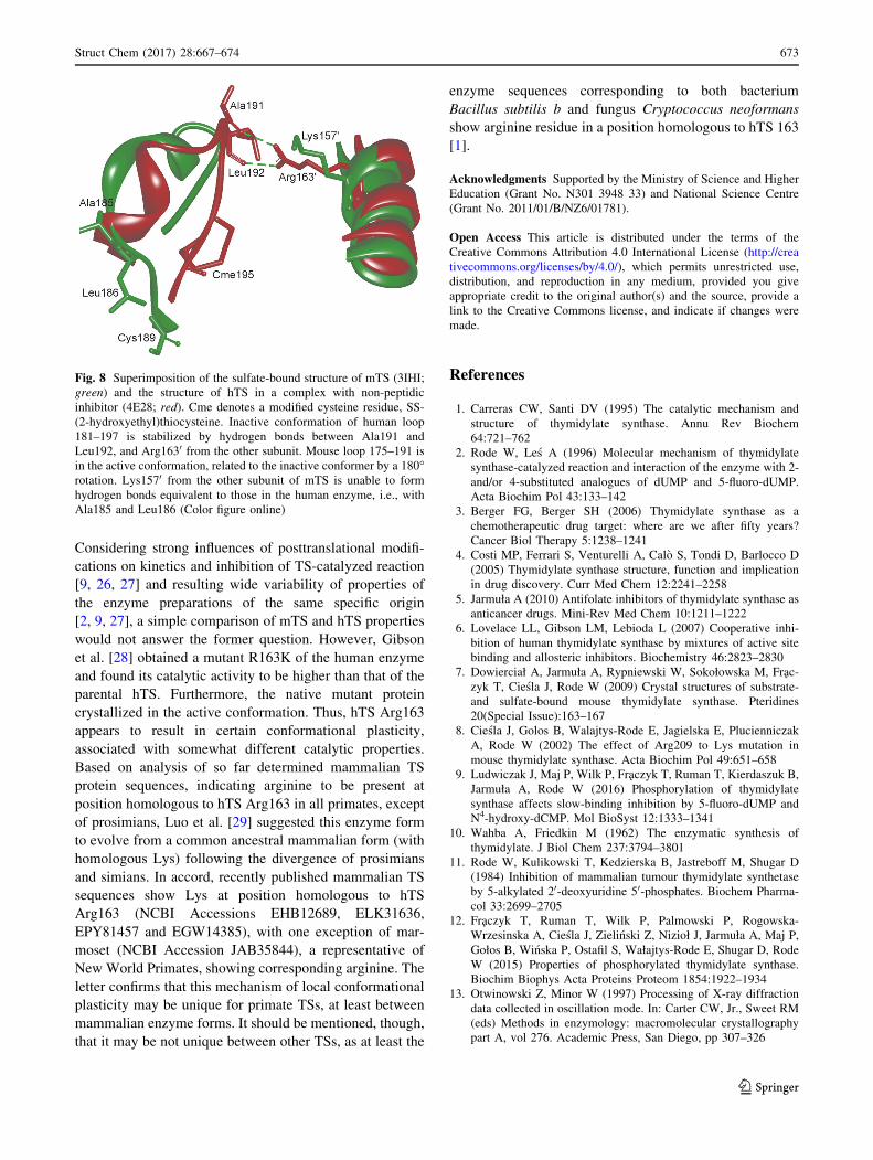
181–197 occupies the inactive conformation (Fig. 7).
In the human TS, the inactive conformer is stabilized by
a pair of hydrogen bonds from Arg163 to the carbonyls of
Ala191 and Leu192 (Fig. 3). The presence of lysine,
instead of arginine, at the position 157 in mTS (equivalent
to hTS 163) disallows the occurrence of the corresponding
hydrogen bonding (with Ala185 and Leu186) due to shorter
side chain and lower adaptability to multiple H-bonding of
the former, compared to the latter residue, as concluded
from the comparison of the mTS and hTS crystal structures
(Fig. 8). This observation supports the above mentioned
hypothesis by Lovelace et al. [6].
In view of the foregoing conclusion of obvious interest
is potential functional consequence of this mechanism, as
well as possibility to find structural features enabling the
inactive conformation stabilization (arginine residue in a
position homologous to hTS 163) in other TS structures.
Fig. 5 Differences in the conformation of the side chain of Arg44
from AS-flanking loop 41-47. Structures are colored as follows:
mTS—blue, mTS–dUMP—green, mTS–FdUMP–meTHF—red. Pro-
teins are shown as solid ribbons and ligands as sticks. Hydrogen
bonds are shown as dashed lines (Color figure online)
Fig. 6 Superimposition of subunit A from the mTS–FdUMP–meTHF
structure (red) and subunit C from the mTS–dUMP-Tomudex
structure (yellow). Most significant differences can be observed in
the conformations of C-terminus as well as two AS-flanking regions:
41–47 and 100–127. Proteins are shown as solid ribbons and ligands
as sticks (Color figure online)
Fig. 7 Superimposition of the sulfate-bound structures of mTS (3IHI;
green) and hTS (1HW4; red). Shown are fragments containing loops
175–191 from mTS (lime), with catalytic Cys189 inside of the active
site (active conformation), and 181–197 from hTS (pink), with
catalytic Cys195 outside of the active site (inactive conformation).
Cys195 (hTS) and Cys189 (mTS), as well as the sulfate anions from
both structures, are shown as sticks, and the rest of structures as solid
ribbon (Color figure online)
672 Struct Chem (2017) 28:667–674
123
Considering strong influences of posttranslational modifi-
cations on kinetics and inhibition of TS-catalyzed reaction
[9, 26, 27] and resulting wide variability of properties of
the enzyme preparations of the same specific origin
[2, 9, 27], a simple comparison of mTS and hTS properties
would not answer the former question. However, Gibson
et al. [28] obtained a mutant R163K of the human enzyme
and found its catalytic activity to be higher than that of the
parental hTS. Furthermore, the native mutant protein
crystallized in the active conformation. Thus, hTS Arg163
appears to result in certain conformational plasticity,
associated with somewhat different catalytic properties.
Based on analysis of so far determined mammalian TS
protein sequences, indicating arginine to be present at
position homologous to hTS Arg163 in all primates, except
of prosimians, Luo et al. [29] suggested this enzyme form
to evolve from a common ancestral mammalian form (with
homologous Lys) following the divergence of prosimians
and simians. In accord, recently published mammalian TS
sequences show Lys at position homologous to hTS
Arg163 (NCBI Accessions EHB12689, ELK31636,
EPY81457 and EGW14385), with one exception of mar-
moset (NCBI Accession JAB35844), a representative of
New World Primates, showing corresponding arginine. The
letter confirms that this mechanism of local conformational
plasticity may be unique for primate TSs, at least between
mammalian enzyme forms. It should be mentioned, though,
that it may be not unique between other TSs, as at least the
enzyme sequences corresponding to both bacterium
Bacillus subtilis b and fungus Cryptococcus neoformans
show arginine residue in a position homologous to hTS 163
[1].
Acknowledgments Supported by the Ministry of Science and Higher
Education (Grant No. N301 3948 33) and National Science Centre
(Grant No. 2011/01/B/NZ6/01781).
Open Access This article is distributed under the terms of the
Creative Commons Attribution 4.0 International License (http://crea
tivecommons.org/licenses/by/4.0/), which permits unrestricted use,
distribution, and reproduction in any medium, provided you give
appropriate credit to the original author(s) and the source, provide a
link to the Creative Commons license, and indicate if changes were
made.
References
1. Carreras CW, Santi DV (1995) The catalytic mechanism and
structure of thymidylate synthase. Annu Rev Biochem
64:721–762
2. Rode W, Les A (1996) Molecular mechanism of thymidylate
synthase-catalyzed reaction and interaction of the enzyme with 2-
and/or 4-substituted analogues of dUMP and 5-fluoro-dUMP.
Acta Biochim Pol 43:133–142
3. Berger FG, Berger SH (2006) Thymidylate synthase as a
chemotherapeutic drug target: where are we after fifty years?
Cancer Biol Therapy 5:1238–1241
4. Costi MP, Ferrari S, Venturelli A, Calo S, Tondi D, Barlocco D
(2005) Thymidylate synthase structure, function and implication
in drug discovery. Curr Med Chem 12:2241–2258
5. Jarmuła A (2010) Antifolate inhibitors of thymidylate synthase as
anticancer drugs. Mini-Rev Med Chem 10:1211–1222
6. Lovelace LL, Gibson LM, Lebioda L (2007) Cooperative inhi-
bition of human thymidylate synthase by mixtures of active site
binding and allosteric inhibitors. Biochemistry 46:2823–2830
7. Dowierciał A, Jarmuła A, Rypniewski W, Sokołowska M, Frac-
zyk T, Ciesla J, Rode W (2009) Crystal structures of substrate-
and sulfate-bound mouse thymidylate synthase. Pteridines
20(Special Issue):163–167
8. Ciesla J, Golos B, Walajtys-Rode E, Jagielska E, Plucienniczak
A, Rode W (2002) The effect of Arg209 to Lys mutation in
mouse thymidylate synthase. Acta Biochim Pol 49:651–658
9. Ludwiczak J, Maj P, Wilk P, Fraczyk T, Ruman T, Kierdaszuk B,
Jarmuła A, Rode W (2016) Phosphorylation of thymidylate
synthase affects slow-binding inhibition by 5-fluoro-dUMP and
N4-hydroxy-dCMP. Mol BioSyst 12:1333–1341
10. Wahba A, Friedkin M (1962) The enzymatic synthesis of
thymidylate. J Biol Chem 237:3794–3801
11. Rode W, Kulikowski T, Kedzierska B, Jastreboff M, Shugar D
(1984) Inhibition of mammalian tumour thymidylate synthetase
by 5-alkylated 20-deoxyuridine 50-phosphates. Biochem Pharma-
col 33:2699–2705
12. Fraczyk T, Ruman T, Wilk P, Palmowski P, Rogowska-
Wrzesinska A, Ciesla J, Zielinski Z, Nizioł J, Jarmuła A, Maj P,
Gołos B, Winska P, Ostafil S, Wałajtys-Rode E, Shugar D, Rode
W (2015) Properties of phosphorylated thymidylate synthase.
Biochim Biophys Acta Proteins Proteom 1854:1922–1934
13. Otwinowski Z, Minor W (1997) Processing of X-ray diffraction
data collected in oscillation mode. In: Carter CW, Jr., Sweet RM
(eds) Methods in enzymology: macromolecular crystallography
part A, vol 276. Academic Press, San Diego, pp 307–326
Fig. 8 Superimposition of the sulfate-bound structure of mTS (3IHI;
green) and the structure of hTS in a complex with non-peptidic
inhibitor (4E28; red). Cme denotes a modified cysteine residue, SS-
(2-hydroxyethyl)thiocysteine. Inactive conformation of human loop
181–197 is stabilized by hydrogen bonds between Ala191 and
Leu192, and Arg1630 from the other subunit. Mouse loop 175–191 is
in the active conformation, related to the inactive conformer by a 180�rotation. Lys1570 from the other subunit of mTS is unable to form
hydrogen bonds equivalent to those in the human enzyme, i.e., with
Ala185 and Leu186 (Color figure online)
Struct Chem (2017) 28:667–674 673
123
14. The CCP4 suite: programs for protein crystallography (1994)
Collaborative computational project, number 4. Acta Crystallogr
D Biol Crystallogr 50:760–763
15. Phan J, Koli S, Minor W, Dunlap RB, Berger SH, Lebioda L
(2001) Human thymidylate synthase is in the closed conformation
when complexed with dUMP and Raltitrexed, an antifolate drug.
Biochemistry 40:1897–1902
16. Almog R, Waddling CA, Maley F, Maley G, Van Roey P (2001)
Crystal structure of a deletion mutant of human thymidylate
synthase D(7–29) and its ternary complex with Tomudex and
dUMP. Prot Sci 10:988–996
17. Sotelo-Mundo RR, Ciesla J, Dzik JM, Rode W, Maley F, Maley
GF, Hardy LW, Montfort WR (1998) Crystal structures of rat
thymidylate synthase inhibited by Tomudex, a potent anticancer
drug. Biochemistry 38:1087–1094
18. Dowierciał A,Wilk P, Rypniewski W, Rode W, Jarmuła A (2014)
Crystal structure of mouse thymidylate synthase in tertiary
complex with dUMP and raltitrexed reveals N-terminus archi-
tecture and two different active site conformations. BioMed Res
Int, Article ID 945803, pp 1–7
19. Perryman SM, Rossana C, Deng TL, Vanin EF, Johnson LF
(1986) Sequence of a cDNA for mouse thymidylate synthase
reveals striking similarity with the prokaryotic enzyme. Mol Biol
Evol 3:313–321
20. Ciesla J, Weiner KX, Weiner RS, Reston JT, Maley GF, Maley F
(1995) Isolation and expression of rat thymidylate synthase
cDNA: phylogenetic comparison with human and mouse
thymidylate synthases. Biochim Biophys Acta 1261:233–242
21. Dabrowska M, Jagielska E, Ciesla J, Płucienniczak A, Kwia-
towski J, Wranicz M, Boireau P, Rode W (2004) Trichinella
spiralis thymidylate synthase: cDNA cloning and sequencing,
and developmental pattern of mRNA expression. Parasitology
128:209–221
22. Jarmuła A, Anulewicz R, Les A, Cyranski MK, Adamowicz L,
Bretner M, Felczak K, Kulikowski T, Krygowski TM, Rode W
(1998) Crystal structures of 5-fluoro-dUrd and its 2 and/or 4-thio
analogues: models of substituted dUMP pyrimidine ring inter-
acting with thymidylate synthase. Biochim Biophys Acta
1382:277–286
23. Jarmuła A, Cyranski MK, Les A, Krygowski TM, Rode W (1998)
Interaction of thymidylate synthase with 5-fluoro-substituted
dUMP analogues in view of the pyrimidine ring structure. Pol J
Chem 72:1958–1962
24. Luo B, Repalli J, Yousef A-M, Johnson SR, Lebioda L, Berger
SH (2011) Human thymidylate synthase with loop 181–197 sta-
bilized in an inactive conformation: ligand interactions, phos-
phorylation, and inhibition profiles. Protein Sci 20:87–94
25. Phan J, Steadman DJ, Koli S, Ding WC, Minor W, Dunlap RB,
Berger SH, Lebioda L (2001) Structure of human thymidylate
synthase suggests advantages of chemotherapy with noncompet-
itive inhibitors. J Biol Chem 276:14170–14177
26. Ciesla J, Fraczyk T, Zielinski Z, Sikora J, Rode W (2006) Altered
mouse leukemia L1210 thymidylate synthase, associated with
cell resistance to 5-fluoro-dUrd, is not mutated but rather reflects
posttranslational modification. Acta Biochim Pol 53:189–198
27. Fraczyk T, Ruman T, Wilk P, Palmowski P, Rogowska-
Wrzesinska A, Ciesla J, Zielinski Z, Nizioł J, Jarmuła A, Maj P,
Gołos B, Winska P, Ostafil S, Wałajtys-Rode E, Shugar D, Rode
W (2015) Properties of phosphorylated thymidylate synthase.
Biochim Biophys Acta: Proteins Proteom 1854:1922–1934
28. Gibson LM, Lovelace LL, Lebioda L (2008) The R163 K mutant
of human thymidylate synthase is stabilized in an active con-
formation: structural asymmetry and reactivity of cysteine 195.
Biochemistry 47:4636–4643
29. Luo B, Johnson SR, Lebioda L, Berger SH (2011) Evolution of
metamorphism in thymidylate synthases within the primate lin-
eages. J Mol Evol 72:306–314
674 Struct Chem (2017) 28:667–674
123