Embed Size (px)
Citation preview

Algae 2017, 32(1): 15-28https://doi.org/10.4490/algae.2017.32.1.31
Open Access
Research Article
Copyright © 2017 The Korean Society of Phycology 15 http://e-algae.org pISSN: 1226-2617 eISSN: 2093-0860
Morphological, molecular, and chromosomal identification of dwarf haploid parthenosporophytes of Tauya basicrassa (Phaeophyceae, Laminariales) from the Sea of Okhotsk
Tatyana A. Klochkova1,2, Nina G. Klochkova2, Norishige Yotsukura3 and Gwang Hoon Kim1,*1Department of Biology, Kongju National University, Kongju 32588, Korea2Kamchatka State Technical University, Petropavlovsk-Kamchatsky 683003, Russia3Field Science Center for Northern Biosphere, Hokkaido University, Sapporo 060-0809, Japan
Morphological, molecular and chromosomal studies were carried out on Tauya basicrassa, an endemic kelp species
distributed on the northern continental coast of the Sea of Okhotsk in Russia. The sporophytes of T. basicrassa grow up
to 3-6 m long, 1.8-2.2 m wide, and 6.5-7 kg wet weight. The thallus has a blade with very thick narrow basal portion and
thinner and much broader upper portion, which usually splits into 3 bullated lobes. A dwarf laminariacean alga, which
did not show any morphological similarity to the other species of the order Laminariales, was found from the same lo-
cality. The blade of this alga is thin and soft, reached 26-34 cm long and 6-6.5 cm wide and had 4 longitudinal rows of
bullations that covered the entire blade. Molecular analysis showed that the dwarf alga has 100% sequence identity in
plastid-encoded RuBisCo spacer, mitochondrial cytochrome c oxidase subunit 1 and nuclear-encoded rDNA genes with
normal sporophytes of T. basicrassa, indicating that they are different life forms of the same species. Fluorescent DAPI
staining showed that the nucleus in the normal sporophyte was 50-65% larger than those of the dwarf ones. Chromosome
count using acetocarmine staining showed n = ca. 20 for the normal sporophytes of T. basicrassa and n = ca. 10 for the
dwarf one. These results suggest that the dwarf thallus is a haploid parthenosporophyte of T. basicrassa, which developed
in nature. This is the first evidence of parthenosporophytes of the laminariacean algae occurring naturally in the field.
Key Words: chromosome; endemic kelp; Laminariales; molecular phylogeny; morphology; parthenosporophyte; Sea of Okhotsk; Tauya basicrassa
INTRODUCTION
The brown algae belonging to kelp species are one of
the most intensively studied seaweeds due to their wide
commercial application and consumption as food and
medicine. It was believed that the generic composition
of kelp in the world ocean was established by the end of
last century (Wynne 1982), therefore new discovery of
the endemic genus, Tauya Kloczcova et Krupnova from
Russia containing a single species, T. basicrassa, was un-
expected and exciting (Klochkova and Krupnova 2004).
The genus Tauya was described from Taui Bay (Russian:
Tauyskaya Guba) located on the northern continental
coast of the Sea of Okhotsk. This remote and sparsely
populated northern area has harsh, almost Arctic climate
(Klochkova et al. 2010, 2012, 2013), but at the same time
Received October 20, 2016, Accepted January 31, 2017
*Corresponding Author
E-mail: [email protected]: +82-41-850-8504, Fax: +82-41-850-8479
This is an Open Access article distributed under the terms of the Creative Commons Attribution Non-Com-
mercial License (http://creativecommons.org/licenses/by-nc/3.0/) which permits unrestricted non-commercial use, distribution, and reproduction in any medium, provided the original work is properly cited.
Algae 2017, 32(1): 15-28
https://doi.org/10.4490/algae.2017.32.1.31 16
chrome c oxidase subunit 1 (COI) and nuclear-encoded
rDNA sequences. Unexpectedly, they turn out to be ge-
netically identical, indicating that they are different life
forms of the same species. Ploidy levels of both morpho-
types were analyzed using fluorescent DAPI staining and
chromosome counting.
MATERIALS AND METHODS
Sample collection and specimen observation
Samples of Tauya basicrassa were collected with SCU-
BA diving and also cast ashore during hydrobiological
expedition held in July 2008 on the northern continental
coast of the Sea of Okhotsk (Belij 2013, Klochkova et al.
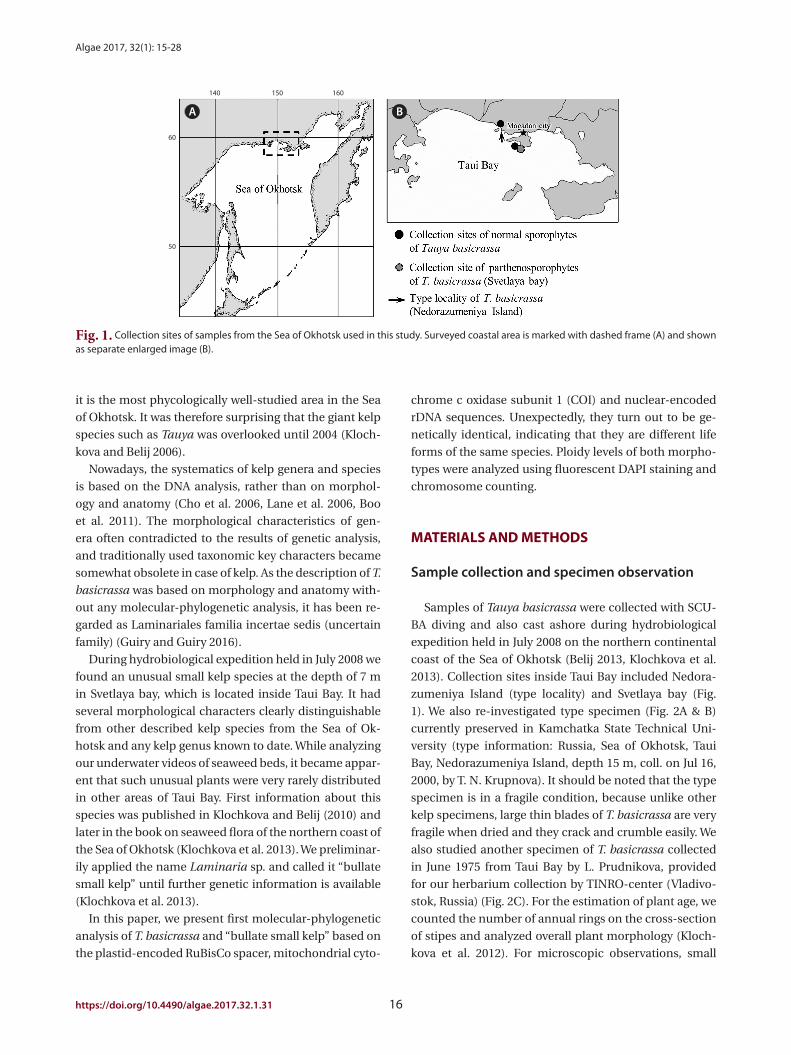
2013). Collection sites inside Taui Bay included Nedora-
zumeniya Island (type locality) and Svetlaya bay (Fig.
1). We also re-investigated type specimen (Fig. 2A & B)
currently preserved in Kamchatka State Technical Uni-
versity (type information: Russia, Sea of Okhotsk, Taui
Bay, Nedorazumeniya Island, depth 15 m, coll. on Jul 16,
2000, by T. N. Krupnova). It should be noted that the type
specimen is in a fragile condition, because unlike other
kelp specimens, large thin blades of T. basicrassa are very
fragile when dried and they crack and crumble easily. We
also studied another specimen of T. basicrassa collected
in June 1975 from Taui Bay by L. Prudnikova, provided
for our herbarium collection by TINRO-center (Vladivo-
stok, Russia) (Fig. 2C). For the estimation of plant age, we
counted the number of annual rings on the cross-section
of stipes and analyzed overall plant morphology (Kloch-
kova et al. 2012). For microscopic observations, small
it is the most phycologically well-studied area in the Sea
of Okhotsk. It was therefore surprising that the giant kelp
species such as Tauya was overlooked until 2004 (Kloch-
kova and Belij 2006).
Nowadays, the systematics of kelp genera and species
is based on the DNA analysis, rather than on morphol-
ogy and anatomy (Cho et al. 2006, Lane et al. 2006, Boo
et al. 2011). The morphological characteristics of gen-
era often contradicted to the results of genetic analysis,
and traditionally used taxonomic key characters became
somewhat obsolete in case of kelp. As the description of T.
basicrassa was based on morphology and anatomy with-
out any molecular-phylogenetic analysis, it has been re-
garded as Laminariales familia incertae sedis (uncertain
family) (Guiry and Guiry 2016).
During hydrobiological expedition held in July 2008 we
found an unusual small kelp species at the depth of 7 m
in Svetlaya bay, which is located inside Taui Bay. It had
several morphological characters clearly distinguishable
from other described kelp species from the Sea of Ok-
hotsk and any kelp genus known to date. While analyzing
our underwater videos of seaweed beds, it became appar-
ent that such unusual plants were very rarely distributed
in other areas of Taui Bay. First information about this
species was published in Klochkova and Belij (2010) and
later in the book on seaweed flora of the northern coast of
the Sea of Okhotsk (Klochkova et al. 2013). We preliminar-
ily applied the name Laminaria sp. and called it “bullate
small kelp” until further genetic information is available
(Klochkova et al. 2013).
In this paper, we present first molecular-phylogenetic
analysis of T. basicrassa and “bullate small kelp” based on
the plastid-encoded RuBisCo spacer, mitochondrial cyto-
Fig. 1. Collection sites of samples from the Sea of Okhotsk used in this study. Surveyed coastal area is marked with dashed frame (A) and shown as separate enlarged image (B).
A B
50
60
140 150 160

Klochkova et al. Tauya basicrassa from the Sea of Okhotsk
17 http://e-algae.org
A
C D
B
E
Fig. 2. Sporophytic plants of Tauya basicrassa from the Sea of Okhotsk. (A) Drawing of the authentic specimen showing a deep-water plant with large undissected blade with several longitudinal clefts (Nedorazumeniya Island). (B) Drawing of holotype specimen collected from the upper sub-tidal zone (reproduced with permission from Klochkova and Krupnova 2004). (C) Dry specimen collected by L. Prudnikova on June 1975 from Taui Bay (Sea of Okhotsk). In herbarium specimens, the basal part of blade becomes much darker. (D & E) Dorsal and ventral sides of the basal part of same blade, showing a concave-convolute hood-like structure with bullations. Three distinct portions are marked with numerals. Scale bars represent: A, 50 cm; B, 4 cm; C, 7 cm; D & E, 2 cm.

Algae 2017, 32(1): 15-28
https://doi.org/10.4490/algae.2017.32.1.31 18
from the NCBI; total alignment was 744 nt-long with 125
indels/gaps. RuBisco alignment was 653 nt-long with 20
indels/gaps. The trees were generated using Bayesian
phylogenetic analysis (MrBayes 3.2.2) (Huelsenbeck and
Ronquist 2001) using a GTR substitution model, 3,000,000
generations, sub-sampled every 1,000 generations and a
burn-in length of 300,000 generations. Maximum likeli-
hood analysis used RAxML 7.2.8 (Stamatakis 2014) using a
GTR + gamma model. Bootstrap support values (%) were
calculated based on 500 bootstrap replicates. Our new
sequences have been deposited in GenBank under the
following accession numbers: KY434160 (rDNA, normal
sporophyte), KY434161 (rDNA, parthenosporophyte),
KY434162 (RuBisCo, normal sporophyte), KY434163
(RuBisCo, parthenosporophyte), KY434164 (COI, normal
sporophyte), KY434165 (COI, parthenosporophyte).
Nuclei staining and chromosome observation
For nuclei staining, 1 mg mL-1 stock solution of DAPI
(4′,6-diamidino-2-phenylindole; Sigma, St. Louis, MO,
USA) dissolved in distilled water was applied to the
cross-sectioned materials incised from different parts
of the blades at a concentration of 5 µg mL-1 for 5 to 10
min and heated in a microwave for a few seconds. The
stained nuclei were examined under a UV filter. In both
types of plants (large and dwarf blades), the nuclei were
marked and their areas were compared. On the total, we
measured 20 nuclei in each type of plant and compared
their surface areas. For observation of chromosomes,
aceto-carmine solution (carmine 2 g, glacial acetic acid
45 mL, ddH2O 50 mL) was prepared by boiling for 2-3 h
and filtering through several layers of the filter paper. For
chromosome staining, 2-3 drops of aceto-carmine solu-
tion were applied to the rehydrated and cross-sectioned
tissue taken from the meristem zone. Fine sections were
put on a slide glass and covered with a cover slip, and
heated over the boiler for 1-2 min. Thereafter, the sec-
tions were strongly squashed between the slide glass and
the cover slip. Chromosomes thus stained were examined
with Olympus BX50 microscope while still immersed in
the stain. On the total, we marked and counted chromo-
somes in 7 and 10 nuclei of the large (i.e., normal spo-
rophytes) and dwarf (i.e., parthenosporophytes) plants
of T. basicrassa, respectively. Because the chromosomes
spread out in three dimensions, the numbers were count-
ed by tracing and marking individual chromosomes
through stacks of consecutively taken images (Mann and
Poulĺčková 2010). First, the labeling was accomplished for
all nuclei in both types of plants and then counting was
performed.
pieces of plants were incised from different parts of the
blades. They were rehydrated in sterilized seawater, cut
with a fine razor blade, and observed under a light mi-
croscope. Micrographs were taken with Olympus DP50
digital camera affixed to an Olympus BX50 microscope
(Olympus, Tokyo, Japan) using Viewfinder Lite and Stu-
dio Lite computer programs (Better Light Inc., Placerville,
CA, USA).
Molecular identification and phylogenetic anal-ysis
For extraction of genomic DNA, pieces of plants in-
cised from different parts of the blades were rehydrated
in sterilized seawater and their surface was thoroughly
cleaned with a fine brush. They were transferred to 2.0-
mL cryotubes and frozen in liquid nitrogen. The best re-
sults were obtained when tissue for DNA extraction was
obtained from the basal part of blade. The cryotubes con-
taining pieces of plants were shaken at 5,000 rev min-1 for
2 min with a cell homogenization machine. The DNA was
then extracted using an Invisorb Spin Plant Mini Kit (In-
vitek, Berlin-Buch, Germany) with some modifications of
the manufacturer’s protocol that we applied in case of ex-
tracting DNA from the kelp samples. The plastid-encoded
RuBisCo spacer was amplified using primer sets RS1-RS2,
KR3-KR4, nuclear-encoded rDNA was amplified with the
primer sets LB1-BC2 (ITS1 region) and YB1-LB2 (ITS2 re-
gion), and mitochondrial COI gene was amplified using
the primer set GazF2-GazR2 (Saunders and Druehl 1993,
Yoon and Boo 1999, Yoon et al. 2001, Lane et al. 2006,
2007). TaKaRa Ex Taq polymerase (Takara Biomedicals,
Otsu, Japan) was used for the PCR amplifications. The
following PCR program was used: initial denaturation at
95°C for 4 min, followed by 35 cycles of denaturation at
94°C for 30 s, annealing at 55°C for 30 s, extension at 72°C
for 1.5 min and a final extension at 72°C for 10 min. PCR
products were checked on an agarose gel for length, con-
centration, and purity. Sequencing was performed com-
mercially (Cosmogenetech Co., Ltd., Seoul, Korea). On the
total, we sequenced 981 nt for rDNA regions in both sam-
ples (18S rRNA, ITS1, 5.8S rRNA, ITS2, and 26S rRNA). For
RuBisCo spacer, 633 nt were sequenced including partial
rbcL and rbcS, and for COI gene, 396 nt were sequenced.
Our new sequences and sequences from GenBank (Na-
tional Center for Biotechnology Information 2016) were
aligned using Geneious (ver. 7.1.8; Biomatters, Auckland,
New Zealand) and the alignments were refined by eye.
For rDNA alignment, our sequences were trimmed up to
619 nt to match the alignment length of other sequences

Klochkova et al. Tauya basicrassa from the Sea of Okhotsk
19 http://e-algae.org
hydrodynamics, at depths of 4-22 m, on rocky substrate
covered with sand and silt. It has preference for the cold-
water bottom layer of the seawater stratum, forming
mono-species communities with an average biomass up
to 7.5 kg m-2 or growing in groups in mixed communi-
ties among other laminariacean algae. Under favorable
conditions, the largest plants reach 3-6 m long, 1.8-2.2 m
wide, and 6.5-7 kg in wet weight.
Our survey on the continental coast of the Sea of Ok-
hotsk revealed that T. basicrassa has a broader distribu-
tion than previously thought and grows not only in Taui
Bay, but in the proximate more southern and northern
regions. However, the gigantic plants develop only in the
type locality, i.e., in the area close to Nedorazumeniya
Island, because of special bottom relief and increased
levels of nutrients in this place, which induce an unlim-
ited growth of plants. The plants have smaller sizes not
exceeding 1-1.5 m at length in places with high waves at
depths of 5-8 m. In some coastal areas, T. basicrassa oc-
cupies the sub-tidal fringe, becoming stubby and not ex-
ceeding 45-65 cm at length. As estimated from studying
the morphology of plants and characteristic annual rings
on the cross-section of stipes, T. basicrassa can live for no
less than 4 years.
Tauya basicrassa (parthenosporophytes). Thallus of
this small yellowish-brown kelp has a small holdfast with
weakly branched haptera, 1.7 cm-long stipe and a thin,
soft, bullate blade reaching 26-34 cm long and 6-6.5 cm
wide. Sculptured bullations are arranged into 4 longitudi-
nal rows, covering the entire blade, forming very distinct
longitudinal as well as transverse rows (Fig. 4A & B). The
margins of blade are mostly strait, with several large folds
in the upper portion. On overall, blades are thin (up to
102 and 86-99 μm in the basal and upper portions, re-
spectively), becoming slightly thicker towards the edges
(Fig. 4D & E); cortex is 9.6 μm thick, composed of one
layer of pigmented isodiametric cells, covered with an
entire thickened mucilaginous covering on the outer sur-
face (Fig. 4F & G). Meristoderm is composed of 2-3 layers
of large thick-walled cells. Medullar is weakly developed,
almost absent. As seen in cross-sections, some cells are
filled with brown-red content (Fig. 4D-F). In fresh plants
extracted from the sea, the basal part of blade in not dif-
ferent in color than the rest of the blade (Fig. 4B), whereas
in dried plants the basal part becomes much darker (low-
er 1/4 of blade) than the upper part (Fig. 4C). Only sterile
plants were collected. As estimated from studying their
morphology, these plants were at least 1 year old.
RESULTS
Morphology and anatomy, ecology and distribu-tion
Tauya basicrassa (normal sporophytes). Large sporo-
phytic plants have unique morphology of blade (Table 1,
Figs 2A-C & 3A); it is very wide, entire or bearing longitu-
dinal clefts in young plants and dissected into 3 or more
lobes with age. In fresh plants extracted from the sea, the
blades’ basal part is narrow, thick, cartilaginous and light-
er in color than the rest of the blade (Fig. 3A & B), look-
ing like a concave-convolute hood with bullations (Fig.
2D & E). On the contrary, in dried plants the basal part
becomes much darker, whereas the upper part becomes
lighter (Fig. 2C). The blade’s upper part bears 8-9 rows
of bullations, is very fragile and crumbles easily, so that
only the rigid basal part with stipe remains (Fig. 2D & E).
Stipe is very short, flattened in its upper portion; haptera are
rigid, thick and branched (Fig. 3B). In adult plants, the blade’s
thickness is 1,300-1,360 μm in the basal part and 345-352
μm in the upper part; the medullar layer is 141 and 64 μm
thick in the basal and upper parts, respectively. Cortex is
12.8 μm thick and consists of one layer of pigmented iso-
diametric cells; beneath the cortex layer very large thick-
walled cells are distributed.
Development of bullations begins in the juvenile
plants (Fig. 3C & D). On the blade, bullations are local-
ized between smooth and more thickened ribbon-like
stripes; the mature plants usually have 6 of such stripes.
Sizes of bullations become gradually larger according to
the increase of the blade’s width. During the reproductive
period occurring from August to early September, spo-
rangial sori begin to develop on these ribbon-like stripes
(Fig. 3E), and later the sori spread outside the stripes.
In T. basicrassa, morphological differentiation of thalli
begins early in plants’ development (Fig. 3C & D). Al-
though its anatomy is similar to other laminariacean
algae, it has some specific characters such as glandular
cells (Fig. 3F) and large mucilage ducts up to 77-128 μm
in size in the cross-section (Fig. 3G & H), which spread on
the whole basal part and meristem zone of the blade. The
mucilage ducts form a conductive system in the tissue
(network of channels), which acts as an elastic armature,
reinforcing the blade on its surface. The mucilage ducts
are absent in the upper part of blade.
In Taui Bay, T. basicrassa inhabits lower border of the
phytal zone and is found in places with lower level of

Algae 2017, 32(1): 15-28
https://doi.org/10.4490/algae.2017.32.1.31 20
A C DB
E
GF H
Fig. 3. The morphology and anatomy of normal sporophytes of Tauya basicrassa from the Sea of Okhotsk. (A) Mature plant freshly extracted from the sea. (B) Stipe is short and flattened in the upper portion, and haptera are rigid and branched as seen on the cross-section. (C & D) Juvenile plants at different state of maturity, showing two (C) and four (D) rows of developing bullations. (E) Sporangial sori in mature plants (arrows, reproduced from Klochkova et al. 2013). (F) Cross-section of blade in the thickenned basal part, showing glandular cells (dashed arrows) and large mucilage ducts (solid arrows). (G) Tangential longitudinal section showing conductive system formed with mucilage ducts, which look similar to a network of channels (solid arrow). Numerous glandular cells can be seen (dashed arrow). (H) Cross-section of mucilage duct. Scale bars represent: A & E, 10 cm; B-D, 5 cm; F, 500 µm; G, 200 µm; H, 100 µm.

Klochkova et al. Tauya basicrassa from the Sea of Okhotsk
21 http://e-algae.org
A C
D
B
E
GF
Fig. 4. The morphology and anatomy of parthenosporophytes of Tauya basicrassa from the Sea of Okhotsk. (A) Underwater photograph of mature plant attached to sea bottom. (B) Freshly extracted plant showing four rows of bullations (A and B are reproduced from Klochkova et al. 2013). (C) Same plant in dried condition showing dark basal part of the blade. (D & E) The blades are thin and become slightly thicker towards the edges; some cells in the meristoderm are filled with brown-red content (arrows). (F & G) Cross-sections of the middle part of blade, showing pigmented cell, large cells in the meristoderm and very thin, almost undeveloped medullar. m, meristoderm; md, meddular; pc, pigmented cell. Scale bars represent: B & C, 2 cm; D & E, 100 µm; F & G, 50 µm.
Algae 2017, 32(1): 15-28
https://doi.org/10.4490/algae.2017.32.1.31 22
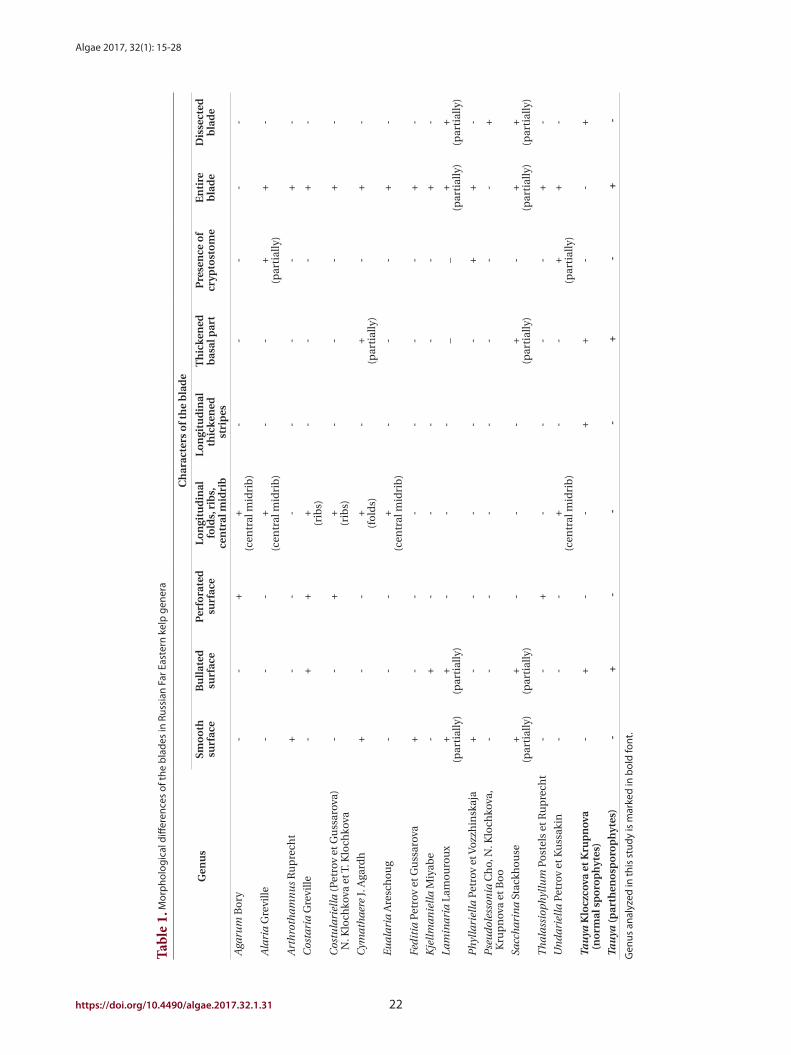
Tabl
e 1.
Mor
pho
logi
cal d
iffer
ence
s of
the
bla
des
in R
ussi
an F
ar E
aste
rn k
elp
gen
era
Gen
us
Ch
arac
ters
of t
he
bla
de
Smo
oth
su
rfac
eB
ull
ated
su
rfac
eP
erfo
rate
d
surf
ace
Lo
ngi
tud
inal
fo
lds,
rib
s,
cen
tral
mid
rib
Lo
ngi
tud
inal
th
icke
ned
st
rip
es
Th
icke
ned
b
asal
par
tP
rese
nce
of
cryp
tost
om
eE
nti
re
bla
de
Dis
sect
ed
bla
de
Aga
rum
Bo
ry-
-+
+(c
entr
al m
idri
b)
--
--
-
Ala
ria
Gre
ville
--
-+
(cen
tral
mid
rib
)-
-+
(par
tial
ly)
+-
Art
hro
tham
nu
s R
up
rech
t+
--
--
--
+-
Cos
tari
a G
revi
lle-
++
+(r
ibs)
--
-+
-
Cos
tula
riel
la (
Petr
ov e
t Gu
ssar
ova)
N
. Klo
chko
va e
t T. K
loch
kova
--
++
(rib
s)-
--
+-
Cym
ath
aere
J. A
gard
h+
--
+(f
old
s)-
+(p
arti
ally
)-
+-
Eu
alar
ia A
resc
ho
ug
--
-+
(cen
tral
mid
rib
)-
--
+-
Fed
itia
Pet
rov
et G
uss
arov
a+
--
--
--
+-
Kje
llm
anie
lla
Miy
abe
-+
--
--
-+
-
Lam
inar
ia L
amo
uro
ux
+(p
arti
ally
)+
(par
tial
ly)
--
-_
_+
(par
tial
ly)
+(p
arti
ally
)
Ph
ylla
riel
la P
etro
v et
Voz
zhin
skaj
a+
--
--
-+
+-
Pse
ud
oles
son
ia C
ho,
N. K
loch
kova
, K
rup
nov
a et
Bo
o-
--
--
--
-+
Sacc
har
ina
Stac
kho
use
+(p
arti
ally
)+
(par
tial
ly)
--
-+
(par
tial
ly)
-+
(par
tial
ly)
+(p
arti
ally
)
Th
alas
siop
hyl
lum
Po
stel
s et
Ru
pre
cht
--
+-
--
-+
-
Un
dar
iell
a P
etro
v et
Ku
ssak
in-
--
+(c
entr
al m
idri
b)
--
+(p
arti
ally
)+
-
Tau
ya K
locz
cova
et K
rup
nov
a
(no
rmal
sp
oro
ph
ytes
)-
+-
-+
+-
-+
Tau
ya (p
arth
eno
spo
rop
hyt
es)
- +
--
-+
-+
-
Gen
us a
naly
zed
in th
is s
tudy
is m
arke
d in
bol
d fo
nt.

Klochkova et al. Tauya basicrassa from the Sea of Okhotsk
23 http://e-algae.org
Nuclei staining and chromosome observation
Fluorescent DAPI staining showed that the volume of
nuclei in the normal sporophytes of T. basicrassa was by
50-65% larger than in the parthenosporophytes (Fig. 8A
& B). Chromosomes from the cells in meristem zone of
both plants stained with acetocarmine were observed. It
was quite difficult to examine the exact shape of chromo-
somes, as the thick cell walls prevented use of the normal
“squash preparation” methods. The shape was dot-like
or slightly elongated in both species, sizes were identical,
and the numbers were n = ca. 20 in T. basicrassa normal
sporophyte and n = ca. 10 in the parthenosporophyte
(Fig. 8C-E).
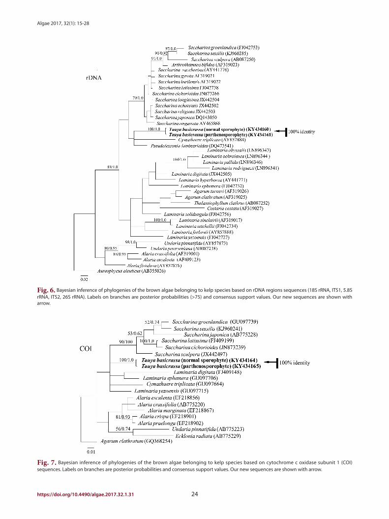
Molecular identification
Phylogenetic analysis using plastid-encoded RuBisCo
spacer, rDNA and COI showed that the giant sporophytes
of T. basicrassa and bullate small kelp were identical (Figs
5-7). In each tree, they formed a separate well-support-
ed clade and did not fall into any known algal genus se-
quenced to date. In RuBisCo tree, the “Tauya” clade posi-
tioned between the clade formed by five different species
from Lessoniaceae and Cymathaere triplicata (Postels et
Ruprecht) Agardh from Laminariaceae. In rDNA and COI
trees, Tauya belonged to the family Laminariaceae.
Fig. 5. Bayesian inference of phylogenies of the brown algae belonging to kelp species based on plastid-encoded RuBisCo spacer sequences. Labels on branches are posterior probabilities (>75) and consensus support values. Our new sequences are shown with arrow.
Algae 2017, 32(1): 15-28
https://doi.org/10.4490/algae.2017.32.1.31 24
Fig. 6. Bayesian inference of phylogenies of the brown algae belonging to kelp species based on rDNA regions sequences (18S rRNA, ITS1, 5.8S rRNA, ITS2, 26S rRNA). Labels on branches are posterior probabilities (>75) and consensus support values. Our new sequences are shown with arrow.
Fig. 7. Bayesian inference of phylogenies of the brown algae belonging to kelp species based on cytochrome c oxidase subunit 1 (COI) sequences. Labels on branches are posterior probabilities and consensus support values. Our new sequences are shown with arrow.

Klochkova et al. Tauya basicrassa from the Sea of Okhotsk
25 http://e-algae.org
cality of T. basicrassa was the small area on the sea bottom
located inside Taui Bay between Nedorazumeniya Island
and continental coast (mainland). This area not exceed-
ing 1/3 of a square kilometer looks like a deep trench in
the sea, with a substrate made with rocks, pebbles and
sand. Because of such peculiar bottom relief, large depth,
and overall appearance of the coastline, seawater mixing
in the bottom layer is weak, resulting in increased nutri-
ents concentration, which in turn causes an unlimited
growth of T. basicrassa sporophytes. In some sites of this
bottom, it formed mono-species communities at 15-17 m
deep.
Some kelp species have been reported to have narrow
distribution (Petrov and Gussarova 1972, Petrov and Kus-
sakin 1997, Lee and Yoon 1998, Kawai et al. 2008, Klochko-
va and Klochkova 2010). Other laminariacean algae have
DISCUSSION
Morphological characteristics of the genus Tauya are
clearly different from other laminariacean species (Kloch-
kova and Krupnova 2004), but it has long been treated as
Laminariales familia incertae sedis (uncertain family) be-
cause there has been no molecular data supporting the
establishment of the genus (Guiry and Guiry 2016). Our
molecular analysis based on rDNA and COI genes sug-
gested attribution of Tauya to the family Laminariaceae,
which is also in agreement with morphological charac-
ters of this genus.
T. basicrassa was originaly described as an endangered
endemic species with very narrow distribution on a small
area of sea bottom characterized by specific hydrological
conditions (Klochkova and Krupnova 2004). The type lo-
Fig. 8. Fluorescent DAPI staining of nuclei and acetocarmine staining of chromosomes of normal sporophytes and parthenosporophytes of Tauya basicrassa. (A) Large nuclei of normal sporophytes. (B) Small nuclei of parthenosporophytes. (C) Selected through-focus images of chromosomes in the large nuclei of normal sporophytes and chromosome counting. (D & E) Selected through-focus images of chromosomes in the small nuclei of parthenosporophytes and chromosome counting. Scale bars represent: A-E, 5 µm.
A
C
D
B
E

Algae 2017, 32(1): 15-28
https://doi.org/10.4490/algae.2017.32.1.31 26
ogy was dramatically different from normal sporophytes
of T. basicrassa our molecular data showed that the dwarf
one is just a different life form of T. basicrassa. Compara-
tive study of their karyotypes showed that this morpho-
logical variation is due to the difference in ploidy levels.
Chromosome number of dwarf plants was half of that in
the normal sporophytes of T. basicrassa. Although no re-
productive structures were found on the collected mate-
rials, the dwarf thallus has probably derived parthenoge-
netically from unfertilized eggs of T. basicrassa.
In laboratory culture, unfertilized eggs of laminari-
acean algae are known to develop parthenogenetically,
but those plants were usually shaped abnormal and
died ephemerally (Ar Gall et al. 1996). Development of
parthenosporophytes under laboratory conditions has
been reported in many kelp species (Fang and Dai 1980,
Lewis et al. 1993, Oppliger et al. 2007, Shan et al. 2013). In
Saccharina japonica (Areschoug) Lane, Mayes, Druehl
et Saunders, female parthenosporophytes developed
zoospores, which grew into microscopic female game-
tophytes, produced eggs later, fused with male sperms
of this species and developed into normal diploid spo-
rophytes (Lewis et al. 1993). To the best of our knowledge,
our results are the first example of parthenogenesis and /
or apogamy in the laminariacean algae demonstrated to
occur naturally in the field. It is noteworthy that the par-
thenosporophytes were at least 1 year old because they
were overwintered plants. Further studies on the life his-
tory of T. basicrassa using laboratory culture may reveal
the mechanisms that govern differential morphogenesis
occurring in sporophytes and gametophytes of laminari-
acean algae.
ACKNOWLEDGEMENTS
This work was supported by a National Research Foun-
dation of Korea Grant (NRF-2015M1A5A1041804) funded
to GHK. This research was also supported by Golden
Seed Project, Ministry of Agriculture, Food and Rural Af-
fairs, and a grant from Marine Biotechnology Program
(PJT200620, genome analysis of marine organisms and
development of functional applications) funded by Min-
istry of Oceans and Fisheries, Korea. We thank Dr. Belij, M.
N. (MagadanNIRO) for his help during samples collection
from the Taui Bay. We thank Dr. Alejnikova, M. D. (Edito-
rial board of “International Journal on Algae,” Algologia)
for permission to reproduce picture of the holotype.
discontinuous distributions and populations of the same
species can be found at large distances. For example, Ar-
throthamnus kurilensis Ruprecht is mainly distributed on
the Kurile Islands, but another isolated population exists
on the south of Sakhalin Island on a small area of sea floor
characterised by upwelling of cold bottom waters (called
“Makarov’s spot” in Russian) (Klochkova 1998). Lami-
naria gurjanovae Zinova also has discontinuous distri-
bution on the Russian Far East. Thus, a small deep-water
population of T. basicrassa near Nedorazumeniya Island
also seemed like a relict population of once widespread
species. Although all plants from this area became gigan-
tic, the increase in size was accompanied by a significant
decrease in the thickness of blade. Such changes occur in
plants when they grow in seawater with low mobility and
high nutrients concentration, because the lack of laminar
flow, which is supposed to bring nutrients to the blade’s
surface, is compensated by an increased absorbing sur-
face area. For example, we observed very large plants of
Sacharina bongardiana on the north-eastern Kamchatka
in Yuzhnaya Glubokaya Bay in the exit site of underwater
fumaroles (Klochkova and Berezovskaya 1997). Gussaro-
va (1975) reported on seaweed gigantism from the Kurile
Islands due to the enrichment of coastal seawaters with
volcanic ashes.
During hydrobiological expedition on the northern
continental coast of the Sea of Okhotsk (Klochkova et al.
2013) we confirmed that Т. basicrassa does not have wide
distribution in this area. It usually inhabits places with
low level of hydrodynamics, at depths of 4-22 m, showing
preference for the cold bottom layer (12-18 m deep). Its
distribution border usually coincided with the replace-
ment of hard substrates (i.e., rocks, boulders) with soft
substrate (i.e., sand), and most commonly it inhabited
sandy silted bottom with more or less frequent boulders,
to which it attached. It did not grow in clusters of plants
or forming dense covers on the bottoms, and the maxi-
mum number of plants per 1 square meter was 5-7. Under
favorable conditions it formed mono-species communi-
ties with an average biomass of 7.5 kg m-2.
A dwarf kelp, which is morphologically unassignable to
any known species of laminariacean algae, was found on
rocks and boulders at 4-7 m deep in the same locality. This
dwarf laminariacean alga distributed sparsely in that area
and never grew in groups (Klochkova et al. 2013). During
analysis of the underwater videos of seaweed beds from
other areas of Taui Bay, we also found several more plants
with same size and morphology. Although their morphol-

Klochkova et al. Tauya basicrassa from the Sea of Okhotsk
27 http://e-algae.org
inariaceaen alga from Taujskaya Bay (Sea of Okhotsk).
Bull. Kamch. State Tech. Univ. [Vestnik KamchatGTU]
11:55-58 (in Russian).
Klochkova, N. G. & Berezovskaya, V. A. 1997. The seaweeds of
Kamchatka’s shelf: biology, distribution, chemical com-
position. Dalnauka, Vladivostok, 153 pp. (in Russian).
Klochkova, N. G. & Klochkova, T. A. 2010. Costulariella, a new
substitute name for Costularia Ju. Petrov et I. Gussarova
(Laminariales, Phaeophyceae). Algae 25:183-185.
Klochkova, N. G. & Krupnova, T. N. 2004. New and interesting
taxa of laminarialeaen algae (Laminariales, Phaeophyta)
of Far Eastern seas of Russia. Tauya basicrassa Kloczc.
et Krupn. gen. et sp. nov. Algology 14:86-94 (bilingual,
Russian-English).
Klochkova, T. A., Belij, M. N. & Klochkova, N. G. 2013. Chap-
ter 3. Seaweeds of the Sea of Okhotsk. In Volobuev, V. V.
& Klochkova, N. G. (Eds.) Seaweeds of the Northern Part
of the Sea of Okhotsk and Their Role as a Substrate for the
Herring Spawning. Federal Agency of Fishery of Russian
Federation, Magadan Scientific-Research Institute of
Fishery and Oceanography (MagadanNIRO), Magadan,
pp. 21-140 (bilingual, Russian-English).
Klochkova, T. A., Kim, G. H., Belij, M. N. & Klochkova, N. G.
2012. Morphology and phytogeography of Laminaria
appressirhiza and L. inclinatorhiza (Phaeophyceae)
from the Sea of Okhotsk. Algae 27:139-153.Klochkova, T. A., Kim, G. H., Lee, K. M., Choi, H. -G., Belij,
M. N. & Klochkova, N. G. 2010. Brown algae (Phaeo-
phyceae) from Russian Far Eastern seas: re-evaluation
of Laminaria multiplicata Petrov et Suchovejeva. Algae
25:77-87.
Lane, C. E., Lindstrom, S. C. & Saunders, G. W. 2007. A mo-
lecular assessment of northeast Pacific Alaria species
(Laminariales, Phaeophyceae) with reference to the util-
ity of DNA barcoding. Mol. Phylogenet. Evol. 44:634-648.
Lane, C. E., Mayes, C., Druehl, L. D. & Saunders, G. W. 2006.
A multi-gene molecular investigation of the kelp (Lami-
nariales, Phaeophyceae) supports substantial taxonom-
ic re-organization. J. Phycol. 42:493-512.
Lee, Y. -P. & Yoon, J. -T. 1998. Taxonomy and morphology
of Undaria (Alariaceae, Phaeophyta) in Korea. Algae
13:427-446.
Lewis, R. J., Jiang, B. Y., Neushul, M. & Fei, X. G. 1993. Haploid
parthenogenetic sporophytes of Laminaria japonica
(Phaeophyceae). J. Phycol. 29:363-369.
Mann, D. G. & Poulĺčková, A. 2010. Mating system, auxospor-
ulation, species taxonomy and evidence for homoploid
evolution in Amphora (Bacillariophyta). Phycologia
49:183-201.
National Center for Biotechnology Information (NCBI).
REFERENCES
Ar Gall, E. (Le Gall, Y.), Asensi, A., Marie, D. & Kloareg, B. 1996.
Parthenogenesis and apospory in the Laminariales: a
flow cytometry analysis. Eur. J. Phycol. 31:369-380.
Belij, M. N. 2013. Seaweeds of the northern part of the Sea
of Okhotsk and their role as a substrate for the herring
spawning. Federal Agency of Fishery of Russian Federa-
tion, Magadan Scientific-Research Institute of Fishery
and Oceanography (MagadanNIRO), Magadan, 193 pp.
(in Russian).
Boo, G. H., Lindstrom, S. C., Klochkova, N. G., Yotsukura, N.,
Yang, E. C., Kim, H. G., Waaland, J. R., Cho, G. Y., Miller,
K. A. & Boo, S. M. 2011. Taxonomy and biogeography of
Agarum and Thalassiophyllum (Laminariales, Phaeo-
phyceae) based on sequences of nuclear, mitochondrial,
and plastid markers. Taxon 60:831-840.
Cho, G. Y., Klochkova, N. G., Krupnova, T. N. & Boo, S. M.
2006. The reclassification of Lessonia laminarioides
(Laminariales, Phaeophyceae): Pseudolessonia gen. nov.
J. Phycol. 42:1289-1299.
Fang, Z. X. & Dai, J. X. 1980. Use of haploid phases in the ge-
netic study of Laminaria japonica. Acta Genet. Sin. 7:19-
25.
Guiry, M. D. & Guiry, G. M. 2016. AlgaeBase. World-wide
electronic publication, National University of Ireland,
Galway. Available from: http://www.algaebase.org. Ac-
cessed Oct 10, 2016.
Gussarova, I. S. 1975. Macroalgae of the sub-tidal zone of
Iturup, Urup and Simushir Islands (large Kurile Islands).
Novosti Sistematiki Nizshih Rastenii [News on System-
atics of Non-vascular Plants] 12:111-118 (in Russian).
Huelsenbeck, J. P. & Ronquist, F. 2001. MRBAYES: Bayesian
inference of phylogenetic trees. Bioinformatics 17:754-
755.Kawai, H., Hanyuda, T., Lindeberg, M. & Lindstrom, S. C.
2008. Morphology and molecular phylogeny of Aureo-
phycus aleuticus gen. et sp. nov. (Laminariales, Phaeo-
phyceae) from the Aleutian Islands. J. Phycol. 44:1013-
1021.
Klochkova, N. G. 1998. Flora of macroalgae of the Russian
Far East. Extended Abstract of National Doctoral Degree
Dissertation, Vladivostok, 49 pp. (in Russian).
Klochkova, N. G. & Belij, M. N. 2006. Additional informa-
tion to the description of Tauya basicrassa Kloczcova et
Krupnova from the Sea of Okhotsk. In Tokranov, A. M.
(Ed.) Conservation of Biodiversity of Kamchatka and
Coastal Waters. Petropavlovsk-Kamchatsky Press, Pet-
ropavlovsk-Kamchatsky, pp. 391-394 (in Russian).
Klochkova, N. G. & Belij, M. N. 2010. Finding an unusual lam-

Algae 2017, 32(1): 15-28
https://doi.org/10.4490/algae.2017.32.1.31 28
riety breeding and sporeling production in the brown
seaweed Undaria pinnatifida (Phaeophyceae): crossing
female gametophytes from parthenosporophytes with
male gametophyte clones. Phycol. Res. 61:154-161.
Stamatakis, A. 2014. RAxML version 8: a tool for phylogenetic
analysis and post-analysis of large phylogenies. Bioin-
formatics 30:1312-1313.
Wynne, M. J. 1982. Phaeophyceae. In Parker, S. P. (Ed.) Synop-
sis and Classification of Living Organisms. McGraw-Hill,
New York, NY, pp. 115-125.
Yoon, H. S. & Boo, S. M. 1999. Phylogeny of Alariaceae (Pha-
eophyta) with special reference to Undaria based on
sequences of the RuBisCo spacer region. Hydrobiologia
398/399:47-55.
Yoon, H. S., Lee, J. Y., Boo, S. M. & Bhattacharya, D. 2001. Phy-
logeny of Alariaceae, Laminariaceae, and Lessoniaceae
(Phaeophyceae) based on plastid-encoded RuBisCo
spacer and nuclear-encoded ITS sequence compari-
sons. Mol. Phylogenet. Evol. 21:231-243.
2016. GenBank. Available from: http//www.ncbi.nlm.
nih.gov. Accessed Oct 10, 2016.
Oppliger, L. V., Correa, J. A. & Peters, A. F. 2007. Parthenogen-
esis in the brown alga Lessonia nigrescens (Laminariales,
Phaeophyceae) from central Chile. J. Phycol. 43:1295-
1301.
Petrov, Yu. E. & Gussarova, I. S. 1972. New species and genus
of laminariacean alga from Simushir Island. Novosti
Sistematiki Nizshih Rastenii [News on Systematics of
Non-vascular Plants] 9:39-44.
Petrov, Yu. E. & Kussakin, O. G. 1997. Undariella kurilensis:
new species and genus of laminariacean alga from the
littoral zone of Yankich Island (Kurile Islands). Biol. Mor.
[Russ. J. Mar. Biol.] 23:79-83 (bilingual, Russian-English).
Saunders, G. W. & Druehl, L. D. 1993. Nucleotide sequences
of the internal transcribed spacers and 5.8S rRNA genes
from Alaria marginata and Postelsia palmaeformis
(Phaeophyta: Laminariales). Mar. Biol. 115:347-352.
Shan, T. F., Pang, S. J. & Gao, S. Q. 2013. Novel means for va-