Embed Size (px)
Citation preview

© Yenchitsomanus et al | Journal of Molecular and Genetic Medicine | December 2005 | Vol 1, No 2 | 49-62 | OPEN ACCESS
49
REVIEW
Molecular mechanisms of autosomal dominant and recessive distal renal tubular
acidosis caused by SLC4A1 (AE1) mutations
Pa-thai Yenchitsomanus1,2,3,*
, Saranya Kittanakom4, Nanyawan Rungroj
3, Emmanuelle Cordat
4,
Reinhart A F Reithmeier4
1Division of Medical Molecular Biology and
2BIOTEC-Medical Biotechnology Unit,
3Division of Molecular Genetics,
Department of Research and Development, Faculty of Medicine Siriraj Hospital, Mahidol University, Bangkok 10700,
Thailand. 4Departments of Biochemistry and Medicine, University of Toronto, Toronto, Ontario, M5S 1A8, Canada
*Correspondence to: Pa-thai Yenchitsomanus, Email: [email protected], Tel: +662 4197000 ext 6666-70, Fax: +662
4184793
Journal of Molecular and Genetic Medicine (2005), 1(2), 49-62
© Copyright Pa-thai Yenchitsomanus et al
Reviewers of this article included Professor Oliver Wrong (London, United Kingdom)
(Received 14 July 2005; Revised 06 September 2005; Accepted 13 September 2005; Available online 16 November 2005; Published
30 December 2005)
ABSTRACT
Mutations of SLC4A1 (AE1) encoding the kidney anion (Cl-/HCO3
-) exchanger 1 (kAE1 or band 3) can result
in either autosomal dominant (AD) or autosomal recessive (AR) distal renal tubular acidosis (dRTA). The
molecular mechanisms associated with SLC4A1 mutations resulting in these different modes of inheritance are now being unveiled using transfected cell systems. The dominant mutants kAE1 R589H, R901X and
S613F, which have normal or insignificant changes in anion transport function, exhibit intracellular retention
with endoplasmic reticulum (ER) localization in cultured non-polarized and polarized cells, while the domi-
nant mutants kAE1 R901X and G609R are mis-targeted to apical membrane in addition to the basolateral
membrane in cultured polarized cells. A dominant-negative effect is likely responsible for the dominant dis-
ease because heterodimers of kAE1 mutants and the wild-type protein are intracellularly retained. The reces-
sive mutants kAE1 G701D and S773P however exhibit distinct trafficking defects. The kAE1 G701D mutant
is retained in the Golgi apparatus, while the mis-folded kAE1 S773P, which is impaired in ER exit and is
degraded by proteosome, can only partially be delivered to the basolateral membrane of the polarized cells. In contrast to the dominant mutant kAE1, heterodimers of the recessive mutant kAE1 and wild-type kAE1
are able to traffic to the plasma membrane. The wild-type kAE1 thus exhibits a ‘dominant-positive effect’
relative to the recessive mutant kAE1 because it can rescue the mutant proteins from intracellular retention to be expressed at the cell surface. Consequently, homozygous or compound heterozygous recessive mutations
are required for presentation of the disease phenotype. Future work using animal models of dRTA will pro-
vide additional insight into the pathophysiology of this disease.
KEYWORDS: Distal renal tubular acidosis, kidney anion exchanger, protein trafficking, dominant nega-
tive effect, dominant positive effect
INTRODUCTION
Primary distal renal tubular acidosis (dRTA) is a clinical
syndrome characterized by impairment of acid secretion
by α-intercalated cells in the distal nephron and collecting
tubule, resulting in decreased net acid excretion and inabil-
ity of kidney to lower urinary pH <5.5 in spite of sponta-
neous academia or after acid loading (Batlle et al 2001;
Alper , 2002; Herrin, 2003). The decreased net acid excre-
tion attributable to decreased ammonium and titratable
acid excretion leads to a positive acid balance and hyper-
chloremic metabolic acidosis. The presence of chronic

© Yenchitsomanus et al | Journal of Molecular and Genetic Medicine | December 2005 | Vol 1, No 2 | 49-62 | OPEN ACCESS
50
metabolic acidosis consequently decreases renal excretion
of citrate (hypocitraturia) while increases calcium excre-
tion (hypercalciuria), creating a favorable environment for
urinary stone formation and nephrocalcinosis. The other
associated abnormalities include hypokalemia leading to
muscle weakness, and metabolic bone disease (rickets or
osteomalacia). The disease can be caused either by a de-
fect in the H+-ATPase, which is responsible for acid (H
+)
secretion at the apical membrane of the epithelial α-
intercalated cells or by an abnormality of the kidney anion
exchanger 1 (kAE1) that functions in chloride (Cl-) and
bicarbonate (HCO3-) anion exchange at the basolateral
membrane (Figure 1). The defects of these two proteins
may occur from mutations of the responsible genes caus-
ing hereditary dRTA. Mutations of at least two genes en-
coding two subunits (B1 and a4) of H+-ATPase have been
reported to cause autosomal recessive dRTA with sen-
sorineural deafness or normal hearing (Karet et al, 1999;
Smith et al, 2000; Stover et al, 2002; Ruf et al, 2003). The
genes that encode B1 subunit in V1 (catalytic) domain and
a4 subunit in Vo (transmembrane) domain of H+-ATPase
are ATP6V1B1 (MIM 192132) located on chromosome
2p13.1 and ATP6V0A4 (MIM 605239) situated on chro-
mosome 7q33-q34, respectively. Also, mutations in hu-
man solute carrier family 4, member 1 (SLC4A1) or anion
exchanger 1 (AE1) gene (MIM 109270) may cause dRTA.
SLC4A1 located on chromosome 17q21-q22 encodes both
erythroid (eAE1, band 3) and kidney (kAE1) isoforms of
AE1 protein. Thus, SLC4A1 mutations shows pleiotrophic
effects resulting in two distinct and seemingly unrelated
disorders, red cell abnormalities (e.g., hereditary spherocy-
tosis and ovalocytosis) and dRTA.
An abnormality of kAE1 that mediates chlo-
ride/bicarbonate (Cl-/HCO3-) exchange at the basolateral
membrane of the α-intercalated cells can lead to a defect
in bicarbonate (HCO3-) extrusion across this membrane
and to its intracellular accumulation. To maintain intracel-
lular acid-base and electroneutal balances, the cells would
reserve acid or hydrogen ion (H+) instead of secreting
through the apical membrane. Consequently, the intracel-
lular accumulation of both bicarbonate and hydrogen ions
would inhibit the dissociation of carbonic acid (H2CO3)
(Figure 1). The failure of hydrogen ion (H+) secretion
through the apical membrane into the tubular lumen due to
a kAE1 defect will eventually result in dRTA.
It was previously unknown why mutations of SLC4A1
cause both autosomal dominant (AD) (Bruce et al, 1997;
Jarolim et al, 1998; Karet et al, 1998; Weber et al, 2000;
Sritippayawan et al, 2003; Cheidde et al, 2003) and auto-
somal recessive (AR) dRTA (Tanphaichitr et al, 1998;
Vasuvattakul et al, 1999; Bruce et al, 2000; Ribeiro et al,
2000; Yenchitsomanus et al, 2002; Sritippayawan et al,
2004). During the past several years, several groups have
begun to unravel the molecular mechanisms of dRTA
caused by SLC4A1 mutations presenting in these two dif-
ferent manners of inheritance. We have recently demon-
strated that dominant and recessive dRTA phenotypes as-
sociated with SLC4A1 mutations are determined by differ-
ent behaviors in the intracellular trafficking of het-
erodimers between mutant and wild-type kAE1. The
dominant kAE1 mutants result in intracellular retention of
the wild-type kAE1, the so called ‘dominant-negative ef-
fect’, whereas the wild-type kAE1 exhibits ‘dominant-
positive effect’ relative to the recessive mutant kAE1 be-
cause it can rescue the recessive mutant kAE1 to express
on the cell surface. We have therefore distinguished the
molecular mechanisms of AD and AR dRTA caused by
SLC4A1 mutations.
Figure 1. Schematic diagram of the α-intercalated cell in the distal nephron. H+-ATPase and H+/K+-ATPase, involving in acid (H+)
secretion (in exchange with K+ in the latter), are present at the apical membrane, while kAE1, functioning in chloride/bicarbonate
(Cl-/HCO3-) exchange, is located at the basolateral membrane. H+ and HCO3
- for apical secretion and basolateral reabsorption are
dissociated from H2CO3, generated from hydration of carbon dioxide (CO2), which is catalyzed by carbonic anhydrase (CA) II.

© Yenchitsomanus et al | Journal of Molecular and Genetic Medicine | December 2005 | Vol 1, No 2 | 49-62 | OPEN ACCESS
51
SLC4A1 AND ENCODED PROTEINS
SLC4A1
The human SLC4A1 or AE1 gene encoding the anion ex-
changer 1 (AE1 or band 3) is located on chromosome
17q21-q22 encompassing approximately 20 kb and con-
sisting of 20 exons separated by 19 introns. The gene con-
tains no TATA or CCAAT boxes in its upstream region
(Tanner et al, 1988; Lux et al, 1989; Sahr et al, 1994) but
consists of transcription factor consensus binding sites,
activator protein 1 (AP1), activator protein 2 (AP2),
CACCC boxes, GATA (erythroid factor 1), and E-boxes in
this region. Instead, the TATA and CCAAT boxes are
found in intron 3. The structure of the gene showed exten-
sive similarity to that of mouse AE1, with a high degree of
conservation of exon/intron junctions (Sahr et al, 1994;
Schofield et al, 1994). The human SLC4A1 encodes both
eAE1 and kAE1 isoforms by using different promoters and
alternative splicing. While eAE1 mRNA is transcribed
from all exons by using its upstream promoter, kAE1
mRNA is transcribed by using the promoter in intron 3.
The eAE1 cDNA sequence comprises 4,906 nucleotides
(nt), excluding its poly (A) tail. The kAE1 mRNA lacks
the sequences of exons 1-3 of the eAE1 transcript but con-
tains a part of intron 3 (designated as exon K1) in its 5’
untranslated region (5’ UTR) and the complete exon 4-20
sequences with the translation initiation (ATG) site for
methionine (Met) at codon 66 (Kollert-Jöns et al, 1993).
The downstream end of exon 20 is defined as the RNA
cleavage site or poly A addition site. A consensus poly A
signal (AATAAA) is located 20 nucleotides upstream of
the RNA cleavage site.
Expression and function of eAE1 and kAE1
The human eAE1 or band 3 is a multifunctional polytopic
membrane glycoprotein containing 911 amino acids (Tan-
ner et al, 1988; Lux et al, 1989) with molecular weight
(MW) of approximately 95 kDa carrying a single glycosy-
lation site at Asn642. It is expressed on erythrocytes at a
high copy number of approximately 1x10⁶ copies per cell
and exists in mature red cells as a mixture of dimers and
tetramers. The eAE1 tetramers bind to ankyrin and cy-
toskeleton proteins, a binding which is critical for mainte-
nance of the biconcave disc shape of red blood cells. In
ankyrin-deficient cells, AE1 is present mainly as dimers
(Jennings, 1984). In peripheral tissues, eAE1 functions in
electroneutral anion (Cl-/HCO3
-) exchange by transporting
bicarbonate (HCO3-) out of erythrocytes in exchange for
chloride (Cl-) uptake into the cell, but it operates in the
reverse mode in the lung. kAE1 which lacks 65 N-terminal
amino-acids of eAE1, is expressed at the basolateral sur-
face of the α-intercalated cells in the distal nephron (Alper
et al, 1989; Kollert-Jöns et al, 1993). It provides a major
exit route for HCO3- in exchange for Cl
- across the baso-
lateral membrane of the α-intercalated cells, while hydro-
gen ion (H+) is secreted through the apical membrane of
these cells into the tubular lumen.
Structure of eAE1 and kAE1
eAE1 comprises two main domains that are structurally and
functionally distinct. The 40-kDa N-terminal cytoplasmic
domain, residues 1-360, involved in functions unrelated to
anion transport, but acts as a membrane anchorage site for
the red cell skeleton through the interactions with ankyrin,
band 4.1, and band 4.2. This domain of eAE1 also binds to
the glycolytic enzyme complex (Campanella et al, 2005).
The N-terminal cytoplasmic domain of kAE1, 65 amino
acids shorter than that of eAE1, does not bind to ankyrin or
glycolytic enzymes (Wang et al, 1995; Zhang et al, 2000)
and its interacting proteins in the α-intercalated cells remain
to be identified. The 50-kDa C-terminal integral membrane
domain with 12-13 transmembrane (TM) spans, from resi-
dues 361 to 882, mediates in Cl-/HCO3
- exchange (Groves
and Tanner, 1999). The anion transport is inhibited by 4, 4’-
diisothiocyanostilbene-2-2’, disulfonic acid (DIDS), which
covalently binds to extracellular Lys539 in TM5 and can
crosslink to Lys851 in TM12 (Kawano et al, 1988; Okubo
et al, 1994). Both eAE1 and kAE1 has a short acidic cyto-
plasmic C-terminal tail (residues 883-911), containing bind-
ing sites for carbonic anhydrase (CA) II (Vince and Reith-
meier, 1998; Reithmeier, 2001) and for protein complexes
involving in the targeting of kAE1 to the basolateral mem-
brane of kidney α-intercalated cells (Devonald et al, 2003a;
Toye et al, 2004).
AE1 – a transport metabolon
The short acidic cytoplasmic C-terminal tail (residues 883-
911) of eAE1 and kAE1 binds to CA II. This binding fa-
cilitates and maximizes bicarbonate transport by channel-
ing substrate from CAII directly to AE1 (Reithmeier RAF,
2001; Sterling et al, 2001). Bicarbonate (HCO3-) from the
breakdown of carbonic acid (H2CO3) generated from hy-
dration of carbon dioxide (CO2) by CAII is transported
across the cell membrane in exchange for chloride (Cl-) by
AE1. It was demonstrated in transfected human embryonic
kidney (HEK) 293 cells that the binding between the cyto-
plasmic C-terminal region of AE1 and CAII provides
maximal anion transport activity of AE1 (Sterling et al,
2001; Sterling and Casey, 2002). The binding motif of
AE1 is acidic L886DADD890, interacting with a basic his-
tidine cluster at the N-terminus of CAII (Vince and
Reithmeier, 1998; Vince et al, 2000). Leu886 on AE1 is
highly conserved among anion exchangers (Vince and
Reithmeier, 1998) and important for the binding, suggest-
ing the involvement of a hydrophobic amino acid residue
in the interaction. The binding motif close to the mem-
brane would ideally position CAII for coordinating its
function with AE1. The association of CAII – a soluble
enzyme and AE1 – a membrane transporter is regarded as
an example of a functional transport ‘metabolon’ (Reith-
meier, 2001), a weakly associated protein complex in-
volved in a metabolic and transporting pathway to facili-
tate movement of metabolite directly from one protein to
the next. Treatment of AE1-transfected HEK 293 cells
with the CA inhibitor, azetazolamide, gave rise to almost
complete inhibition of anion transport activity, and block-
ing of CAII binding to AE1 resulted in a decrease in Cl-
/HCO3- transport (Sterling et al, 2001).
Targeting of eAE1 and kAE1
Trafficking of eAE1 from its biosynthesis site and in-
tracellular compartments to the red cell membrane is
facilitated by glycophorin A (GPA), a 36 kDa glycopro-
tein of the red cell membrane (Bruce et al, 1994). The
importance of the N-terminus of kAE1 in protein target-
ing has recently been described (Toye et al, 2004). In

© Yenchitsomanus et al | Journal of Molecular and Genetic Medicine | December 2005 | Vol 1, No 2 | 49-62 | OPEN ACCESS
52
stably transfected Madin-Darby canine kidney
(MDCK) cells expressing the AE1 membrane domain
(residues 361-911), lacking the N-terminal domain, the
protein was localized to the apical membrane, suggest-
ing that elements within the N-terminal sequence are
required for the basolateral targeting of kAE1.
Y359KGL362 is a potential tyrosine-targeting motif in
the N-terminus of kAE1.
A motif (Y904DEV907) is present on the cytoplasmic C-
terminal tail of human AE1. This motif conforms to a
subset of tyrosine-based signals, YXXØ motif (where Y
is tyrosine, X is any amino acid, and Ø is hydrophobic
amino acid) (Canfield et al, 1991), which is known to
be involved in localization to coated pits and clathrin-
mediated endocytosis, and also in basolateral sorting
(Matter et al, 1994; Lin et al, 1997; Distel et al, 1998).
Many membrane proteins are usually sorted to the baso-
lateral surface through interactions of tyrosine-based
signal motifs in their cytoplasmic domains with adap-
tor-protein complexes (Bonifacino and Dell'Angelica,
1999; Folsch et al, 1999). This motif plays an important
role in kAE1 distribution in polarized cells. Tyr904 in
the YXXØ motif of kAE1 has been examined for the
basolateral signal in MDCK cells and rat inner me-
dullary collecting duct (IMCD) cells (Devonald et al,
2003a; Toye et al, 2004). kAE1 with substitution of
Tyr904 by alanine (Y904A) or with an 11 amino acid
deletion (R901X) is mistargeted to the apical membrane
(Devonald et al, 2003a; Toye et al, 2004). The YXXØ
motif is recognized by the µ subunit of the adaptor pro-
tein (AP) complexes of AP-1, AP-2, AP-3, and AP-4,
(Ohno et al, 1995; Bonifacino and Dell'Angelica, 1999;
Bonifacino and Traub, 2003), especially AP-1B which
is specific to polarized epithelial cells (Ohno et al,
1999; Folsch et al, 1999). Nevertheless, AP-1B may not
be involved in kAE1 targeting as shown in a study us-
ing renal epithelial cell line from porcine kidneys
(LLC-PK1) lacking the µ1B subunit (Devonald et al,
2003a; Toye et al, 2004).
The acidic patch (DE) at positions 905-906 following
Tyr904 and the last 4 amino acids (A908MPV911) at the
C-terminus may also be required for trafficking and
localization of kAE1. The A908MPV911 motif has been
proposed as a potential PDZ protein binding domain
(XØXØ; where Ø represents a hydrophobic amino
acid). Replacement of the acidic patch (DE) with two
alanines (AA), or deletion of the last 4 amino acids at
the C-terminus (AE1∆908-911) showed non-polarized
distribution to apical membrane and some intracellular
localization in transfected polarized MDCK and IMCD
cells (Devonald et al, 2003b). A similar result was ob-
tained from the trafficking study of the deletion of 5
amino acids at the C-terminus of AE1 (AE1∆907-911)
in transiently transfected HEK 293 cells and non-
polarized LLC-PK1 cells (Cordat et al, 2003). The mo-
tifs present in the C-terminal tail of kAE1 may also be
involved in endocytosis and retention in the basolateral
membrane via interaction with the actin cytoskeleton.
However, the proteins that interact with these motifs at
the C-terminus of kAE1 in the α-intercalated cells have
not yet been identified.
HUMAN DISEASES CAUSED BY SLC4A1 MUTATIONS
Hereditary spherocytosis and dRTA
Mutations in eAE1 can result in hereditary spherocytosis
(HS), a common inherited hemolytic anemia with the
presence of osmotically fragile spheroidal-shape erythro-
cytes and splenomegaly (Bruce and Tanner, 1999). Ap-
proximately 20% of HS cases are caused by dominantly
inherited heterozygous SLC4A1 mutations (Jarolim et al,
1996; Bruce and Tanner, 1996; Tanner MJ, 1997; Tse and
Lux, 1999). The link between AE1-deficient HS and
dRTA was investigated (Rysava et al, 1997) and it was
found that the patients with AE1-deficient HS had no
dRTA although two out of eight patients studied had an
incomplete form of dRTA, defined by acid secretion de-
fect after an acid load (urine pH >5.5) but absence of clini-
cal manifestation. These two patients, mother and daugh-
ter, carried a previously identified SLC4A1 mutation caus-
ing band 3 PRIBRAM, a substitution of G by T at the po-
sition +1 of intron 12 (IVS12+1G>T), leading to the reten-
tion of intron 12 sequence following the normal codon 477
and encoding 7 novel amino acids and a termination con-
don (TGA) at the position 8th triplet of intron 12 (Jarolim
et al, 1996). This has led to the conclusion that SLC4A1
HS mutations are not normally sufficient to cause full ex-
pression of dRTA (Rysava et al, 1997). However, severe
HS and dRTA with complete absence of eAE1 was found
in the patient with homozygous SLC4A1 V488M mutation
(band 3 Coimbra) (Ribeiro et al, 2000).
The spectrum of AE1 deficiency in HS ranges from 20–
40% decrease to complete absence of red cell AE1 con-
tent. These usually occur from nonsense, frameshift, and
missense mutations of SLC4A1 (Iolascon, et al 1998). The
missense mutations may cause a greater decrease in AE1
expression in red cells, probably due to a dominant-
negative effect (Dhermy et al, 1999). Several SLC4A1
mutations that alter amino acids in the cytoplamic domain
of AE1 may change binding sites for ankyrin, band 4.2, α-
and β-spectrins (Eber et al, 1996; Alloisio et al, 1997).
Additionally, the SLC4A1 E40K and P327R mutations
affecting the cytosolic domain of AE1 are associated with
a decreased band 4.2 without loss of AE1 content (Jarolim
et al, 1992; Rybicki et al, 1993). From studies in AE1
knock-out mice, it has been hypothesized that AE1 defi-
ciency causes reduction of protein–lipid and protein–
protein interactions in the red cell membrane, weakening
the connections between the membrane and cytoskeleton
and thereby causing blebbing of AE1-free portions of
membrane (Peters et al, 1996). This would cause the re-
duction of surface area to volume ratio and the spheroidal
shape of red cells.
The effect of seven SLC4A1 HS-missense mutations
(L707P, R760Q, R760W, R808C, H834P, T837M, and
R870W), located within the transmembrane domains of
AE1, on the biosynthesis and functional expression of
AE1 in transfected HEK 293 cells, has been examined
(Quilty and Reithmeier, 2000). All seven HS mutations
caused the AE1 protein to mis-fold and be retained in-
tracellularly. However, there was no change in the oli-
gomeric state or the half-life of the mutant AE1. Intracel-
lular retention of HS mutant AE1 would lead to destruc-

© Yenchitsomanus et al | Journal of Molecular and Genetic Medicine | December 2005 | Vol 1, No 2 | 49-62 | OPEN ACCESS
53
tion of the protein during erythroid development and
would account for the lack of HS mutant AE1 in the
plasma membrane of the mature red cell. The possible
explanation for lack of the dRTA phenotype of SLC4A1
HS mutations is that while these mutations affect eAE1 in
the erythroid cells, they may have little or no effect on
kAE1 in the kidney α-intercalated cells. However, it is
also likely that some mutations may result in both HS and
AR dRTA, which in the heterozygous condition would
result in HS phenotype without dRTA symptoms. The
combined HS and dRTA symptoms may only appear in
patients carrying two mutant alleles. Homozygous
SLC4A1 V488M mutation causing severe HS and dRTA
(Ribeiro et al, 2000) is a good example that supports this
explanation.
Southeast Asian ovalocytosis and dRTA
Southeast Asian ovalocytosis (SAO) is a morphological
red cell abnormality caused by a mutational deletion of 27
base-pair (bp) in exon 11 of SLC4A1 leading to an in-
frame 9 amino acid deletion involving Ala400-Ala408 of
eAE1, at the junction between the N-terminal domain and
the first transmembrane span (Jarolim et al, 1991). The
mutant protein is inactive for anion transport function and
seems to have an increased tendency to form oligomers
which exhibit increased association with the membrane
cytoskeleton (Liu et al, 1995), increasing membrane rigid-
ity and decreasing red cell deformability. The deletion
impairs the ability of the first transmembrane segment to
integrate into the membrane; however, once integrated it
assumes a transmembrane disposition (Cheung and Reith-
meier, 2005). SAO is widespread in the Southeast Asia
regions including southern Thailand, Malaysia, Indonesia,
the Phillipines, and Papua New Guinea (PNG). Its preva-
lence is about 3% in the southern Thai population
(Yenchitsomanus et al, 2003) but higher in Indonesian
islands and parts of PNG (Nurse et al, 1992) and it is as
high as 35% in the population of the north coast of PNG
(Mgone et al, 1996). SAO is found only in the heterozy-
gous state of SLC4A1 ∆Ala400-∆Ala408 deletion, its ho-
mozygous state is likely to be lethal (Liu et al, 1994;
Mgone et al, 1996), for the fetus carrying homozygous
SAO mutation may not survive in utero. High frequencies
of SLC4A1 SAO mutation in Southeast Asian populations
may result from a selective survival advantage against
malaria infection because the patient with SAO is less sus-
ceptible to cerebral malaria (Allen et al, 1999).
The heterozygous SLC4A1 SAO mutation does not cause
dRTA (Vasuvattakul et al, 1999; Bruce et al, 2000). The
effect of the SAO deletion on stability and trafficking of
AE1 and kAE1 was examined in transfected HEK 293
cells and kAE1 in MDCK cells (Cheung et al, 2005). Ex-
pression levels and stabilities of SAO proteins were sig-
nificantly reduced in HEK 293 cells. The mutant AE1 pro-
tein was retained intracellularly in these cells without de-
tection at the cell surface. The mutant protein is misfolded
but its homodimers and heterodimers with the normal pro-
teins could be formed. While kAE1 was localized to the
cell surface or the basolateral membrane after polarization
of MDCK cells, kAE1 SAO was retained intracellularly.
In co-expression of kAE1 SAO and kAE1 in MDCK cells,
kAE1 SAO was partly retained intracellularly and co-
localized with kAE1 at the cell surface. Thus, Cheung et al
(Cheung et al, 2005) have proposed that in the kidney of
heterozygous SAO patients, homodimers of kAE1 and
heterodimers of kAE1 SAO and kAE1 traffic to the baso-
lateral membrane of the α-intercalated cells, while
homodimers of kAE1 SAO are retained in the endoplasmic
reticulum and rapidly degraded. This would result in suffi-
cient expression of kAE1 to maintain adequate bicarbonate
reabsorption and proton secretion without the phenotype
of dRTA.
However, co-existence of SAO and dRTA results from com-
pound heterozygous SLC4A1 SAO and another dRTA muta-
tion present on the opposite allele, indicating an autosomal
recessive manner of inheritance (Vasuvattakul et al, 1999;
Bruce et al, 2000; Wrong et al, 2002; Yenchitsomanus, 2003).
Compound heterozygous SLC4A1 SAO/G701D mutations
are frequently observed in pediatric patients in the popula-
tions of Thailand and other Southeast Asian countries
(Vasuvattakul et al, 1999; Bruce et al, 2000; Wrong et al,
2002; Yenchitsomanus, 2003). Other genotypes occurring
from compound heterozygous SLC4A1 mutations with or
without SAO were also noted (Bruce et al, 2000; Sritippay-
awan et al, 2004). The details of these compound heterozy-
gous SLC4A1 mutations are discussed in the section of AR
dRTA.
Autosomal dominant distal renal tubular acidosis (AD
dRTA)
The association between SLC4A1 mutations and AD
dRTA has been well documented (Bruce et al, 1997;
Jarolim et al, 1998; Karet et al, 1998; Weber et al, 2000;
Sritippayawan et al, 2003). The most common mutation
resulting in AD dRTA is a missense substitution of
Arg589, a conserved residue located within the region of
TM6 and TM7 of AE1 protein, predominantly by histidine
(R589H) or cysteine (R589C) and rarely by serine
(R589S) (Table 1 and Figure 2). The presence of muta-
tions at the same position in several populations with alle-
lic heterogeneity and at least two de novo mutations (Karet
et al, 1998; Sritippayawan et al, 2003) makes it unlikely
that this frequently observed R589H mutation will result
from founder effect but is more likely to result from recur-
rent mutations. This led to the suggestion that codon 589
(CGC) is a ‘mutational hotspot’ of SLC4A1 (Sritippay-
awan et al, 2003), and the mechanism of recurrent muta-
tions probably involves methylation and deamination that
alter cytosine (C) to thymine (T) in the CpG dinucleotides
(Wrong et al, 2002; Sritippayawan et al, 2003).
The first three SLC4A1 mutations to be identified in AD
dRTA (R589H, R589C, and S613F) were heterozygous,
and had normal red cell morphology and eAE1 content
(Bruce et al, 1997). Red cell anion transport activity of the
patients with the R589H or R589C mutations were slightly
reduced (approximately 80% of normal) while those of the
patients with S613F mutation were greatly increased
(262% of normal). When expressed in Xenopus oocytes,
only eAE1 R589H showed decreased (40% of normal)
chloride influx activity whereas kAE1 R589H showed
normal activity. The kAE1 R589C and S613F mutants did
not show a significantly different transport function com-
pared with wild-type kAE1. Co-expression of wild-type

© Yenchitsomanus et al | Journal of Molecular and Genetic Medicine | December 2005 | Vol 1, No 2 | 49-62 | OPEN ACCESS
54
Table 1. SLC4A1 mutations causing autsomal dominant (AD) and autosomal recessive (AR) distal renal tubular acidosis (dRTA)
Mutations causing AD dRTA Mutations causing AR dRTA
Band 3 PRIBRAMa
R589H
R589C
R589S
G609R
S613F
A858Db
A888L+889X
R901X (Walton)
SAO (∆Ala400-Ala408)c
V488M (Coimbra)
R602H (Songkla I)
G701D (Bangkok I)
S773P (Siriraj I)
∆V850
Notes: aBand 3 PRIBRAM is a mutation owning to a SLC4A1 IVS12+1G>T substitution (see text). Heterozygous band 3 PRIBRAM results in incomplete
dRTA. bHeterozygous A858D mutation causes incomplete dRTA; complete dRTA is occurred from its compound heterozygous conditions with other reces-
sive mutations (e.g. A858D/SAO and ∆V850/A858D). Thus, the assignment of band 3 PRIBRAM and A858D mutations under AD dRTA may be
questionable. cSAO, Southeast Asian ovalocytosis - an in-frame nine-amino acid deletion (∆Ala400-Ala408), is regarded as a mutation causing AR dRTA because its compound heterozygous conditions with G701D or other recessive mutations results in dRTA with ovalocytic red cells. Homozygous SAO has
not been reported, and is believed to be lethal in utero.
(Bruce et al, 1997 & 2000; Cheidde et al, 2003; Jarolim et al, 1998; Karet et al, 1998; Ribeiro et al, 2000; Rungroj et al, 2004; Sritippayawan et al, 2003 & 2004; Tanphaichitr et al, 1998; Vasuvattakul et al, 1999; Weber et al, 2000;Yenchitsomanus et al, 2002; Yenchitsomanus, 2003).
and mutant AE1 did not show any effect on the chloride
uptake function in Xenopus oocyte, suggesting that domi-
nant dRTA does not result from a change in transport ac-
tivity. The conserved Arg589 is located in the region
close to TM6 and TM7, which is important for anion bind-
ing activity, and S613F change may distort the conforma-
tion of the cytoplasmic loop between TM6 and TM7
which contains the putative anion binding site.
Three further unrelated families with the SLC4A1 R589H
mutation associated with AD dRTA were later reported
(Jarolim et al, 1998). The individuals with this mutation
had normal red cell morphology with a slight decrease
(~20%) in sulfate influx activity. kAE1 R589H expressed
in Xenopus oocytes showed a 20-50% reduction in Cl-/Cl
-
and Cl-/HCO3
- exchange but the anion transport activities
between wild-type and mutant eAE1 were not different.
No dominant negative behavior on the anion transport ac-
tivity was observed in the co-expression of wild-type and
mutant eAE1 or kAE1 in the oocytes (Jarolim et al, 1998).
From screening 26 kindreds with primary dRTA for muta-
tions in SLC4A1, Karet et al (Karet et al, 1998) found no
mutations in this gene in any of the kindreds with AR
dRTA, confirmed by linkage analysis. In contrast, het-
erozygous mutations in SLC4A1 were identified in one
dominant dRTA kindred, one sporadic case, and one kin-
dred with two affected brothers. In these individuals,
SLC4A1 R589S, de novo R589H, or R901X due to an in-
tragenic 13-bp duplication resulting in deletion of the last
11 amino acids of AE1 (band 3 Walton), respectively,
were identified, indicating the key role of Arg589 and the
C terminus in normal AE1 function. However, the sugges-
tion in this paper that the defects in SLC4A1 were not re-
sponsible for AR dRTA has been proved incorrect by the
subsequent studies of AR dRTA associated with SLC4A1
mutations in Thai and other Southeast Asian populations
(Tanphaichitr et al, 1998; Vasuvattakul et al, 1999; Bruce
et al 2000; Wrong et al, 2002; Yenchitsomanus et al, 2002;
Yenchitsomanus, 2003) (Table 1).
Two novel SLC4A1 mutations resulting in AD dRTA were
recently reported. A heterozygous novel mutation oc-
curred from a 20-bp deletion in exon 20 of SLC4A1 lead-
ing to mutation in codon 888 followed by a premature ter-
mination codon at position 889 (A888L+889X), truncating
the protein by 23 amino acids, was identified in two af-
fected brothers with dRTA, nephrocalcinosis, and failure
to thrive, and in their father who had incomplete dRTA
(Cheidde et al, 2003). A novel missense G609R mutation
causing AD dRTA was reported in affected members of a
large Caucasian pedigree who all exhibited dRTA with
prominent nephrocalcinosis and progressive renal impair-
ment (Rungroj et al, 2004) and AD dRTA in this family
was not associated with loss of anion transport function of
the mutant protein but associated with its mis-targeting
which will be discussed later.
© Yenchitsomanus et al | Journal of Molecular and Genetic Medicine | December 2005 | Vol 1, No 2 | 49-62 | OPEN ACCESS
55
The results of these studies clearly indicate that SLC4A1
mutations can cause AD dRTA. However, the molecular
mechanism of AD dRTA was mainly unclear. Although
the SLC4A1 mutations were found to be co-segregated
with the disease in the several affected families, the mutant
proteins did not have significantly functional change as
examined in red cells and Xenopus oocytes. This indicated
that the defects are not simply a loss of anion transport
activity but probably caused by other mechanisms, and
that these two cell types might not serve as good systems
for studying the defects, because the mutant AE1 proteins
could move to the surface of the red cells and oocytes,
mediating normal transport activities. It was found in the
subsequent studies using transfected cell systems that traf-
ficking defects of mutant kAE1 proteins are the major
pathogenic mechanisms.
Autosomal recessive distal renal tubular acidosis (AR
dRTA)
The SLC4A1 mutation associated with AR dRTA was first
reported in two Thai sibs (Tanphaichitr et al, 1998) who
had hemolytic anemia and abnormal red cells with xero-
cyte-like dumbbell morphology but normal red cell AE1
content and anion transport activity. They were found to
carry a homozygous SLC4A1 mutation resulting in a sub-
stitution of glycine by aspartic acid at position 701 in the
AE1 protein (G701D), namely band 3 Bangkok I, which
also links to two polymorphisms, M31T and K56E. When
expressed in Xenopus oocyte, both eAE1 and kAE1
G701D showed lack of cell surface expression and anion
transport activity. When co-expressed with a red cell AE1
chaperone, glycophorin A (GPA), the plasma membrane
expression and chloride influx activity of eAE1 and kAE1
G701D could be rescued. Thus, in patients’ red cells
which normally contain GPA, eAE1 G701D will have
surface expression and function, explaining the normal red
cell anion transport activity. The renal phenotype is ex-
plained by the absence of GPA expression in the α-
intercalated cells. Severe hemolytic anemia and red cell
abnormality in the two affected sibs in this family are most
likely due to the presence of both homozygous SLC4A1
G701D mutation and homozygous hemoglobin (Hb) E,
because the finding of severe hemolytic anemia were not
observed, but only slight red cell changes were noted, in
seven patients with homozygous SLC4A1 G701D mutation
without homozygous Hb E from five additional Thai fami-
lies studied (Yenchitsomanus et al, 2002). The homozy-
gous SLC4A1 G701D mutation seems to be a frequent
cause of AR dRTA in Thai pediatric patients from north-
eastern Thailand (Yenchitsomanus et al, 2002).
The presence of both SLC4A1 SAO and G701D mutations
in Thai and Southeast Asian populations leads to the pos-
sibility of compound heterozygosity of these two muta-
tions in the same individual. Two cases of AR dRTA and
SAO resulted from compound heterozygous SLC4A1
SAO/G701D mutations were originally reported in two
Thai families from southern Thailand (Vasuvattakul et al,
1999). These patients were not anemic but had little mor-
phological changes of red cells in addition to SAO, and
had a decrease of red cell sulfate flux of about 40%. As
hemopglobinopathies (e.g., Hb E), and thalassemias are
highly common genetic defects in Thailand and other
Southeast Asian countries and as the interaction between
homozygous SLC4A1 G701D mutation and homozygous
Hb E appeared as an example in the first Thai family re-
ported (Tanphaichitr et al, 1998), other types of genetic
interaction within the same individual would be antici-
pated. Indeed, two additional families with AR dRTA
caused by the compound heterozygous SLC4A1
SAO/G701D genotype from the central Thailand and co-
existence of thalassemias leading to dRTA and anemia are
recently observed by our group (Yenchitsomanus et al,
data to be published). The presence of Thai families with
compound heterozygous SLC4A1 SAO/G701D mutations
most likely occur from a mixture between the northeastern
Thai population that has a high frequency of the G701D
mutation, and the southern Thai population that has a high
frequency of SLC4A1 SAO (Yenchitsomanus et al, 2003).
The southward migration of the SLC4A1 G701D allele
seems to reach as far as Malaysia.
Three patients with AR dRTA with compound heterozy-
gous SLC4A1 SAO/G701D mutations were later reported
in two Malaysian families, and AR dRTA in another pa-
tient from a Malaysian family resulted from compound
heterozygous SLC4A1 SAO/A858D mutations (Bruce et
al, 2000). In the same study, AR dRTA associated with
other genotypes including SAO/∆V850, ∆V850/∆V850,
and ∆V850/A858D was observed in patients from six
Papua New Guinean families. Hemolytic anemia and ab-
normal red cell properties were noted in these patients.
Red cell and Xenopus oocyte expression studies showed
that the AE1 ∆V850 and A858D mutant proteins have
greatly decreased anion transport when present as com-
pound heterozygotes (∆V850/A858D, SAO/∆V850 or
SAO/A858D) with only 3% of the normal red cell sulfate
efflux for SAO/A858D, the lowest anion transport activity
reported. Similar to the heterozygous SLC4A1 mutation
causing band 3 PRIBRAM (Rysava et al, 1997), the het-
erozygous A858D mutation was found to be associated
with incomplete dRTA. The complete dRTA was ob-
served in compound heterozygous (∆V850/A858D or
SAO/A858D) genotypes. The mutant AE1 A858D protein
seems to possess a greater structural or functional abnor-
mality than other mutant AE1 proteins resulting in AR
dRTA. SLC4A1 A858D has been, however, assigned as a
mutation causing AD dRTA by the first reporting group
(Bruce et al, 2000). It should be noted that the dominant
and recessive diseases are generally defined by clinical
phenotypes, which may be variable in expressivity and
penetrance and can also be modified by the effect of other
genes. On the other hand, abnormal laboratory findings
can be observed in the heterozygous carriers of recessive
diseases that usually present normal phenotypes.
The presence of the homozygous V488M (band 3 Coim-
bra) mutation causing severe HS and dRTA in a young
Portuguese child (Ribeiro et al, 2000) further confirms the
AR form of dRTA associated with SLC4A1 mutations.
Two additional novel SLC4A1 mutations in 3 patients with
AR dRTA from 2 unrelated Thai families were recently
reported by our group (Sritippayawan et al, 2004). In the
first family, the patient with dRTA, rickets, failure to
thrive and nephrocalcinosis, had novel compound het-
erozygous G701D/S773P mutations. In the second family,
© Yenchitsomanus et al | Journal of Molecular and Genetic Medicine | December 2005 | Vol 1, No 2 | 49-62 | OPEN ACCESS
56
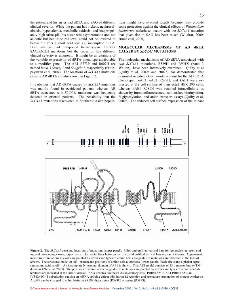
the patient and his sister had dRTA and SAO of different
clinical severity. While the patient had rickets, nephrocal-
cinosis, hypokalemia, metabolic acidosis, and inappropri-
ately high urine pH, his sister was asymptomatic and not
acidotic but her urine pH level could not be lowered to
below 5.5 after a short acid load i.e. incomplete dRTA.
Both siblings had compound heterozygous SLC4A1
SAO/R602H mutations but the cause of this different
clinical severity is unknown. It might be an example of
the variable expressivity of dRTA phenotype attributable
to a modifier gene. The AE1 S773P and R602H are
named band 3 Siriraj I and Songkla I, respectively (Sritip-
payawan et al, 2004). The locations of SLC4A1 mutations
causing AR dRTA are also shown in Figure 2.
It is obvious that AD dRTA caused by SLC4A1 mutations
was mainly found in occidental patients whereas AR
dRTA associated with SLC4A1 mutations was frequently
detected in oriental patients. The possibility that the
SLC4A1 mutations discovered in Southeast Asian popula-
tions might have evolved locally because they provide
some protection against the clinical effects of Plasmodium
falciparum malaria as occurs with the SLC4A1 mutation
that gives rise to SAO has been raised (Wilairat, 2000;
Bruce et al, 2000).
MOLECULAR MECHANISMS OF AD dRTA
CAUSED BY SLC4A1 MUTATIONS
The molecular mechanisms of AD dRTA associated with
two SLC4A1 mutations, R589H and R901X (band 3
Walton), have been intensively examined. Quilty et al
(Quilty et al, 2002a and 2002b) has demonstrated that
dominant negative effect would account for the AD dRTA
phenotype. eAE1, eAE1 R589H, and kAE1 were ex-
pressed at the cell surface of transfected HEK 293 cells,
whereas kAE1 R589H was retained intracellularly as
shown by immunofluorescence, cell surface biotinylation,
N-glycosylation, and anion transport assays (Quilty et al,
2002a). The reduced cell surface expression of the mutant
Figure 2. The SLC4A1 gene and locations of mutations (upper panel). Filled and unfilled vertical bars (or rectangle) represent cod-
ing and non-coding exons, respectively. Horizontal lines between the filled and unfilled vertical bars represent introns. Approximate
locations of mutations in exons are pointed by arrows and types of amino acid change due to mutations are indicated at the tails of
arrows. The structural model of AE1 protein and positions of amino acid alterations (lower panel). Each circle and alphabet repre-
sent amino acid in AE1. An incomplete N-terminal domain of AE1 is shown. This AE1 model consists of 13 transmembrane (TM) domains (Zhu et al, 2003). The positions of amino acid change due to mutations are pointed by arrows and types of amino acid al-
terations are indicated at the tails of arrows. SAO denotes Southeast Asian ovalocytosis. PRIBRAM is AE1 PRIBRAM (an
IVS12+1G>T substitution causing an mRNA splicing defect with intron 12 retention and premature termination of protein synthesis),
Arg589 can be changed to either histidine (R589H), cysteine (R589C) or serine (R589S).

© Yenchitsomanus et al | Journal of Molecular and Genetic Medicine | December 2005 | Vol 1, No 2 | 49-62 | OPEN ACCESS
57
kAE1 was likely due to its retention in the endoplasmic
reticulum (ER). Co-expression of kAE1 R589H reduced
the cell surface expression of wild-type kAE1 and eAE1
due to heterodimer formation and a dominant-negative
effect. The mutant eAE1 and kAE1 were not grossly mis-
folded as they could bind to an inhibitor affinity resin.
Other SLC4A1 mutations at the same position (R589C and
R589S) also prevented the targeting of kAE1 to the cell
surface, indicating that the normal Arg589, which is evolu-
tionary conserved, is important for proper trafficking. In
studies using transfected and virally infected MDCK cells
originating from principal cells, two groups have similarly
observed that the mutant kAE1 R589H and kAE1 S613F
retain in ER of the non-polarized cells and also that they
distribute throughout the cells with a failure to reach the
cell surface in the polarized cells (Toye et al, 2004; Cordat
et al, 2005). A co-expression study in MDCK cells showed
that the kAE1 R589H similarly retained the wild-type
kAE1 intracellularly, probably through heterodimer forma-
tion (Cordat et al, 2005).
The properties of kAE1 R901X expressed in Xenopus oo-
cytes, non-polarized MDCK, and HEK 293 cells have
been examined (Toye et al, 2002; Quilty et al, 2002b).
kAE1 R901X had a normal chloride transport activity
when expressed in Xenopus oocytes. While normal kAE1
was expressed at the cell surface in the cultured kidney
cell line, the kAE1 R901X was retained intracellularly
within the cultured cells, indicating that the C-terminal tail
of AE1, which is truncated in kAE1 R901X, is required for
its movement to the cell surface in kidney cells. It was
proposed (Toye et al, 2002) but not proved that kAE1
R901X gives rise to dominant dRTA by inhibiting the
movement of normal kAE1 to the cell surface, resulting
from the association of the normal and mutant proteins in
kAE1 hetero-oligomers. It was truly proved by Quilty et
al (Quilty et al, 2002b) that this dominant-negative effect
was due to hetero-oligomer formation of the mutant and
wild-type proteins, because co-expression of the wild-type
and mutant kAE1, or eAE1 R901X, indeed resulted in
intracellular retention of the wild-type proteins in a pre-
medial Golgi compartment in the expression experiments
using transiently transfected HEK 293 cells. A series of
truncations at the C-terminus of AE1 also resulted in im-
paired ER exit, depending on the extent of the truncation
(Cordat et al, 2003). Intracellular retention of kAE1 in the
renal α-intercalated cells by the dRTA mutant would ac-
count for the impaired bicarbonate extrusion to inter-
stitium and acid secretion into the urine that is characteris-
tic of dRTA.
The distribution of epitope-tagged full-length wild-type
and AE1 R901X was examined in polarized MDCK cells
and rat IMCD cells (which express AE1) (Devonald et al,
2003a). In both cell types, the wild-type AE1 localized to
the basolateral plasma membrane domain whereas AE1
R901X was found at both the basolateral and apical cell
surfaces as well as intracellularly, suggesting that AD
dRTA is caused not only by a decrease of AE1 protein at
the basolateral surface but also by its inappropriate pres-
ence at the apical surface. The apical chloride and bicar-
bonate flux would alter electrochemical balance across the
cell, impairing both unidirectional basolateral bicarbonate
extrusion and apical proton secretion. The non-polarized
distribution of mutant AE1 could be explained by the loss
of a basolateral targeting signal present in the deleted por-
tion of its C terminus. The motif YDEV (904–907) pre-
sent in the tail of AE1 conforms to a subset of tyrosine-
based targeting motifs, YXXØ. When Tyr904 was mu-
tated to alanine (Y904A), the distribution of protein was
similar to that of AE1 R901X. YXXØ motifs interact with
µ subunits of adaptor-protein complexes, one of which,
AP-1B, is specific to polarized epithelial cells. Whether
sorting of AE1 depends on AP-1B or not was investigated
in the proximal renal tubular cell line, LLC-PK1, which
was reported to lack the µ1B subunit and thus cannot form
AP-1B complexes. The results suggested that an adaptor
protein other than AP-1B is involved in basolateral sorting
of AE1 (Devonald et al, 2003a).
The mis-targeting to the apical membrane of kAE1 R901X
in stably transfected polarized MDCKI cells was also con-
firmed in a subsequent study, and it was similarly demon-
strated that the 11 C-terminal residues of kAE1 containing
a tyrosine-dependent basolateral targeting signal was not
recognized by µ1B-containing AP-1 adaptor complexes
(Toye et al, 2004). In addition, in the absence of the N-
terminus of kAE1, the C-terminus was not sufficient to
localize kAE1 to the basolateral membrane, suggesting
that a determinant within the kAE1 N-terminus co-
operates with the C-terminus for kAE1 basolateral local-
ization (Toye et al, 2004 and 2005).
A missense SLC4A1 mutation, glycine 609 to arginine
(G609R), giving rise to AD dRTA was reported in an ex-
tended Caucasian family (Rungroj et al, 2004). The kAE1
G609R expressed on the cell surface of Xenopus oocytes
maintained normal anion transport function. In contrast to
the normal basolateral localization of wild-type kAE1, the
mutant kAE1 G609R was partailly located at the apical
and sub-apical membrane, and also at the basolateral
membrane in polarized MDCK cells, similar to the kAE1
R901X. Gly609 may play a role in kAE1 protein traffick-
ing. Since this residue is close to Arg589 and Ser613
where their missense mutations cause AD dRTA, this re-
gion of AE1 may be involved in the protein trafficking or
sorting process (Rungroj et al, 2004). The kAE1 G609R
has not been examined for a dominant-negative effect,
which might explain the dominant dRTA phenotype. In
analogy to the kAE1 R901X, kAE1 G609R, which was
similarly mis-targeted in the polarized MDCK cells, may
also intracellularly retain the wild-type kAE1 protein.
MOLECULAR MECHANISMS OF AR dRTA
CAUSED BY SLC4A1 MUTATIONS
Based on the previous study of kAE1 G701D in Xenopus
oocyte (Tanphaicitr et al, 1998; Bruce et al, 2000) showing
functional mutant protein with defective trafficking to the
cell surface, we propose two possible models for the mo-
lecular mechanism of AR dRTA caused by SLC4A1 muta-
tions. Firstly, in the heterozygous state, the mutant kAE1
per se is unable to form homo- and heterodimers and so
would be unable to traffic to the cell surface. Thus, only
the mutant kAE1 is defective, while the wild-type kAE1 is
still intact. The wild-type kAE1 would be able to dimerize

© Yenchitsomanus et al | Journal of Molecular and Genetic Medicine | December 2005 | Vol 1, No 2 | 49-62 | OPEN ACCESS
58
and traffic to the cell surface, and sufficiently maintain the
normal transport function. And, secondly, in the het-
erozygous state, the mutant kAE1 might be able to form
homo- and heterodimers, but only the homodimer would
be defective in trafficking to the cell surface. In the sec-
ond model, the heterodimer of the mutant and wild-type
kAE1 proteins would be able to traffic to the cell surface
and would not exhibit the dominant-negative effect, as
observed in AD dRTA.
Since a novel compound heterozygous SLC4A1
G701D/S773P mutation was recently reported by our
group in the Thai patient with AR dRTA (Sritippayawan et
al, 2004), the S773 and G701D mutations were then used
as examples for the study in order to elucidate the molecu-
lar mechanism of AR dRTA caused by SLC4A1 mutations,
and to examine the two proposed models. The biosynthe-
sis and trafficking of kAE1 S773P were studied in tran-
siently transfected HEK 293 cells, expressing the mutant
alone or in combination with wild-type kAE1 or kAE1
G701D (Kittanakom et al, 2004). It was found that kAE1
S773P was expressed at only a third the level of the wild-
type kAE1, had a two-fold decrease in its half-life, and
was targeted for degradation by the proteasome. The mu-
tant protein could not be detected at the plasma membrane
of transfected HEK 293 cells and showed predominant
immunolocalization in the ER of both HEK 293 and LLC-
PK1 cells.
The oligosaccharide on a kAE1 S773P N-glycosylation
mutant (N555) was not processed to the complex form
indicating impaired exit from the ER. The kAE1 S773P
Figure 3. The molecular and cellular model for dominant and recessive dRTA in polarized epithelial cells. Schematic model of
epithelial cells expressing wild-type kAE1, dominant or recessive mutants in homozygous (left model) or heterozygous state (right
model). Dimers of wild-type kAE1 (burgundy ovals) traffic to the basolateral membrane (solid line) while dominant dRTA mutants
(brown ovals) are retained in the ER. Recessive dRTA mutants (green ovals) are partially impaired (dotted lines) in their exit from
the ER but can either traffic to the basolateral membrane or are retained in the Golgi apparatus. Heterodimers of wild-type kAE1 and dominant kAE1 mutant are retained in the ER, while heterodimers of wild-type kAE1 and recessive kAE1 mutants can traffic to the
basolateral membrane. Theoretically, the proportions of wild-type kAE1 homodimer, heterodimer, and mutant kAE1 homodimer are
25%, 50% and 25%, respectively. Therefore, 25% of kAE1 in the dominant model and 75% of kAE1 in the recessive dRTA model
traffic to the plasma membrane. TJ denotes tight junction.

© Yenchitsomanus et al | Journal of Molecular and Genetic Medicine | December 2005 | Vol 1, No 2 | 49-62 | OPEN ACCESS
59
was not properly folded as it showed decreased binding to
an inhibitor affinity resin and increased sensitivity to pro-
teases. The kAE1 G701D also exhibited defective traffick-
ing to the plasma membrane. The kAE1 S773P was able
to form homodimers and heterodimers with wild-type
kAE1 or with kAE1 G701D. Heterodimers of wild-type
kAE1 with kAE1 S773P or G701D, in contrast to the
dominant mutant kAE1 R589H, were delivered to the
plasma membrane (Figure 3). Thus, the second proposed
model is likely to be the true one, and the wild-type kAE1
seems to show a ‘dominant-positive effect’ in rescuing the
recessive mutant kAE1 trafficking to the plasma mem-
brane, in contrast with the dominant mutant kAE1 result-
ing in a ‘dominant-negative effect’ when heterodimerized
with the wild-type kAE1.
In our recent study in MDCK cells (Cordat et al, 2005), we
found that the recessive mutants, kAE1 S773P and
G701D, showed distinct trafficking defects. The mis-
folded recessive mutant kAE1 S773P, while largely re-
tained in the ER in non-polarized MDCK cells, was pre-
dominantly targeted to the basolateral membrane in polar-
ized cells while kAE1 G701D was retained in the Golgi in
both non-polarized and polarized cells. The co-expression
study in MDCK cells confirmed the previous study in
HEK 293 cells that the dominant mutant kAE1 R589H
retained wild-type kAE1 protein intracellularly while the
recessive kAE1 mutants did not. The co-expression of
kAE1 S773P and G701D in the same cells showed some
co-localization of S773P with G701D in the Golgi, but
kAE1 S773P could still traffic to the basolateral mem-
brane. Also, no kAE1 G701D was detected at the cell
surface, suggesting that kAE1 S773P did not assist the
intracellularly retained kAE1 G701D to traffic to the cell
surface as did the wild-type kAE1, despite their ability to
oligomerize in the MDCK cells. This result suggests that
in the patients with compound heterozygous SLC4A1
G701D/S773P mutations, only the mis-folded
S773P/S773P homodimers which may not properly func-
tion can reach the basolateral membrane of the kidney α-
intercalated cells, resulting in the development of dRTA.
CONCLUSIONS
The molecular mechanisms of AD and AR dRTA caused
by SLC4A1 mutations have now been elucidated (Figure
3). The modes of inheritance of dRTA associated SLC4A1
mutations are primarily dictated by the position of amino
acid alterations in the encoded kAE1 protein and on some
occasions by the type of amino acid replacement, affecting
kAE1 folding and molecular structure without signifi-
cantly changing anion exchange function. The structural
alterations of mutated kAE1 in both phenotypic conditions
do not generally interfere with its dimerization as homo-
and heterodimers. However, they affect intracellular traf-
ficking of the mutant kAE1 homodimers from the ER and
trans-Golgi network to the plasma membrane. Between
two extremes of normally trafficking of wild-type-kAE1
homodimer and abnormally trafficking of mutant-kAE1
homodimer, trafficking ability of the heterodimer between
wild-type and mutant kAE1 in heterozygous conditions
exhibits a key role in determining the dominant or reces-
sive nature of the phenotype. In AD dRTA, mutant kAE1
in the heterodimer induces a trafficking defect of wild-type
kAE1, so called the ‘dominant-negative effect’, leading to
the AD dRTA phenotype. By contrast, in AR dRTA,
wild-type kAE1 in the heterodimer corrects the trafficking
defect of mutant kAE1, a newly described ‘dominant-
positive effect’, producing the AR dRTA phenotype. In
the case of AD dRTA, the mutant (R589H, S613F, and
R901X) homodimers are retained in the ER, as are het-
erodimers with the wild-type kAE1. The retention of the
wild-type protein by the heterodimer formation does not
allow sufficient kAE1 to be localized to the basolateral
membrane. In some cases (R901X and G609R), the
dominant mutants can exit the ER and are partially mis-
sorted to the apical membrane, which would also impair
the basolateral bicarbonate reabsorption and apical acid
secretion of the α-intercalated cells. In the case of AR
dRTA, the mutant homodimers can traffic to the Golgi
(G701D) or the basolateral membrane (S773P), but since
the latter protein is mis-folded, no transport activity is
achieved. Heterodimers of AR dRTA mutants can form
with the wild-type kAE1, but in this case can traffic to the
basolateral membrane. The wild-type homodimers that
form can traffic normally to the basolateral membrane. In
the heterozygous state, sufficient wild-type kAE1, either
as a homodimer or as a heterodimer with an AR dRTA
mutant, would be present to maintain sufficient bicarbon-
ate reabsorption and acid secretion.
All these findings may serve as an example in the elucida-
tion of molecular mechanisms of AD and AR phenotypes
of the same disease caused by defects of a common gene;
to our knowledge this is the first time that the phenotypes
of the two different modes of inheritance of the same dis-
ease are clearly explained in term of the biochemical prop-
erties of the homodimers and heterodimers of the encoded
wild-type and mutant proteins.
ACKNOWLEDGEMENTS
The authors thank the Thailand National Center for Ge-
netic Engineering and Biotechnology (BIOTEC) under the
National Science and Technology Development Agency
(NSTDA), the Thailand Research Fund (TRF), and the
Canadian Institutes of Health Research (CIHR) for sup-
porting this work. E.C. and S.K. are post-doctoral fellows
supported by CIHR. S.K. and N.R. were formerly sup-
ported by the TRF-Royal Golden Jubilee PhD Scholarship.
Dr Prida Malasit and Professor Prapon Wilairat, the re-
cipients of TRF-Senior Research Scholar Award, are
thanked for their supports to PY.
STATEMENT OF COMPETING INTERESTS
The authors declared no competing interests. LIST OF ABBREVIATIONS
AD; Autosomal dominant
AE; Anion exchanger
AP; Activator protein or adaptor protein
AR; Autosomal recessive
CA; Carbonic anhydrase
DIDS; 4, 4’-diisothiocyanostilbene-2-2’, disulfonic acid
© Yenchitsomanus et al | Journal of Molecular and Genetic Medicine | December 2005 | Vol 1, No 2 | 49-62 | OPEN ACCESS
60
dRTA; Distal renal tubular acidosis
ER; Endoplasmic reticulum
GPA; Glycophorin A
HS; Hereditary spherocytosis
HEK; Human embryonic kidney
IMCD; Inner medullary collecting duct
LLC-PK1; Renal epithelial cell line from porcine kidneys
MDCK; Madin-Darby canine kidney cells
PNG; Papua New Guinea
SAO; Southeast Asian ovalocytosis
SLC4A1; Solute carrier family 4, member 1
TM; Transmembrane
UTR; Untranslated region
REFERENCES
Allen SJ, O'Donnell A, Alexander ND et al. 1999. Prevention of
cerebral malaria in children in Papua New Guinea by Southeast
Asian ovalocytosis band 3. Am J Trop Med Hyg, 60, 1056-1060.
Alloisio N, Texier P, Vallier A et al. 1997. Modulation of clinical
expression and band 3 deficiency in hereditary spherocytosis.
Blood, 90, 414-420.
Alper SL. 2002. Genetic diseases of acid-base transporters. Annu
Rev Physiol, 64, 899-923.
Alper SL, Natale J, Gluck S, Lodish HF and Brown D. 1989.
Subtypes of intercalated cells in rat kidney collecting duct de-
fined by antibodies against erythroid band 3 and renal vacuolar
H+-ATPase. Proc Natl Acad Sci USA, 86,5429-533.
Batlle D, Ghanekar H, Jain S and Mitra A. 2001. Hereditary dis-
tal renal tubular acidosis: new understandings. Annu Rev Med,
52,471-484.
Bonifacino JS and Dell'Angelica EC. 1999. Molecular bases for
the recognition of tyrosine-based sorting signals. J Cell Biol,
145, 923-926.
Bonifacino JS and Traub LM. 2003. Signals for sorting of trans-
membrane proteins to endosomes and lysosomes. Annu Rev
Biochem, 72, 395-447.
Bruce LJ, Groves JD, Okubo Y, Thilaganathan B and Tanner MJ.
1994. Altered band 3 structure and function in glycophorin A-
and B-deficient (MkMk) red blood cells. Blood, 84, 916-922.
Bruce LJ and Tanner MJ. 1996. Structure-function relationships of
band 3 variants. Cell Mol Biol (Noisy-le-grand), 42, 953-973.
Bruce LJ, Cope DL, Jones GK et al. 1997. Familial distal renal tubu-
lar acidosis is associated with mutations in the red cell anion ex-
changer (Band 3, AE1) gene. J Clin Invest, 100, 1693-1707.
Bruce LJ and Tanner MJ. 1999. Erythroid band 3 variants and
disease. Baillieres Best Pract Res Clin Haematol, 12, 637-654.
Bruce LJ, Wrong O, Toye AM et al. 2000. Band 3 mutations,
renal tubular acidosis and South-East Asian ovalocytosis in Ma-
laysia and Papua New Guinea: loss of up to 95% band 3 trans-
port in red cells. Biochem J, 350, 41-51.
Campanella ME, Chu H and Low PS. 2005. Assembly and regu-
lation of a glycolytic enzyme complex on the human erythro-
cyte membrane. Proc Natl Acad Sci USA, 102, 2402-2407.
Canfield WM, Johnson KF, Ye RD, Gregory W and Kornfeld S.
1991. Localization of the signal for rapid internalization of the
bovine cation-independent mannose 6-phosphate/insulin-like
growth factor-II receptor to amino acids 24-29 of the cytoplas-
mic tail. J Biol Chem, 266, 5682-5688.
Cheidde L, Vieira TC, Lima PR, Saad ST and Heilberg IP. 2003.
A novel mutation in the anion exchanger 1 gene is associated
with familial distal renal tubular acidosis and nephrocalcinosis.
Pediatrics, 112, 1361-1367.
Cheung JC and Reithmeier RA. 2005. Membrane integration and
topology of the first transmembrane segment in normal and
Southeast Asian ovalocytosis human erythrocyte anion ex-
changer 1. Mol Memb Biol, 22, 203-214.
Cheung JC, Cordat E and Reithmeier RA. 2005. Trafficking
defects of the Southeast Asian ovalocytosis deletion mutant of
anion exchanger 1 membrane proteins. Biochem J (in press).
Cordat E, Li J and Reithmeier RA. 2003. Carboxyl-terminal
truncations of human anion exchanger impair its trafficking to
the plasma membrane. Traffic, 4, 642-651.
Cordat E, Kittanakom S, Yenchitsomanus P et al. 2005. Domi-nant and recessive distal renal tubular acidosis mutations of
AE1 induce distinct trafficking defects in MDCK cells. Traffic
(revised).
Devonald MA, Smith AN, Poon JP, Ihrke G and Karet FE.
2003a. Non-polarized targeting of AE1 causes autosomal domi-
nant distal renal tubular acidosis. Nat Genet, 33, 125-127.
Devonald MA, Rungroj N and Karet FE. 2003b. Molecular analysis
of basolateral targeting region in the c-terminal cytosolic domain of
AE1. J Am Soc Nephrol, 14, Abstracts Issue, 560A.
Dhermy D, Burnier O, Bourgeois M and Grandchamp B. 1999.
The red blood cell band 3 variant (band 3 Biceetrel: R490C) associated with dominant hereditary spherocytosis causes de-
fective membrane targeting of the molecule and a dominant
negative effect. Mol Membr Biol, 16, 305-312.
Distel B, Bauer U, Le Borgne R and Hoflack B. 1998. Baso-
lateral sorting of the cation-dependent mannose 6-phosphate
receptor in Madin-Darby canine kidney cells. Identification of a basolateral determinant unrelated to clathrin-coated pit localiza-
tion signals. J Biol Chem, 273, 186-193.
Eber SW, Gonzalez JM, Lux ML et al. 1996. Ankyrin-1 muta-
tions are a major cause of dominant and recessive hereditary
spherocytosis. Nat Genet, 13, 214-218.
Folsch H, Ohno H, Bonifacino JS and Mellman I. 1999. A novel clathrin adaptor complex mediates basolateral targeting in po-
larized epithelial cells. Cell, 99, 189-198.
Groves JD and Tanner MJ. 1999. Topology studies with biosyn-
thetic fragments identify interacting transmembrane regions of
the human red-cell anion exchanger (band 3; AE1). Biochem J, 344, 687-697.
Herrin J. 2003. Renal tubular acidosis, Lippincott William &
Wilkins, Philadelphia.
Iolascon A, Miraglia del Giudice E, Perrotta S, Alloisio N, Morle
L and Delaunay J. 1998. Hereditary spherocytosis: from clinical
to molecular defects. Haematologica, 83, 240-257. Jarolim P, Palek J, Amato D et al. 1991. Deletion in erythrocyte
band 3 gene in malaria-resistant Southeast Asian ovalocytosis.
Proc Natl Acad Sci U S A, 88, 11022-11026.
Jarolim P, Palek J, Rubin HL, Prchal JT, Korsgren C and Cohen
CM. 1992. Band 3 Tuscaloosa: Pro327----Arg327 substitution
in the cytoplasmic domain of erythrocyte band 3 protein associ-
ated with spherocytic hemolytic anemia and partial deficiency
of protein 4.2. Blood, 80, 523-529.
Jarolim P, Murray JL, Rubin HL, et al. 1996. Characterization of
13 novel band 3 gene defects in hereditary spherocytosis with
band 3 deficiency. Blood, 88, 4366-4374.
Jarolim P, Shayakul C, Prabakaran D et al. 1998. Autosomal domi-
nant distal renal tubular acidosis is associated in three families with
heterozygosity for the R589H mutation in the AE1 (band 3) Cl-
/HCO3- exchanger. J Biol Chem, 273, 6380-6388.
Jennings ML. 1984. Oligomeric structure and the anion transport
function of human erythrocyte band 3 protein. J Membr Biol,
80, 105-117.
Karet FE, Gainza FJ, Gyory AZ et al. 1998. Mutations in the
chloride-bicarbonate exchanger gene AE1 cause autosomal
dominant but not autosomal recessive distal renal tubular acido-
sis. Proc Natl Acad Sci USA, 95, 6337-6342.
Karet FE, Finberg KE, Nelson RD et al. 1999. Mutations in the
gene encoding B1 subunit of H+-ATPase cause renal tubular
acidosis with sensorineural deafness. Nat Genet, 21, 84-90.
Kawano Y, Okubo K, Tokunaga F, Miyata T, Iwanaga S and
Hamasaki N. 1988. Localization of the pyridoxal phosphate
binding site at the COOH-terminal region of erythrocyte band 3
protein. J Biol Chem, 263, 8232-8238.

© Yenchitsomanus et al | Journal of Molecular and Genetic Medicine | December 2005 | Vol 1, No 2 | 49-62 | OPEN ACCESS
61
Kittanakom S, Cordat E, Akkarapatumwong V, Yenchitsomanus
P and Reithmeier RA. 2004. Trafficking defects of a novel
autosomal recessive distal renal tubular acidosis mutant
(S773P) of the human kidney anion exchanger (kAE1). J Biol
Chem, 279, 40960-40971.
Kollert-Jöns A, Wagner S, Hubner S, Appelhans H and Drenck-
hahn D. 1993. Anion exchanger 1 in human kidney and oncocy-
toma differs from erythroid AE1 in its NH2 terminus. Am J
Physiol, 265, F813-F821.
Lin S, Naim HY and Roth MG. 1997. Tyrosine-dependent baso-
lateral sorting signals are distinct from tyrosine-dependent in-
ternalization signals. J Biol Chem, 272, 26300-26305.
Liu SC, Jarolim P, Rubin HL et al. 1994. The homozygous state
for the band 3 protein mutation in Southeast Asian Ovalocyto-
sis may be lethal. Blood, 84, 3590-3591.
Liu SC, Palek J, Yi SJ et al. 1995. Molecular basis of altered red
blood cell membrane properties in Southeast Asian ovalocyto-
sis: role of the mutant band 3 protein in band 3 oligomerization
and retention by the membrane skeleton. Blood, 86, 349-358.
Lux SE, John KM, Kopito RR and Lodish HF. 1989. Cloning and
characterization of band 3, the human erythrocyte anion- exchange
protein (AE1). Proc Natl Acad Sci USA, 86, 9089-9093.
Matter K and Mellman I. 1994. Mechanisms of cell polarity:
sorting and transport in epithelial cells. Curr Opin Cell Biol, 6,
545-554.
Mgone CS, Koki G, Paniu MM et al. 1996. Occurrence of the
erythrocyte band 3 (AE1) gene deletion in relation to malaria
endemicity in Papua New Guinea. Trans R Soc Trop Med Hyg,
90, 228-231.
Nurse GT, Coetzer TL and Palek J. 1992. The elliptocytoses,
ovalocytosis and related disorders. Baillieres Clin Haematol, 5,
187-207.
Ohno H, Stewart J, Fournier MC et al. 1995. Interaction of tyro-
sine-based sorting signals with clathrin-associated proteins. Sci-
ence, 269, 1872-1875.
Ohno H, Tomemori T, Nakatsu F et al. 1999. Mu1B, a novel
adaptor medium chain expressed in polarized epithelial cells.
FEBS Lett, 449, 215-220.
Okubo K, Kang D, Hamasaki N and Jennings ML. 1994. Red
blood cell band 3. Lysine 539 and lysine 851 react with the
same H2DIDS (4,4'-diisothiocyanodihydrostilbene-2,2'-
disulfonic acid) molecule. J Biol Chem, 269, 1918-1926.
Peters LL, Shivdasani RA, Liu SC et al. 1996. Anion exchanger
1 (band 3) is required to prevent erythrocyte membrane surface
loss but not to form the membrane skeleton. Cell, 86, 917-927.
Quilty JA and Reithmeier RA. 2000. Trafficking and folding
defects in hereditary spherocytosis mutants of the human red
cell anion exchanger. Traffic, 12, 987-998.
Quilty JA, Li J and Reithmeier RA. 2002a. Impaired trafficking of
distal renal tubular acidosis mutants of the human kidney anion ex-
changer kAE1. Am J Physiol Renal Physiol, 282, F810-820.
Quilty JA, Cordat E and Reithmeier RA. 2002b. Impaired trafficking
of human kidney anion exchanger (kAE1) caused by hetero-
oligomer formation with a truncated mutant associated with distal
renal tubular acidosis. Biochem J, 368(Pt 3), 895-903.
Reithmeier RAF. 2001. A membrane metabolon linking carbonic
anhydrase with chloride/bicarbonate anion exchangers. Blood
Cells Mol Dis, 27, 85-89.
Ribeiro ML, Alloisio N, Almeida H et al. 2000. Severe heredi-
tary spherocytosis and distal renal tubular acidosis associated
with the total absence of band 3. Blood, 96, 1602-1604.
Ruf R, Rensing C, Topaloglu R et al. 2003. Confirmation of the
ATP6B1 gene as responsible for distal renal tubular acidosis.
Pediatr Nephrol, 18, 105-109.
Rungroj N, Devonald MA, Cuthbert AW et al. 2004. A novel
missense mutation in AE1 causing autosomal dominant distal
renal tubular acidosis retains normal transport function but is
mistargeted in polarized epithelial cells. J Biol Chem,
279,13833-13838.
Rybicki AC, Qiu JJ, Musto S, Rosen NL, Nagel RL and
Schwartz RS. 1993. Human erythrocyte protein 4.2 deficiency
associated with hemolytic anemia and a homozygous 40 glu-
tamic acid-->lysine substitution in the cytoplasmic domain of
band 3 (band 3 Montefiore). Blood, 81, 2155-2165.
Rysava R, Tesar V, Jirsa M, Jr., Brabec V and Jarolim P. 1997.
Incomplete distal renal tubular acidosis coinherited with a mu-
tation in the band 3 (AE1) gene. Nephrol. Dial. Transplant, 12,
1869-1873.
Sahr KE, Taylor WM, Daniels BP, Rubin HL and Jarolim P.
1994. The structure and organization of the human erythroid
anion exchanger (AE1) gene. Genomics, 24, 491-501.
Schofield AE, Martin PG, Spillett D and Tanner MJ. 1994. The
structure of the human red blood cell anion exchanger (EPB3,
AE1, band 3) gene. Blood, 84, 2000-2012.
Smith AN, Skaug J, Choate KA et al. 2000. Mutations in
ATP6N1B, encoding a new kidney vacuolar proton pump 116-
kD subunit, cause recessive distal renal tubular acidosis with
preserved hearing. Nat Genet, 26, 71-75.
Sritippayawan S, Kirdpon S, Vasuvattakul S et al. 2003. A de
novo R589C mutation of anion exchanger 1 causing distal renal
tubular acidosis. Pediatr Nephrol, 18, 644-648.
Sritippayawan S, Sumboonnanonda A, Vasuvattakul S et al.
2004. Novel compound heterozygous SLC4A1 mutations in
Thai patients with autosomal recessive distal renal tubular aci-
dosis. Am J Kidney Dis, 44, 64-70.
Sterling D and Casey JR. 2002. Bicarbonate transport proteins.
Biochem Cell Biol, 80, 483-497.
Sterling D, Reithmeier RA and Casey JR. 2001. A transport me-
tabolon. Functional interaction of carbonic anhydrase II and chlo-
ride/bicarbonate exchangers. J Biol Chem, 276, 47886-47894.
Stover EH, Borthwick KJ, Bavalia C, et al. 2002. Novel
ATP6V1B1 and ATP6V0A4 mutations in autosomal recessive
distal renal tubular acidosis with new evidence for hearing loss.
J Med Genet, 39,796-803.
Tanner MJ, Martin PG and High S. 1988. The complete amino
acid sequence of the human erythrocyte membrane anion-
transport protein deduced from the cDNA sequence. Biochem J,
256, 703-712.
Tanner MJA. 1997. The structure and function of band 3 (AE1):
recent developments (review). Mol Membr Biol, 14, 155-165.
Tanphaichitr VS, Sumboonnanonda A, Ideguchi H et al. 1998.
Novel AE1 mutations in recessive distal renal tubular acidosis.
Loss-of- function is rescued by glycophorin A. J Clin Invest,
102, 2173-2179.
Toye AM, Bruce LJ, Unwin RJ, Wrong O and Tanner MJ. 2002.
Band 3 Walton, a C-terminal deletion associated with distal re-
nal tubular acidosis, is expressed in the red cell membrane but
retained internally in kidney cells. Blood, 99, 342-347.
Toye AM, Banting G and Tanner MJ. 2004. Regions of human kid-
ney anion exchanger 1 (kAE1) required for basolateral targeting of
kAE1 in polarised kidney cells: mis-targeting explains dominant
renal tubular acidosis (dRTA). J Cell Sci, 117, 1399-1410.
Toye AM, Ghosh S, Young MT et al. 2005. Protein-4.2 associa-
tion with band 3 (AE1, SLCA4) in Xenopus oocytes: effects of
three natural protein-4.2 mutations associated with hemolytic
anemia. Blood, 105,4088-4095.
Tse WT and Lux SE. 1999. Red blood cell membrane disorders.
Br J Haematol, 104, 2-13.
Vasuvattakul S, Yenchitsomanus PT, Vachuanichsanong P et al.
1999. Autosomal recessive distal renal tubular acidosis associated
with Southeast Asian ovalocytosis. Kidney Int, 56, 1674-1682.
Vince JW and Reithmeier RAF. 1998. Carbonic anhydrase II
binds to the carboxyl terminus of human band 3, the erythro-
cyte Cl-/HCO3- exchanger. J Biol Chem, 273, 28430-28437.
Vince JW, Carlsson U and Reithmeier RAF. 2000. Localization
of the Cl-/HCO3- anion exchanger binding site to the amino-
terminal region of carbonic anhydrase II. Biochemistry, 39,
13344-13349.

© Yenchitsomanus et al | Journal of Molecular and Genetic Medicine | December 2005 | Vol 1, No 2 | 49-62 | OPEN ACCESS
62
Wang CC, Moriyama R, Lombardo CR and Low PS. 1995. Par-
tial characterization of the cytoplasmic domain of human kid-
ney band 3. J Biol Chem, 270, 17892-17897.
Weber S, Soergel M, Jeck N and Konrad M. 2000. Atypical dis-
tal renal tubular acidosis confirmed by mutation analysis. Pedi-
atr Nephrol, 15, 201-204.
Wilairat P. 2000. Renal tubular acidosis, anion exchanger 1 (AE1) mutations, Southeast Asian ovalocytosis: a malaria con-
nection? FAOPS Newsletter, 9, 3-8.
Wrong O, Bruce LJ, Unwin RJ, Toye AM and Tanner MJ. 2002.
Band 3 mutations, distal renal tubular acidosis, and Southeast
Asian ovalocytosis. Kidney Int, 62, 10-19.
Yenchitsomanus PT, Vasuvattakul S, Kirdpon S et al. 2002.
Autosomal recessive distal renal tubular acidosis caused by
G701D mutation of anion exchanger 1 gene. Am J Kidney Dis,
40, 21-29.
Yenchitsomanus P, Sawasdee N, Paemanee A et al. 2003. Anion
exchanger 1 mutations associated with distal renal tubular aci-dosis in the Thai population. J Hum Genet, 48, 451-456.
Yenchitsomanus P. 2003. Human anion exchanger1 mutations
and distal renal tubular acidosis. Southeast Asian J Trop Med
Public Health, 34, 651-658.
Zhang D, Kiyatkin A, Bolin JT and Low PS. 2000. Crystallographic
structure and functional interpretation of the cytoplasmic domain of erythrocyte membrane band 3. Blood, 96, 2925-2933.
Zhu Q, Lee DW and Casey JR. 2003. Novel topology in C-
terminal region of the human plasma membrane anion ex-
changer, AE1. J Biol Chem, 278, 3112-3120.
SHORT COPYRIGHT STATEMENT
This is an open access article, published under the terms of the
Licence for Users available at http://www.libpubmedia.co.uk/
MedJ/LicenceForUsers.pdf. This licence permits non-commercial use, distribution and reproduction of the article, pro-
vided the original work is appropriately acknowledged with cor-
rect citation details.