Embed Size (px)
Citation preview

Molecular Dynamics/Order Parameter Extrapolation
for Bionanosystem Simulations
YINGLONG MIAO, PETER J. ORTOLEVA
Center for Cell and Virus Theory, Department of Chemistry, Indiana University, Bloomington,Indiana 47405
Received 28 January 2008; Revised 30 April 2008; Accepted 5 June 2008DOI 10.1002/jcc.21071
Published online 17 July 2008 in Wiley InterScience (www.interscience.wiley.com).
Abstract: A multiscale approach, molecular dynamics/order parameter extrapolation (MD/OPX), to the all-atom
simulation of large bionanosystems is presented. The approach starts with the introduction of a set of order parame-
ters (OPs) automatically generated with orthogonal polynomials to characterize the nanoscale features of bionanosys-
tems. The OPs are shown to evolve slowly via Newton’s equations, and the all-atom multiscale analysis (AMA)
developed earlier (Miao and Ortoleva, J Chem Phys 2006, 125, 44901) demonstrates the existence of their stochastic
dynamics, which serve as the justification for our MD/OPX approach. In MD/OPX, a short MD run estimates the
rate of change of the OPs, which is then used to extrapolate the state of the system over time that is much longer
than the 10214 second timescale of fast atomic vibrations and collisions. The approach is implemented in NAMD
and demonstrated on cowpea chlorotic mottle virus (CCMV) capsid structural transitions (STs). It greatly accelerates
the MD code and its underlying all-atom description of the nanosystems enables the use of a universal interatomic
force field, avoiding recalibration with each new application as needed for coarse-grained models. The source code
of MD/OPX is distributed free of charge at https://simtk.org/home/mdopx and a web portal will be available via
http://sysbio.indiana.edu/virusx.
q 2008 Wiley Periodicals, Inc. J Comput Chem 30: 423–437, 2009
Key words: bionanosystems; CCMV; structural transitions; order parameters; all-atom multiscale analysis MD/OPX
Introduction
Bionanostructures are key elements of many systems of interest
in the pure and applied life sciences. Natural examples include
viruses, ribosomes, and other subcellular features. Engineered
bionanosystems include vaccines (i.e., diminished viruses), nano-
particles functionalized for medical imaging and thermal cancer
treatments, and nanocapsules for targeted delivery of therapeutic
agents. These systems are typically supramillion atoms in size.
They reside at the interface between the highly atomic fluctuat-
ing and the near-macroscopic deterministic regimes and their
atomic-scale detail is vital to their function. As a consequence,
they display dual atomistic/macroscopic behaviors on multiple
spatial and temporal scales, which must be understood simulta-
neously.
Our objective is to develop a methodology to simulate these
systems that builds in a strategy designed to address the special
characteristics of the multiple scale processes and the cross-talk
among them as follows. Atomic-scale fluctuations of nanosystems
provide the entropy involved in the thermal average forces driving
their nanoscale dynamics. Conversely, their overall transforma-
tions modify the statistics of the atomic-scale fluctuations.
Viruses present an excellent example of this interscale cross-
talk. They display a variety of structural dynamical phenomena
and, hence, are used to motivate this study. Many viruses
undergo STs, i.e., large conformational changes in their architec-
ture, in response to changes in their microenvironment condi-
tions. The latter include temperature, pH, ionic strength, and cat-
ion concentrations, as well as contact with a host cell membrane.
For example, native cowpea chlorotic mottle virus (CCMV) cap-
sid swells dramatically as the pH is increased from 5.0 to 7.5 in
the absence of divalent cations.1–5 Nudaurelia capensis x virus
(NxV)6–9 and HK97 bacteriophage10,11 undergo large structural
changes during capsid maturation. Poliovirus undergoes irrevers-
ible STs upon maturation or receptor-mediated cell entry.12–16
Such STs are believed to play significant roles in viral life-circle
events, such as virus attachment to cell membranes and genome
release from the viral capsid into a host cell. As characterized
quantitatively via electron cryomicroscopy (cryoEM) and X-ray
crystallography, these STs are typically accomplished through a
series of distinct steps involving intact translation and rotation
of protein structural units (e.g., protomers, pentameric, and hex-
americ capsomers), and those in poliovirus and NxV are trig-
gered by irreversible bond-cleavage of capsid proteins.
Correspondence to: P. J. Ortoleva; e-mail: [email protected] (http://
sysbio.indiana.edu)
q 2008 Wiley Periodicals, Inc.

In principle, many aspects of viral STs, assembly, and disas-
sembly can be understood in terms of the dynamics of a set of
atoms evolving classically through an interatomic force field.
The benefit of such an approach is that it enables a general, pa-
rameter-free, predictive nanosystem model. The challenge is to
implement this approach as a practical computational algorithm.
A central objective of the approach presented here is to capture
atomic-scale detail and yet simulate whole-virus dynamics.
Theoretical approaches have been developed to simulate viral
processes. These include coarse-grained models,17,18 symmetry-
constrained models,19 computational molecular dynamics
(MD),19–32 and normal mode analysis33–36 as reviewed ear-
lier.37,38 However, coarse-grained models miss the atomic detail
needed for interpreting drug and cell surface receptor interac-
tions and do not allow the universality afforded by an intera-
tomic force field. Normal mode analysis is based on single-well
(harmonic) potential approximations. It provides some insights
into viral STs but cannot be used to simulate STs, and the calcu-
lated vibrational normal modes cannot describe the diffusive
(friction-dominated) motion that dominates slow viral processes;
thus, it cannot address the very local and highly nonlinear nature
of the drug-virus interaction or the initiation and propagation of
a structural transition across a virus. Direct MD is limited to
biologically uninteresting timescales. For example, NAMD,39 a
high-performance parallel MD code, has been used to simulate a
complete satellite tobacco mosaic virus (STMV). The simulation
took 50 days on a SGI 1,024 processor Altrix system using 256
processors and 128 GB of memory but only captured a physical
time of 55 ns.20 However, viral STs take milliseconds to minutes
or longer.7 With such an approach, it would take about 2,500
years to obtain a physical result. Clearly, a speed-up of at least
a factor of 10,000 over MD is needed to make all-atom simula-
tion a viable approach. In summary, previous approaches do not
simultaneously capture atomic-scale detail and whole-virus,
long-time behaviors in a nonsymmetry-constrained fashion, or
account for the highly nonlinear and the often symmetry-broken
nature of many key bionanosystem phenomena. The challenge
remains to develop methods that preserve the atomic–nanoscale
cross-talk underlying the dynamics of bionanosystems.
Here, we present molecular dynamics/order parameter extrapo-
lation (MD/OPX), an approach for all-atom simulation of large
bionanosystems with the potential to overcome the shortcomings
of the methods noted earlier. MD/OPX is based on our all-atom
multiscale theory of nanosystem dynamics.37,38 It integrates the
following elements: (i) automated construction of order parame-
ters (OPs) that characterize the nanoscale structural features of
nanosystems, (ii) a demonstration based on Newton’s equations
that prove that the OPs evolve slowly relative to the 10214 s time-
scale of fast atomic vibrations and collisions, (iii) rigorous multi-
scale techniques that can be used to derive a stochastic (Fokker-
Planck or Smoluchowski) equation and equivalent Langevin equa-
tions of the dynamics of these OPs starting from the Liouville
equation, (iv) the use of eqs. (i) to (iii) to justify the suggestion of
the equation-free multiscale (EFM) analysis approach developed
by Kevrekidis et al.,40 that short bursts of MD simulations can be
used to extrapolate coarse variables (i.e. OPs) over large time
intervals and thus advance the nanostructure over long time, and
(v) an implementation of eqs. (i) to (iv) that is optimized and sta-
bilized via an adaptive dual timestep procedure and microscopic
configuration-corrected extrapolation using our automatically gen-
erated OPs. The MD/OPX approach is essentially equivalent to
solving the Langevin equations, except that the friction coeffi-
cients and thermal-average forces are evaluated implicitly.
In the following, we generate general OPs automatically for
bionanosystems in the following section, and we present MD/
OPX as an approach to solving the stochastic dynamics of these
OPs implicitly in the section on molecular dynamics/order pa-
rameter extrapolation and demonstrate MD/OPX on CCMV cap-
sid STs in the Application to CCMV Capsid Structural Transi-
tions section. Finally, the conclusions are drawn.
General Order Parameters for Bionanosystems
The order parameters (OPs) for bionanosystems considered in
this study have the following properties:
� capture overall nanoscale features of the nanosystems;
� vary much more slowly in time than the 10214 s timescale of
fast atomic vibrations and collisions;
� can be expressed in terms of the atomic positions and
momenta; and
� can be generated via a readily implemented, general procedure
capturing complex features of interest in virology and cell
physiology.
A brief survey of the types of phenomena these OPs are
intended to capture sets the stage for our formulation.
A nanoparticle in an aqueous medium has internal dynamics,
e.g., the STs of a viral capsid or genome. As the nanoparticle
migrates across the system, it often has a closely associated
layer of water molecules. The motion of the nanoparticle may
excite hydrodynamic modes in the host fluid. Multiple nanopar-
ticles, viruses, nanocapsules with therapeutic payloads, and other
elements may interact, constituting a complex, composite of
nanoscale subunits that may repel each other, bind, coalesce or
self-assemble into nanomaterials. We seek a method to construct
OPs that can be programmed to capture the salient features of
such systems and phenomena. Our algorithm is as follows.
Consider a system embedded in a box (a cube for simplicity
of presentation) of volume L3. Basis functions uk(x) (e.g., poly-
nomials or harmonic functions) labeled with integer index k are
introduced such that
ZL=2
�L=2
dxukðxÞ uk0 ðxÞ ¼ dkk0 (1)
for Kronecker delta dkk0. Composite functions Uk ð~sÞ are defined
such that
Ukð~s Þ ¼ uk1ðxÞuk2
ðyÞuk3ðzÞ; (2)
where ~s ¼ ðx; y; zÞ for box centered at ~s ¼~0. According to the
space-warping method,41 a nanostructure embedded in a space (~shere) is considered to be a deformation of a reference space ~s 0.
The deformation of space is used to introduce OPs via
424 Miao and Ortoleva • Vol. 30, No. 3 • Journal of Computational Chemistry
Journal of Computational Chemistry DOI 10.1002/jcc

~s ¼Xk
Ukð~s 0Þ~Wk: (3)
As the ~Wk change, ~s-space is deformed, and so is the nanosys-
tem embedded in it. The ~Wk constitute a set of vector OPs that
serve as the starting point of our all-atom multiscale approach
provided that they can be related to the atomic configuration of
the nanosystem and can be shown to evolve slowly.
Each atom in the system is moved via the above deformation
by evolving ~Wk. However, given a finite truncation of the k sum
in [eq. (3)], there will be some residual displacement of individ-
ual atoms that is not accounted for. Denoting this residual for
atom i as ~ri, we write
~si ¼Xk
Ukð~s 0i Þ~Wk þ~ri: (4)
This gives ~si in the deformed state for the instantaneous values
of the ~Wk and ~ri, and the reference configuration ~s 0. If a suffi-
cient number of terms are retained in the sum and the basis
functions are chosen properly, then the ~ri will be small. The
exception to this theme is for cases where the interdiffusion of
molecules is a key part of the phenomenon of interest, e.g., vis-
cous drug molecules in an aqueous medium. This follows
because there is an angstrom distortion of space needed to keep
track of the motion of each small molecule relative to its neigh-
bors. In this case, we expect that the ~ri could grow as square
root of time, i.e., to display typical random walk behaviors.
However, even this case could be addressed as the system con-
figuration is extremely stray, so that the reference configuration
~s 0i ði ¼ 1; 2; . . .Þ could be regularly updated at a type of
‘‘piecewise continuation’’ in time by monitoring the ~ri in the
course of a simulation.
To evolve ~Wk via Newtonian mechanics and thereby start our
multiscale analysis, ~Wk must be expressed in terms of the ~si. Let
mi be the mass of atom i and m be the mass of a typical atom.
Multiplying eq. (4) by miUqð~s 0i Þ=m and summing over the N
atoms (i ¼ 1, 2, . . ., N) in the system yields
Xk
Bqk~Wk ¼
L3
N
XNi¼1
mi
mUq
�~s 0i
�~si; Bqk ¼
L3
N
XNi¼1
mi
mUq
�~s 0i
�Uk
�~s 0i
�:
(5)
The ~ri contribution is neglected in arriving at this definition of~Wk as ~ri fluctuates with i and hence with space, while the basis
functions that capture overall nanostructural features vary
smoothly by design, i.e., capture overall features such as nano-
particle position, orientation, size, and shape. Thus, [eq. (5)] is
not an approximation; rather, the above discussion is a way to
argue for the definition [eq. (5)] of OPs that captures coherent
behaviors of a nanosystem. With this definition of ~Wk, eq. (5) is
an exact relationship, since the ~ri correct errors in the displaced
atomic positions over and above the coherent contribution from
the ~Wk sum.
Our approach has several conceptual and technical advan-
tages. In the above, we include all atoms in the system, e.g.,
those in a nanoparticle and those in its microenvironment. This
allows the formulation to capture the boundary layer of water
that tends to move with a nanoparticle due to the viscous nature
of water at the nanoscale. Thus, the method captures hydrody-
namic modes in the host medium and layers of water bound to
the nanoparticle. This formulation also allows for the description
of multiple nanoparticles, viral capsids composed of protein cap-
somers, or a virus interacting with a host cell membrane. The
orthogonality of the basis functions [eq. (1)] implies that the ma-
trix B is nearly diagonal, and the OPs can easily be computed
numerically in terms of the atomic positions. Especially when
most of the space in the system is occupied with atoms, the isum is essentially a Monte Carlo integration. The orthogonality
of the normalized basis functions implies that Bqk � dqk and [eq.
(5)] can be approximated as
~Wq �L3
N
XNi¼1
mi
mUq
�~s0i
�~si: (6)
In our implementation of the method, we provide both options
to solve a linear system of equations in eq. (5) for ~Wk or com-
pute them directly via eq. (6). Inclusion of mi in the above
expressions gives the OPs the character of generalized center-of-
mass (CM) variables. Thus, if Uk is constant, then ~Wk is propor-
tional to the CM.
In the above formulation, we did not make any assumptions
about the structure of the nanosystem. Thus, there may be one
nanoparticle in an aqueous host, multiple nanoparticles, a virus
and a cell membrane, or a liposome with its therapeutic payload.
The ability to address composite nanosystems is a great benefit
of the present formulation.
Newton’s equations and statistical considerations can be used
to argue that the above OPs ~Wk evolve slowly. As d~si=dt ¼~pi=mi for momentum ~pi of atom i, one has
d~Wk
dt¼
~Pk
Nm; ~Pk � L3
XNi¼1
Uq
�~s 0i
�~pi: (7)
While ~Wk has a sum of N atoms many of which have similar
directions due to the smooth variation in Uk with ~s0i , the
momenta have fluctuating direction and tend to cancel near equi-
librium. Hence, the thermal average of ~Pk is small and, hence,~Wk tend to evolve slowly and the ratio of the characteristic time
of ~Wk to that of fast atomic vibrations and collisions should be
on the order of the number of atoms in the system, i.e., O(N).
This argument has been validated via simulations as will be dis-
cussed in the Application to CCMV Capsid Structural Transi-
tions section.
Molecular Dynamics/Order Parameter
Extrapolation
Newton’s equations show that if Uk vary smoothly on a nanome-
ter scale, then the OPs ~Wk evolve on a timescale longer than the
10214 s timescale of fast atomic vibrations and collisions. In this
case, our AMA37,38,42 demonstrates the existence of a stochastic
(Fokker-Planck or Smoluchowski) equation and equivalent Lan-
425Molecular Dynamics/Order Parameter Extrapolation for Bionanosystem Simulations
Journal of Computational Chemistry DOI 10.1002/jcc

gevin equations for the dynamics of these OPs ~Wk. In this sec-
tion, we will present MD/OPX, a computational approach to
solving the Langevin equations implicitly without the need to
construct the thermal-average forces and friction coefficients.
Since an MD simulation generates the evolution of the all-
atom configuration of a nanostructure, it yields our OPs via eq.
(5). In a small time interval dt (e.g., 100 MD timesteps), an MD
code output can be used to generate ~Wkðtþ dtÞ. Then, the
change ~Wkðtþ dtÞ � ~WkðtÞ divided by dt represents the deter-
ministic part of the Langevin dynamics. With this, one approxi-
mates ~Wkðtþ DtÞ for a timestep Dt via ~Wkðtþ DtÞ � ~WkðtÞ þDt½~Wkðtþ dtÞ � ~WkðtÞ�=dt, where Dt � dt but smaller than the
characteristic time for OP dynamics. In this way, one may
advance ~Wk via a sequence of dual (dt, Dt) cycles.
To implement this dual timestep algorithm, we use eq. (4) to
reconstruct the all-atom configuration after the OP advancement
to t þ Dt, taking the residual ~ri to be its value at t þ dt. We
justify the use of ~riðtþ dtÞ to approximate ~riðtþ DtÞ because
we find that (i) with a sufficient number of basis functions Uk
chosen, the ~ri are small, and (ii) various ways (e.g., energy min-
imization and short MD run) can be used to anneal them to be
consistent with states of nanostructures likely to appear under bi-
ological conditions.
In the present implementation of MD/OPX, we use NAMD
as our platform. A Fortran program was written to read NAMD
output all-atom structures, calculate the resultant OPs by solving
eq. (5), extrapolate the OPs for Dt, generate an atomic configura-
tion at t þ Dt with the extrapolated OPs, and put the result all-
atom structure back into NAMD to start the next (dt, Dt) cycle.
The flowchart in Figure 1a shows this MD/OPX work flow.
Starting from a PDB structure, an initial minimization and an
initial MD run are applied before MD/OPX cycling commences.
A variety of steps were taken to optimize and stabilize the
MD/OPX computations. First, the OPX over Dt deforms all
Figure 1. (a) schematic flowchart of MD/OPX and (b) its implementation with dynamic adaptive run
parameter adjustment in whcih Energy minimization and a short dt0 MD run are applied to anneal the
simulated all-atom structure after OPX and run parameters (notably dt and Dt) are adjusted dynami-
cally according to structural and dynamic indicators to optimize the balance between program accuracy
and CPU speed.
426 Miao and Ortoleva • Vol. 30, No. 3 • Journal of Computational Chemistry
Journal of Computational Chemistry DOI 10.1002/jcc

space continuously. This can lead to unphysical atomic struc-
tures, such as stretched bonds, unreasonable bond angles and
dihedrals, close/overlapping atoms, etc. Thus, we perform an
energy minimization and a short MD run to anneal these
unphysical structures after obtaining the all-atom structure that
is generated from extrapolated OPs. Next, we implement an
adaptive cycling. Simulation parameters including dt, Dt, the
amount of energy minimization steps and the length of the short
MD run (dt0) for structure annealing are adjusted dynamically
during program running to optimize the balance between accu-
racy and CPU speed. Total forces on atoms of the simulated
structure are loaded, and atomic accelerations are calculated as
indicators of the unphysical structures after OPX. Specifically,
the average atomic acceleration of the evolving structure after
OPX and the maximum atomic acceleration after energy minimi-
zation are used as the indicators for on-the-fly parameter control.
If the indicators are within the chosen boundaries, our code
increments Dt/dt ratio by a factor of 1.1 to accelerate the simula-
tion. If they are beyond the critical values, the Dt/dt ratio is
decreased by a factor of 0.5. The difference between the acceler-
ation and deceleration feature was found to stabilize and opti-
mize the overall algorithm. The objective of our adaptive proce-
dure is to dynamically seek the optimum, i.e., to minimize the
total CPU time for a given biological time by varying the run
parameters. The final flowchart is shown in Figure 1b.
Our MD/OPX implementation greatly enhances the utility of
standard MD codes for large bionanosystem applications. Options
provided by MD/OPX include: (i) adaptive choice of dt and Dt,(ii) different types of basis functions Uk, (iii) automated choice
of the number of OPs, and (iv) compute ~Wk at the beginning and
end of the dt MD interval and use them for simple Euler extrapo-
lation or compute ~Wk multiple times during the dt MD interval
and then smooth the ~Wk profiles via least squares fitting to better
estimate d~Wk=dt for carrying out the extrapolation to t þ Dt.Several cautionary notes are in order. The deterministic
forces of the Langevin equations contribute to ~Pk (associated
momenta of ~Wk) to order Dt, while the random forces contribute
to order O(Dt1/2). Thus, the cumulative effect of the random
Langevin force can be overestimated by the MD/OPX scheme.
This difficulty is overcome by proper choice of dt and Dt.Another source of error is that dt might not be large enough. If
dt is too short, this would in effect violate the assumed equiva-
lence between the long-time and ensemble averages on which
our derivation of the Langevin equations is based.37 We address
this by performing simulations to determine an acceptable dt, for
which results become insensitive to further dt increases. Thus, dtcannot be too short, but the extrapolation scheme requires that
dt also be short relative to the characteristic time for which the
OPs change appreciably. Thus, one must make use of the adapt-
ive choice of dt and Dt provided in MD/OPX to achieve an
acceptable balance between accuracy and efficiency.
Application to CCMV Capsid
Structural Transitions
In this section, the background of CCMV capsid STs is briefly
reviewed. Computations of structural OPs generated with Legen-
dre polynomials are shown to capture the nanoscale dynamics of
capsomers in a CCMV capsid during its expansion. An explora-
tory MD/OPX simulation on the swollen form of CCMV capsid
for 1 ns is analyzed quantitatively, and the results are shown to
agree with those of a direct MD simulation. The MD/OPX per-
formance is presented along with simulation timing results, and
its potential optimization is discussed.
CCMV Capsid STs
CCMV is a member of the bromovirus group of the Bromoviri-
dae family. Its genome consists of four positive-sense single-
stranded RNA molecules, two of which are encapsulated sepa-
rately in two virions and the remaining two form a third type of
particle together. Because the purified RNA and coat protein of
CCMV can reassemble in vitro to produce infectious virions,
CCMV is an excellent system for studying protein–protein and
protein–RNA interactions, which provide important information
in the assembly and disassembly of icosahedral viruses, ribo-
somes, and other bionanosystems.
The crystal structure of wild-type CCMV was solved at 3.2-
A resolution by X-ray crystallography.1 Its capsid is comprised
of 180 chemically identical protein subunits that form a 286-A
diameter icosahedral shell displaying a T ¼ 3 quasi-symmetry.
Each protein subunit is composed of 190 amino acids taking
three quasi-equivalent positions on the capsid surface; one copy
of the three quasi-threefold related subunits, called a protomer,
is shown in Figure 2a with the three subunits colored in blue
(A), red (B), and green (C). The native capsid (Fig. 2b) is organ-
ized in 12 pentameric and 20 hexameric capsomers. There are
five A subunits in each pentamer, three B, and three C subunits
in each hexamer.
Like many plant viruses, the morphology and stability of
CCMV is affected by conditions in the host medium (e.g., pH,
temperature, and ionic strength).1,3 Native CCMV is stable in a
compact form around pH 5.0. When pH is raised to 7.0 at low
ionic strength (<0.2 M) in the absence of divalent cations, the
capsid undergoes a 10% radial expansion at the quasi-threefold
axes. In the expansion scheme proposed by Liu et al.,3 the swol-
len capsid (Fig. 2c) can be generated by taking the pentamers
and hexamers through the following rigid-body changes from
their native configurations, translate pentamers by 24 A radially,
and rotate them counter-clockwise by 98 around their fivefold
axes, and translate hexamers by 21 A radially and rotate them
counter-clockwise by 88 around their three-fold axes. This
scenario provides a good test for our MD/OPX methodology.
OPs Capturing Capsomer Dynamics During
CCMV Capsid Expansion
To demonstrate the viability of our OPs, four intermediate struc-
tures and the final swollen one were generated from native
CCMV capsid equally distributed along its expansion path and
OPs generated with orthonormalized Legendre polynomials for
the five atomic structures were calculated by setting the native
capsid as the reference configuration. Results show that our
structural OPs are able to capture the nanoscale dynamics of the
capsomers, i.e., radial translation and rotation around their
symmetric axes, during the capsid expansion as follows.
427Molecular Dynamics/Order Parameter Extrapolation for Bionanosystem Simulations
Journal of Computational Chemistry DOI 10.1002/jcc

Starting from the native CCMV capsid, five steps were
applied to transform the native capsid structure into the swollen
structure with 4.8-A translation and 1.88 rotation for pentamers
and 4.25-A translation and 1.68 rotation for hexamers in each
step. The inner shell view of the five result structures are shown
in Figures 3a1 to 3a5. With these five atomic structures, we cal-
culated the OPs using eq. (5) and then the coherent contribution
to their atomic coordinates, i.e., the first term on the RHS of eq.
(4). These coherent structures generated with OPs were com-
pared with the five original structures to investigate the perform-
ance of the OPs.
Four sets of calculations were performed to evaluate the
effects of varying the number of OPs, notably 33, 43, 53, and 63.
For n3 OPs, Legendre polynomials of order (0, 1, . . ., n 2 1) in
Figure 2. (a) Crystal structure of a CCMV protomer with its three quasi-threefold related subunits col-
ored in blue (A), red (B) and green (C). (b) Native CCMV capsid organized in 12 pentameric and 20
hexameric capsomers with five blue A subunits in each pentamer and three red B and three green C
subunits in each hexamer. (c) The swollen CCMV capsid was generated computationally by rigid-body
translations and rotations of the pentamers and hexamers according to the expansion scheme proposed
by Liu et al.3
428 Miao and Ortoleva • Vol. 30, No. 3 • Journal of Computational Chemistry
Journal of Computational Chemistry DOI 10.1002/jcc

the X, Y, and Z directions were used (see the General Order Pa-
rameters for Bionanosystems section). Figures 3b1 to 3b5 show
a sample set of structures visualized with the coherent contribu-
tion of atomic coordinates calculated from 33 OPs. They are
found to reflect the overall expansion of the capsid, although the
openings formed around the quasi-threefold axes during the
Figure 3. (a1) to (a5) in the left column show the inner shell view of the four intermediate CCMV
capsid structures and the final swollen one generated from the native capsid and (b1) to (b5) in the
right column show their corresponding coherent structures calculated from 33 OPs. [Color figure can
be viewed in the online issue, which is available at www.interscience.wiley.com.]
429Molecular Dynamics/Order Parameter Extrapolation for Bionanosystem Simulations
Journal of Computational Chemistry DOI 10.1002/jcc

expansion are lost. As the number of OPs is increased from 33
to 63, the calculated coherent structures become closer to their
original atomic structures.
To focus on the translation and rotation of capsomers, one
pentamer and one hexamer were extracted from the five capsid
structures along the expansion pathway, as well as their coherent
structures calculated from 33, 43, 53, and 63 OPs. The center of
mass (CM) positions of the protein subunits included in the pen-
tamer from these structures were visualized in Figure 4a and
similarly for the hexamer in Figure 4b. In the figure, the black
bead represents the CM position of the capsid, which stays at
the origin for all structures; blue beads represent CM positions
of the protein subunits in the chosen pentamer and hexamer
extracted from the five original capsid structures, while the red,
green, cyan, and purple beads correspond to the protein subunits
from the five coherent structures calculated from 33, 43, 53, and
63 OPs. The top view of the blue beads slightly off the symme-
try axes of the pentamer and hexamer in Figures 4a and 4b
shows their radial translation and rotation accompanying the
capsid expansion. The trajectories of the other four sets of beads
show that the translation of the pentamer and hexamer during
the capsid expansion can be captured with just 33 OPs (red
beads), and the accuracy increases with the number of OPs, and
the partial of the pentamer and hexamer rotation is captured
with 53 (cyan beads) and 63 (purple beads) OPs, while it is not
revealed with 33 (red beads) or 43 (green beads) OPs. This can
be explained by noting that CCMV capsid has 12 pentamers and
20 hexamers, and their total translation and rotation degrees of
freedom (DoF) are both 96, which is smaller than 53 and 63, but
greater than 33 and 43. When the number of OPs is smaller than
the total translation DoF of capsomers (i.e., preserved structural
units defined here), partial of the capsomer translation is cap-
tured, and when the number of OPs exceeds the translation DoF,
the capsomer rotation starts to be captured. This suggests that
our OPs capture the ST dynamics when enough OPs are used.
The positions and orientations of beads in a coarse-grained
Figure 3. (continued)
430 Miao and Ortoleva • Vol. 30, No. 3 • Journal of Computational Chemistry
Journal of Computational Chemistry DOI 10.1002/jcc

model are in a sense a subset of our OPs. However, as we con-
struct our OPs from the all-atom configuration of the nanosys-
tem, universal interatomic force fields can be used for our MD/
OPX simulations without recalibration of force field parameters
as needed for coarse-grained models. Furthermore, our approach
is free from assumptions on the identity of structural units, i.e.,
forces and energies dictate which structural units are preserved
along the transition pathway. This greatly enhances the predic-
Figure 5. Ribbon representations of the output structures of the swollen CCMV capsid after 1-ns sim-
ulation using MD (a) and MD/OPX (b).
Figure 4. Demonstration of using OPs to capture the translation and rotation of a pentamer (a) and a
hexamer (b) during the native CCMV capsid expansion: the black bead represents the CM position of
the capsid, which stays at the origin for all structures; blue beads represent CM positions of the protein
subunits in the chosen pentamer and hexamer from the five original capsid structures; while the red,
green, cyan, and purple beads correspond to the protein subunits from the five coherent capsid struc-
tures calculated from 33, 43, 53, and 63 OPs.
431Molecular Dynamics/Order Parameter Extrapolation for Bionanosystem Simulations
Journal of Computational Chemistry DOI 10.1002/jcc
tive potential of our methodology over coarse-grained phenome-
nological bead models.
Comparison of MD and MD/OPX Simulations
on CCMV Capsid
Even though the native CCMV capsid in an aqueous medium is
known to undergo a swelling process in response to changes in
host medium conditions, direct MD simulations show that when
the N-terminal residues are omitted from the capsid proteins by
only using the X-ray structure downloaded from PDB (ID:
1CWP), the native CCMV capsid does not undergo significant
structural changes over 10 ns in vacuum. To study the evolution
of our OPs in the General Order Parameters for Bionanosystems
section and demonstrate our MD/OPX methodology, we started
with the swollen CCMV capsid structure constructed as
described earlier and then simulated its ensuing shrinkage in
vacuum. For the MD/OPX simulation, Legendre polynomials of
order (0, 1, 2) were used to generate 33 OPs for capturing capsid
shrinkage dynamics. One thousand MD/OPX cycles were run
with each cycle composed of 100 1-fs MD steps, i.e., dt ¼ 100
fs and one OPX of 900 equivalent MD steps, i.e., Dt ¼ 1000 fs.
Results, including snapshots of the simulated trajectories, time
courses of the capsid radii, root mean square deviation (RMSD)
from the starting structure, individual OPs and program timing
are compared with those of a direct MD simulation as follows.
The structures after 1-ns simulation using MD (a) and MD/
OPX (b) are shown in Figure 5. To quantitatively compare
them, the profile of the average and minimum and maximum
radii of the capsid backbone (the distance between one of the
protein backbone atoms and the capsid CM) over time are pre-
sented in Figure 6. These radius values for MD/OPX and MD
simulation are seen to be in good agreement. In both simula-
tions, the average capsid radius is decreased from 141 A to
about 136 A (i.e., by 3.5%). The contributions from MD and
OPX to the overall capsid shrinkage in the MD/OPX simulation
are computed as 85.2% for OPX and the rest as 14.8% for MD
(values are obtained through averaging over 1000 MD-OPX
cycles). Given the fact that each MD/OPX cycle is composed of
100 1-fs MD steps and one OPX of 900 equivalent MD steps,
the capsid shrinkage captured by OPX (85.2%) is close to its
simulated time portion (90%) and the contribution is significant.
Note that the contribution from OPX to the bionanosystem dy-
namics in MD/OPX simulations is proportional to its simulated
time portion, i.e., as the ratio of the OP timestep to the MD run
time increases in each cycle, the contribution from OPX
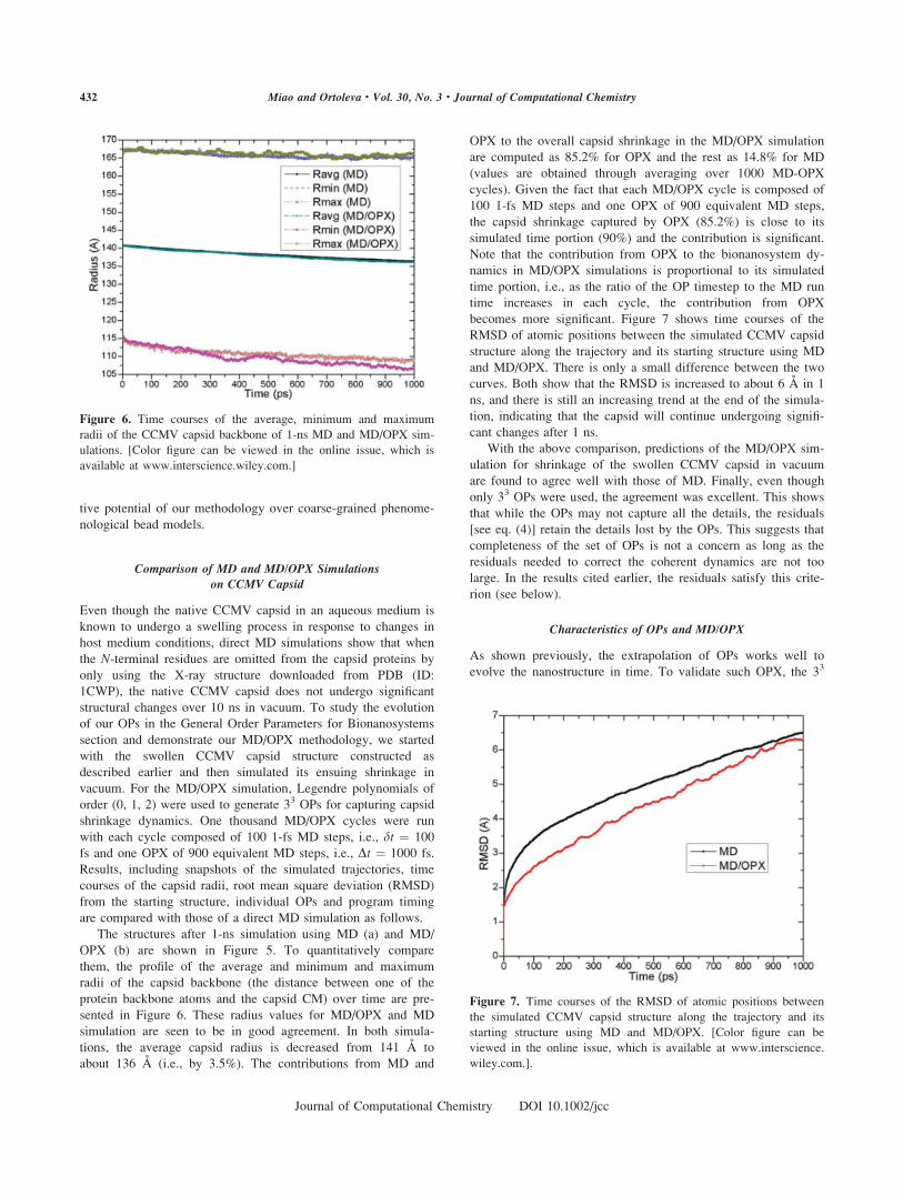
becomes more significant. Figure 7 shows time courses of the
RMSD of atomic positions between the simulated CCMV capsid
structure along the trajectory and its starting structure using MD
and MD/OPX. There is only a small difference between the two
curves. Both show that the RMSD is increased to about 6 A in 1
ns, and there is still an increasing trend at the end of the simula-
tion, indicating that the capsid will continue undergoing signifi-
cant changes after 1 ns.
With the above comparison, predictions of the MD/OPX sim-
ulation for shrinkage of the swollen CCMV capsid in vacuum
are found to agree well with those of MD. Finally, even though
only 33 OPs were used, the agreement was excellent. This shows
that while the OPs may not capture all the details, the residuals
[see eq. (4)] retain the details lost by the OPs. This suggests that
completeness of the set of OPs is not a concern as long as the
residuals needed to correct the coherent dynamics are not too
large. In the results cited earlier, the residuals satisfy this crite-
rion (see below).
Characteristics of OPs and MD/OPX
As shown previously, the extrapolation of OPs works well to
evolve the nanostructure in time. To validate such OPX, the 33
Figure 6. Time courses of the average, minimum and maximum
radii of the CCMV capsid backbone of 1-ns MD and MD/OPX sim-
ulations. [Color figure can be viewed in the online issue, which is
available at www.interscience.wiley.com.]
Figure 7. Time courses of the RMSD of atomic positions between
the simulated CCMV capsid structure along the trajectory and its
starting structure using MD and MD/OPX. [Color figure can be
viewed in the online issue, which is available at www.interscience.
wiley.com.].
432 Miao and Ortoleva • Vol. 30, No. 3 • Journal of Computational Chemistry
Journal of Computational Chemistry DOI 10.1002/jcc

Figure 8. Time courses of CCMV capsid OPs corresponding to Legendre polynomials in the X, Y,
and Z directions of the MD simulation: (a) three OPs labeled with indices {1, 0, 0} in X-direction
(X100), {0, 1, 0} in Y-direction (Y010) and {0, 0, 1} in Z-direction (Z001), reflecting an isotropic
shrinkage of the capsid can be readily extrapolated to long time on a timescale of nanoseconds, (b)
other OPs fluctuating rapidly around zero over 1 ns, and (c) a closer look at them from 60 to 100 ps,
showing that their characteristic time is roughly 10 ps. [Color figure can be viewed in the online issue,
which is available at www.interscience.wiley.com.]
433Molecular Dynamics/Order Parameter Extrapolation for Bionanosystem Simulations
Journal of Computational Chemistry DOI 10.1002/jcc

OPs generated with Legendre polynomials of order (0, 1, 2) in
X, Y, and Z directions for the 1-ns MD simulation output trajec-
tory of the swollen CCMV capsid are plotted versus time. Figure
8a shows time courses of the capsid OPs labeled with indices
(1, 0, 0) in the X-direction (0, 1, 0) for Y and (0, 0, 1) for Z(X100, Y010, and Z001), reflecting an isotropic shrinkage of the
capsid. The OPs vary slowly and can be readily extrapolated to
a long time on a timescale of nanoseconds. Other OPs shown in
Figure 8b are seen to fluctuate rapidly around zero over 1 ns. A
closer look at these OPs from 60 to 100 ps (Fig. 8c) shows that
their values are comparatively stable on a timescale of 10 ps,
which implies that their characteristic time periods are 10 ps or
longer. This means that all the OPs display much less stochastic
behavior than do individual atoms and have much longer charac-
teristic time than the 10214 s timescale of fast atomic vibrations
and collisions as expected; thus, the OPs satisfy one of the crite-
ria for the applicability of MD/OPX. The origin of the OP fluc-
tuations in Figure 8b is likely that they indicate the elastic vibra-
tions of the capsid in free space. We expect that when the capsid
is placed in an aqueous medium, frictional effects will dampen
these fluctuations so that the higher order OPs have longer char-
acteristic time and can also be extrapolated over larger time
intervals.
Another basis for MD/OPX is identified by investigating the
coherent and residual contributions to the atomic configuration
along its simulated trajectory. Figure 9 shows a snapshot of the
CCMV capsid after 1 ps MD/OPX simulation with atomic coor-
dinates given by their coherent coordinates plus the residuals.
Similar to the results presented in the OPs Capturing Capsomer
Dynamics During CCMV Capsid Expansion section, the coher-
ent coordinates calculated with 33 OPs are found to reflect the
overall capsid structure with the residuals close to zero. This
suggests that the slow collection motions in viral STs are cap-
tured with coherent coordinates computed through the slowly-
varying OPs, while the fast motions including atomic vibrations
and collisions are accounted for by the rapidly-fluctuating resid-
uals.
With the characteristic time of OPs determined to be in the
range of 10 ps to 1 ns, the adaptive procedure described in the
Molecular Dynamics/Order Parameter Extrapolation section has
been implemented to find the timestep of OPs allowed for MD/
OPX simulations. While it varies with other simulation parame-
ters, for a simulation using 100 1-fs MD steps (i.e., dt0 ¼ 100
fs) to anneal the resultant structure of OPX and keeping dt as
100 fs, the OP timestep Dt displays a typical value of 1.8 ps.
With another additional 1000 energy minimization steps after
OPX to anneal the structure, the optimized OP timestep becomes
5 ps. Further optimization of all run parameters is under investi-
gation.
To test the stability of MD/OPX, a 16 ns simulation was run
on the swollen CCMV capsid, and the results (see Fig. 10) show
that the average radius of the capsid backbone decreases from
141 to 132 A, the minimum capsid radius drops comparatively
fast in the beginning of the simulation from 116 to 110 A in
Figure 8. (continued)
434 Miao and Ortoleva • Vol. 30, No. 3 • Journal of Computational Chemistry
Journal of Computational Chemistry DOI 10.1002/jcc

2 ns and decreases more slowly for the rest of the time to 103 A
at 16 ns, while the maximum capsid radius keeps fluctuating
with an overall decreasing trend from 166 to 163 A. As a conse-
quence, the capsid thickens from 50 to 60 A during its shrink-
age. The change in the rate of capsid shrinkage can be explained
by the fact that with the swollen capsid approaching its native
state near equilibrium, the rate of progression decreases. This
also proves that our MD/OPX approach is stable for long-time
bionanosystem simulations.
MD/OPX Performance and Potential Optimization
The simulations in this study were run on the Big Red cluster at
Indiana University with 64 IBM processors for parallel MD and
one processor for the serial OPX code. Timing results show that
about 0.097 s are needed for one MD step and 15 s for one
OPX step. As a result, the OPX in each cycle is 372 times faster
than the MD run for the 900 equivalent MD steps. For a 1-ns
simulation, MD/OPX is 9.76 times faster than the direct MD.
Because the overall speedup of MD/OPX over MD is directly
proportional to the Dt/dt ratio, the above performance results can
be improved by a factor of 5 by increasing the OP timestep to 5
ps as obtained from the adaptive MD/OPX simulations. How-
ever, additional energy minimization and short MD run are
needed in the present implementation to anneal the OPX result
structure and they downgrade the program running performance.
The present implementation of MD/OPX makes use of the Tcl
scripting in NAMD. Atomic structures are transferred ineffi-
ciently via file saving and reading between NAMD and the For-
tran OPX program, and for the adaptive procedure, forces on all
atoms are loaded to calculate their accelerations as indicators
redundantly. These can be avoided by integrating MD/OPX into
the core code of NAMD so that the indicators used to judge the
structure of OPX can be queried efficiently, and less computa-
tion is expected for annealing the structure, i.e., fewer energy
minimization steps and shorter MD run. With this and the Dt/dtratio obtained as 50, the overall acceleration of MD/OPX over
MD is expected to reach 50. Also as the OPs we constructed
display different timescales (10 ps to 1 ns), multiple timesteps
can be implemented to further optimize the procedure and a
larger average timestep for OPs can be obtained to accelerate
the simulations.
Conclusions
A computational approach, MD/OPX, is developed to simulate
large bionanosystems. It makes use of a short MD run to esti-
mate the rate of change of OPs constructed automatically with
orthogonal polynomials, which is then used to extrapolate the
state of the system over long time. The approach is based on
our AMA theory for dynamical nanosystems. The all-atom for-
mulation enables the development of a universal simulator,
Figure 9. A snapshot of the CCMV capsid after 1-ps MD/OPX simulation showing that the atomic
coordinates are equal to their coherent contribution plus the residuals. [Color figure can be viewed in
the online issue, which is available at www.interscience.wiley.com.]
Figure 10. Time courses of the average, minimum, and maximum
radii of the CCMV capsid backbone of a 16-ns MD/OPX simulation.
[Color figure can be viewed in the online issue, which is available
at www.interscience.wiley.com.]
435Molecular Dynamics/Order Parameter Extrapolation for Bionanosystem Simulations
Journal of Computational Chemistry DOI 10.1002/jcc

avoiding the need for recalibration with each new application.
Thereby, an understanding of bionanosystems is achieved based
on the principles of molecular physics. The approach greatly
extends the realm of applicability of standard MD packages.
Unique features of MD/OPX as implemented include:
� automated construction of a set of OPs capturing the struc-
ture of complex bionanosystems and their nanoscale dynam-
ics;
� a rigorous demonstration of the timescale separation between
the characteristic time of the OPs and atomistic dynamics and
the implied existence of Langevin equations for their stochas-
tic dynamics;
� capturing all-atom detail in whole-nanostructure simulations;
and
� optimization of a dual timestep evolution algorithm via
dynamic adaptation of simulation run parameters.
The OPs constructed with orthonormalized Legendre polyno-
mials were demonstrated to be capable of capturing the nano-
scale dynamics of capsomers during native CCMV capsid
expansion. MD/OPX was then tested on shrinkage of the swol-
len CCMV capsid in vacuum. The simulation results are shown
to agree well with those of a direct MD simulation. As imple-
mented here, MD/OPX is 9.76 times faster than NAMD. With
potential optimization, the overall acceleration of MD/OPX over
NAMD can be improved to about 50 for simulating the CCMV
capsid in vacuum. The slowly varying characteristics of OPs
revealed and verified in this study justify the development of a
direct solver of the Langevin equations for the OPs. A theoreti-
cal speed-up of the solver over MD by a factor of O(N) is
expected for large bionanosystems composed of N atoms with
larger timesteps applied for the time integration and a much
smaller number of unknown variables to solve, though this theo-
retical speed-up is overestimated due to the computations needed
for constructing the frictions coefficients and thermal average
forces in the early rapidly changing stage of the OP evolution.
While the CCMV capsid used to demonstrate our MD/OPX
approach has 432,120 atoms, many systems of biological interest
are supramillion atoms in size (e.g., large viruses and cell mem-
branes in host media) and as the size of the simulated system
increases, the simulation speed-up of our multiscale approach
over direct MD will become greater. MD/OPX and the direct
solver of the Langevin equations for the OPs based on our
AMA methodology hold great promise for long-time simulations
of large bionanosystems. Their benefits for health sciences and
biotechnology include the computer-aided design of antiviral
drugs and vaccines, functionalizing nanoparticles for medical
imaging and thermal cancer treatments, and designing nanocap-
sules (e.g., viral capsids and liposomes) for delivery of therapeu-
tic agents.
Acknowledgments
Thanks to the U.S. Air Force Research laboratories at Wright
Patterson Air Force Base for their support through the Student
Research Participant Program from the Oak Ridge Institute of
Science and Education (ORISE), a seed grant from National
Institutes of Health (NIH) roadmap for medical research (grant
U54 GM072970) through Standford University via SimBios and
the support from Indiana University College of Arts and Scien-
ces and the METACyt project through the Center for Cell and
Virus Theory.
References
1. Speir, J. A.; Munshi, S.; Wang, G. J.; Baker, T. S.; Johnson, J. E.
Structure 1995, 3, 63.
2. Johnson, J. E.; Speir, J. A. J Mol Biol 1997, 269, 665.
3. Liu, H. J.; Qu, C. X.; Johnson, J. E.; Case, D. A. J Struct Biol 2003,
142, 356.
4. Liepold, L. O.; Revis, J.; Allen, M.; Oltrogge, L.; Young, M.; Doug-
las, T. Phys Biol 2005, 2, S166.
5. Speir, J. A.; Bothner, B.; Qu, C.; Willits, D. A.; Young, M. J.; John-
son, J. E. J Virol 2006, 80, 3582.
6. Canady, M. A.; Tihova, M.; Hanzlik, T. N.; Johnson, J. E.; Yeager,
M. J Mol Biol 2000, 299, 573.
7. Canady, M. A.; Tsuruta, H.; Johnson, J. E. J Mol Biol 2001, 311,
803.
8. Taylor, D. J.; Krishna, N. K.; Canady, M. A.; Schneemann, A.;
Johnson, J. E. J Virol 2002, 76, 9972.
9. Taylor, D. J.; Wang, Q.; Bothner, B.; Natarajan, P.; Finn, M. G.;
Johnson, J. E. Chem Commun 2003, 2770.
10. Lee, K. K.; Tsuruta, H.; Hendrix, R. W.; Duda, R. L.; Johnson, J. E.
J Mol Biol 2005, 352, 723.
11. Wikoff, W. R.; Conway, J. F.; Tang, J.; Lee, K. K.; Gan, L.; Cheng,
N.; Duda, R. L.; Hendrix, R. W.; Steven, A. C.; Johnson, J. E.
J Struct Biol 2006, 153, 300.
12. Fricks, C. E.; Hogle, J. M. J Virol 1990, 64, 1934.
13. Belnap, D. M.; Filman, D. J.; Trus, B. L.; Cheng, N. Q.; Booy, F.
P.; Conway, J. F.; Curry, S.; Hiremath, C. N.; Tsang, S. K.; Steven,
A. C.; Hogle, J. M. J Virol 2000, 74, 1342.
14. Belnap, D. M.; McDermott, B. M.; Filman, D. J.; Cheng, N.; Trus,
B. L.; Zuccola, H. J.; Racaniello, V. R.; Hogle, J. M.; Steven, A. C.
Proc Natl Acad Sci 2000, 97, 73.
15. Tsang, S. K.; McDermott, B. M.; Racaniello, V. R.; Hogle, J. M.
J Virol 2001, 75, 4984.
16. Hogle, J. M. Annu Rev Microbiol 2002, 56, 677.
17. Arkhipov, A.; Freddolino, P. L.; Schulten, K. Structure 2006, 14,
1767.
18. Harries, D.; May, S.; Gelbart, W. M.; Ben-Shaul, A. Biophys J
1998, 75, 159.
19. Speelman, B.; Brooks, B. R.; Post, C. B. Biophys J 2001, 80, 121.
20. Freddolino, P. L.; Arkhipov, A. S.; Larson, S. B.; McPherson, A.;
Schulten, K. Structure 2006, 14, 437.
21. Durup, J. J Phys Chem 1991, 95, 1817.
22. Tuckerman, M. E.; Berne, B. J. J Chem Phys 1991, 95, 8362.
23. Tuckerman, M. E.; Berne, B. J.; Martyna, G. J. J Chem Phys 1991,
94, 6811.
24. Askar, A.; Space, B.; Rabitz, H. J Phys Chem 1995, 99, 7330.
25. Reich, S. Physica D 1995, 89, 28.
26. Phelps, D. K.; Post, C. B. J Mol Biol 1995, 254, 544.
27. Phelps, D. K.; Rossky, P. J.; Post, C. B. J Mol Biol 1998, 276,
331.
28. Elezgaray, J.; Sanejouand, Y. H. Biopolymers 1998, 46, 493.
29. Elezgaray, J.; Sanejouand, Y. H. J Comput Chem 2000, 21, 1274.
30. Feenstra, K. A.; Hess, B.; Berendsen, H. J. C. J Comput Chem
1999, 20, 786.
31. Sorensen, M. R.; Voter, A. F. J Chem Phys 2000, 112, 9599.
436 Miao and Ortoleva • Vol. 30, No. 3 • Journal of Computational Chemistry
Journal of Computational Chemistry DOI 10.1002/jcc

32. Chun, H. M.; Padilla, C. E.; Chin, D. N.; Watanabe, M.; Karlov, V.
I.; Alper, H. E.; Soosaar, K.; Blair, K. B.; Becker, O. M.; Caves, L.
S. D.; Nagle, R.; Haney, D. N.; Farmer, B. L. J Comput Chem
2000, 21, 159.
33. Tama, F.; Brooks, C. L. J Mol Biol 2002, 318, 733.
34. Tama, F.; Brooks, C. L. J Mol Biol 2005, 345, 299.
35. van Vlijmen, H. W. T.; Karplus, M. J Chem Phys 2001, 115,
691.
36. van Vlijmen, H. W. T.; Karplus, M. J Mol Biol 2005, 350, 528.
37. Miao, Y.; Ortoleva, P. J Chem Phys 2006, 125, 44901.
38. Miao, Y.; Ortoleva, P. J. J Chem Phys 2006, 125, 214901.
39. Phillips, J. C.; Braun, R.; Wang, W.; Gumbart, J.; Tajkhorshid, E.;
Villa, E.; Chipot, C.; Skeel, R. D.; Kale, L.; Schulten, K. J Comput
Chem 2005, 26, 1781.
40. Kevrekidis, I. G.; Gear, C. W.; Hummer, G. Aiche J 2004, 50,
1346.
41. Jaqaman, K.; Ortoleva, P. J. J Comput Chem 2002, 23, 484.
42. Shreif, Z.; Ortoleva, P. J Stat Phys 2008, 130, 669.
437Molecular Dynamics/Order Parameter Extrapolation for Bionanosystem Simulations
Journal of Computational Chemistry DOI 10.1002/jcc


![CHOPtrey: contextual online polynomial extrapolation for ... · In [10], context-based extrapolation is exclusively intended for FMU models and extrapolation is per-formed on integration](https://img.dokumen.tips/doc/110x75/5eab92861431d863cb1b1b5b/choptrey-contextual-online-polynomial-extrapolation-for-in-10-context-based.jpg)