Embed Size (px)
Citation preview
Hematology 2003 279
Molecular Diagnostics
Rita M. Braziel, Margaret A. Shipp, Andrew L. Feldman, Virginia Espina, Mary Winters,Elaine S. Jaffe, Emanuel F. Petricoin III, and Lance A. Liotta
ies would be applied to define a patient’s disease pro-file. This profile could be used not only to confirm andrefine primary diagnoses, but potentially might also beused to determine the risk for development of cancer;to detect cancer at an early, more curable stage; to pre-dict drug efficacy against a cancer and also drug toxic-ity; to improve disease staging for determination of riskof distant spread and subsequent relapse; and to assessresponse to therapy by monitoring for residual disease.How close are we in 2003 to providing our patientswith this type of individualized medical approach? Un-fortunately, it must be acknowledged that we are stillfar from this ideal. Thus far, few of the exciting scien-tific advances of the recent past have been translatedinto improved patient care or better technology formolecular diagnostics. Nonetheless, the field of mo-lecular diagnostics is growing, adapting, changing con-stantly, and beginning to permeate virtually every area ofmedicine. Table 1 provides a listing of some of the cur-rent and anticipated techniques used in molecular diag-nostics today. These techniques are discussed below.
I. MOLECULAR DIAGNOSIS OF
HEMATOPOIETIC DISORDERS
Rita M. Braziel, MD*
The monumental research advances in genomic andprotein research over the past several years have madeit possible to envision, in the not too distant future, thedevelopment of medical care that is truly tailored toeach individual patient. Clinicians are anxious to in-corporate this new knowledge into the selection of morespecifically targeted therapies, and there is generalagreement that new research insights need to be trans-lated into useful clinical tests. Ideally, information fromall available studies, including traditional morphologicand immunophenotypic findings as well as data fromnew genomic, proteomic, and pharmacogenomic stud-
* Oregon Health and Sciences University, Department ofPathology, L471, 3191 SW Sam Jackson Park Rd, PortlandOR 97239
It is increasingly evident that molecular diagnos-tics, that is, the use of diagnostic testing tounderstand the molecular mechanisms of anindividual patient’s disease, will be pivotal in thedelivery of safe and effective therapy for manydiseases in the future. A huge body of new infor-mation on the genetic, genomic and proteomicprofiles of different hematopoietic diseases isaccumulating. This chapter focuses on newtechnologies and advancements in understandingthe molecular basis of hematologic disorders,providing an overview of new information and itssignificance to patient care.
In Section I, Dr. Braziel discusses the impactof new genetic information and research technolo-gies on the actual practice of diagnostic molecularhematopathology. Recent and projected changesin methodologies and analytical strategies usedby clinical molecular diagnostics laboratories forthe evaluation of hematologic disorders will be
discussed, and some of the challenges to clinicalimplementation of new molecular information andtechniques will be highlighted.
In Section II, Dr. Shipp provides an update oncurrent scientific knowledge in the genomicprofiling of malignant lymphomas, and describessome of the technical aspects of gene expressionprofiling. Analysis methods and the actual andpotential clinical and therapeutic applications ofinformation obtained from genomic profiling ofmalignant lymphomas are discussed.
In Section III, Dr. Liotta presents an update onproteomic analysis, a new and very active area ofresearch in hematopoietic malignancies. Hedescribes new technologies for rapid identificationof different important proteins and protein net-works, and the potential therapeutic and prognos-tic value of the elucidation of these proteins andprotein pathways in the clinical care of patientswith malignant lymphomas.
280 American Society of Hematology
Tests for Genome-Wide Screening forChromosomal Abnormalities
Routine cytogenetics is the traditional method for asurvey of genome-wide chromosomal abnormalities, butstandard karyotyping studies, even with chromosomebanding, miss many subtle chromosomal abnormalities.Other methodologies for genomic profiling of chromo-somal abnormalities have been developed, which haveconsiderably augmented our knowledge of the geneticfeatures of various hematopoietic malignancies. Theseassays for genomic profiling are based on screening ofchromosomes or DNA for loss or gain of chromosomesor genes, in contrast to gene-expression profiling per-formed on RNA. These assays detect changes in chro-mosome/gene location and number, not gene expres-sion and function; for all of these studies, at least fordisomic loci, the normal reference copy number is 2.Spectral karyotyping (SKY) and comparative genomichybridization (CGH) are complementary fluorescentmolecular genetic techniques for detection of wholegenome chromosomal abnormalities. With SKY, 24differentially labeled painting probes representing allchromosomes are cohybridized, Fourier spectroscopyis used to distinguish the different spectrally overlap-ping probes, and special imaging software is used foranalysis. This technique has been found to greatly fa-cilitate the detection of many previously cryptic chro-mosomal translocations and rearrangements, and is al-ready available for clinical purposes in many institu-tions.1-3 CGH uses the hybridization of differentiallylabeled tumor DNA and reference DNA to produce a
map of the DNA copy number changes in the tumorgenome.3-4 CGH assays are not yet available for rou-tine clinical use, but technical permutations of this re-search methodology are reputedly in the pipeline forclinical laboratories. A variant of CGH, called matrixCGH, uses genomic cDNA fragments instead of thechromosome targets used in standard CGH, and evenmore powerful is the use of arrayed cDNA sequenceswith CGH. These latter techniques allow detection ofunknown amplified genes, not just gene regions, andprovide even higher resolution for identification of ge-nomic imbalances.
Tests Targeting SpecificChromosomal Abnormalities
Multiple methods can be used for the detection of spe-cific chromosomal abnormalities, including variouspermutations of the polymerase chain reaction (PCR),Southern blotting, and fluorescence in situ hybridiza-tion (FISH) with molecular probes. PCR-based meth-ods, often multiplexed, have been the screening test ofchoice for most molecular laboratories if the chromo-somal abnormality of interest was amenable to PCRanalysis. However, as the number of genes importantin diagnosis and prediction of prognosis has increasedalmost exponentially over the past few years, a differ-ent molecular testing algorithm has evolved for hemato-poietic malignancies. The development, validation, andmaintenance of numerous PCR analyses for detectionof the ever-increasing important chromosomal abnor-malities in hematopoietic malignancies is simply notpractical for most laboratories. Fortunately, the devel-opment of molecular probes for use in FISH assays hasprovided a valuable alternative method to standard PCRanalyses. FISH assays are not as sensitive as PCR as-says, but FISH analyses are used predominantly at di-agnosis and relapse, a time when only a low level ofanalytical sensitivity is needed since there are usuallyhigh levels of abnormal cells. The use of FISH assaysfor molecular evaluation of malignant lymphomas andleukemias has increased remarkably over the past 1–2years, and has blurred the lines between classical cyto-genetics and molecular pathology.
FISH is a very useful technique for detection oftargeted chromosomal abnormalities. It can be done onblood, bone marrow, tissue touch preparations, bodyfluids, and even paraffin-embedded fixed tissue, so it isapplicable to many specimen types. FISH overcomesone of the biggest problems with routine cytogeneticanalysis of many lymphoma and chronic leukemiasamples (i.e. the need for metaphases), as FISH can bedone with either metaphase or interphase preparations.In FISH assays, the target is usually nuclear DNA of
Table 1. Summary of techniques for molecular analysis ofhematopoietic disorders.
Tests for Genome-Wide Screening of Chromosomal Abnormalities
Spectral karyotyping (multicolor fluorescence in situhybridization)
Comparative genomic hybridization (research)
Tests Targeting Specific Chromosomal Abnormalities
PCR (polymerase chain reaction analysis of DNA)
RT-PCR (reverse transcriptase PCR analysis of RNA)
Real-time PCR (automated PCR )
Genotyping for single nucleotide polymorphisms (PCR-SSP)
Fluorescence in situ hybridization (FISH)
Tests for Gene Expression Profiling
Global microarrays (research)
Focused microarrays (research)
Microarray of amplified RNA from microdissection (research)
Molecular Tests for Minimal Residual Disease Detection
Nested PCR
Quantitative real-time PCR (Q-PCR or Q-RT-PCR)
Hematology 2003 281
interphase or metaphase cells attached to glass micro-scope slides. Most FISH assays are based on the abilityof single stranded DNA to bind (hybridize) to comple-mentary DNA, although some RNA FISH assays areavailable.5 The molecular test probes (DNA) can belabeled with biotin or digoxigenin-labeled nucleotidesand detected with fluorophor-conjugated antibodies, ormay be directly fluorophor-labeled. With the use of dualor triple pass filters, multicolor FISH can be done.6,7
There are several different strategies for the de-sign of FISH assays. Single fusion-dual color FISH as-says for translocations utilize 2 probe hybridization tar-gets located on 1 side of each of the 2 genetic break-points; the usual level of false positive background cellsfrom incidental overlap of signal in this type of assay is5%–10%. Dual fusion-dual color FISH assays for trans-location utilize large probes that span 2 breakpoints onthe different chromosomes. Dual fusion-dual colorFISH is optimal for detection of low levels of nucleipossessing a simple balanced translocation, as it greatlyreduces the number of normal background nuclei withan abnormal signal pattern. FISH using dual color-breakapart probes is very useful in the evaluation of genesknown to have multiple translocation partners; the dif-ferently colored probes hybridize to targets on oppo-site sides of the breakpoint in the known gene.Multicolor FISH using 3 to 4 differently colored probescan be done in selected cases to determine the overlapof different genetic abnormalities in different cell popu-
lations. FISH with centromeric probes is useful for de-tection of changes in chromosome number (i.e., mono-somy, diploidy, trisomy).6-8
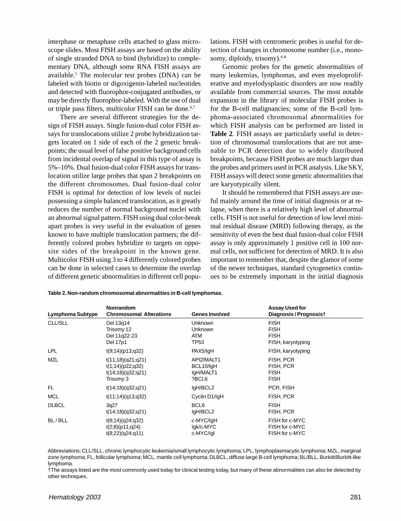
Genomic probes for the genetic abnormalities ofmany leukemias, lymphomas, and even myeloprolif-erative and myelodysplastic disorders are now readilyavailable from commercial sources. The most notableexpansion in the library of molecular FISH probes isfor the B-cell malignancies; some of the B-cell lym-phoma-associated chromosomal abnormalities forwhich FISH analysis can be performed are listed inTable 2. FISH assays are particularly useful in detec-tion of chromosomal translocations that are not ame-nable to PCR detection due to widely distributedbreakpoints, because FISH probes are much larger thanthe probes and primers used in PCR analysis. Like SKY,FISH assays will detect some genetic abnormalities thatare karyotypically silent.
It should be remembered that FISH assays are use-ful mainly around the time of initial diagnosis or at re-lapse, when there is a relatively high level of abnormalcells. FISH is not useful for detection of low level mini-mal residual disease (MRD) following therapy, as thesensitivity of even the best dual fusion-dual color FISHassay is only approximately 1 positive cell in 100 nor-mal cells, not sufficient for detection of MRD. It is alsoimportant to remember that, despite the glamor of someof the newer techniques, standard cytogenetics contin-ues to be extremely important in the initial diagnosis
Table 2. Non-random chromosomal abnormalities in B-cell lymphomas.
Nonrandom Assay Used forLymphoma Subtype Chromosomal Alterations Genes Involved Diagnosis / Prognosis†
CLL/SLL Del 13q14 Unknown FISHTrisomy 12 Unknown FISHDel 11q22-23 ATM FISHDel 17p1 TP53 FISH, karyotyping
LPL t(9;14)(p13;q32) PAX5/IgH FISH, karyotyping
MZL t(11;18)(q21;q21) API2/MALT1 FISH, PCRt(1;14)(p22;q32) BCL10/IgH FISH, PCRt(14;18)(q32;q21) IgH/MALT1 FISHTrisomy 3 ?BCL6 FISH
FL t(14;18)(q32;q21) IgH/BCL2 PCR, FISH
MCL t(11;14)(q13;q32) Cyclin D1/IgH FISH, PCR
DLBCL 3q27 BCL6 FISHt(14;18)(q32;q21) IgH/BCL2 FISH, PCR
BL / BLL t(8;14)(q24;q32) c-MYC/IgH FISH for c-MYCt(2;8)(p11;q24) Igk/c-MYC FISH for c-MYCt(8;22)(q24;q11) c-MYC/Igl FISH for c-MYC
Abbreviations: CLL/SLL, chronic lymphocytic leukemia/small lymphocytic lymphoma; LPL, lymphoplasmacytic lymphoma; MZL, marginalzone lymphoma; FL, follicular lymphoma; MCL, mantle cell lymphoma; DLBCL, diffuse large B-cell lymphoma; BL/BLL, Burkitt/Burkitt-likelymphoma.†The assays listed are the most commonly used today for clinical testing today, but many of these abnormalities can also be detected byother techniques.
282 American Society of Hematology
and follow-up of patients with hematopoietic malig-nancies. Focusing only on tests that target specific ge-netic abnormalities, like FISH and PCR, can result inthe failure to detect the additional important cytoge-netic abnormalities that may be present initially or thatmay occur following therapy. For example, the needfor intermittent cytogenetic analysis is very clear inchronic myelogenous leukemia (CML) patients. A num-ber of these CML patients have developed clonal karyo-typic abnormalities in Philadelphia chromosome–nega-tive cells while on therapy with imatinib mesylate; theseabnormalities would not have been detected by FISHor PCR analyses for BCR/ABL.9
Genotyping for single nucleotide polymorphisms(SNPs) is relevant to both research and routine molecu-lar diagnosis. Allele-specific PCR amplification tech-niques using sequence-specific primers (PCR-SSP) arewidely employed for detection of SNPs in the genesthat encode immunogenic proteins such as alloantigens.The status of certain alloantigens (i.e., human leuko-cyte antigens, blood group antigens, human platelet al-loantigens) is frequently investigated by this method-ology before organ transplantation. Technical advancesin genotyping of SNPs have improved the ability toperform testing for HLA and blood group antigens onsmall samples of DNA, such as those obtained frompatients with low leukocyte counts.10,11
Gene Expression ProfilingDespite glowing predictions of future clinical utility andmultiple published reports on the use of microarraysfor gene expression profiling (GEP) in lymphomas andleukemias,12-18 no microarrays are available yet formolecular diagnostics. However, focused arrays utiliz-ing fewer, but highly significant genes, are currentlyavailable for research studies and are in the develop-ment pipeline for B-cell non-Hodgkin lymphomas andacute leukemias. The diffuse large B-cell lymphomasare likely to be the lymphoma subtype for which fo-cused microarrays will first be used for routine clinicalpurposes. Needless to say, molecular laboratories areanxiously awaiting the shift of this technology into theclinical arena.
Although microarrays for GEP have not yet madeit into the clinical molecular laboratory, informationgained from GEP data is impacting clinical laboratorytesting. One example is flow cytometric analyses ofZAP-70 expression in chronic lymphocytic leukemias/small lymphocytic lymphomas (CLL/SLL). Pilot GEPstudies in patients with CLL/SLL identified genes thatwere differentially expressed between leukemic clonesthat did not have mutated IgH
V regions and those that
did. The best discriminator was a gene called ZAP-70;
a high level of ZAP-70 expression correctly predictedunmutated IgHv gene status in most patients.19,20 Thisis clinically relevant because the absence of somaticmutations in the variable regions of the IgH gene hasbeen determined to have adverse prognostic significancein CLL/SLL patients; those with unmutated IgHv re-gions often have progressive disease while those withmutated IgHv regions often pursue a more indolentcourse. Since molecular testing for IgHv mutation sta-tus involves multiple complex PCR reactions and se-quencing procedures, the analysis is impractical forclinical testing. Fortunately, the ZAP-70 protein isreadily detected by either flow cytometric analysis orimmunohistochemical staining,20 and these proceduresare currently being set up in many clinical laboratoriesin lieu of molecular analysis for somatic mutation ofthe IgH
V regions or microarray GEP.
There are some caveats about GEP data; these maybe part of the reason for delay in implementation ofthis technique in clinical practice. Numerous GEP da-tabases are now available in the public domain for dif-ferent lymphomas, acute leukemias, and myelomas, andmultiple publications have resulted from independentanalyses of these databases. It turns out that differentinvestigators do not always find the same results anddraw the same conclusions from analyses of the samedatabases. This has made it apparent that, because ofthe extreme complexity, there are potential problemswith analyses of GEP microarrays and databases thatcan produce erroneous GEP results.21,22 A few of theproblems that have been described include samplingvariability of tumors, chip differences and defects, dif-ferences and biases in analysis of GEP data, and sourcesof systematic error in microarray analysis. Clearly, sift-ing the real GEP changes from artifacts and noise inmicroarray experiments is often difficult, but rapid iden-tification and neutralization of spurious results is es-sential to prevent them from becoming accepted facts.Another potential problem with GEP is the possibilityof missing relevant cell populations present at a lowlevel in the tumor specimen. Since GEP provides anaverage expression profile for an entire cellular popu-lation, small subpopulations of important cells are un-likely to be recognized. The application of new meth-ods for microdissection, followed by RNA amplifica-tion, would allow targeting of specific populations ofinterest that were previously missed by GEP. In con-clusion, there are enough problems with GEP micro-arrays and interpretations that it is important to havevalidation of significant GEP changes from more thanone laboratory/database before important clinical de-cisions are based on this data.
Hematology 2003 283
Molecular Tests for MinimalResidual Disease Detection
Although many patients with hematologic malignan-cies achieve a complete clinical remission (CR) andeven a complete pathologic remission by standard mor-phologic and immunologic criteria, a relatively highproportion of them will ultimately relapse. The sourceof this relapse is clearly from a persistent malignantcellular population that is present at a low level, belowthe limit of detection by standard techniques. For thisreason, considerable effort has been devoted by mo-lecular laboratories in the past 5 to 10 years to developnew molecular techniques to increase the sensitivity ofdetection of neoplastic cells. The application of thesetechniques has demonstrated the presence of residualneoplastic cells in many patients in CR. This reservoirof neoplastic cells, detected only by sensitive molecu-lar methods, is commonly referred to as minimal re-sidual disease (MRD). The detection of MRD in a vari-ety of hematologic malignancies suggests that obtain-ing a molecular remission should be a goal of therapy,and the results of most studies of MRD detection sup-port this concept. However, it has still not been clearlyestablished for many hematologic malignancies thatpatients with only a few residual malignant cells, de-tected only by very sensitive techniques, will benefitfrom additional therapy.
If achieving a molecular remission is confirmed tobe an important goal following therapy for most hema-tologic malignancies, as seems likely, then MRD test-ing will become a much larger component of testing inmolecular diagnostics laboratories. Ideally, techniquesused for MRD detection should have a sensitivity levelin the 105 to 106 range, be applicable to almost all pa-tients with the disease, provide some quantification ofthe target, and be rapid, inexpensive, readily standard-ized, and disease-specific. Also of critical importancefor the clinical utility of tests for MRD detection is goodinterlaboratory reproducibility and standardization ofreporting. In reality, most commonly used molecularanalyses for MRD detection do not meet many of thesecriteria. A particular problem for clinicians is the lackof standardization of testing techniques and primersbetween laboratories, which essentially mandates follow-up testing for MRD be performed in the laboratory thatdid the previous testing to allow comparison of results.With frequent shifts in patient locations and changing in-surance carrier requirements, sending follow-up speci-mens to the same laboratory may be impossible.
Only a few commonly used techniques are sensi-tive enough for detection of MRD in leukemias andlymphomas. Nested PCR and quantitative real-time PCRcan be used for disease-associated translocations, with-
out the need for patient-specific primers. If the malig-nant clone does not carry a good translocation targetfor PCR analysis, patient-specific gene rearrangementsmay be targeted, using either nested or quantitative real-time PCR. Nested PCR analyses can detect up to 1malignant cell in 106 normal cells. Quantitative real-time PCR assays, with a sensitivity of 1 in 104-105, arealmost as sensitive as the nested PCR. A substantivenumber of studies of MRD detection have been per-formed in only a few hematopoietic malignancies, spe-cifically chronic myelogenous leukemia, follicular lym-phoma, and childhood acute lymphoblastic leukemia.The different methods used for detection of MRD inthese 3 different hematopoietic malignancies are dis-cussed below.
Chronic myelogenous leukemiasWith imatinib mesylate therapy, a complete cytogeneticresponse (CCR) can be achieved for most patients withnewly diagnosed chronic myelogenous leukemia(CML).23 Quantitative real-time RT-PCR analysis (Q-RT-PCR) is most often used to monitor for MRD inpatients who have achieved a CCR by bone marrowcytogenetics and/or FISH. Interestingly, Q-RT-PCRmonitoring for BCR/ABL can be performed on eitherperipheral blood or bone marrow; comparable resultshave been found on analysis of simultaneous blood andmarrow specimens24 (R Braziel, unpublished data). Thisfacilitates follow-up of imatinib-treated CML patients.Real-time PCR is a relatively new molecular techniquethat allows simultaneous PCR amplification and detec-tion of target DNA or cDNA sequences. The specimenis normalized against an internal control, typically asingle copy gene; for CML MRD testing, ABL orG6PDH is typically used as the internal control. A stan-dard curve is made from a dilution series of a BCR/ABL-positive cell line, and the amount of residual leu-kemia cells is calculated by using this standard curve(Figure 1; see Appendix, page 600). Advantages ofreal-time PCR over standard nested PCR for BCR/ABLinclude a decreased turnaround time, decreased chancefor post-PCR contamination, decreased variability ofresults because the data collection occurs in the expo-nential phase of the PCR reaction, high throughput, andthe possibility of obtaining quantitative results. Real-time PCR procedures are much more amenable tointerlaboratory standardization than nested PCR analy-ses. The major disadvantage of real-time PCR testingis the inability to compare the size of any detected re-arrangements to that of the original malignant clonewithout additional testing. However, this is not a prob-lem with MRD testing in CML, as there is not a back-ground population of normal cells carrying the BCR/
284 American Society of Hematology
ABL translocation. Quantitative real-time PCR tech-nology can be used with many translocation targets,and can also be used for antigen receptor gene rear-rangement analysis. The determination of the trend inthe quantitative numbers of residual BCR/ABL-posi-tive cells over a period of time is thought to provideimportant therapeutic information in the follow-up ofCML patients.25,26 Optimal methods for quantitativereal-time PCR detection of MRD in CML patients havenot yet been established, and this testing is currentlyperformed mainly in a few reference laboratories.
Once again, the importance of remembering thelimitations of very targeted molecular testing in CMLmust be stressed. The clinician must be alert for devel-opment of clonal abnormalities in BCR/ABL negativecells9 or the development of resistance to imatinibmesylate. The presence of mutations or amplificationof BCR/ABL is known to be associated with resistanceto imatinib mesylate in CML patients,27 and testing forthese abnormalities is likely to become a standard partof the evaluation of CML patients in the future, at leastfor those who fail to achieve and maintain a CCR.
Follicular lymphomasThe recent use of therapeutic modalities such as au-tologous bone marrow transplant following ex vivopurging of bone marrow B cells, monoclonal antibodytherapy, and vaccine therapy has resulted in improvedclinical outcomes of patients with follicular lympho-mas (FL). PCR analysis for MRD performed on serialbone marrow samples in treated FL patients in com-plete remission has shown that some patients do achievea molecular remission, and that the failure to achieveor maintain a molecular remission is predictive of re-lapse.28-34 Although the optimal methodology and tim-ing for detection of MRD has yet to be determined, thet(14;18)(q32;q21)–IgH/BCL2 translocation, seen in80%-90% of FL, is a good target for MRD detection.Unlike MRD testing in CML, in patients with FL, bonemarrow analysis is clearly more sensitive for detectionof MRD than peripheral blood. Many FL patients clearFL cells from the blood, while they still have persistentmarrow involvement.
Nested PCR assays have been used historically andremain the most sensitive methodology for FL MRDdetection; nested PCR can detect one translocation-car-rying cell in 106 normal cells and is still used in manylaboratories for detection of this translocation. How-ever, other molecular laboratories have switched fromnested PCR to quantitative real time PCR. Real-timePCR for IgH/BCL2 is less labor-intensive and lacks therisk of contamination of standard nested PCR, but doeshave an analytical sensitivity that is usually at least 1
log less than that of nested PCR for IgH/BCL2. An ad-ditional and under-recognized problem in interpreta-tion of real-time PCR analyses for MRD in FL is thepotential for false positive results from occasional be-nign IgH/BCL2 translocation-carrying cells. These arepresent in 30-40% of normal individuals, and with ahighly sensitive test, a false positive result could occur ifa comparison to the original FL clone is not made.35-37
This comparison is readily performed with nested PCR(Figure 2), but would require substantial additionalmolecular analysis with real-time PCR. Using a quan-titative real-time PCR method on serial bone marrowsfor MRD detection in FL, determining a trend over time,may obviate the necessity for this additional testing.Additional studies are needed to evaluate the clinicalefficacy of MRD detection in FL patients in general, andto compare the relative clinical value of the nested andquantitative real-time PCR MRD detection methods.
Precursor B-cell lymphoblastic leukemiasMultiple large prospective studies have clearly demon-strated the high prognostic value of MRD monitoringin children with precursor B-cell lymphoblastic leuke-mias (pre-B-ALL).38-43 In childhood pre-B-ALL, stud-ies of MRD have generally targeted patient-specific IgHantigen receptor gene rearrangements. This methodtakes advantage of the fingerprint-like sequences of thejunctional regions of rearranged IgH genes, which dif-fer in length and composition for each lymphocyteclone. To obtain these sequences, standard IgH PCRanalysis is performed at diagnosis and/or relapse andthe PCR products are Southern blotted, followed bysequencing of junctional regions of the clonal IgH re-arrangements. The different IgH rearrangements arethen used for design of patient-specific oligonucleotideprimers that are subsequently used in real-time PCRassays to follow the patient. Patient-specific IgH prim-ers increase PCR sensitivity up to 1000-fold comparedto standard consensus primers for IgV
H gene rearrange-
ments; reactive background rearranged B cells do notobscure the clonal PCR products. At the present time,patient/clone-specific IgH PCR is not practical outsideof a funded clinical trial setting, but this techniqueclearly offers the best potential for a sensitive, specific,and rapid analysis method that could be used over thecourse of therapy in most patients with pre-B-ALL.
Indeed, this same type of patient/clone-specific IgHPCR technology could be used for MRD detection inmost other B-cell lymphomas (BCL) also, in which ei-ther no translocation-associated molecular event isavailable for MRD testing or the recurrent transloca-tions occur in too low a proportion of the BCL subtypeto be clinically useful. Standard IgH gene PCR with
Hematology 2003 285
consensus rather than patient-specific IgH primers canusually detect only 1 malignant cell in 102-3 normal cells,so the only sufficiently sensitive and specific methodof testing for MRD detection in most BCL is thereforethe use of patient/clone-specific Ig gene rearrange-ments. Quantitative real-time PCR techniques usingstandard IgH primers may provide some early infor-mation about the trend of the disease course over time,but will become negative when the patient could stillhave substantial residual disease. The combination ofpatient-specific IgH primers and quantitative real-timePCR could make a major contribution to the achieve-ment of standardized MRD detection in BCL.
ConclusionWhether offering or ordering a molecular test, the phy-sician should know the circumstances in which the testshould be ordered, the circumstances in which the test
would not be useful, the advantages and the limitationsof the test, and how to interpret the results. Many clini-cians and pathologists are unfamiliar with moleculartests for hematologic malignancies, and misinterpretresults of molecular testing. To avoid this, cliniciansmust be knowledgeable about the molecular test theyare ordering and cautious about overinterpretation ofresults. Physicians ordering molecular tests must beprepared to offer counseling on them, either personallyor by referral. Good patient consent forms for molecu-lar testing are crucial, and should explain to patientsthe meaning of a positive test, a negative test, and aninconclusive test. The consent form should inform thepatient that the test could uncover other clinically rel-evant information, things that were not even beinglooked for.
Clinical molecular laboratories today are faced withtwo daunting tasks. First and foremost is the necessity
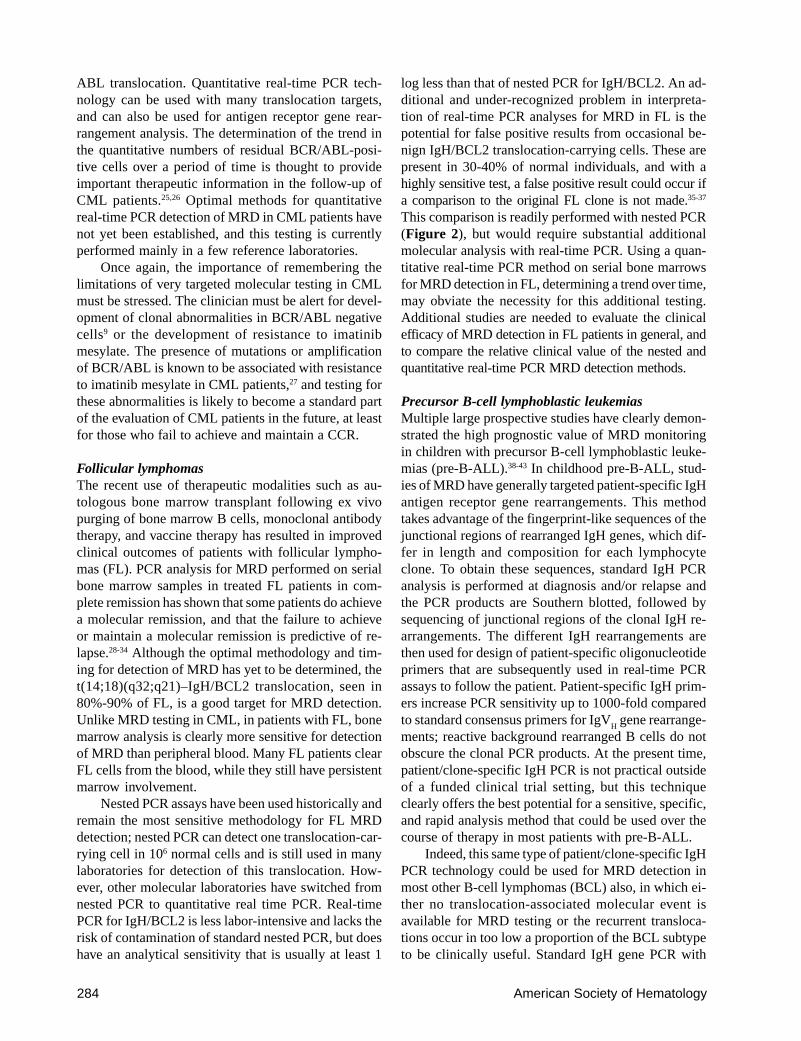
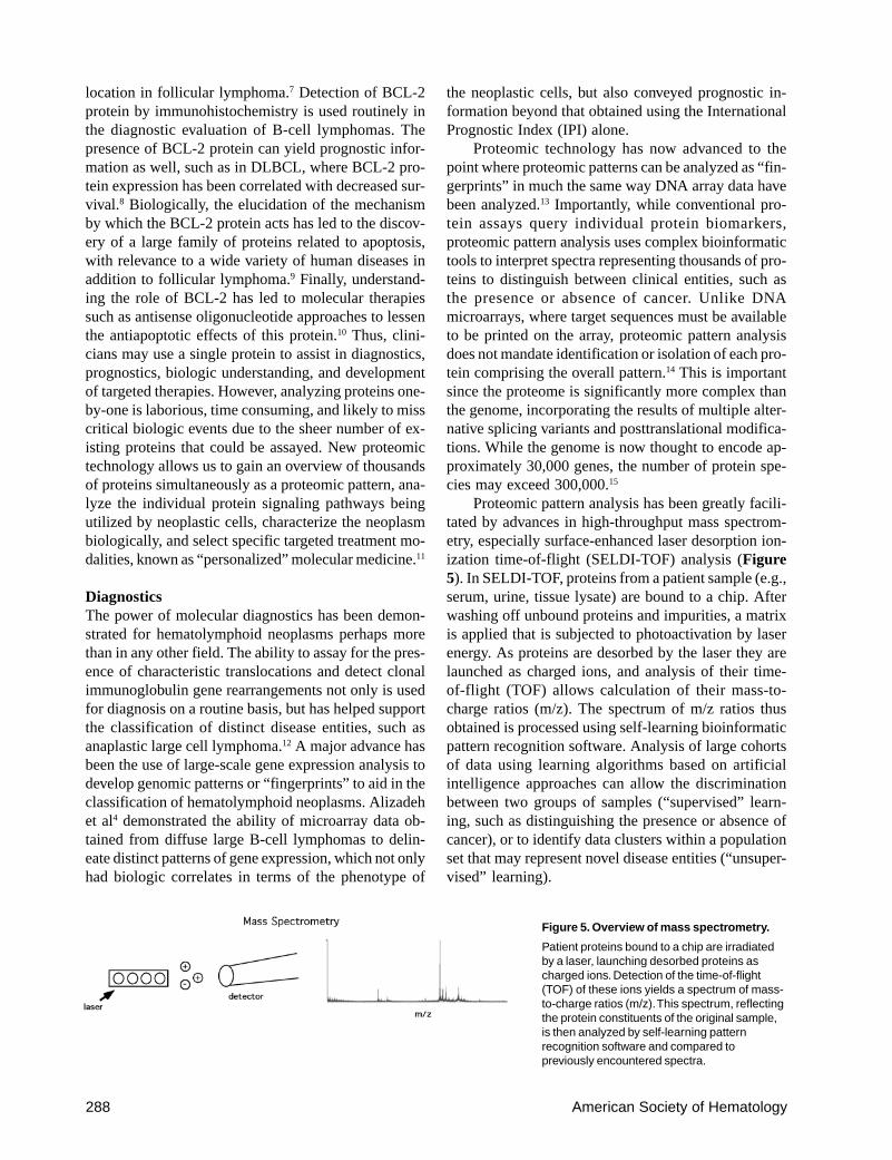
Figure 2. An example of nested polymerase chain reaction (PCR) analysis for detection of minimal residual disease (MRD) inmarrows from 2 follicular lymphoma (FL) patients.
Each sample is subjected to PCR for β-globin housekeeping gene and IgH/BCL2; PCR products undergo gel electrophoresis and (+)bands are detected by ethidium bromide staining (center gels = β-globin and bottom gels = IgH/BCL2 MBR). A band is seen only if anappropriate product is detected; specificity of bands is confirmed by Southern blot using BCL2 probe (top autoradiographs).
(Panel A ): Multiple controls are run in parallel with patient samples (see box insert). (+) controls are serial dilutions of an IgH/BCL2 (+)cell line (lanes 4-7). The 106 dilution is run twice; as shown, a (+) result is often detected in only 1of these samples at the lower limit ofanalytical test sensitivity.
(Panels B andC ): Parallel PCR analyses of 5 sequential marrows from 2 different FL patients are shown. For each patient, sample Awas at diagnosis, before any treatment, B was 1-2 months after completion of CHOP chemotherapy but before treatment with anti-CD20monoclonal antibody, and marrows C, D, and E were obtained at 2 months, 6 months, and 12 months after CD20 therapy. PCR of allclinical samples is run in duplicate. The patient in Panel B has a (+) nested PCR for IgH/BCL2 MBR at diagnosis and after CHOP, but themarrow becomes PCR (–) by 2 months after CD20 therapy and remains (–) at 6 and 12 months. The patient in Panel C is also PCR (+) atdiagnosis and after CHOP, but also has a (+) band in 1 lane at 2 months after CD20 therapy. The (+) band is confirmed to represent abenign IgH/BCL2-carrying cell, not MRD, as it is not present in the duplicate sample and is clearly a different size from the patient’s FLclone. Note that results are more easily interpreted on the BCL2-probed Southern blots than on the ethidium bromide–stained gels.
286 American Society of Hematology
of expanding test menus to meet the increasing clinicaldemand for testing for new genetic markers in hemato-logic disorders. Expanding test menus to meet clinicalneeds will require technical advances, as the currenttechnology in clinical molecular laboratories does notallow rapid screening for a broad panel of relevant genesat a reasonable cost. However, equipment developmenthas not been aimed at clinical laboratories, which tra-ditionally have low budgets for new equipment, but atlarge pharmaceutical companies with abundant cash fornew purchases. This disconnect must clearly be ad-dressed if clinical molecular testing is to be advanced,but even if the technological bottleneck preventingtranslation of new genetic knowledge to the clinicalarena is alleviated, the lack of a reasonable level ofreimbursement for molecular testing in general is stilla major roadblock to successful implementation of newtechniques for clinical molecular diagnostic testing. Inmost cases, the amount reimbursed for molecular diag-nostic testing in hematopoietic malignancies in theUnited States today is inadequate to even cover the costsof test reagents. Little progress in the ideal model of“personalized” medicine will occur if this lack of fund-ing persists.
II. MOLECULAR SIGNATURES OF LYMPHOID
MALIGNANCIES: IDENTIFICATION OF NOVEL DISEASE
SUBTYPES AND RATIONAL THERAPEUTIC TARGETS
Margaret A. Shipp, MD*
Lymphoid malignancies are currently classified on thebasis of morphology, immunophenotype, genetic fea-tures, clinical characteristics, and possible normal cellsof origin.1 With the sequencing of the human genomeand associated development of representative DNAmicroarrays, it is now possible to obtain broad-basedtranscriptional profiles of specific lymphoid malignan-cies and previously unidentified disease subtypes.
The most commonly used platforms for gene ex-pression profiling are cDNA and oligonucleotidemicroarrays (Figure 3; see Appendix, page 600).2 WithcDNA arrays, polymerase chain reaction (PCR) prod-ucts of cDNA clones are spotted on filters or glass slides.A potential advantage of cDNA arrays is that they canbe designed to address specific biologic questions. Forexample, the recently described “lymphochip” cDNAarrays are enriched for genes with documented impor-tance in lymphocyte biology.2 Oligonucleotide
microarrays include oligonucleotide probes depositedor synthesized directly on the surface of a silicon wa-fer. Oligonucleotide microarrays can potentially offeradditional specificity by tailoring probes to reducecross-hybridization and discern splice variants.2 A com-mon oligonucleotide array platform also facilitates com-parisons across datasets of different tumor types3 (Fig-ure 3; see Appendix, page 600).
Two main approaches have been used to analyzegene expression datasets: unsupervised and supervisedlearning (Table 3).2 Unsupervised learning methodsaggregate samples into groups based on the overall simi-larity of their gene expression profiles without a prioriknowledge of specific relationships (Table 3). Com-monly used unsupervised learning algorithms includeself-organizing maps (SOMs), hierarchical clustering,and probabilistic clustering (Table 3). In contrast, su-pervised learning techniques group tumors based onknown differences (i.e., cured versus fatal disease) anddevelop transcriptional profiles of the defined groups(Table 3 and Figure 4; see Appendix, page 601). Fre-quently used supervised learning algorithms includeweighted voting, k-NN, support vector machine (SVM),and IBM SPLASH (Table 3).
One of the lymphoid malignancies in which geneexpression profiling has been informative is diffuse largeB-cell lymphoma (DLBCL). The most common lym-phoid malignancy in adults, DLBCL comprises almost40% of all lymphoid tumors. Although a subset of DLBCLpatients can be cured with standard adriamycin-con-taining combination chemotherapy, the majority die oftheir disease. Robust clinical prognostic factor modelssuch as the International Prognostic Index can be usedto identify patients who are less likely to be cured withstandard therapy.4 However, such models do not pro-vide specific insights regarding more effective treat-ment strategies. For these reasons, additional insights
* Dana-Farber Cancer Institute, 44 Binney Street, RoomD940, Boston MA 02115-6084
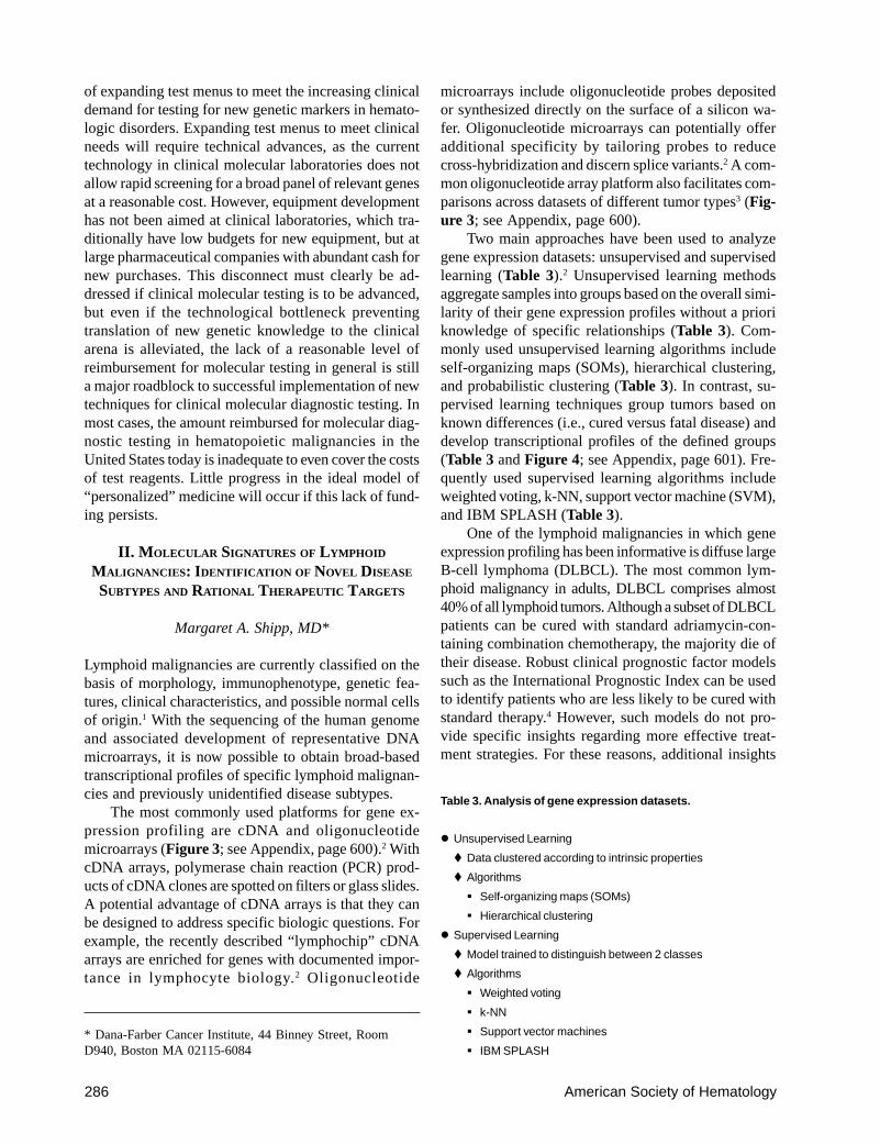
Table 3. Analysis of gene expression datasets.
� Unsupervised Learning
� Data clustered according to intrinsic properties
� Algorithms
� Self-organizing maps (SOMs)
� Hierarchical clustering
� Supervised Learning
� Model trained to distinguish between 2 classes
� Algorithms
� Weighted voting
� k-NN
� Support vector machines
� IBM SPLASH
Hematology 2003 287
into molecular bases for the observed clinical hetero-geneity in DLBCL are critically needed. In addition,the multiple genetic abnormalities associated with sub-sets of DLBCL reflect additional molecular heteroge-neity in this disease.5,6
Investigators have utilized gene expression profil-ing to elucidate molecular bases for observed differ-ences in DLBCL, identifying possible normal cells oforigin,7 tumors with different responses to standardcombination chemotherapy,8,9 novel rational treatmenttargets,8 and related disease entities (Savage et al, un-published material). For these reasons, the lessons fromgene expression profiling in DLBCL are likely to bebroadly applicable to other lymphoid malignancies.
In one of the earliest applications of gene expres-sion profiling, cDNA microarrays (lymphochips) andunsupervised learning techniques (hierarchical cluster-ing) were used to characterize the transcriptional pro-files of DLBCL and normal lymphocytes, includinggerminal center (GC) B cells and in vitro activated pe-ripheral blood B cells.7 In a pilot study, subsets ofDLBCLs were found to share gene expression patternswith normal GC B cells or in vitro activated PB B cells.7
In an expanded analysis, a refined cell-of-origin signa-ture (100 genes that distinguished GC-B-cell-like andactivated-B-cell-like lymphomas at a significance levelof P < .001) was used to identify tumors with featuresof above-mentioned normal B cells and a third unre-lated subset.9
Additional investigators have utilized supervisedlearning methods to develop transcriptional profiles ofcured versus fatal/refractory DLBCLs.8 Genes impli-cated in outcome signatures included ones that regu-late B-cell receptor signaling, critical serine/threoninephosphorylation pathways, and apoptosis.8 Two of thegenes and pathways identified in this supervised out-come analysis8 have already been credentialed as pos-sible rational therapeutic targets in DLBCL. In addi-tional analyses, a combination of unsupervised and su-pervised learning methods were used to develop aDLBCL outcome model that included the cell-of-ori-gin distinction and additional parameters including HLAclass II expression and indices of proliferation.9
These extremely powerful computational strategiesprovide new mechanisms for identifying discrete sub-sets of DLBCL and other lymphoid malignancies.10 Thenext challenges will be to link the molecular signaturesof cell-of-origin and prognosis in lymphoid tumors withimplicated biological pathways, specific pathogeneticmechanisms,11,12,13 and associated rational targets oftherapy.14
III. PROTEOMIC ANALYSIS OF
HEMATOLYMPHOID NEOPLASMS:DIAGNOSTIC, BIOLOGIC, AND
THERAPEUTIC IMPLICATIONS
Andrew L. Feldman, MD, Virginia Espina, MS,Mary Winters, BS, Elaine S. Jaffe, MD, Emanuel F.Petricoin III, PhD, and Lance A. Liotta, MD, PhD*
Hematolymphoid neoplasms are responsible for over60,000 deaths annually in the United States, and arethe most commonly occurring cancers in children.1
Despite these sobering statistics, it is within this fieldthat molecular medicine has made its earliest and great-est strides, the promise of which is just beginning to berealized. The past few decades have seen the discoveryof the t(9;22) BCR/ABL translocation in chronic my-elogenous leukemia (CML),2 the characterization of therole in apoptosis of the BCL-2 family of proteins,3 theuse of microarray analysis to delineate new subsets ofdiffuse large B-cell lymphoma (DLBCL),4 and the in-troduction of novel biologic agents such as rituximab5
and imatinib,6 which already have had far-ranging im-pact in reducing the burden of cancer in selected pa-tients. The field of hematolymphoid neoplasms remainsfertile ground for the application of technology in themolecular diagnosis, characterization, and treatment ofhuman disease.
Overview of ProteomicsThe functional effectors of cellular pathways and pro-cesses are proteins. While these proteins are encodedby the genome, only a subset of the possible proteinproducts of the genetic code are produced, and the func-tional status of these proteins often depends heavily onposttranslational modifications that are not reflected intheir genomic sequences. Thus, while significant ad-vances have been made from the analysis of the ge-nome and its transcribed complement of mRNA, theprotein end products of these processes are the effectorarm of cellular events and offer an in vivo, functionallyrelevant window into the workings of the cell. The studyof this wide complement of proteins derived from thegenome is known as proteomics, and the proteins col-lectively are called the proteome.
The analysis of proteins is used daily in the clini-cal diagnosis and treatment of hematolymphoid neo-plasms. An example is BCL-2 protein, an antiapoptoticprotein overexpressed as a result of the t(14;18) trans-
* National Cancer Institute, National Institutes of Health, 10Center Drive, MSC 1500, Bethesda MD 20892-1500
288 American Society of Hematology
location in follicular lymphoma.7 Detection of BCL-2protein by immunohistochemistry is used routinely inthe diagnostic evaluation of B-cell lymphomas. Thepresence of BCL-2 protein can yield prognostic infor-mation as well, such as in DLBCL, where BCL-2 pro-tein expression has been correlated with decreased sur-vival.8 Biologically, the elucidation of the mechanismby which the BCL-2 protein acts has led to the discov-ery of a large family of proteins related to apoptosis,with relevance to a wide variety of human diseases inaddition to follicular lymphoma.9 Finally, understand-ing the role of BCL-2 has led to molecular therapiessuch as antisense oligonucleotide approaches to lessenthe antiapoptotic effects of this protein.10 Thus, clini-cians may use a single protein to assist in diagnostics,prognostics, biologic understanding, and developmentof targeted therapies. However, analyzing proteins one-by-one is laborious, time consuming, and likely to misscritical biologic events due to the sheer number of ex-isting proteins that could be assayed. New proteomictechnology allows us to gain an overview of thousandsof proteins simultaneously as a proteomic pattern, ana-lyze the individual protein signaling pathways beingutilized by neoplastic cells, characterize the neoplasmbiologically, and select specific targeted treatment mo-dalities, known as “personalized” molecular medicine.11
DiagnosticsThe power of molecular diagnostics has been demon-strated for hematolymphoid neoplasms perhaps morethan in any other field. The ability to assay for the pres-ence of characteristic translocations and detect clonalimmunoglobulin gene rearrangements not only is usedfor diagnosis on a routine basis, but has helped supportthe classification of distinct disease entities, such asanaplastic large cell lymphoma.12 A major advance hasbeen the use of large-scale gene expression analysis todevelop genomic patterns or “fingerprints” to aid in theclassification of hematolymphoid neoplasms. Alizadehet al4 demonstrated the ability of microarray data ob-tained from diffuse large B-cell lymphomas to delin-eate distinct patterns of gene expression, which not onlyhad biologic correlates in terms of the phenotype of
the neoplastic cells, but also conveyed prognostic in-formation beyond that obtained using the InternationalPrognostic Index (IPI) alone.
Proteomic technology has now advanced to thepoint where proteomic patterns can be analyzed as “fin-gerprints” in much the same way DNA array data havebeen analyzed.13 Importantly, while conventional pro-tein assays query individual protein biomarkers,proteomic pattern analysis uses complex bioinformatictools to interpret spectra representing thousands of pro-teins to distinguish between clinical entities, such asthe presence or absence of cancer. Unlike DNAmicroarrays, where target sequences must be availableto be printed on the array, proteomic pattern analysisdoes not mandate identification or isolation of each pro-tein comprising the overall pattern.14 This is importantsince the proteome is significantly more complex thanthe genome, incorporating the results of multiple alter-native splicing variants and posttranslational modifica-tions. While the genome is now thought to encode ap-proximately 30,000 genes, the number of protein spe-cies may exceed 300,000.15
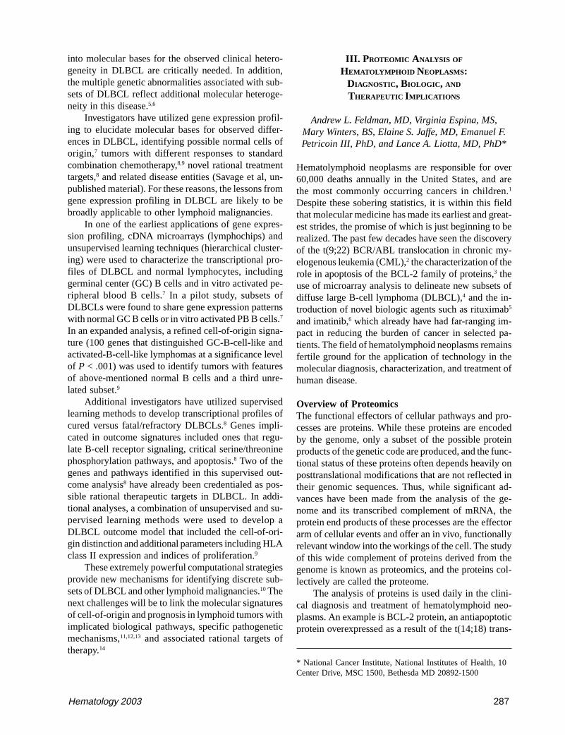
Proteomic pattern analysis has been greatly facili-tated by advances in high-throughput mass spectrom-etry, especially surface-enhanced laser desorption ion-ization time-of-flight (SELDI-TOF) analysis (Figure5). In SELDI-TOF, proteins from a patient sample (e.g.,serum, urine, tissue lysate) are bound to a chip. Afterwashing off unbound proteins and impurities, a matrixis applied that is subjected to photoactivation by laserenergy. As proteins are desorbed by the laser they arelaunched as charged ions, and analysis of their time-of-flight (TOF) allows calculation of their mass-to-charge ratios (m/z). The spectrum of m/z ratios thusobtained is processed using self-learning bioinformaticpattern recognition software. Analysis of large cohortsof data using learning algorithms based on artificialintelligence approaches can allow the discriminationbetween two groups of samples (“supervised” learn-ing, such as distinguishing the presence or absence ofcancer), or to identify data clusters within a populationset that may represent novel disease entities (“unsuper-vised” learning).
Figure 5. Overview of mass spectrometry.
Patient proteins bound to a chip are irradiatedby a laser, launching desorbed proteins ascharged ions. Detection of the time-of-flight(TOF) of these ions yields a spectrum of mass-to-charge ratios (m/z). This spectrum, reflectingthe protein constituents of the original sample,is then analyzed by self-learning patternrecognition software and compared topreviously encountered spectra.
Hematology 2003 289
An example of the power of this approach is therecent report of the ability to characterize the proteomicspectra derived from sera of women with and withoutovarian cancer.16 SELDI-TOF mass spectra were usedas a training set to develop an artificial intelligence al-gorithm that, when applied to blinded samples, couldaccurately identify 100% of patients with ovarian can-cer and 95% of controls. This approach was sensitiveenough to accurately identify all cancer patients, eventhose with stage I disease; specificity was demonstratedby including women with benign ovarian conditions inthe control group, as well as finding the algorithm in-capable of detecting the presence of cancer in sera frompatients with prostate cancer.17 A recent improvement inmass spectrometry allowed correct identification of serafrom ovarian cancer patients and controls with 100% sen-sitivity and 100% specificity.14
The classification of hematolymphoid neoplasmshas evolved from a system based on morphology aloneto one based on cell of origin, the determination ofwhich is aided by detection of lineage-specific proteinmarkers using flow cytometry or immunohistochemis-try.18 This classification also utilizes detection of addi-tional protein markers which are not lineage-specificbut rather relate to the pathogenetic mechanism of thedisease (e.g., BCL-2) or other clinically relevant ge-netic events (e.g., p53 mutations19). In many cases, pan-els of antibodies are chosen in part due to limitations ofthe techniques employed, such as assaying cell surfacemolecules by flow cytometry, or ability of antibodiesto detect antigens in paraffin-embedded tissue sections.The use of proteomic patterns representing tens of thou-sands of protein species may be an enormously power-ful tool to complement the ongoing efforts to classifyhematolymphoid neoplasms in ways that are biologi-cally accurate and clinically relevant.
Additional ways that proteomic profiles might beused diagnostically include screening for minimal re-sidual disease after treatment,20 screening for the de-velopment of neoplasia in high-risk populations (e.g.,posttransplant),21 screening for transformation of a low-grade neoplasm into a high-grade one,22 and character-izing/predicting responses to therapeutic interventions.13
For example, lymphoma cells in vitro have been shownto demonstrate characteristic proteomic patterns afterexposure to chemotherapeutic agents,23 and character-ization of in vivo patterns may serve as an early predic-tor of response and/or toxicity.
Molecular Characterization and TreatmentAs previously mentioned, proteomic profiles can haveimportant diagnostic and prognostic implications with-out reference to the individual proteins which consti-
tute these profiles.14 Even at the single protein level,the protein CD20 has been used widely as a marker ofB cells and a target for anti-B-cell monoclonal anti-body therapy,5 although these uses are not primarilybased on its specific cellular function. Clearly, how-ever, the widespread analysis of the proteome will yieldextensive data regarding function and utilization of criti-cal protein pathways, as was discussed for the BCL-2protein, and is expected to have far-ranging implica-tions for the identification of molecular targets for path-way-specific biologic therapy.
The ability to analyze the nature of the proteomein human tissues has been greatly facilitated by rapiddevelopments in the field of protein microarrays.24 Thepower of this technique has been enhanced by the de-velopment of laser capture microdissection,25 in whichsubpopulations of cells, such as lymphoid follicles,26
can be isolated from tissue sections using a laser pulse.Protein lysates prepared from these samples then canbe robotically applied in miniature dilution curves to asolid phase array with multiple other samples. Hundredsof replicate arrays can be generated and probed for theexpression of a large complement of proteins using spe-cific antibodies, including those that differentially rec-ognize cleaved and/or phosphorylated forms of key sig-nal transduction molecules.27 In this way, proteinmicroarrays can identify the particular signaling path-ways utilized by a population of neoplastic cells to tai-lor specific targeted therapy to modulate the functionof these pathways.
One particularly attractive use of this technologyis characterizing the status of apoptotic pathways inhematolymphoid neoplasms. Apoptosis is an essentialelement for the normal development and maintenanceof the immune system.28 The development of a healthyimmune repertoire is a highly stringent process, neces-sitating the elimination of most developing lympho-cytes; conversely, the ability to avoid apoptosis is criti-cal to the rapid expansion of quiescent lymphocytes inresponse to foreign antigen. Families of proteins re-lated to apoptosis, such as the BCL-2 family, thereforeinclude both proapoptotic and antiapoptotic membersto aid in the homeostatic regulation of these immuneprocesses. Perturbation of these homeostatic mecha-nisms is exemplified by the t(14;18) translocation infollicular lymphoma, leading to overexpression of BCL-2. Further studies have indicated that antiapoptotic sig-nals from BCL-2 are important in nonfollicular neo-plasms as well, in which complex interactions amongmultiple BCL-2 family members appear to be critical.29
A downstream level of regulation is the inhibitor ofapoptosis protein (IAP) family, which exerts its effectby inhibiting effector caspases.30 Because multiple pro-
290 American Society of Hematology
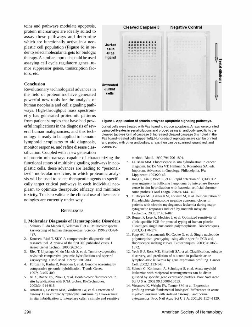
teins and pathways modulate apoptosis,protein microarrays are ideally suited toassay these pathways and determinewhich are functionally active in a neo-plastic cell population (Figure 6) in or-der to select molecular targets for biologictherapy. A similar approach could be usedassaying cell cycle regulatory genes, tu-mor suppressor genes, transcription fac-tors, etc.
ConclusionRevolutionary technological advances inthe field of proteomics have generatedpowerful new tools for the analysis ofhuman neoplasia and cell signaling path-ways. High-throughput mass spectrom-etry has generated proteomic patternsfrom patient samples that have had pow-erful implications in the diagnosis of sev-eral human malignancies, and this tech-nology is ready to be applied to hemato-lymphoid neoplasms to aid diagnosis,monitor response, and refine disease clas-sification. Coupled with a new generationof protein microarrays capable of characterizing thefunctional status of multiple signaling pathways in neo-plastic cells, these advances are leading to “personal-ized” molecular medicine, in which proteomic analy-sis will be used to select therapeutic agents to specifi-cally target critical pathways in each individual neo-plasm to optimize therapeutic efficacy and minimizetoxicity. Trials to validate the clinical use of these tech-nologies are currently under way.
REFERENCES
I. Molecular Diagnosis of Hematopoietic Disorders1. Schrock E, du Manoir S, Veldman T, et al. Multicolor spectral
karyotyping of human chromosomes. Science. 1996;273:494-497.
2. Knutsen, Ried T. SKY. A comprehensive diagnostic andresearch tool. A review of the first 300 published cases. JAssoc Genet Technol. 2000;26:3-15.
3. Ried T, Liyanage M, du Manoir S, et al. Tumor cytogeneticsrevisited: comparative genomic hybridization and spectralkaryotyping. J Mol Med. 1997;75:801-814.
4. Forozan F, Karhu R, Kononen J, et al. Genome screening bycomparative genomic hybridization. Trends Genet.1997;13:405-409.
5. Xi X, Roane DS, Zhou J, et al. Double-color fluorescence insitu hybridization with RNA probes. BioTechniques.2003;34:914-918.
6. Anastasi J, Le Beau MM, Vardiman JW, et al. Detection oftrisomy 12 in chronic lymphocytic leukemia by fluorescencein situ hybridization to interphase cells: a simple and sensitive
method. Blood. 1992;79:1796-1801.7. Le Beau MM. Fluorescence in situ hybridization in cancer
diagnosis. In: De Vita VT, Hellman S, Rosenberg SA, eds.Important Advances in Oncology. Philadelphia, PA:Lippincott; 1993:29-45.
8. Jiang F, Lin F, Price R, et al. Rapid detection of IgH/BCL2rearrangement in follicular lymphoma by interphase fluores-cence in situ hybridization with bacterial artificial chromo-some probes. J Mol Diagn. 2002;4:144-149.
9. O’Dwyer ME, Gatter KM, Loriaux M, et al. Demonstration ofPhiladelphia chromosome negative abnormal clones inpatients with chronic myelogenous leukemia during majorcytogenetic responses induced by imatinib mesylate.Leukemia. 2003;17:481-487.
10. Bugert P, Lese A, Meckies J, et al. Optimized sensitivity ofallele-specific PCR for prenatal typing of human plateletalloantigen single nucleotide polymorphisms. Biotechniques.2003;35:170-174.
11. Papp AC, Pinsonneault JK, Cooke G, et al. Single nucleotidepolymorphism genotyping using allele-specific PCR andfluorescence melting curves. Biotechniques. 2003;34:1068-1072.
12. Yeoh E-J, Ross ME, Shurtleff SA, et al. Classification, subtypediscovery, and prediction of outcome in pediatric acutelymphoblastic leukemia by gene expression profiling. CancerCell. 2002;1:133-143.
13. Schoch C, Kohlmann A, Schnittger S, et al. Acute myeloidleukemias with reciprocal rearrangements can be distin-guished by specific gene expression profiles. Proc Natl AcadSci U S A. 2002;99:10008-10013.
14. Virtaneva K, Wright FA, Tanner SM, et al. Expressionprofiling reveals fundamental biological differences in acutemyeloid leukemia with isolated trisomy 8 and normalcytogenetics. Proc Natl Acad Sci U S A. 2001;98:1124-1129.
Figure 6. Application of protein arrays to apoptotic signaling pathways.
Jurkat cells were treated with Fas ligand to induce apoptosis. Arrays were printedusing cell lysates in serial dilutions and probed using an antibody specific to thecleaved (active) form of caspase 3. Increased cleaved caspase 3 is noted in theFas ligand–treated cells (upper left). Hundreds of replicate arrays can be printedand probed with other antibodies; arrays then can be scanned, quantified, andcompared.
Hematology 2003 291
15. Alizadeh AA, Eisen MB, Davis RE, et al. Distinct types ofdiffuse large B-cell lymphoma identified by gene expressionprofiling. Nature. 2000;403:503-511.
16. Rosenwald A, Wright G, Chan WC, et al. The use of molecu-lar profiling to predict survival after chemotherapy for diffuselarge B-cell lymphoma. N Engl J Med. 2002;346:1937-1947.
17. Shipp MA, Ross KN, Tamayo P, et al. Diffuse large B-celllymphoma outcome prediction by gene-expression profilingand supervised machine learning. Nat Med. 2002;8:68-74.
18. Rosenwald A, Wright G, Wiestner A, et al. The proliferationgene expression signature is a quantitative integrator ofoncogenic events that predicts survival in mantle celllymphoma. Cancer Cell. 2003;3:185-197.
19. Rosenwald A, Alizadeh AA, Widhopf G, et al. Relation ofgene expression phenotype to immunoglobulin mutationgenotype in B-cell chronic lymphocytic leukemia. J Exp Med.2001;194:1639-1647.
20. Wiestner A, Rosenwald A, Barry TS, et al. ZAP-70 expressionidentifies a chronic lymphocytic leukemia subtype withunmutated immunoglobulin genes, inferior clinical outcome,and distinct gene expression profile. Blood. 2003;101:4944-4951.
21. Tseng GC, Oh MK, Rohlin L, et al. Issues in cDNAmicroarray analysis: quality filtering, channel normalization,models of variations and assessment of gene effects. NucleicAcids Res. 2001;29:2549-2557.
22. Zian J, Kluger Y, Yu H, et al. Identification and correction ofspurious spatial correlations in microarray data.Biotechniques. 2003;35:42-48.
23. Deininger MWN, Goldman JM, Melo JV. The molecularbiology of chronic myeloid leukemia. Blood. 2000;96:3343-3356.
24. LeGouill S, Talmant P, Milpied N, et al. Fluorescence in situhybridization on peripheral-blood specimens is a reliablemethod to evaluate cytogenetic response in chronic myeloidleukemia. J Clin Oncol. 2000;18:1535-1538.
25. Olavarria E, Kanfer E, Szydlo R, et al. Early detection ofBCR/ABLl transcripts by quantitative reverse-transcriptase-polymerase chain reaction predicts outcome after allogeneicstem cell transplantation for chronic myeloid leukemia. Blood.2001;97:1560-1565.
26. Radich JP, Gooley T, Bryant E, et al. The significance of bcr-abl molecular detection in chronic myeloid leukemia patient“late”, 18 months or more after transplantation. Blood.2001;98:1701-1707.
27. Branford S, Zbigniew R, Walsh S, et al. Detection of BCR-ABL mutations in patients with CML treated with imatinib isvirtually always accompanied by clinical resistance, andmutations in the ATP phosphate-binding loop (P-loop) areassociated with a poor prognosis. Blood. 2003;102:276-283.
28. Gribben JG, Neuberg D, Freedman AS, et al. Detection bypolymerase chain reaction of residual cells with the bcl-2translocation is associated with increased risk of relapse afterautologous bone marrow transplantation for B-cell lymphoma.Blood. 1993;81:3449-3457.
29. Moos M, Schulz R, Martin S, et al. The remission statusbefore and the PCR status after high-dose therapy withperipheral blood stem cell support are prognostic factors forrelapse-free survival in patients with follicular non-Hodgkin’slymphoma. Leukemia. 1998;12:1971-1976.
30. Apostolidis J, Gupta RK, Grenzelias D, et al. High-dosetherapy with autologous bone marrow support as consolida-tion of remission in follicular lymphoma: long-term clinicaland molecular follow-up. J Clin Oncol. 2000;96:864-869.
31. Czuczman MS, Grillo-Lopez AJ, McLaughlin P, et al. Clearing
of cells bearing the bcl-2 [t(14;18)] translocation from bloodand marrow of patients treated with rituximab alone or incombination with CHOP chemotherapy. Ann Oncol.2001;12:109-114.
32. Cabanillas F, McLaughlin P, Hagemeister F, et al. Molecularresponses with FND+Rituxan chemoimmunotherapy for stageIV indolent follicular non-Hodgkin’s lymphoma. Blood.2000;96:331a (abstract #1429).
33. Lopez-Guillermo A, Cabanillas F, McLaughlin P, et al. Theclinical significance of molecular response in indolentfollicular lymphomas. Blood. 1998;91:2955-2960.
34. Bendandi M, Gocke CD, Kobin CB, et al. Complete molecularremissions induced by patient-specific vaccination plusgranulocyte-monocyte colony-stimulating factor againstlymphoma. Nat Med. 1999;5:1171-1177.
35. Segal GH, Jorgensen SM, Braylan RC. Standard polymerasechain reaction analysis does not detect t(14;18) in reactivelymphoid hyperplasia. Arch Pathol Lab Med. 1994;118:791-794.
36. Limpens J, De Jong D, van Krieken JHJM, et al. Bcl-2/JHrearrangements in benign lymphoid tissue with follicularhyperplasia. Oncogene. 1991;6:2271-2276.
37. Limpens J, Stad R, Vos C, et al. Lymphoma-associatedtranslocation t(14;18) in blood B cells in normal individuals.Blood. 1995;85:2528-2536.
38. Eckert C, Landt O, Seeger K, et al. Potential of LightCyclertechnology for quantification of minimal residual disease inchildhood acute lymphoblastic leukemia. Leukemia.2000;14:316-323.
39. Goulden NJ, Knechtil CJC, Garland RJ, et al. Minimalresidual disease analysis for the prediction of relapse inchildren with standard-risk acute lymphoblastic leukaemia. BrJ Haematol. 1998;100:235-244.
40. Szczepanski T, Willemse MJ, Brinkhof B, et al. Comparativeanalysis of Ig and TCR gene rearrangements at diagnosis andat relapse of childhood precursor-B-ALL provides improvedstrategies for selection of stable PCR targets for monitoring ofminimal residual disease. Blood. 2002;99:2315-2323.
41. Van Dongen JJM, Macintyre EA, Gabert JA, et al. Standard-ized RT-PCR analysis of fusion gene transcripts fromchromosome aberrations in acute leukemia for detection ofminimal residual disease. Leukemia. 1999;13:a901-1928.
42. Van der Velden VHJ, Willemse JM, van der Schoot CE, et al.Immunoglobulin kappa deleting element rearrangements inprecursor B acute lymphoblastic leukemia are stable targetsfor detection of minimal residual disease by real-timequantitative PCR. Leukemia. 2002;16:928-936.
43. van Belzen N, Hupkes PE, Doekharen D, et al. Detection ofminimal disease using rearranged immunoglobulin heavychain genes from intermediate- and high-grade B cell non-Hodgkin’s lymphoma. Leukemia. 1997;11:1742-1752.
II. Molecular Signatures of Lymphoid Malignan-cies: Identification of Novel Disease Subtypesand Rational Therapeutic Targets
1. Jaffe ES, Harris NL, Stein H, Vardiman JW. World HealthOrganization Classification of Tumours. Pathology andGenetics of Tumours of Haematopoietic and LymphoidTissues. Lyon, France: IARC Press; 2001.
2. Ramaswamy S, Golub TR. DNA microarrays in clinicaloncology. J Clin Oncol. 2002;20:1932-1941.
3. Ramaswamy S, Ross KN, Lander ES, Golub TR. A molecularsignature of metastasis in primary solid tumors. Nat Genet.2003;33:49-54.
4. Shipp M, Harrington D, Anderson J, et al. A predictive model
292 American Society of Hematology
for aggressive non-Hodgkin’s lymphoma: The InternationalNHL Prognostic Factors Project. N Engl J Med.1993;329:987-994.
5. Kuppers R, Dalla-Favera R. Mechanisms of chromosomaltranslocations in B cell lymphomas. Oncogene. 2001;20:5580-5594.
6. Pasqualucci L, Neumeister P, Goossens T, et al.Hypermutation of multiple proto-oncogenes in B-cell diffuselarge-cell lymphomas. Nature. 2001;412:341-346.
7. Alizadeh A, Elsen M, Davis R, et al. Distinct types of diffuselarge B-cell lymphoma identified by gene expressionprofiling. Nature. 2000;4051:503-511.
8. Shipp MA, Ross KN, Tamayo P, et al. Diffuse large B-celllymphoma outcome prediction by gene-expression profilingand supervised machine learning. Nat Med. 2002;8:68-74.
9. Rosenwald A, Wright G, Chan WC, et al. The use of molecu-lar profiling to predict survival after chemotherapy for diffuselarge B-cell lymphoma. N Engl J Med. 2002;346:1937-1947.
10. Rosenwald A, Wright G, Wiestner A, et al. The proliferationgene expression signature is a quantitative integrator ofoncogenic events that predicts survival in mantle celllymphoma. Cancer Cell. 2003;3:185-197.
11. Yeoh EJ, Ross ME, Shurtleff SA, et al. Classification, subtypediscovery, and prediction of outcome in pediatric acutelymphoblastic leukemia by gene expression profiling. CancerCell. 2002;1:133-143.
12. Wessendorf S, Schwaenen C, Kohlhammer H, et al. Hiddengene amplifications in aggressive B-cell non-Hodgkinlymphomas detected by microarray-based comparativegenomic hybridization. Oncogene. 2003;22:1425-1429.
13. Martinez-Climent JA, Alizadeh AA, Segraves R, et al.Transformation of follicular lymphoma to diffuse large celllymphoma is associated with heterogeneous seet of DNA copynumber and gene expression alterations. Blood.2003;101:3109-3117.
14. Bohen SP, Troyanskaya O, Alter O, et al. Variation in geneexpression patterns in follicular lymphoma and the responseto rituximab. Proc Natl Acad Sci U S A. 2003;100:1926-1930.
III. Proteomic Analysis of HematolymphoidNeoplasms: Diagnostic, Biologic, andTherapeutic Implications
1. Greenlee RT, Hill-Harmon MB, Murray T, Thun M. Cancerstatistics, 2001. CA Cancer J Clin. 2001;51:15-36.
2. Nowell PC, Hungerford DA. A minute chromosome in humanchronic granulocytic leukemia. Science. 1960;132:1497-1500.
3. Vaux DL, Cory S, Adams JM. Bcl-2 gene promoteshaemopoietic cell survival and cooperates with c-myc toimmortalize pre-B cells. Nature. 1988;335:440-442.
4. Alizadeh AA, Eisen MB, Davis RE, et al. Distinct types ofdiffuse large B-cell lymphoma identified by gene expressionprofiling. Nature. 2000;403:503-511.
5. Plosker GL, Figgitt DP. Rituximab: a review of its use in non-Hodgkin’s lymphoma and chronic lymphocytic leukaemia.Drugs. 2003;63:803-843.
6. Deininger MW, Druker BJ. Specific targeted therapy ofchronic myelogenous leukemia with imatinib. Pharmacol Rev.2003;55:401-423.
7. Tsujimoto Y, Finger LR, Yunis J, Nowell PC, Croce CM.Cloning of the chromosome breakpoint of neoplastic B cellswith the t(14;18) chromosome translocation. Science.1984;226:1097-1099.
8. Gascoyne RD, Adomat SA, Krajewski S, et al. Prognosticsignificance of Bcl-2 protein expression and Bcl-2 gene
rearrangement in diffuse aggressive non-Hodgkin’s lym-phoma. Blood. 1997;90:244-251.
9. Baliga BC, Kumar S. Role of Bcl-2 family of proteins inmalignancy. Hematol Oncol. 2002;20:63-74.
10. Wang H, Prasad G, Buolamwini JK, Zhang R. Antisenseanticancer oligonucleotide therapeutics. Curr Cancer DrugTargets. 2001;1:177-196.
11. Liotta LA, Kohn EC, Petricoin EF. Clinical proteomics:personalized molecular medicine. JAMA. 2001;286:2211-2214.
12. Jaffe ES. Anaplastic large cell lymphoma: the shifting sandsof diagnostic hematopathology. Mod Pathol. 2001;14:219-228.
13. Petricoin EF, Zoon KC, Kohn EC, Barrett JC, Liotta LA.Clinical proteomics: translating benchside promise intobedside reality. Nat Rev Drug Discov. 2002;1:683-695.
14. Conrads TP, Zhou M, Petricoin EF, Liotta LA, Veenstra TD.Cancer diagnosis using proteomic patterns. Expert Rev MolDiagn. 2003;3:411-420.
15. Venter JC, Adams MD, Myers EW, Li PW, Mural RJ, SuttonGG, Smith HO, Yandell M, Evans CA, Holt RA, et al. Thesequence of the human genome. Science. 2001;291:1304-1351.
16. Petricoin EF, Ardekani AM, Hitt BA, et al. Use of proteomicpatterns in serum to identify ovarian cancer. Lancet.2002;359:572-577.
17. Wulfkuhle JD, Liotta LA, Petricoin EF. Proteomic applicationsfor the early detection of cancer. Nat Rev Cancer. 2003;3:267-275.
18. Jaffe ES, Harris NL, Stein H, Vardiman JW, eds. Pathologyand genetics of tumours of haematopoietic and lymphoidtissues. World Health Organization Classification of Tumours.Lyon, France: IARC Press; 2001.
19. Wilson WH, Teruya-Feldstein J, Fest T, et al. Relationship ofp53, bcl-2, and tumor proliferation to clinical drug resistancein non-Hodgkin’s lymphomas. Blood. 1997;89:601-609.
20. van der Velden VH, Hochhaus A, Cazzaniga G, Szczepanski T,Gabert J, Van Dongen JJ. Detection of minimal residualdisease in hematologic malignancies by real-time quantitativePCR: principles, approaches, and laboratory aspects.Leukemia. 2003;17:1013-1034.
21. Curtis RE, Travis LB, Rowlings PA, et al. Risk oflymphoproliferative disorders after bone marrow transplanta-tion: a multi-institutional study. Blood. 1999;94:2208-2216.
22. Elenitoba-Johnson KS JS, Abbott RT, Palais RA, et al.Involvement of multiple signaling pathways in follicularlymphoma transformation: p38-mitogen-activated proteinkinase as a target for therapy. Proc Natl Acad Sci U S A.2003;100:7259-7264.
23. Poirier F, Joubert-Caron R, Labas V, Caron M. Proteomicanalysis of a lymphoma-derived cell line (DG75) followingtreatment with a demethylating drug: modification ofmembrane-associated proteins. Proteomics. 2003;3:1028-1036.
24. Liotta LA, Espina V, Mehta AI, et al. Protein microarrays:meeting analytical challenges for clinical applications. CancerCell. 2003;3:317-325.
25. Emmert-Buck MR, Bonner RF, Smith PD, et al. Laser capturemicrodissection. Science. 1996;274:998-1001.
26. Cong P, Raffeld M, Teruya-Feldstein J, Sorbara L, Pittaluga S,Jaffe ES. In situ localization of follicular lymphoma:description and analysis by laser capture microdissection.Blood. 2002;99:3376-3382.
27. Paweletz CP, Charboneau L, Bichsel VE, et al. Reverse phaseprotein microarrays which capture disease progression showactivation of pro-survival pathways at the cancer invasion

Hematology 2003 293
front. Oncogene. 2001;20:1981-1989.28. Opferman JT, Korsmeyer SJ. Apoptosis in the development
and maintenance of the immune system. Nat Immunol.2003;4:410-415.
29. Hersey P, Zhang XD. Overcoming resistance of cancer cells toapoptosis. J Cell Physiol. 2003;196:9-18.
30. Shi Y. Mechanisms of caspase activation and inhibition duringapoptosis. Mol Cell. 2002;9:459-470.