Embed Size (px)
Citation preview
1
Molecular characterization of carbamoyl-phosphate synthetase (CPS1)
deficiency using human recombinant CPS1 as a key tool
Carmen Diez-Fernandez1,2, Ana I Martínez,2 Satu Pekkala,2,3 Belén Barcelona,1,2,4 Isabel Pérez-
Arellano,2,4 Ana María Guadalajara,2 Marshall Summar,5 Javier Cervera,1,2,4* Vicente Rubio1,4*
1Instituto de Biomedicina de Valencia (IBV-CSIC), Spain; 2Centro de Investigación Príncipe
Felipe, Valencia, Spain; 3Current address: Department of Health Sciences, University of
Jyväskylä, Finland; 4Group 739, CIBERER, ISCIII, Spain; 5Childrens National Medical Center,
Washington DC, USA
CD-F and AIM have contributed equally to this work
JC and VR have contributed equally to this work
*Correspondencing authors:
Vicente Rubio or Javier Cervera
Instituto de Biomedicina de Valencia
C/ Jaime Roig 11
Valencia-46010, Spain
E-mail: [email protected], [email protected]
2
ABSTRACT
The urea cycle disease carbamoyl-phosphate synthetase deficiency (CPS1D) has been
associated with many mutations in the CPS1 gene [Häberle et al. Hum Mutat 2011; 32:579-589].
The disease-causing potential of most of these mutations is unclear. To test the mutations effects,
we have developed a system for recombinant expression, mutagenesis, and purification of human
carbamoyl-phosphate synthetase 1 (CPS1), a very large, complex and fastidious enzyme. The
kinetic and molecular properties of recombinant CPS1 are essentially the same as for natural
human CPS1. Glycerol partially replaces the essential activator N-acetyl-L-glutamate (NAG),
opening possibilities for treating CPS1D due to NAG site defects. The value of our expression
system for elucidating the effects of mutations is demonstrated with eight clinical CPS1
mutations. Five of these mutations decreased enzyme stability, two mutations drastically
hampered catalysis, and one vastly impaired NAG activation. In contrast, the polymorphisms
p.Thr344Ala and p.Gly1376Ser had no detectable effects. Site-limited proteolysis proved the
correctness of the working model for the human CPS1 domain architecture generally used for
rationalizing the mutations effects. NAG and its analogue and orphan drug N-carbamoyl-L-
glutamate, protected human CPS1 against proteolytic and thermal inactivation in the presence of
MgATP, raising hopes of treating CPS1D by chemical chaperoning with N-carbamoyl-L-
glutamate.
Keywords: urea cycle, CPS1 deficiency, hyperammonemia, carbamylglutamate
3
INTRODUCTION
Carbamoyl-phosphate synthetase 1 (CPS1) deficiency (CPS1D; MIM# 237300) is a rare
autosomal recessive inborn error of the urea cycle [Häberle et al., 2011], the cycle that detoxifies
the neurotoxin ammonia produced in body protein catabolism. Unless promptly treated, the
hyperammonemia caused by CPS1D can lead to encephalopathy, coma and death or mental
retardation [Brusilow and Horwich, 2001; Häberle et al., 2012]. The time of onset and severity of
the presentation appear related to the amount of residual activity of the enzyme in the liver [Shih,
1976].
Human CPS1 (hCPS1), a 1462-amino acid, 160-kDa multidomain mitochondrial liver
and intestinal enzymatic protein, catalyzes the complex 3-step reaction that is the first of the urea
cycle [Pierson and Brien, 1980; Rubio, 1993; Rubio et al., 1981, Pekkala et al., 2010]:
NAG 2ATP + NH3+ HCO3
- → 2ADP + Pi + CP
(NAG = N-acetyl-L-glutamate; essential activator of CPS1; CP = carbamoyl-phosphate)
The CPS1 gene (MIM# 608307) spans ∼120 kb, it maps to 2q35 [Summar et al., 1995],
comprising 38 exons and 37 introns [Summar et al., 2003]. More than 230 genetic lesions have
been reported in CPS1D, with little recurrence, since most mutations are "private" to individual
families [Häberle et al., 2011], with about 140 of these mutations being missense changes for
which the disease-causing role has not been proven in most cases.
In an earlier study [Yefimenko et al., 2005] we attempted to infer the disease-causing
potential of missense mutations found in patients with CPS1D by introducing them in
recombinantly expressed Escherichia coli CPS, studying experimentally the consequences of
such introduction on the activity or the stability of the purified enzyme. Although useful, this
approach had obvious drawbacks due to the limited (∼40%) sequence identity [Nyunoya et al.,
4
1985] and the large functional differences between the bacterial and human CPSs. These
differences include the use and the lack of use by bacterial CPS of, respectively, glutamine and
NAG [Meister, 1989], while CPS1 cannot use glutamine as ammonia donor, utilizing ammonia
with high affinity, and it needs NAG as an essential allosteric activator without which it is
inactive [Rubio et al., 1981, 1983a]. In fact, the role of the CPS1 N-terminal 40-kDa region (Fig.
1A), corresponding to the glutamine-splitting, small subunit of bacterial CPS [Meister, 1989;
Nyunoya et al., 1985] is uncertain in CPS1, and thus, the impact of hCPS1 mutations affecting
this region cannot be inferred from E. coli CPS studies. Furthermore, the unique characteristics
among CPSs of the NAG activation of CPS1 makes difficult to infer from bacterial CPS studies
the impact of hCPS1 mutations mapping in the C-terminal ∼20-kDa of this enzyme, since this
region (the allosteric or regulatory domain, Fig. 1A) hosts the site for NAG [Pekkala et al., 2009,
Rodríguez-Aparicio et al., 1989] and must generate the allosteric signal that shift CPS1 from
inactive to active.
We now exploit our recent success in producing recombinant rodent CPS1 in a
baculovirus/insect cell system [Pekkala et al., 2009], to develop a similar system for recombinant
production of pure human CPS1 (rhCPS1). Although harboring an N-terminal His-tag to help
purification, rhCPS1 is proven here to have the same properties and characteristics as CPS1
purified from human liver [Pierson and Brien, 1980; Rubio et al. 1981] and the same domain
composition as the well-studied rodent enzyme [Evans and Balon, 1988; Marshall and Fahien,
1988; Powers-Lee and Corina, 1986]. We demonstrate the value of this expression system for
testing the functional impact of missense mutations found in CPS1D, thus helping infer the
disease-causing role of these mutations. Furthermore, we show that NAG and its analogue and
orphan drug N-carbamyl-L-glutamate (NCG) importantly decrease hCPS1 susceptibility to
5
proteolytic attack and thermal inactivation, raising hopes that NCG might be used as a chemical
chaperone for treating CPS1D due to misfolding-causing mutations. The present results have
also produced novel information on the significance of the N-terminal and C-terminal domains
of CPS1.
PATIENTS AND METHODS
Patients and CPS1 mutations
The eight missense mutations chosen (Table 1) were reported [Eeds et al., 2006; Finckh
et al., 1998; Kurokawa et al., 2007; Summar, 1998] in seven CPS1D patients with neonatal
presentations, implying high disease severity. Patients 4 and 1 (Table 1) were, respectively,
homozygous or compound heterozygous for one or two missense mutations, whereas in patients
2 and 6 the mutation was detected in mRNA studies that failed to detect a second mutant allele.
The other three patients (Table 1) carried in one allele a missense mutation and in the other a
truncation-causing change (nonsense changes in patients 3 and 7; a frameshift in patient 5) that,
because of the large protein region deleted, should cause enzyme inactivation. The PolyPhen-2
(http://genetics.bwh.harvard.edu/pph2) [Adzhubei et al., 2010] and MutPred
(http://mutpred.mutdb.org) [Li et al., 2009] servers assigned these mutations with a high
probability of being pathogenic, whereas they made predictions of benignity for two
polymorphisms causing non-synonymous amino acid substitutions [Finckh et al., 1998; Summar
et al., 2003] that are studied also here as negative controls (Table 1).
All the mutations dealt with here were already included in the locus-specific database for
CPS1 (http://www.lovd.nl/CPS1). Amino acid conservation (Table 1) was determined by
6
ClustalW sequence alignment of either CPS1, CPSIII or other CPSs from 15, 6 and 270 species,
respectively.
Recombinant human CPS1 production
Human CPS1 cDNA [Haraguchi et al., 1991] (GenBank entry NM_001875.4), was
generated from human liver mRNA [Summar et al., 2003] as two complementary fragments by
two RT-PCR reactions with appropriate primers. After sequential incorporation of these
fragments into pcDNA3.1 (from Invitrogen), the complete CPS1 cDNA was reconstructed within
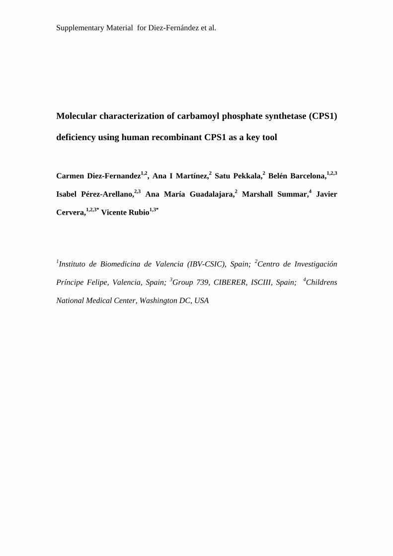
this plasmid by exploiting a unique HindIII CPS1 site, yielding pcDNA3.1-hCPS1. Then (Supp.
Fig. S1) a 3985 bp fragment comprising the CPS1 open reading frame (ORF) from base 580
onwards (base 1 is the A of the translation initiation codon) was excised from this plasmid by
BamHI and EcoRI, and was ligated into pFastBac. The ORF encoding mature CPS1 (lacking the
N-terminal mitochondrial targeting sequence, bp 1-117) was completed by in-frame ligation of a
PCR-generated fragment comprising bp 118-579 of the CPS1 cDNA (primers: Cloning_F and
Cloning_R, Supp. Table S1; they incorporate a BamHI site for cloning). This yielded pFastBac-
CPS1, which encodes mature hCPS1 (amino acids 40-1500) preceded N-terminally by the His6-
tag MSYYHHHHHHDYDIPTTENLYFQGAMDP. Site-directed mutagenesis of pFastBac-CPS1
was performed by the overlapping extension method (Quickchange kit from Stratagene) using
the forward and reverse primers given in Supp. Table S1. The correctness of the constructs, the
presence of the desired mutation, and the absence of unwanted mutations, were corroborated by
sequencing.
For producing rhCPS1 (Supp. Fig. S1), we used the commercial Bac-to-Bac® Baculovirus
Expression System (Invitrogen), following the manufacturer's directions. In short, E. coli Max
7
Efficiency DH10Bac cells (Invitrogen), transformed with pFastBac-CPS1, were grown on LB-
agar containing 50/7/10/40/100 μg/ml of, respectively, kanamycin/
gentamycin/tetracyclin/IPTG/Bluo-Gal. Individual white colonies were inoculated into 5-ml LB
medium with the same antibiotics, cultured overnight, and the bacmid was isolated. The
baculovirus was produced by transfecting Sf9 insect cells with CPS1 cDNA-carrying (proven by
PCR) bacmid, using Cellfectin/Grace medium (5 hours, 27ºC), followed by 3-day culture (27ºC,
six-well plate) in Sf900 medium (Invitrogen) containing 0.1% Pluronic F-68, 50 U/ml penicillin
and 50 μg/ml streptomycin. The culture was centrifuged, and the supernatant was used for
baculovirus enrichment by infecting with it (1:60 dilution) a suspension of 1.5x10⁶ Sf9 cells/ml.
After 48-hour culturing (27ºC, orbital shaking at 125 rpm) and centrifugation, the supernatant
was used to inoculate (1:50 dilution) a fresh cell suspension for CPS1 production, collecting the
cells by centrifugation after 3 days of culture as above.
Enzyme purification
Unless indicated, all steps (Supp. Fig. S1) were at 4ºC. To purify rhCPS1 (wild-type or
mutant forms), the insect cell pellet from a 50-ml culture was suspended in 3 ml of a lysis
solution [50 mM glycyl-glycine, pH 7.4, 1 mM dithiothreitol (DTT), 10% glycerol, 20 mM KCl,
0.1% Triton X-100, 5 μM E-64 protease inhibitor and 1% of the protease inhibitor cocktail for
His-tagged proteins (Sigma product P8849)] and was thawed (melting ice) and frozen (liquid
nitrogen or dry CO2-acetone mixture) three times. After 10-min centrifugation (16,000xg) and
supernatant filtration through a 0.22 μm membrane, the supernatant was applied to a HisTrap HP
1-ml column fitted in an ÄKTA FPLC system (GE Healthcare) that was equilibrated with 50
mM glycyl-glycine pH 7.4, 1 mM DTT, 10 % glycerol, 0.5 M NaCl, and 20 mM imidazole.
8
After a 10-ml column wash, a 15-ml linear gradient of 20-500 mM imidazole in the same
solution was applied. The CPS1-containing fractions (monitored by SDS-PAGE) were pooled,
concentrated to 3-5 mg protein/ml by centrifugal ultrafiltration (100-kDa cutoff membrane,
Amicon Ultra, Millipore), enriched with 10% extra glycerol and 1 mM extra DTT, and frozen at
-80ºC.
Rat liver CPS1, and E. coli-expressed recombinant Enterococcus faecalis ornithine
transcarbamylase (OTC) [Barcelona-Andrés et al., 2002] were purified as reported [Alonso and
Rubio, 1983; Marshall and Cohen, 1972].
Enzyme activity assays
In the standard CPS1 assay, CP was converted to citrulline, which was measured
colorimetrically [Pekkala et al., 2009]. The enzyme was incubated 10 min at 37ºC in an assay
mixture containing 50 mM glycyl-glycine pH 7.4, 70 mM KCl, 1 mM DTT, 20 mM MgSO4, 5
mM ATP, 35 mM NH4Cl, 50 mM KHCO3, 10 mM NAG, 5 mM L-ornithine and 4 U/ml OTC.
When the concentration of a substrate was varied, other substrates were kept at the
concentrations given above (unless indicated), with MgSO4 being in 15 mM excess over ATP.
Since NCG yields color in the citrulline assay, when testing the NCG concentration-
dependence of CPS1 activity, we used a continuous pyruvate kinase/lactate dehydrogenase
coupled assay in which ADP production was monitored as NADH oxidation at 340 nm
[Guthorlein and Knappe, 1968]. The assay (37ºC) used the same solution as the standard assay
except for the lack of OTC and ornithine and the inclusion of 2.5 mM phosphoenolpyruvate, 0.25
mM NADH, 40 μg/ml pyruvate kinase and 25 μg/ml lactate dehydrogenase. The kinetic
parameters for NAG were identical, within experimental error, in this assay and in the standard

9
assay. This NADH oxidation-coupled assay, without NH4Cl, was used for measuring the HCO3--
dependent ATPase partial reaction of CPS1. For measurement of the partial reaction of ATP
synthesis from CP and ADP, an NADP reduction-coupled assay [Yefimenko et al., 2005] was
used, monitoring the absorbance at 340 nm in a mixture at 37ºC containing 50 mM glycylglycine
pH 7.4, 0.1 M KCl, 15 mM MgSO4, 15 mM glucose, 0.5 mM ADP, 5 mM CP, 10 mM NAG, 1
mM NADP, 1 mM DTT, 0.1 mg/ml hexokinase and 25 μg/ml glucose-6-phosphate
dehydrogenase.
One CPS1 unit produces per minute 1 μmol citrulline or 2 μmol ADP. The program
GraphPad Prism (GraphPad Software, San Diego, California) was used for curve fitting.
Other techniques
SDS-PAGE [Laemmli, 1970] was performed in 8% polyacrylamide gels, with Coomassie
staining or, for cell extracts, by western blotting/immunostaining (ECL system, GE Healthcare),
utilizing an anti-rat liver CPS1 first antibody [Alonso et al., 1989]. Western blots of rat liver
CPS1 protease digests (Fig. 1) were stained with immunoperoxidase [Alonso et al., 1989] using
as first antibodies rabbit antisera against the electrophoretically separated [Amero et al., 1994]
N-terminal 40-kDa or C-terminal 120-kDa moieties of rat liver CPS1 (produced by limited
elastase digestion [Marshall and Fahien, 1988]). Protein was determined according to [Bradford,
1976] using bovine serum albumin as standard.
RESULTS AND DISCUSSION
Producing human CPS1 in a baculovirus/insect cell system
10
In the liver, CPS1 is produced as a precursor that is matured by cleavage of its N-terminal
38-39 amino acids upon entry to the mitochondria [Ryall et al., 1985]. We cloned for
recombinant expression the cDNA for human liver CPS1 without the N-terminal 39 codons,
which were replaced by a 28-codon N-terminal His6-tag. This allowed testing the effects of the
mutations on the mature form of the enzyme (the functional one in vivo). The tag simplified and
speeded CPS1 purification, what is important, given the instability of mammalian CPS1
[Raijman and Jones, 1976] and its sensitivity to proteolytic attack [Guadalajara et al., 1983]. The
CPS1 expressed here carries the more frequent Thr form of the p.Thr1406Asn polymorphism
(rs1047891, Ensemble database; allelic frequencies of T/N forms, ∼0.7/0.3) that has been
associated with increased frequency of some vascular pathologies possibly related to decreased
citrulline levels and nitric oxide production (see for example [Pearson et al., 2001]).
The procedure used (Supp. Fig. S1), optimized for highest rhCPS1 production, had as
important elements the use of insect cells cultured for two weeks after unfreezing, and the
infection of the cells with a nominal virus-to-cell ratio of 2 in the final CPS1-production step,
leaving 72 hours the infected cells in culture (27ºC, orbital shaking, 125 rev/min), in either 50 or
200 ml of medium, before cell harvesting. Purification was possible weeks after harvesting by
freezing cells pellets at -80ºC.
Prior studies with liver-purified hCPS1 [Pierson and Brien, 1980; Rubio et al., 1981] and
even more extensive studies with rat liver CPS1 [Alonso et al., 1992; Guadalajara et al., 1983;
Guthöhrlein and Knappe, 1968; Marshall and Fahien, 1985, 1988; Raijman and Jones, 1976]
showed that mammalian CPS1 is highly instable, requiring precautions to avoid oxidation,
proteolytic cleavage and inactivation of unknown cause but preventable by glycerol. Taking into
account these factors, we used for cell extract preparation and in subsequent steps a neutral
11
medium at 4ºC containing 10% glycerol, 1 mM DTT and a very extensive protease inhibitor
cocktail, using a fast 3-step purification protocol consisting of cell disruption by freeze-thawing
in 0.1% Triton X-100-containing CPS1-protecting solution, centrifugal clarification and 0.22
μm-pore membrane filtration, and a final step of fast Ni-affinity column chromatography with
imidazole gradient elution.
rhCPS1, which represented ∼10% of the soluble cell protein (Supp. Fig. S1, left track of
the gel), was largely soluble (not illustrated) and yielded after the column chromatography step
∼15 mg per L of cell culture, of highly active (Table 2), homogeneous and pure enzyme as
shown by the finding in SDS-PAGE of a single band migrating as expected for its sequence-
deduced mass (163.9 kDa). (Supp. Fig. S1, right track of the gel).
Recombinant human CPS1 represents well the natural liver enzyme
It was important to ascertain that rhCPS1 closely represents in all its properties natural
hCPS, particularly since, to minimize enzyme inactivation, the N-terminal His6-tag was not
removed. Similarly to liver CPS1, rhCPS1 has an essential requirement for NAG (Fig. 2A). Its
specific activity (Table 2) is similar to that reported for liver-isolated human CPS1 [Pierson and
Brien, 1980; Rubio et al., 1981]. Apparent Km values for ATP, HCO3- and NH4
+, and the Ka for
NAG and for the drug analogue of NAG, NCG, are also within published value ranges for
natural hCPS1 (Table 2 and Fig. 2) [Pierson and Brien, 1980; Rubio et al., 1981].
rhCPS1 also presents the same oligomeric state as liver-derived hCPS [Rubio et al.,
1981]. Gel exclusion chromatography experiments (Fig. 3A) reveal highly similar peaks and
estimated masses for the natural human enzyme and for rHCPS1: in both cases the apparent mass
12
exceeds by <30% that of the monomer, indicating that the enzyme consists of monomers in rapid
equilibrium with dimers, with strong predominance of the monomers.
Glycerol partially replaces NAG in the activation of human CPS1.
We also investigated potentially crucial traits of hCPS1 that had not been amenable to
investigation until now. Thus, we show here (Fig. 3B, right panel) that hrCPS1 is activated by
glycerol in the absence of NAG, whereas in the presence of NAG is inhibited by increasing
concentrations of glycerol. Similar observations had been made with rat liver CPS1 (Fig. 2B, left
panel and [Britton et al., 1981; Rubio et al., 1983b]). The substantial activation attained with
glycerol and the fact that this polyol appears to activate CPS1 without specifically binding to the
NAG site [Rubio et al., 1983b] make conceivable the possibility of developing treatments for
CPS1D patients with a damaged NAG site, in which CPS1 could be activated by compounds that
do not bind to the NAG site.
Limited proteolysis reveals the hCPS1 domain organization.
The domain composition of hCPS1 had not been made amenable to investigation until
now. The structure of E. coli CPS [Thoden et al., 1997] had revealed a multidomain organization
that was anticipated [Rubio, 1993] by the results of limited proteolysis and of other studies with
rodent and E. coli CPSs [Cervera et al., 1993; Evans and Balon, 1988; Marshall and Fahien,
1988; Powers-Lee and Corina, 1986; Rodríguez-Aparicio et al., 1989; Rubio et al., 1991].
Limited proteolysis studies with rodent CPS1 revealed four points of preferential proteolytic
cleavage that appear to correspond to exposed sequences linking adjacent globular domains, and
which are differentially cleaved by elastase, trypsin and chymotrypsin [Powers-Lee and Corina,
13
1986; Marshall and Cohen, 1988] (Fig. 1). We observed limited tryptic or elastase digestion
patterns with hrCPS1 that fully comply with the reported fragmentation patterns for rat liver
CPS1 (Figs. 1B and C) [Marshall and Fahien, 1988; Powers-Lee and Corina, 1986]. These
findings support the existence of the same domain organization and architecture in human and rat
CPS1, also supporting the similarity of this architecture with that of E. coli CPS [Rubio et al.,
1991; Thoden et al., 1997]. Interestingly, as with rat liver CPS1 [Marshall and Fahien, 1988],
chymotrypsin inactivated rhCPS1 (Fig. 1D, plot) while decreasing very little CPS1 polypeptide
size (Fig. 1D, gels), in agreement with prior experiments with rat liver CPS1 that showed that the
enzyme is cleaved very close to its C-terminus [Marshall and Fahien, 1988].
Testing the effects of clinical CPS1D mutations on enzyme functionality
The present results show that the properties of rhCPS1 mirror those of natural CPS1,
supporting the use of rhCPS1 for testing the impact of clinical mutations on enzyme function and
stability. We demonstrate this use here with eight missense mutations found in seven neonatal
CPS1D patients (Table 1 and Fig. 1A) [Eeds et al., 2006; Finckh et al., 1998; Kurokawa et al.,
2007; Summar, 1998] and with two trivial polymorphisms (Table 1 and Fig. 1A) [Finckh et al.,
1998; Summar et al., 2003]. Of these ten amino acid substitutions, two map in the bicarbonate
phosphorylation domain (a catalytic domain) and are therefore likely to hamper activity. The
other eight changes were selected because they map in CPS1 regions of unclear or unique
function for which bacterial CPS would not be a good model. Thus, four of them map in the
Glnase-like subdomain and another four in the C-terminal domain (Fig. 1A). In any case, E. coli
CPS would not have been an optimal model for nine of these amino acid substitutions, since only
one affected residue is strictly conserved in all CPSs (Table 1).
14
As expected, the polymorphisms p.Thr344Ala (c.1030A>G) and p.Gly1376Ser
(c.4126G>A), mapping respectively in the Glnase-like and the NAG-binding domains (Fig. 1A),
caused no negative effects on enzyme production (Fig. 4A), activity (Fig. 4C), kinetic parameters
for each substrate or for NAG (Table 3), or on thermal stability (Fig. 4D). In contrast, two
clinical mutations affecting respectively these same regions, p.Leu390Arg (c.1169T>G) and
p.Leu1381Ser (c.4142T>C) (Fig. 1A), were clearly disease-causing, since they induced strong
CPS1 instability as revealed by western blotting of insect cell extracts that showed (Fig. 4B)
proteolytic digestion bands instead of the clear-cut CPS1 band observed with the wild-type
enzyme. Indeed, these two mutations, which replace hydrophobic residues (residues of
hydrophobic nature are found at these positions in all known CPS sequences, Table 1) by polar
residues of larger (p.Leu390Arg) or smaller (p.Leu1381Ser) size, were predicted by the MutPred
server to be associated with loss of stability (Table 1).
The other six mutations studied (Table 1) were sufficiently stable to allow purification
(Fig. 4A). The p.Ala438Pro (c.1313G>C) and p.Thr544Met (c.1631C>T) mutations, which
affect the bicarbonate-phosphorylation domain, and the p.Thr1443Ala (c.4327A>G) mutation,
which affects the C-terminal domain, greatly decreased enzyme activity, to undetectable or
nearly undetectable values (Fig. 4C), clearly indicating that they are disease-causing. Although
having no detectable activity in the assay for the complete reaction (detection limit, 1% of the
activity of wild-type rhCPS1), the p.Ala438Pro mutant catalyzed the partial reaction of ATP
synthesis from ADP and CP (not shown) that is the reversal of the final step of the CPS1 reaction
(a three-step reaction: 1- bicarbonate phosphorylation; 2- carbamate production from
carboxyphosphate and ammonia; and 3- carbamate phosphorylation) but, as expected from the
domain that is affected by the mutation, it failed to catalyze the bicarbonate-dependent ATPase
15
partial reaction that reflects the bicarbonate phosphorylation step [Metzenberg et al., 1958; Rubio
et al. 1981].
The large decrease in the activity of the p.Thr544Met mutant was shown to be due (Table
3 and Fig. 2) to the combination of a ∼60-fold increase in the apparent Km for bicarbonate, a ∼20-
fold increase in the Ka for NAG and a ∼4-fold respective decrease and increase in the apparent
Vmax and Km for ammonia. Except the increase in the KaNAG, these kinetic aberrations stem from
the fact that this mutation affects a domain that catalyzes the initial two steps of the reaction,
involving as substrates ATP, bicarbonate (step 1) and ammonia (step 2) [Rubio 1993; Thoden et
al., 1997]. The increased KaNAG cannot be attributed to direct changes in the NAG site, which sits
on another enzyme domain [Rodríguez-Aparicio et al. 1989], but to the hampering of the cross-
talk between the bicarbonate phosphorylation domain and the NAG binding domain that results
in a large increase in affinity for NAG when both ATP and bicarbonate are bound [Alonso and
Rubio, 1983].
The decrease in enzyme activity caused by the p.Thr1443Ala mutation, (Fig. 4C) is
accounted by a nearly 200-fold increase in the KaNAG and by a nearly 10-fold decrease in the
apparent Vmax (Table 3 and Fig. 2A). The localization of the NAG-binding domain justifies these
effects if the mutation hampers NAG binding and the transmission of the regulatory signal from
the NAG site to both phosphorylation domains. Indeed, the two partial reactions of the enzyme,
which reflect the two phosphorylation steps, were undetectable in this mutant (results not
shown).
The other three mutations examined here, p.Asn355Asp (c.1063A>G) and p.Tyr389Cys
(c.1166A>G) which affect the Glnase-like domain and coexist in patient 1, and p.Ala1378Thr
(c.4132G>A), which maps on the NAG-binding domain, appear to have too little an effect on
16
enzyme activity or on kinetic parameters to justify the neonatal deficiency (Fig. 4C, Table 3).
The largest changes observed with these mutations were a ∼4-fold decrease in Vmax and a nearly
3-fold increase in the Ka for NAG, occuring with the p.Asn355Asp mutation (Table 3). However,
thermal inactivation assays (Fig. 4D) revealed that these mutations substantially decreased the
thermal stability of rhCPS1, particularly p.Asn355Asp, which lowered ∼11ºC the mid-
inactivation temperature. In contrast, the temperature dependence of enzyme inactivation was
identical for the two polymorphisms and for the wild-type enzyme (Fig. 4D). The combined
effects of the decrease in Vmax and the modest increase in KaNAG with the p.Asn355Asp mutant,
together with the decreased enzyme stability, may result in enzyme deficiency. This may also be
the case with the p.Tyr389Cys mutation, which decreased ∼40% enzyme activity (Fig. 4C) and
caused a substantial, although less drastic effect on thermal inactivation. Finally, with
p.Ala1378Thr, the deficiency could be due to the combination of the decreased stability and a
twofold increase in apparent Km for ATP (Table 3). The fact that these last two mutations are,
respectively, only one and three positions away from Leu390 and Leu1381, two residues for
which their p.Leu390Arg and p.Leu1381Ser mutations were found to cause dramatic loss of
enzyme stability (see above), lends further support to the view that these mutations may hamper
sufficiently enzyme stability "in vivo" to cause enzyme deficiency.
Influence of the substrates and of NAG on the resistance of human CPS1 to proteolytic or
thermal inactivation
From all of the above, enzyme destabilization appears a crucial element in the causation
of CPS1D with five of the eight missense mutations studied here (p.Asn355Asp, p.Tyr389Cys,
p.Leu390Arg, p.Ala1378Thr and p.Leu1381Ser). Therefore, enzyme stabilization by ligands
17
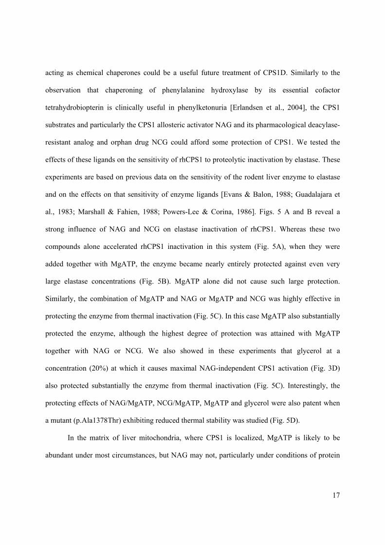
acting as chemical chaperones could be a useful future treatment of CPS1D. Similarly to the
observation that chaperoning of phenylalanine hydroxylase by its essential cofactor
tetrahydrobiopterin is clinically useful in phenylketonuria [Erlandsen et al., 2004], the CPS1
substrates and particularly the CPS1 allosteric activator NAG and its pharmacological deacylase-
resistant analog and orphan drug NCG could afford some protection of CPS1. We tested the
effects of these ligands on the sensitivity of rhCPS1 to proteolytic inactivation by elastase. These
experiments are based on previous data on the sensitivity of the rodent liver enzyme to elastase
and on the effects on that sensitivity of enzyme ligands [Evans & Balon, 1988; Guadalajara et
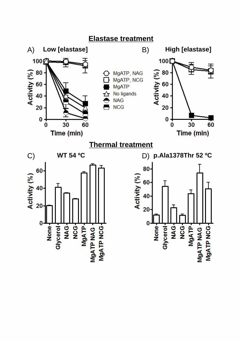
al., 1983; Marshall & Fahien, 1988; Powers-Lee & Corina, 1986]. Figs. 5 A and B reveal a
strong influence of NAG and NCG on elastase inactivation of rhCPS1. Whereas these two
compounds alone accelerated rhCPS1 inactivation in this system (Fig. 5A), when they were
added together with MgATP, the enzyme became nearly entirely protected against even very
large elastase concentrations (Fig. 5B). MgATP alone did not cause such large protection.
Similarly, the combination of MgATP and NAG or MgATP and NCG was highly effective in
protecting the enzyme from thermal inactivation (Fig. 5C). In this case MgATP also substantially
protected the enzyme, although the highest degree of protection was attained with MgATP
together with NAG or NCG. We also showed in these experiments that glycerol at a
concentration (20%) at which it causes maximal NAG-independent CPS1 activation (Fig. 3D)
also protected substantially the enzyme from thermal inactivation (Fig. 5C). Interestingly, the
protecting effects of NAG/MgATP, NCG/MgATP, MgATP and glycerol were also patent when
a mutant (p.Ala1378Thr) exhibiting reduced thermal stability was studied (Fig. 5D).
In the matrix of liver mitochondria, where CPS1 is localized, MgATP is likely to be
abundant under most circumstances, but NAG may not, particularly under conditions of protein
18
restriction as when a urea cycle deficiency is suspected. Therefore, under these circumstances the
administration of NCG might help protect the enzyme from thermal inactivation or from
proteolytic degradation. Therefore, studies on the effects of NCG on CPS1 stability “in vivo” are
warranted. Indeed, a genetically demonstrated CPS1D patient has been documented to respond to
NCG administration [Williams et al., 2010].
Final comments
rhCPS1 is shown here to mirror the natural human enzyme or the rat liver enzyme in all
aspects analyzed, including substrate and activator kinetics, oligomeric form, domain
composition, sensitivity to proteases and protection thereof by ligands, and ability of glycerol to
replace NAG as a CPS1 activator. Our confirmation that the domain organization is that reported
for other CPSs supports the present structural rationalizations of the effects of missense changes
in CPS1 [Häberle et al., 2011; Martínez et al., 2010]. Since recombinant production of hCPS1
permits the introduction of amino acid changes at will, the way is now open for testing the
functional impact of missense mutations found in CPS1D. It would be desirable to compare the
present system and even to complement it with the one using Schyzosaccharomyces pombe as the
expression host of hCPS1 [Ahuja and Powers-Lee, 2008]. Although data on this system are very
limited, the simultaneous use of both expression systems may prove desirable for maximizing the
number of clinical mutants that can be tested. After all, both expression systems are heterologous
with respect to hCPS1 and therefore the possibility cannot be excluded that some mutant forms
can be expressed in one system but not in the other.
With the present system, clear-cut correlations between given missense mutations and
specific molecular phenotypes can be established, hopefully shedding light on the degree of
19
severity of different mutations. Indeed, the lack of detectable effects of the two polymorphisms
studied here, and the severity of the effects demonstrated for six of the eight mutations analyzed,
clearly indicate that the experimental studies with rhCPS1 mutants expressed “in vitro” identify
disease-causing mutations. Even with the two mutations that caused the less drastic effects, they
triggered some negative changes on enzyme activity and/or stability that were not observed with
the two polymorphisms. In any case, the extension of the present pilot study to larger series of
clinical CPS1D mutations should permit deeper ascertaining of the sensitivity of our approach to
identify disease-causing mutations and for estimating their actual severity.
Our results shed also light on the role of the Glnase-like domain of CPS1 and on the
reasons for its preservation despite the fact that CPS1 does not use glutamine [Rubio et al.,
1981]. The observations that the amino acid substitutions p.Asn355Asp and p.Tyr398Cys,
mapping in the Glnase-like domain of CPS1 (Fig. 1A), do not inactivate CPS1 or cause dramatic
changes in Km values for the substrates (Table 3), agrees with the general belief that this domain
is not directly involved in the enzyme reaction. However, these mutations, as well as another two
mutations (p.S123F and p.H337R) introduced previously in the N-terminal region of rat CPS1
[Pekkala et al., 2010] resulted in 40-75% reduction in enzyme activity (see for the present
mutations Fig. 4C). These results also agree with a study [Ahuja and Powers-Lee, 2008] in which
hCPS1 lacking the entire N-terminal region exhibited a 700-fold reduction in enzyme activity,
although the very drastic change of deleting 25% of the protein molecule may render difficult the
interpretation of this extreme degree of inactivation. In any case, the changes revealed by our
present studies and by earlier studies with single amino acid substitutions affecting this CPS1
domain clearly support an activating role of this N-terminal region on CP synthesis, which is
catalyzed by the C-terminal moiety of the enzyme [Cervera et al., 1993; Rubio, 1993]. Such
20
activation appears reminiscent of the one caused by the small subunit of E. coli CPS on the
catalysis by the large subunit of the reaction from ammonia [Meister, 1989]. The Glnase-like
domain also stabilizes the enzyme, since the p.Leu390Arg, p.Asn355Asp and p.Tyr389Cys
mutations decrease CPS1 stability. Again this role is reminiscent of the strong stabilization
triggered in E. coli CPS upon association of the small and large subunits [Cervera et al., 1993].
The activation of CP synthesis, and the enhanced enzyme stability, may be sufficiently important
advantages to warrant retention of the N-terminal region in CPS1.
Our present findings also shed light on the roles of the C-terminal domain. We localized
"in silico" the NAG site in the crystal structure of this domain [Pekkala et al., 2009], providing as
experimental support for this localization the results of photoaffinity labeling with N-
chloroacetyl-L-glutamate and of site-directed mutagenesis of rat liver CPS1 [Pekkala et al.,
2009]. We now provide even more direct proof for such localization with hCPS1, the enzyme for
which the crystal structure of the C-terminal domain was determined. Thus, among all the
mutations studied here, the one mapping closest to the proposed NAG site, p.Thr1443Ala,
produces by far the most drastic decrease in the apparent affinity for NAG (two orders of
magnitude decrease) (Table 3). In our proposed NAG binding site [Pekkala et al., 2009],
Thr1443 is close to the bound activator, adjacent to a residue of the NAG site and to one of the
three lid residues that cover the bound NAG molecule (Supp. Fig. S2). The importance of
Thr1443 for effector regulation of CPS1 is highlighted also by the observation that
phosphorylation of the equivalent residue in hamster CPSII (a component of CAD, the
trifunctional enzyme involved in pyrimidine biosynthesis), Ser1406 [Simmer et al., 1990],
hampers CPSII allosteric regulation by its negative effector UTP [Carrey et al., 1985]. As NAG
in CPS1, UTP binds to the C-terminal domain of CPSII [Liu et al. 1994].
21
The present data also evidence that the C-terminal domain is an important determinant for
CPS1 stability, since two mutations mapping in this domain, p.Ala1378Thr and p.Leu1381Ser,
which affect the inner face of a helix from the outer layer of the αβα sandwich conforming this
domain (Supp. Fig. S2), substantially or very drastically destabilized the enzyme. This important
impact on enzyme stability clearly supports a high degree of integration of the C-terminal
domain in the CPS1 architecture. Thus, despite the multidomain character of CPS1, the
architecture of this enzyme would appear to be highly cooperative, explaining the influence of
NAG binding on events that occur far away from the C-terminal domain, such as the activation
of both phosphorylation domains [Rubio et al., 1983a].
ACKNOWLEDGMENTS
Supported by grants from the Fundación Alicia Koplowitz 2011, Prometeo 2009/051 from the
Valencian Government, and BFU2011-30407 and SAF2010-17933 from the Science Department
of the Spanish Government. CD and AIM had FPU (Spanish Government) and CIPF-Bancaixa
fellowships.
REFERENCES
Adzhubei IA, Schmidt S, Peshkin L, Ramensky VE, Gerasimova A, Bork P, Kondrashov AS,
Sunyaev SR. 2010. A method and server for predicting damaging missense mutations.
Nature Methods 7:248-249.
Ahuja V, Powers-Lee SG. 2008. Human carbamoyl-phosphate synthetase: insight into N-
acetylglutamate interaction and the functional effects of a common single nucleotide
polymorphism. J Inherit Metab Dis 31:481-491.
22
Alonso E, Girbés J, García-España A, Rubio V. 1989. Changes in urea cycle-related metabolites
in the mouse after combined administration of valproic acid and an amino acid load. Arch
Biochem Biophys 272:267-273.
Alonso E, Cervera J, García-España A, Bendala E, Rubio V. 1992. Oxidative inactivation of
carbamoyl phosphate synthetase (ammonia). Mechanism and sites of oxidation, degradation
of the oxidized enzyme, and inactivation by glycerol, EDTA, and thiol protecting agents. J
Biol Chem 267:4524-4532.
Alonso E, Rubio V. 1983. Binding of N-acetyl-L-glutamate to rat liver carbamoyl phosphate
synthetase (ammonia). Eur J Biochem 135:331-337.
Amero SA, James TC, Elgin SC. 1994. Production of antibodies using proteins in gel bands.
Methods Mol Biol 32:401-406.
Barcelona-Andrés B, Marina A, Rubio V. 2002. Gene structure, organization, expression, and
potential regulatory mechanisms of arginine catabolism in Enterococcus faecalis. J
Bacteriol 184:6289-6300.
Bradford MM. 1976. A rapid and sensitive method for the quantitation of microgram quantities
of protein utilizing the principle of protein-dye binding. Anal Biochem 72:248-254.
Britton HG, Rubio V, Grisolia S. 1981. Synthesis of carbamoyl phosphate by carbamoyl
phosphate synthetase I in the absence of acetylglutamate. Activation of the enzyme by
cryoprotectants. Biochem Biophys Res Commun 99:1131-1137.
Brusilow SW, Horwich AL. 2001. Urea cycle enzymes. In: Scriver CR, Beaudet AL, Sly WS,
Valle D, editors; Child B, Kinzler KW, Vogelstein B, associated editors. The Metabolic and
Molecular Bases of Inherited Disease, 8e. New York: McGraw-Hill. Vol 2, p 1909-1963.
23
Carrey EA, Campbell DG, Hardie DG. 1985. Phosphorylation and activation of hamster
carbamyl phosphate synthetase II by cAMP-dependent protein kinase. A novel mechanism
for regulation of pyrimidine nucleotide biosynthesis. EMBO J 4:3735-3742.
Cervera J, Conejero-Lara F, Ruiz-Sanz J, Galisteo ML, Mateo PL, Lusty CJ, Rubio V. 1993. The
influence of effectors and subunit interactions on Escherichia coli carbamoyl-phosphate
synthetase studied by differential scanning calorimetry. J Biol Chem 268:12504-12511.
Eeds AM, Hall LD, Yadav M, Willis A, Summar S, Putnam A, Barr F, Summar ML. 2006. The
frequent observation of evidence for nonsense-mediated decay in RNA from patients with
carbamyl phosphate synthetase I deficiency. Mol Genet Metab 89:80-86.
Erlandsen H, Pey AL, Gámez A, Pérez B, Desviat LR, Aguado C, Koch R, Surendran S, Tyring
S, Matalon R, Scriver CR, Ugarte M et al. (2004) Correction of kinetic and stability defects
by tetrahydrobiopterin in phenylketonuria patients with certain phenylalanine hydroxylase
mutations. Proc Natl Acad Sci U S A. 101:16903-16908.
Evans DR, Balon MA. 1988. Controlled proteolysis of ammonia-dependent carbamoyl-
phosphate synthetase I from Syrian hamster liver. Biochim Biophys Acta 953:185-196.
Finckh U, Kohlschutter A, Schafer H, Sperhake K, Colombo JP, Gal A. 1998. Prenatal diagnosis
of carbamoyl-phosphate synthetase I deficiency by identification of a missense mutation in
CPS1. Hum Mutat 12:206-211.
Guadalajara AM, Rubio V, Grisolía S. 1983. Inactivation of carbamoyl phosphate synthetase
(ammonia) by elastase as a probe to investigate binding of the substrates. Biochem Biophys
Res Commun. 117:238-244.
24
Guthöhrlein G, Knappe J. 1968. Structure and function of carbamoylphosphate synthase. I.
Transitions between two catalytically inactive forms and the active form. Eur J Biochem
7:119-127.
Häberle J, Shchelochkov OA, Wang J, Katsonis P, Hall L, Reiss S, Eeds A, Willis A, Yadav M,
Summar S; Urea Cycle Disorders Consortium, Lichtarge O et al. 2011. Molecular defects in
human carbamoyl phosphate synthetase I: mutational spectrum, diagnostic and protein
structure considerations. Hum Mutat 32:579-585.
Häberle J, Boddaert N, Burlina A, Chakrapani A, Dixon M, Huemer M, Karall D, Martinelli D,
Sanjurjo Crespo P, Santer R, Servais A et al.. 2012. Suggested Guidelines for the Diagnosis
and Management of Urea Cycle Disorders. Orphanet J Rare Dis 7:32.
Haraguchi Y, Uchino T, Takiguchi M, Endo F, Mori M, Matsuda I. 1991. Cloning and sequence
of a cDNA encoding human carbamyl phosphate synthetase I: molecular analysis of
hyperammonemia. Gene 107:335-340.
Kurokawa K, Yorifuji T, Kawai M, Momoi T, Nagasaka H, Takayanagi M, Kobayashi K,
Yoshino M, Kosho T, Adachi M, Otsuka H, Yamamoto S, et al.. 2007. Molecular and
clinical analyses of Japanese patients with carbamoylphosphate synthetase 1 (CPS1)
deficiency. J Hum Genet 52:349-354.
Laemmli UK. 1970. Cleavage of structural proteins during the assembly of the head of
bacteriophage T4. Nature 227:680-685.
Li B, Krishnan VG, Mort ME, Xin F, Kamati KK, Cooper DN, Mooney SD, Radivojac P. 2009.
Automated inference of molecular mechanisms of disease from amino acid substitutions.
Bioinformatics 25: 2744-2750.

25
Liu X, Guy HI, Evans DR. 1994. Identification of the regulatory domain of the mammalian
multifunctional protein CAD by the construction of an Escherichia coli hamster hybrid
carbamyl-phosphate synthetase. J Biol Chem 269:27747-27755.
Marshall M, Cohen PP. 1972. Ornithine transcarbamylase from Streptococcus faecalis and
bovine liver. I. Isolation and subunit structure. J Biol Chem. 247:1641-1653.
Marshall M, Fahien LA. 1985. Proximate sulfhydryl groups in the acetylglutamate complex of
rat carbamylphosphate synthetase I: their reaction with the affinity reagent 5'-p-
fluorosulfonylbenzoyladenosine. Arch Biochem Biophys 241:200-214.
Marshall M, Fahien LA. 1988. Proteolysis as a probe of ligand-associated conformational
changes in rat carbamyl phosphate synthetase I. Arch Biochem Biophys 262:455-470.
Martínez AI, Pérez-Arellano I, Pekkala S, Barcelona B, Cervera J. 2010. Genetic, structural and
biochemical basis of carbamoyl phosphate synthetase 1 deficiency. Mol Genet Metab
101:311-323.
Meister A. 1989. Mechanism and regulation of the glutamine-dependent carbamyl phosphate
synthetase of Escherichia coli. Adv Enzymol Relat Areas Mol Biol 62:315-374.
Metzenberg RL, Marshall M, Cohen PP. 1958. Carbamyl phosphate synthetase: studies on the
mechanism of action. J Biol Chem 233:1560-1564.
Nyunoya H, Broglie KE, Widgren EE, Lusty CJ. 1985. Characterization and derivation of the
gene coding for mitochondrial carbamyl phosphate synthetase I of rat. J Biol Chem
260:9346-9356.
Pearson DL, Dawling S, Walsh WF, Haines JL, Christman BW, Bazyk A, Scott N, Summar ML.
2001. Neonatal pulmonary hypertension--urea-cycle intermediates, nitric oxide production,
and carbamoyl-phosphate synthetase function. N Engl J Med. 344:1832-1838.
26
Pekkala S, Martinez AI, Barcelona B, Gallego J, Bendala E, Yefimenko I, Rubio V, Cervera J.
2009. Structural insight on the control of urea synthesis: identification of the binding site for
N-acetyl-L-glutamate, the essential allosteric activator of mitochondrial carbamoyl-
phosphate synthetase1. Biochem J 424:211-220.
Pekkala S, Martínez AI, Barcelona B, Yefimenko I, Finckh U, Rubio V, Cervera J. 2010.
Understanding carbamoyl-phosphate synthetase I (CPS1) deficiency by using expression
studies and structure-based analysis. Hum Mutat 31:801-808.
Pierson DL, Brien JM. 1980. Human carbamylphosphate synthetase I. Stabilization, purification,
and partial characterization of the enzyme from human liver. J Biol Chem 255:7891-7895.
Powers-Lee SG, Corina K. 1986. Domain structure of rat liver carbamoyl phosphate synthetase I.
J Biol Chem 261:15349-15352.
Raijman L, Jones ME. 1976. Purification, composition, and some properties of rat liver carbamyl
phosphate synthetase (ammonia). Arch Biochem Biophys 175:270-278.
Rodriguez-Aparicio LB, Guadalajara AM, Rubio V. 1989. Physical location of the site for N-
acetyl-L-glutamate, the allosteric activator of carbamoyl phosphate synthetase, in the 20-
kilodalton COOH-terminal domain. Biochemistry 28:3070-3074.
Rubio V. 1993. Structure-function studies in carbamoyl phosphate synthetases. Biochem Soc
Trans 21:198-202.
Rubio V, Britton HG, Grisolia S. 1983a. Mitochondrial carbamoyl phosphate synthetase activity
in the absence of N-acetyl-L-glutamate. Mechanism of activation by this cofactor. Eur J
Biochem 134:337-343.
Rubio V, Britton HG, Grisolía S. 1983b. Activation of carbamoyl phosphate synthetase by
cryoprotectants. Mol Cell Biochem 53-54:279-298.
27
Rubio V, Cervera J, Lusty CJ, Bendala E, Britton HG. 1991. Domain structure of the large
subunit of Escherichia coli carbamoyl phosphate synthetase. Location of the binding site for
the allosteric inhibitor UMP in the COOH-terminal domain. Biochemistry 30:1068-1075.
Rubio V, Ramponi G, Grisolia S. 1981. Carbamoyl-phosphate synthetase I of human liver.
Purification, some properties and immunological cross-reactivity with the rat liver enzyme.
Biochim Biophys Acta 659:150-160.
Ryall J, Nguyen M, Bendayan M, Shore GC. 1985. Expression of nuclear genes encoding the
urea cycle enzymes, carbamoyl-phosphate synthetase I and ornithine carbamoyl transferase,
in rat liver and intestinal mucosa. Eur J Biochem 152:287-292.
Shih VE. 1976 Congenital hyperammonemic syndromes. Clin Perinatol 3:3-14.
Simmer JP, Kelly RE, Rinker AG Jr, Scully JL, Evans DR. 1990. Mammalian carbamyl
phosphate synthetase (CPS). DNA sequence and evolution of the CPS domain of the Syrian
hamster multifunctional protein CAD. J Biol Chem 265:10395-10402.
Summar ML. 1998. Molecular genetic research into carbamoyl-phosphate synthase I: molecular
defects and linkage markers. J Inherit Metab Dis 21 Suppl 1:30-39.
Summar ML, Dasouki MJ, Schofield PJ, Krishnamani MR, Vnencak-Jones C, Tuchman M, Mao
J, Phillips JA 3rd. 1995. Physical and linkage mapping of human carbamyl phosphate
synthetase I (CPS1) and reassignment from 2p to 2q35. Cytogenet Cell Genet 71:266-267.
Summar ML, Hall LD, Eeds AM, Hutcheson HB, Kuo AN, Willis AS, Rubio V, Arvin MK,
Schofield JP, Dawson EP. 2003. Characterization of genomic structure and polymorphisms
in the human carbamyl phosphate synthetase I gene. Gene 311:51-57.
28
Thoden JB, Holden HM, Wesenberg G, Raushel FM, Rayment I. 1997. Structure of carbamoyl
phosphate synthetase: a journey of 96 Å from substrate to product. Biochemistry 36:6305-
6316.
Williams M, Huijmans JGM, van Diggelen OP, van der Low EJTM, de Klerk JBC, Haeberle J
(2010) Carbamoyl phosphate synthetase I (CPS 1) deficiency: treatment with carglumic acid
(Carbaglu) J Inherit Metab Dis 33 (Suppl 1):S118
Yefimenko I, Fresquet V, Marco-Marín C, Rubio V, Cervera J. 2005. Understanding carbamoyl-
phosphate synthetase deficiency: impact of clinical mutations on enzyme functionality. J
Mol Biol 349:127-141.

29
LEGENDS TO FIGURES
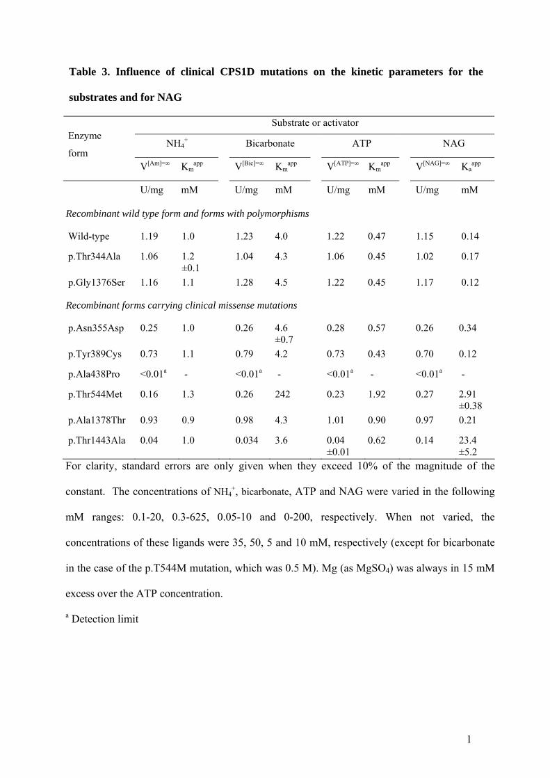
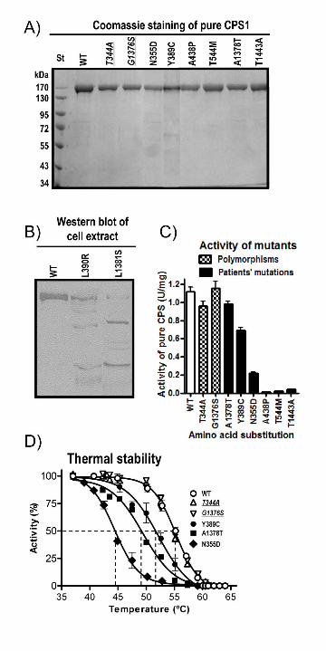
Figure 1. Domain composition of human CPS1. A: Linear scheme of mature CPS1, highlighting
four interdomain linkers that are cleaved by the indicated proteases as identified in rodent CPS1,
with the corresponding fragments masses (in kDa) above them [Marshall and Fahien, 1988].
Chymo, chymotrypsin. Trypsin(2) denotes cleavage after scission at the other tryptic site. The
bars shaded grey schematize the 40-kDa N-terminal and the 120-kDa C-terminal CPS1 moieties
that correspond to the small and large subunits of E. coli CPS, respectively. Polyclonal rabbit
antibodies raised against these isolated moieties are called Anti-40 and Anti-120. Functional
domains are shown in background texture and are identified. Glnase-like corresponds to the
Glnase domain of bacterial CPS, but has no known function in CPS1. ??, unknown function. A
dashed line separates two domains composing a proteolytic domain. The two polymorphisms (in
italic and grey background) and the eight clinical mutations studied here are mapped in the CPS1
polypeptide with banners. B, C and D: Top part, fragments generated with trypsin, elastase or
chymotrypsin [Marshall and Fahien, 1988]. The cleavage points are identified with the ABCD
notation used in panel A. Each tryptic fragment is called T1-T3, elastase fragments E1-E4 and
the chymotryptic one C1. Their approximate masses and their reactivity with Anti-40 and Anti-
120 antibodies are given. The 20 kDa C-terminal fragment is rapidly degraded [Marshall and
Fahien, 1988] and is shown crossed. Lower parts, SDS-PAGE of digested recombinant human
CPS1 and, for comparison, of rat liver CPS1, stained with Coomassie or by immunoperoxidase
with Anti-40 or Anti-120 after western blotting (only done with the rat enzyme). Digestions of
CPS1 (1.3-2 mg/ml) were at 37º, for 15-30 min with the indicated protease (4-16 μg/ml;
pancreatic, from Boheringer Mannheim or Sigma) in 35 mM Tris-HCl pH 7.4, 9% glycerol, 1.5

30
mM DTT, 20 mM KCl and 10 mM NAG. The enzyme was preincubated at least 15 min at 37ºC
prior to the addition of the protease. This addition was considered time zero. Fragments are
identified in the gels as T1-T3, E1-E4 or C1. Note in (D) that while chymotrypsin inactivates rat
and human CPS1 (see the plot), there is little decrease (∼2 kDa) in polypeptide mass (top panels),
corresponding to the loss of approximately 12 residues from the enzyme C-terminus documented
earlier for rat CPS1 [Marshall and Fahien, 1988].
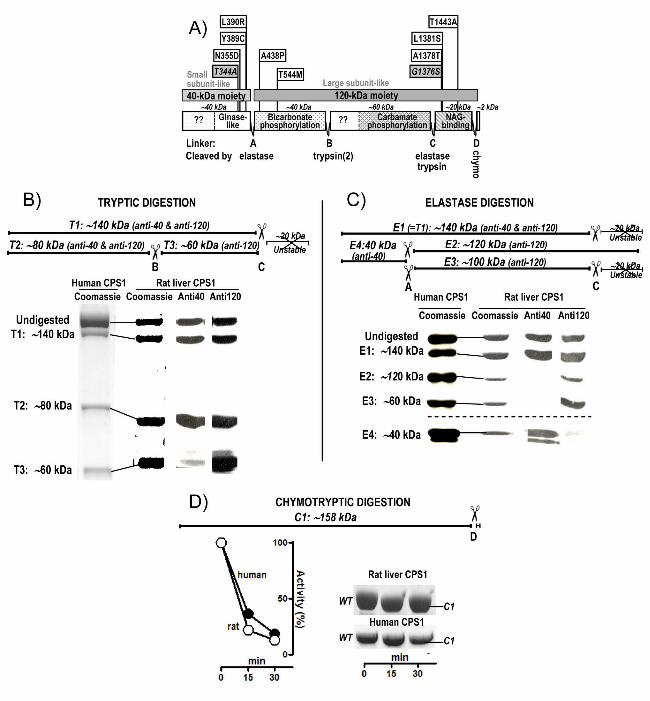
Figure 2. Dependence of CPS1 activity on the concentrations of NAG and bicarbonate for non-
mutated (WT) CPS1 and for the indicated mutant forms. The curves fitted to the data are
hyperbolae for the kinetic constants given in Table 3. The insets expand the curves for the
mutants, to demonstrate the hyperbolic kinetics despite the decreased activity. A: NAG varied. In
the case of the p.Thr544Met mutant the concentration of bicarbonate was fixed at 0.5 M. B:
bicarbonate varied.
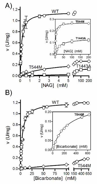
Figure 3. Recombinant human CPS1 replicates the natural enzyme in the oligomeric state and
the ability to be activated and inhibited by glycerol. A: Gel exclusion chromatography analysis.
The upper lines are the semilogarithmic plots of the masses of marker proteins (closed circles)
versus their elution volumes. Peaks of CPS1 activity (left panel, open symbols) or of CPS1
protein (peak in the right panel) are shown below them. The mass of CPS1, estimated by
interpolation, is shown above a vertical line emerging from the peak. Left panel, results with the
enzyme purified from human liver [Rubio et al., 1981], using a conventional 0.9 × 56 cm column
of Sephadex G-200 run at 23ºC. For further details, including the list and masses of standards
used, see [Rubio et al., 1981]. Right panel, present results with the recombinant human enzyme
31
under essentially the same conditions as those in [Rubio et al., 1981] except for the use of a
Superdex 200HR (10/300) column mounted on an Äkta fast protein liquid chromatography
system, at a flow rate of 0.5 ml/min, the application of 0.02 ml of a 3 mg/ml solution of human
recombinant CPS1 or appropriate amounts of protein standards, and the continuous monitoring
of the optical absorption at 280 nm. Protein standards for this panel were (masses are given in
parenthesis, in kDa) bovine pancreatic ribonuclease A (13.7), bovine serum albumin (66.4), yeast
alcohol dehydrogenase dimer (73.4) and tetramer (146.8), β-amylase (223.8) and ferritin (440).
B: Effects of the addition of glycerol on CPS1 activity in the presence or in the absence of 10
mM NAG as indicated (closed and open circles, respectively). Velocities, measured as ADP
production, are expressed as a percentage of the velocity in the presence of 10 mM NAG and in
the absence of glycerol. Left and right panels, results with the rat liver enzyme and with the
recombinant human enzyme, respectively.
Figure 4. Production and properties of the enzyme forms with the amino acid substitutions
studied here. A: SDS-PAGE (8% polyacrylamide, Coomassie staining) of purified human
recombinant CPS1, either wild-type (WT) or carrying the indicated mutations. Polymorphisms
are shown in italic script and underlined. St, protein markers with masses indicated on the side.
B: Western blot of cell extracts (∼20 μg protein per well) of Sf9 cells infected with baculoviruses
encoding either wild-type human CPS1 or its p.Leu390Arg or p.Leu1381Ser mutants. A
polyclonal rabbit antiserum against rat liver CPS1 was used for immunostaining. C: Enzyme
activity (standard assay conditions) of the purified wild-type or mutant human CPS1 forms. The
bars for the wild-type enzyme, for the two polymorphisms and for the forms carrying clinical
32
mutations are filled in white, checkerboard and black, respectively. Error bars give standard
errors. D: Thermal stability of wild-type recombinant CPS1, and of the forms carrying either the
polymorphisms (in italic and underlined), or the indicated clinical mutations. The enzyme, at 0.5-
1 mg/ml in a solution of 50 mM glycyl-glycine pH 7.4, 20 mM KCl an 20% glycerol, was heated
15 min at the indicated temperature, then rapidly cooled to 0ºC, and its activity determined
immediately at 37ºC. The horizontal dashed line marks 50% inactivation, whereas the vertical
dashed lines cross the X-axis at the temperature at which 50% inactivation occurs for each
enzyme form.
Figure 5. Effects of ligands on the inactivation of rhCPS1 by elastase or by heating. When
indicated, NAG, NCG and ATP were added, at 10 mM concentrations. When ATP was added,
MgSO4 was also added at a concentration of 20 mM. Incubations were terminated by dilution in
the continuous enzyme activity assay, monitoring ADP production at 37ºC. Results are given as
percentages of the activity not having undergone the corresponding proteolytic or heating
treatment. A and B: Digestions of rhCPS1 with respective elastase concentrations of 10 μg/ml or
50 μg/ml. Other conditions were as in Fig. 1D. C and D: Thermal inactivation of the wild-type
or the p.Ala1378Thr mutant forms of rhCPS1 (both used at 0.1 mg/ml concentrations) after 15-
min incubation at the indicated temperatures in a solution of 50 mM glycyl-glycine pH 7.4, 20
mM KCl, 1 mM DTT with the indicated ligands or with 20% glycerol.
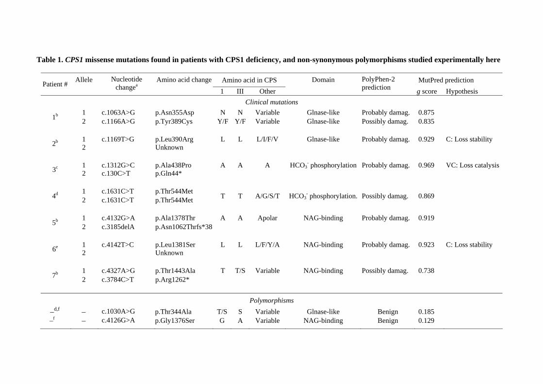
Table 1. CPS1 missense mutations found in patients with CPS1 deficiency, and non-synonymous polymorphisms studied experimentally here
Amino acid in CPS MutPred prediction Patient # Allele
Nucleotide changea
Amino acid change 1 III Other
Domain
PolyPhen-2 prediction g score Hypothesis
Clinical mutations 1 c.1063A>G p.Asn355Asp N N Variable Glnase-like Probably damag. 0.875 1b 2 c.1166A>G p.Tyr389Cys Y/F Y/F Variable Glnase-like Possibly damag. 0.835
1 c.1169T>G p.Leu390Arg L L L/I/F/V Glnase-like Probably damag. 0.929 C: Loss stability 2b 2 Unknown
1 c.1312G>C p.Ala438Pro A A A HCO3
- phosphorylation Probably damag. 0.969 VC: Loss catalysis 3c 2 c.130C>T p.Gln44*
1 c.1631C>T p.Thr544Met 4d 2 c.1631C>T p.Thr544Met T T A/G/S/T HCO3- phosphorylation. Possibly damag. 0.869
1 c.4132G>A p.Ala1378Thr A A Apolar NAG-binding Probably damag. 0.919 5b 2 c.3185delA p.Asn1062Thrfs*38
1 c.4142T>C p.Leu1381Ser L L L/F/Y/A NAG-binding Probably damag. 0.923 C: Loss stability 6e 2 Unknown
1 c.4327A>G p.Thr1443Ala T T/S Variable NAG-binding Possibly damag. 0.738 7b 2 c.3784C>T p.Arg1262*
Polymorphisms
–d,f – c.1030A>G p.Thr344Ala T/S S Variable Glnase-like Benign 0.185 –f – c.4126G>A p.Gly1376Ser G A Variable NAG-binding Benign 0.129
All patients were reported to exhibit neonatal presentations. Amino acids are shown in three-letter code for the human substitutions and in one-
letter code for occurrence in the various CPSs. Variable denotes the occurrence at a given position in the indicated group of CPSs, of >4 types of
amino acids with no constant chemical characteristics (polar, apolar, charged, etc). When the second allele is not a missense change, no data are
given on residue conservation and pathogenicity potential. For localization of the CPS1 domains and the mapping of the mutations in the CPS1
polypeptide, see Fig. 4A. PolyPhen-2 grades the probability of a damaging effect of an amino acid substitution, as probably damaging, possibly
damaging and benign. MutPred gave a g score corresponding to the probability that a given amino acid substitution was deleterious/disease-
associated. When indicated this server was very confident (VC) or confident (C) that the indicated changes caused loss of stability or loss of a
catalytic residue.
a cDNA reference sequence NM_001875.4 (GenBank). +1 corresponding to the A of the translation initiation codon in the reference sequence
(according to journal guidelines under www.hgvs.org/mutnomen).
bEeds et al., 2006.
c Kurokawa et al., 2007.
d Finckh et al., 1998.
e Summar, 1998.
f Summar et al., 2003.
1
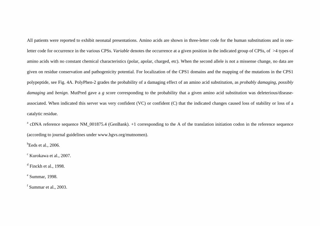
Table 2. Comparison of the activity, the apparent Km values for the substrates and
the Ka values for N-acetyl-L-glutamate (NAG) or for its analog N-carbamyl-L-
glutamate (NCG), for recombinant or natural human CPS1
Apparent Km or Ka value (mM) Source
Activity
U/mg ATP HCO3- NH4
+ NAG NCG
Recombinanta 1.1 0.5 4.0 1.0 0.14 2.0
Liverb 1.5 1.1 6.7 0.8 0.10 -
Liverc 1.5 0.3 2.2 1.3 0.15 2.0 aPresent work.
bPierson and Brien, 1980.
cRubio et al., 1981.
1
Table 3. Influence of clinical CPS1D mutations on the kinetic parameters for the
substrates and for NAG
Substrate or activator
NH4+ Bicarbonate ATP NAG
Enzyme
form V[Am]=∞
Kmapp
V[Bic]=∞ Km
app V[ATP]=∞
Kmapp
V[NAG]=∞ Ka
app
U/mg mM U/mg mM U/mg mM U/mg mM
Recombinant wild type form and forms with polymorphisms
Wild-type 1.19 1.0 1.23 4.0 1.22 0.47 1.15 0.14
p.Thr344Ala 1.06 1.2 ±0.1
1.04 4.3 1.06 0.45 1.02 0.17
p.Gly1376Ser 1.16 1.1 1.28 4.5 1.22 0.45 1.17 0.12
Recombinant forms carrying clinical missense mutations
p.Asn355Asp 0.25 1.0 0.26 4.6 ±0.7
0.28 0.57 0.26 0.34
p.Tyr389Cys 0.73 1.1 0.79 4.2 0.73 0.43 0.70 0.12
p.Ala438Pro <0.01a - <0.01a - <0.01a - <0.01a -
p.Thr544Met 0.16 1.3 0.26 242 0.23 1.92 0.27 2.91 ±0.38
p.Ala1378Thr 0.93 0.9 0.98 4.3 1.01 0.90 0.97 0.21
p.Thr1443Ala 0.04 1.0 0.034 3.6 0.04 ±0.01
0.62 0.14 23.4 ±5.2
For clarity, standard errors are only given when they exceed 10% of the magnitude of the
constant. The concentrations of NH4+, bicarbonate, ATP and NAG were varied in the following
mM ranges: 0.1-20, 0.3-625, 0.05-10 and 0-200, respectively. When not varied, the
concentrations of these ligands were 35, 50, 5 and 10 mM, respectively (except for bicarbonate
in the case of the p.T544M mutation, which was 0.5 M). Mg (as MgSO4) was always in 15 mM
excess over the ATP concentration.
a Detection limit
A)Elastase treatment
Low [elastase] High [elastase]B)
Thermal treatmentD) p.Ala1378Thr 52 ºCC) WT 54 ºC
Supplementary Material for Diez-Fernández et al.
Molecular characterization of carbamoyl phosphate synthetase (CPS1)
deficiency using human recombinant CPS1 as a key tool
Carmen Diez-Fernandez1,2, Ana I Martínez,2 Satu Pekkala,2 Belén Barcelona,1,2,3
Isabel Pérez-Arellano,2,3 Ana María Guadalajara,2 Marshall Summar,4 Javier
Cervera,1,2,3* Vicente Rubio1,3*
1Instituto de Biomedicina de Valencia (IBV-CSIC), Spain; 2Centro de Investigación
Príncipe Felipe, Valencia, Spain; 3Group 739, CIBERER, ISCIII, Spain; 4Childrens
National Medical Center, Washington DC, USA
Supplementary Material for Diez-Fernández et al.
Supp. Figure S1. Diagram schematizing the steps of the production of recombinant
human CPS1. The gel (SDS-PAGE, Coomassie staining, the arrows give the positions
of protein standards of the indicated polypeptide masses) illustrates the presence of
abundant soluble CPS1 protein in the postcentrifugal supernatant of the cell extract (left
track) and the essential homogeneity of the purified protein (right track).
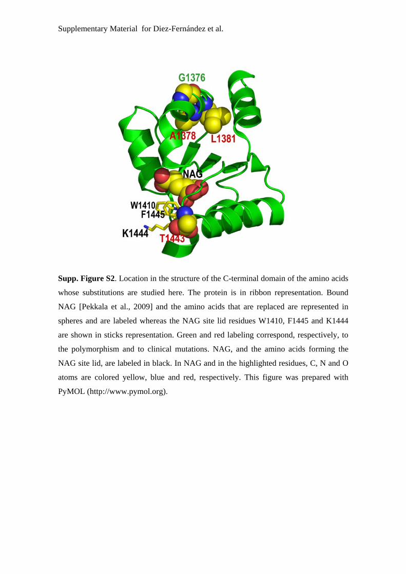
Supplementary Material for Diez-Fernández et al.
Supp. Figure S2. Location in the structure of the C-terminal domain of the amino acids
whose substitutions are studied here. The protein is in ribbon representation. Bound
NAG [Pekkala et al., 2009] and the amino acids that are replaced are represented in
spheres and are labeled whereas the NAG site lid residues W1410, F1445 and K1444
are shown in sticks representation. Green and red labeling correspond, respectively, to
the polymorphism and to clinical mutations. NAG, and the amino acids forming the
NAG site lid, are labeled in black. In NAG and in the highlighted residues, C, N and O
atoms are colored yellow, blue and red, respectively. This figure was prepared with
PyMOL (http://www.pymol.org).
Supplementary Material for Diez-Fernández et al.
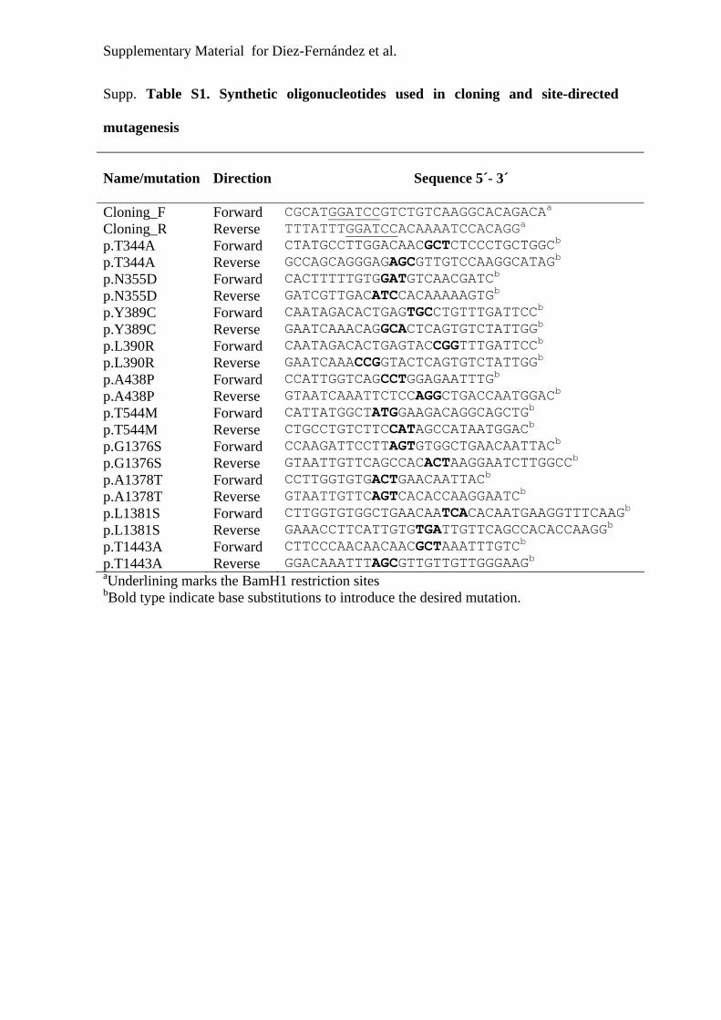
Supp. Table S1. Synthetic oligonucleotides used in cloning and site-directed
mutagenesis
Name/mutation
Direction Sequence 5´- 3´
Cloning_F Forward CGCATGGATCCGTCTGTCAAGGCACAGACAa Cloning_R Reverse TTTATTTGGATCCACAAAATCCACAGGa p.T344A Forward CTATGCCTTGGACAACGCTCTCCCTGCTGGCb p.T344A Reverse GCCAGCAGGGAGAGCGTTGTCCAAGGCATAGb p.N355D Forward CACTTTTTGTGGATGTCAACGATCb p.N355D Reverse GATCGTTGACATCCACAAAAAGTGb p.Y389C Forward CAATAGACACTGAGTGCCTGTTTGATTCCb p.Y389C Reverse GAATCAAACAGGCACTCAGTGTCTATTGGb p.L390R Forward CAATAGACACTGAGTACCGGTTTGATTCCb p.L390R Reverse GAATCAAACCGGTACTCAGTGTCTATTGGb p.A438P Forward CCATTGGTCAGCCTGGAGAATTTGb p.A438P Reverse GTAATCAAATTCTCCAGGCTGACCAATGGACb p.T544M Forward CATTATGGCTATGGAAGACAGGCAGCTGb p.T544M Reverse CTGCCTGTCTTCCATAGCCATAATGGACb p.G1376S Forward CCAAGATTCCTTAGTGTGGCTGAACAATTACb p.G1376S Reverse GTAATTGTTCAGCCACACTAAGGAATCTTGGCCb p.A1378T Forward CCTTGGTGTGACTGAACAATTACb p.A1378T Reverse GTAATTGTTCAGTCACACCAAGGAATCb p.L1381S Forward CTTGGTGTGGCTGAACAATCACACAATGAAGGTTTCAAGb p.L1381S Reverse GAAACCTTCATTGTGTGATTGTTCAGCCACACCAAGGb p.T1443A Forward CTTCCCAACAACAACGCTAAATTTGTCb p.T1443A Reverse GGACAAATTTAGCGTTGTTGTTGGGAAGb aUnderlining marks the BamH1 restriction sites bBold type indicate base substitutions to introduce the desired mutation.

![Mitomycins syntheses: a recent update · 2009. 8. 12. · 3-amino-5-hydroxybenzoic acid 20 (AHBA), D-glucosamine 21 and carbamoyl phosphate (Scheme 4) [29-32]. The key interme-diate,](https://img.dokumen.tips/doc/110x75/60b772c4d068841bb818084d/mitomycins-syntheses-a-recent-update-2009-8-12-3-amino-5-hydroxybenzoic-acid.jpg)