Embed Size (px)
Citation preview

of July 11, 2018.This information is current as
Immune SystemMolecular Basis of DNA Recognition in the
Maninjay K. Atianand and Katherine A. Fitzgerald
http://www.jimmunol.org/content/190/5/1911doi: 10.4049/jimmunol.1203162
2013; 190:1911-1918; ;J Immunol
Referenceshttp://www.jimmunol.org/content/190/5/1911.full#ref-list-1
, 29 of which you can access for free at: cites 87 articlesThis article
average*
4 weeks from acceptance to publicationFast Publication! •
Every submission reviewed by practicing scientistsNo Triage! •
from submission to initial decisionRapid Reviews! 30 days* •
Submit online. ?The JIWhy
Subscriptionhttp://jimmunol.org/subscription
is online at: The Journal of ImmunologyInformation about subscribing to
Permissionshttp://www.aai.org/About/Publications/JI/copyright.htmlSubmit copyright permission requests at:
Email Alertshttp://jimmunol.org/alertsReceive free email-alerts when new articles cite this article. Sign up at:
Print ISSN: 0022-1767 Online ISSN: 1550-6606. Immunologists, Inc. All rights reserved.Copyright © 2013 by The American Association of1451 Rockville Pike, Suite 650, Rockville, MD 20852The American Association of Immunologists, Inc.,
is published twice each month byThe Journal of Immunology
by guest on July 11, 2018http://w
ww
.jimm
unol.org/D
ownloaded from
by guest on July 11, 2018
http://ww
w.jim
munol.org/
Dow
nloaded from

Molecular Basis of DNA Recognition in the ImmuneSystemManinjay K. Atianand and Katherine A. Fitzgerald
Recognition of microbial nucleic acids is one strategy bywhich mammalian hosts respond to infectious agents.Intracellular DNA that is introduced into cells duringinfection elicits potent inflammatory responses by trig-gering the induction of antiviral type I IFNs and thematuration and secretion of inflammatory cytokines,such as TNF-a, IL-1b, and IL-18. In addition, if nu-cleases, such as DNase II or DNase III (Trex1), fail toclear self-DNA, accumulated DNA gains access to in-tracellular compartments where it drives inflammatoryresponses leading to autoimmune disease. In this re-view, we discuss a rapidly evolving view of how cyto-solic DNA-sensing machineries coordinate antimicrobialimmunity and, if unchecked, lead to autoimmune dis-ease. The Journal of Immunology, 2013, 190: 1911–1918.
The innate immune system is the first line of defenseagainst infectious agents. Germline-encoded patternrecognition receptors (PRRs), including TLRs, NOD-
like receptors (NLRs), retinoic acid inducible-I (RIG-I)–likereceptors, and C-type lectins, recognize a wide range of mi-crobial products, often referred to as microbe-associated mo-lecular patterns (1). Recognition of microbe-associated molecularpatterns by these surveillance receptors turns on signaling path-ways that coordinate transcription of hundreds of inflammatorygenes, the products of which control infection directly andmarshal the T and B cells of the adaptive immune system (2).In addition to classical microbial products, such as bacterialLPS or lipoproteins, microbial nucleic acids have emerged asmajor triggers of innate immune defenses.The best-characterized nucleic acid sensors are a subset of
TLRs, type I transmembrane receptors localized to the endo-somal compartment that sense dsRNA (TLR3) (3), ssRNA(TLR7 and TLR8) (4–6), and hypomethylated CpG DNA(TLR9) (7, 8). TLRs induce type I IFN and other inflam-matory genes via Toll/IL-1R domain containing adaptor
molecules, such as MyD88 (TLR7/8/9) or TRIF for TLR3(9). Cytosolic RNA sensors, such as RIG-I and melanomadifferentiation-associated protein 5, have also been identified(10, 11); they signal via a unique adaptor mitochondrial an-tiviral signaling (MAVS; also known as IFN-b promoterstimulator-1) to mediate NF-kB and IRF-dependent tran-scription of inflammatory genes (12).The cellular machinery that senses cytosolic DNA is still
being elucidated. Although significant progress has been madein understanding how DNA is recognized and how DNAsignaling ensues and leads to inflammation, the molecular basisof cytosolic DNA recognition in innate immunity is still beingworked out in detail. A number of newmolecules (which will bedescribed below) have been identified that contribute in variousways to recognition and signaling in response to DNA. TLR9expressed in endosomal membranes was the first identifiedDNA receptor that recognizes hypomethylated CpG motifs(7). However, in humans, TLR9 expression is restricted toB cells (13) and plasmacytoid dendritic cells (pDCs) (14)and, therefore, does not account for DNA-induced immuneresponses in other cell types, such as macrophages. Twoseminal studies demonstrated that the targeted delivery ofsynthetic dsDNA or a 45-nt immunostimulatory DNA intothe cytosol of macrophages and dendritic cells (DCs) trig-gered Tank-binding kinase 1 (TBK1)/IRF3-dependent in-duction of type I IFN and other inflammatory genes in aTLR9-independent manner (15, 16). These findings led toa search in many laboratories for sensor(s) that could couplecytosolic DNA recognition to immune signaling. Two con-ceptually distinct signaling pathways have since emerged. Thefirst of these leads to the proteolytic activation of the cysteineprotease caspase-1 associated with maturation and secretion ofthe proinflammatory cytokines IL-1b and IL-18. A secondpathway that is still being worked out leads to the transcrip-tional induction of type I IFN and proinflammatory genes.These two pathways are depicted in their simplest form inFig. 1. In this article, we briefly summarize and discuss thestate of play of DNA sensing and signaling and the functional
Division of Infectious Diseases and Immunology, Department of Medicine, Universityof Massachusetts Medical School, Worcester, MA 01605
Received for publication November 15, 2012. Accepted for publication January 7, 2013.
This work was supported by National Institutes of Health Grants AI093752, AI067497,and AI083713 (to K.A.F.).
Address correspondence and reprint requests to Dr. Katherine A. Fitzgerald, Division ofInfectious Diseases and Immunology, Department of Medicine, University of Massa-chusetts Medical School, LRB, Room 309, 364 Plantation Street, Worcester, MA01605. E-mail address: [email protected]
Abbreviations used in this article: AIM2, absent in melanoma 2; CARD, caspase-1activation and recruitment domain; c-di-AMP, cyclic di-AMP; c-di-GMP, cyclic di-
GMP; cGAMP, cyclic GMP-AMP; CTD, C-terminal domain; DAI, DNA-dependentactivator of IRF; DC, dendritic cell; IFI16, IFN inducible protein 16; LRRFIP1,leucine-rich repeat in flightless-I interacting protein 1; MAVS, mitochondrial antiviralsignaling; NLR, NOD-like receptor; pDC, plasmacytoid dendritic cell; PRR, patternrecognition receptor; PYD, pyrin domain; PYHIN, pyrin and HIN200 domain-con-taining; RIG-I, retinoic acid inducible-I; SLE, systemic lupus erythematosus; STING,stimulator of type I IFN gene; TBK1, Tank-binding kinase 1.
Copyright� 2013 by TheAmerican Association of Immunologists, Inc. 0022-1767/13/$16.00
www.jimmunol.org/cgi/doi/10.4049/jimmunol.1203162
by guest on July 11, 2018http://w
ww
.jimm
unol.org/D
ownloaded from
significance of these event in antimicrobial immunity andautoimmune diseases.
Inflammasome-activating cytosolic DNA sensors
Cytosolic DNA leads to the production of the proin-flammatory cytokines IL-1b and IL-18. IL-1b is secreted bymany cell types and is essential for both the innate andadaptive arms of the immune response (17). IL-1b is im-portant in activating neutrophils, macrophages, DCs, andT cells, whereas IL-18 is crucial for IFN-g production by NKcells and T cells (18, 19). In contrast to the majority of in-flammatory mediators that are regulated transcriptionally, IL-1b and IL-18 are regulated at both the transcriptional andposttranslational levels. Upon transcriptional induction byTLRs and other sensor systems, IL-1b and IL-18 are syn-thesized as inactive precursor proteins, which are subsequentlyprocessed by the cysteine protease caspase-1 (IL-1b convert-ing enzyme) (20). Conversion of procaspase-1 into an enzy-matically active form, caspase-1, occurs upon formation ofa multiprotein complex in the cytosol referred to as the“inflammasome” (21). The nucleotide binding and leucine-rich repeat containing receptor (NLR; aka NOD-like recep-tors) and pyrin and HIN200 domain-containing (PYHIN)family proteins (discussed further below) are known to forminflammasome complexes by recruiting procaspase-1 via anadaptor protein ASC (22). ASC is a bipartite molecule con-taining an N-terminal pyrin domain (PYD) and the C-terminal caspase-1 activation/recruitment domain (CARD);therefore, it acts as an adaptor to bring together NLRs (orPYHIN proteins) and procaspase-1 via homotypic PYD:PYDand CARD:CARD interactions, respectively (Fig. 2). Severalinflammasome complexes have been identified in recent years.Of the known inflammasomes, Nlrp3, absent in melanoma 2(AIM2), and, most recently, IFN inducible protein 16 (IFI16)
inflammasomes, have been linked to immune responses tointracellular DNA, as well as bacterial or DNA virus infec-tions.
The Nlrp3 inflammasome. The Nlrp3 inflammasome is the best-studied inflammasome and is associated with bacterial, viral,parasitic, and fungal infections (23). NLRP3 is also activatedin response to endogenous danger signals (24). The Nlrp3inflammasome in macrophages responds to modified vacciniavirus Ankara strain (25) and adenovirus (26) to facilitatecaspase-1 activation and IL-1b secretion. For adenovirus,intracellular viral DNA, but not viral capsids, activate theNlrp3 inflammasome. However, caspase-1 activation and IL-1b secretion in response to transfected nonviral cytosolic DNA(synthetic, bacterial, or mammalian origin) was Nlrp3 inde-pendent, but ASC dependent, indicating that cytosolic DNA-driven inflammasome activation requires an ASC-dependentsensor (26). Whether adenoviral DNA is the ligand requiredto trigger assembly of the Nlrp3 inflammasome is unclear, butan indirect mode is more likely, as suggested by a recent studythat perturbations in cell membrane associated with adenoviralentry are the trigger for Nlrp3 activation (27). The Nlrp3inflammasome also recognizes influenza A infection, a negativestranded RNA virus (28). Therefore, the Nlrp3 inflammasomeis involved in mediating the inflammatory responses to bothDNA and RNA viruses. It is likely that the Nlrp3 inflammasomesenses cytosolic nucleic acids indirectly.
The AIM2 inflammasome. Several groups independently iden-tified AIM2 as the receptor for cytosolic DNA that leads tocaspase-1 activation and IL-1b secretion (29–31). Contrary toother cytosolic DNA sensors, which are primarily involved inthe induction of type I IFN, AIM2 triggers the activation ofthe inflammasome. Notably, the AIM2 inflammasome is thefirst among all inflammasome-activating proteins identified inwhich a direct receptor–ligand interaction is demonstrated.
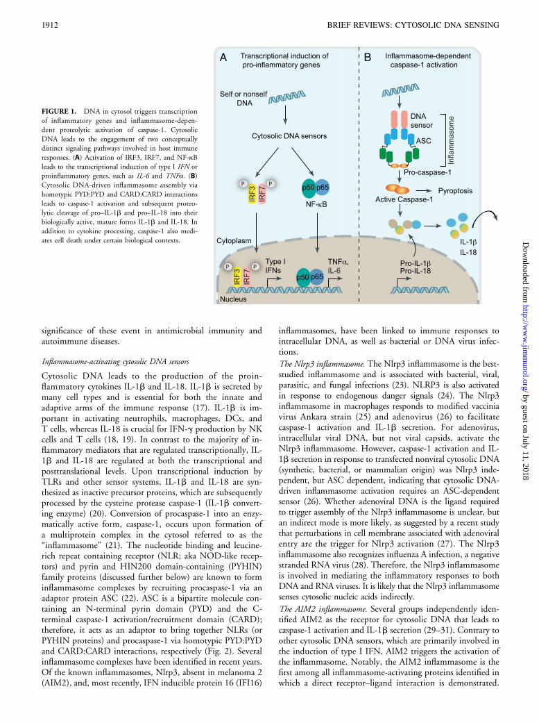
FIGURE 1. DNA in cytosol triggers transcription
of inflammatory genes and inflammasome-depen-
dent proteolytic activation of caspase-1. Cytosolic
DNA leads to the engagement of two conceptually
distinct signaling pathways involved in host immune
responses. (A) Activation of IRF3, IRF7, and NF-kB
leads to the transcriptional induction of type I IFN or
proinflammatory genes, such as IL-6 and TNFa. (B)Cytosolic DNA-driven inflammasome assembly via
homotypic PYD:PYD and CARD:CARD interactions
leads to caspase-1 activation and subsequent proteo-
lytic cleavage of pro–IL-1b and pro–IL-18 into their
biologically active, mature forms IL-1b and IL-18. In
addition to cytokine processing, caspase-1 also medi-
ates cell death under certain biological contexts.
1912 BRIEF REVIEWS: CYTOSOLIC DNA SENSING
by guest on July 11, 2018http://w
ww
.jimm
unol.org/D
ownloaded from

AIM2 binds cytosolic DNA of self and nonself origin,including bacterial, viral, and mammalian DNA, in a sequence-independent manner. Upon DNA binding via its HIN200domain, AIM2 undergoes oligomerization and, thereby,recruits caspase-1 via ASC. In vivo studies in Aim2-deficientmice clearly indicate that the Aim2 inflammasome is essentialand functions in a nonredundant manner for innate defenseresponses to DNA viruses and intracellular bacterial infections(32, 33). Caspase-1 activation and IL-1b/IL-18 secretion inresponse to mouse CMV and vaccinia virus infection in murinemacrophages is dependent upon Aim2 inflammasome (32).Consistently, Aim2-dependent IL-18 secretion and NK cellactivation are essential for an early control of mouse CMVinfection in vivo. Similarly, Aim2 plays a crucial role incontrolling Francisella tularensis infection in vivo, becauseAim2-deficient mice are more susceptible to infection andshow higher bacterial counts in infected organs (33, 34).Activation of the Aim2 inflammasome is also reported inother bacterial infections including Listeria monocytogenes(35, 36) and Mycobacterium tuberculosis (37). How Aim2 isexposed to bacterial DNA during infection is not wellunderstood. There is some evidence that DNA released intothe cytosol following lysosomal bacteriolysis is the trigger forAim2 activation (38). Recent evidence indicates that the AIM2-related protein IFI16 also forms an inflammasome complex
following Kaposi sarcoma–associated herpes virus infectionof endothelial cells (39).
Type I IFN–producing cytosolic DNA sensors
Type I IFN production is another major consequence of cy-tosolic DNA sensing and is essential for antiviral immunity andimmunity to many classes of infectious agents. Although weunderstand in detail how DNA via AIM2 leads to IL-1bproduction, the molecular bases to DNA-induced type I IFNgene transcription is less clear. A core signaling moduleconsisting of stimulator of type I IFN gene (STING)–TBK1–IRF3 is engaged and is absolutely essential for type I IFNresponses to DNA. TBK1, an IKK-related kinase originallycharacterized as the kinase responsible for the phosphorylation-induced activation of IRF3 in TLR and then RIG-I–like re-ceptor signaling, is also central for DNA-signaling pathways(40, 41). Therefore, TBK1 activation acts as a point of con-vergence of multiple PRR-driven pathways that results inIRF3 phosphorylation and transcription of type I IFN genesand related IFN-stimulated genes.Several groups independently identified STING (also
known as MPYS, TMEM173, ERIS, and MITA) as a keycomponent of the DNA-sensing pathway (42–44). STINGwas shown to localize to the endoplasmic reticulum and outermitochondrial membranes via five transmembrane-spanningregions. In response to DNA, STING translocates to peri-nuclear regions where it interacts with TBK1 to relay down-stream signals to IRF3. STING deficiency in macrophages orDCs leads to a markedly impaired type I IFN response to B-DNA and immunostimulatory DNA or to infection withDNA viruses, including HSV-1, human CMV, and vacciniavirus (42, 45). STING is also essential for type I IFN in-duction in response to intracellular bacteria, including F.tularensis (46, 47), L. monocytogenes (46, 48), and Brucellaabortus (49), and in response to extracellular bacteria, such asStreptococcus pneumoniae (50) and Streptococcus pyogenes (51).Initial studies showed that STING also interacted withcomponents of the RNA-recognition machinery, such as RIG-I,where it was linked to type I IFN induction in response tovesicular stomatitis virus, a negative-strand RNA virus (42).However, subsequent studies in STING-deficient cells sug-gested that viruses or ligands that engaged RIG-I have normalIFN responses in STING-deficient murine macrophages (48,52). However, additional studies linked STING to DNA- andRNA-recognition pathways by uncovering a role for it in theactivation of STAT6 and induction of STAT6-dependentchemokines (52).The assumption from these earlier studies is that, analogous
to MyD88 or MAVS, STING functions as an adaptor mol-ecule. This assumption implies that DNA-binding proteinsthat recognize microbial DNA engage STING and activateTBK1 and downstream signaling. Several candidate sensorshave been discovered and implicated to varying degrees in thisDNA-dependent TBK1–IRF3 and type I IFN productionpathway (Fig. 3).
DNA-dependent activator of IRFs. DNA-dependent activator ofIRFs (DAI), also known as Z-DNA–binding protein, ZBP, orDLM1, is an IFN-stimulated gene and the first cytosolicDNA sensor identified (53, 54). DAI binds Z-form DNAvia two N-terminal Z-DNA–binding domains and an adjacentprotein region carrying B-DNA–binding potential. DAI was
FIGURE 2. Cytosolic DNA triggers inflammasome activation. Intracellular
DNA following microbial infection or phagocytosis of immune complexes can
potentially trigger the assembly of either NLR (e.g., NLRP3) or PYHIN
inflammasomes (AIM2 and IFI16). Upon activation, these cytosolic DNA
sensors recruit the inflammasome adaptor ASC to activate caspase-1, which leads
to the processing of pro–IL-1b and pro–IL-18 into their biologically active
forms. Notably, AIM2 and IFI16 bind directly to DNA via its C-terminal
HIN200 domain and therefore act as the true receptors for cytosolic DNA.
However, the precise mechanism triggering Nlrp3 inflammasome assembly and
whether microbial DNA is the ligand for Nlrp3 are still unclear. Interestingly,
IFI16 inflammasome is thought to assemble inside the nucleus in response to
Kaposi sarcoma associated herpes virus infection in endothelial cells.
The Journal of Immunology 1913
by guest on July 11, 2018http://w
ww
.jimm
unol.org/D
ownloaded from

shown to mediate DNA-induced type I IFN production incell-based studies. However, subsequent studies in DAI 2/2
mice revealed that DAI-deficient cells and mice retainnormal type I IFN responses to DNA viruses and varioustypes of synthetic DNA (55), indicating that DAI is eitherdispensable or a redundant sensor of cytosolic DNA. WhetherDAI impacts other aspects of DNA-induced signaling is stillunclear.
RNA polymerase III. Following the discovery of DAI, RNApolymerase III was linked to DNA recognition, adding anotherlayer of complexity to the cytosolic DNA-recognition path-way (56, 57). RNA polymerase III present in the cytosoltranscribes AT-rich DNA into immunostimulatory dsRNAtranscripts characterized by uncapped 59-triphosphate moieties,which act as a ligand for RIG-I (58). Subsequently, RIG-Isignals via MAVS to induce the expression of type I IFN andother cytokines. Therefore, RNA polymerase III does not sensecytosolic DNA directly, but it generates a ligand to engage theRIG-I pathway. Involvement of the RNA polymerase III/RIG-Ipathway in response to AT-rich DNA explains the partialdependence upon MAVS for DNA-induced induction of typeI IFN in certain cell types. The RNA polymerase III pathway isfunctional in both human and mouse cells and is engaged inresponse to the intracellular dsDNA mimetic poly(dA:dT), aswell as with certain DNA viruses, like adenovirus and EBV.Additional studies also linked RNA polymerase III and type IIFN responses during Legionella pneumophila and HSV-1infections; however, there is some controversy over theselatter data (59, 60).
IFI16 and p204. IFI16 was identified as a DNA-binding proteinin human monocytes in affinity-purification studies (61). Like
AIM2, IFI16 is a member of the PYHIN (aka p200 protein)family. IFI16 is primarily localized in the nucleus. In some celltypes, such as macrophages, a small pool of cytoplasmic IFI16colocalizes with transfected DNA or viral DNA that gainsaccess to the cytosolic compartment during HSV-1 infection.In other cell types, such as fibroblasts, which are permissive toHSV1 infection, IFI16 engages viral DNA that accumulatesduring productive infection in the nucleus (62). IFI16 doesnot leave the nucleus in HSV-1–infected human foreskinfibroblast cells; however, this nuclear sensing of HSV-1 requirescytoplasmic STING signaling. Therefore, HSV-1 DNA rec-ognition may differ in permissive versus nonpermissive cells,but, in both cases, IFI16-dependent recognition of HSV-1DNA is coupled to STING signaling. It is important tounderstand the role of nuclear and cytoplasmic IFI16 functionduring HSV infection in these contexts and to better understandhow signaling from these compartments is initiated andregulated. IFI16 has two C-terminal DNA-binding HIN200domains and, like AIM2, it binds DNA via nonsequence-specific DNA recognition accomplished through electrostaticattractions between the positively charged HIN200 domainresidues and the negatively charged dsDNA sugar–phosphatebackbone (63). IFI16 then interacts with STING to activateTBK1 to trigger IFN-b induction. It is unknown whetherIFI16 binds STING directly or indirectly. Humans have 4PYHIN proteins (IFI16, AIM2, MNDA, and IFIX), whereasmice have 13 (64). The murine PYHIN protein Ifi204 (p204)is proposed to function in an analogous manner to IFI16.Knockdown of IFI16 (or Ifi204) compromise DNA-induced,as well as HSV-1–induced, IRF3 activation and IFN induction(61). Evidence is also accumulating for a functional role ofIFI16/Ifi204 in controlling HSV infection, because p204plays a role in resistance to HSV-1 infection in the cornealepithelium (65). Interestingly, the HSV viral nuclear ICP0protein can target IFI16 for degradation, thereby docu-menting a mechanism by which HSV evades IFI16-mediateddetection (62).
Aspartate–glutamate–any amino acid–aspartate/histidine boxhelicases. Aspartate–glutamate–any amino acid–aspartate/histidine box–containing helicases are also beginning toemerge as important players in the recognition of cytosolicnucleic acids. DHX36 and DHX9 have been identified asTLR9-independent, but MyD88-dependent, sensors ofCpG-A and CpG-B DNA, respectively, in human pDCs(66). These two distinct oligodeoxynucleotides are knowninducers of either type I IFN (CpG-A) or proinflammatorycytokines, such as TNF-a and IL-6 (CpG-B), in pDCs(67). Consistently, DHX36 is involved in the productionof IFN-a, whereas DHX9 mediates TNFa/IL-6 productionin HSV-1–infected (or CpG-treated) human pDCs. DHX36and DHX9 interact directly with the Toll/IL-1R domain ofMyD88 to trigger downstream signaling to activate IRF7 andNF-kB p50, respectively. Subsequent studies also ascribeda role for DHX36 and DHX9 in the recognition of cytosolicRNA. DHX36, together with DDX1 and DDX21, formsa complex with the adaptor TRIF to mediate type I IFNinduction in response to synthetic dsRNA mimic polyinosinic-polycytidylic acid (68). Similarly, DHX9 interacts with MAVSto induce a type I IFN in response to dsRNA in myeloid DCs(69). It is unclear whether DHX36 and DHX9 are involved inthe recognition of RNA viruses. In a comprehensive shRNA
FIGURE 3. Cytosolic DNA sensors activate the transcription of type I IFNand other inflammatory genes. Cytosolic DNA of microbial or self-origin is
a potent trigger of type I IFN production via the STING–TBK1–IRF3 axis,
as well as other proinflammatory cytokines (e.g., TNFa and IL-6), by en-
gaging NF-kB signaling. Distinct cytosolic DNA sensors along with their
select activating conditions are shown (these are discussed in detail in the
text). The DNA-induced signaling pathway converges on the adaptor STING
and the kinase TBK1, which phosphorylates IRF3 to mediate downstream
signaling events leading to transcriptional induction of inflammatory genes.
Besides cytosolic DNA, bacterial small molecules c-di-AMP and c-di-GMP
also act as potent stimulators of the type I IFN response by engaging STING
either as a direct sensor or coactivator (discussed in text). Host cells use
distinct nucleases to eliminate both self and nonself DNA from extracellular
space (DNase I), phagolysosomes (DNase II), and cytosol (DNase III; Trex1)
to avoid deleterious effects of excess DNA-induced immune responses.
1914 BRIEF REVIEWS: CYTOSOLIC DNA SENSING
by guest on July 11, 2018http://w
ww
.jimm
unol.org/D
ownloaded from

screen for all 59 members of the aspartate–glutamate–anyamino acid–aspartate/histidine box family, Zhang et al. (70)identified DDX41 as a cytosolic DNA receptor in both mouseand human DCs. DDX41 binds to dsDNA through itshelicase domain and triggers the activation of IRF3, NF-kB,and MAPK signaling. Knockdown of DDX41 in DCs led toimpaired type I IFN and proinflammatory cytokine productionin response to transfected dsDNA or DNA viruses, includingHSV-1 and adenovirus, but not against polyinosinic-polycytidylic acid or influenza virus infection. Moreover,DDX41 binds STING in DCs in response to transfectedpoly(dA:dT) or HSV-1 DNA, suggesting that STING isinvolved in signaling downstream of DDX41 recognitionof the cytosolic DNA. Unlike IFI16, which is a type I IFNinducible gene, DDX41 is expressed at relatively high levelsin immune cells. Therefore, DDX41 was proposed torecognize DNA in the early stages of infection, whereas IFI16,a type I IFN inducible gene, is induced at later stages (as a resultof DDX41-dependent IFN production), where these twosensors together coordinate DNA-induced IFN responses.
Ku70. Several additional DNA-binding proteins have beenlinked to DNA recognition and innate immunity. A recentstudy by Zhang et al. (71) identified Ku70, a component ofthe DNA repair and telomere maintenance pathway, as thenewest member of the cytosolic DNA-sensing machinery.Ku70 was identified as a DNA-binding protein in HEK293cells by DNA-affinity purification followed by mass spec-trometry. Notably, Ku70 is involved in the production oftype III IFN (IFN-l1; also known as IL29), but not type IIFN (IFN-a or IFN-b), in response to a variety of transfectedDNA (.500 bp) in HEK293, murine macrophages, andDCs. Subsequent studies indicated that the DNA-inducedIFN-l1 induction required IRF1 and IRF7 binding to theIFN-l1 promoter, which implicates Ku70 in a signalingpathway involving IRF1/IRF7. Although Ku70 was shownto mediate HSV-induced IFN-l1 induction in HEK293cells, it remains to be fully elucidated whether Ku70 rec-ognizes DNA viruses and bacterial infections in primaryimmune cells. Because the biological activity and the signalingpathway used by both types I and III IFN are very similar, it isalso unclear how the engagement of Ku70-mediated IFN-l1production contributes to the overall DNA-induced innateimmune responses.
Leucine-rich repeat in flightless-I interacting protein 1. Leucine-richrepeat in flightless-I interacting protein 1 (LRRFIP1) wasoriginally identified by Wilson et al. (72) using a cDNAlibrary to screen for cellular interacting proteins of HIV-1RNA; it was shown to bind RNA, as well as DNA witha lower affinity. Subsequent studies indicated that LRRFIP1recognizes both RNA and DNA in the cytosol and enhancesthe transcription of the Ifnb gene through a novel b-catenin–dependent–signaling pathway (73). In a subsequent RNAinterference screen against leucine-rich repeat–containing andleucine-rich repeat–interacting proteins in primary murinemacrophages, Yang et al. (73) identified LRRFIP1 as acytosolic sensor involved in the production of IFN-b in re-sponse to L. monocytogenes or vesicular stomatitis virusinfection. LRRFIP1 is similarly involved in IFN-b productionin response to transfected dsRNA or dsDNA. Interestingly,LRRFIP1 uses b-catenin, an integral component of the Wnt-signaling pathway, to turn on Ifnb gene transcription. Upon
recruitment by LRRFIP1 by unidentified mechanisms, b-cateninis phosphorylated at serine 552, which then translocates to thenucleus and binds to IRF3, leading to enhanced recruitmentof the histone acetyltransferase p300, thereby enhancingtranscription of the Ifnb gene.
Role of STING and DDX41 in sensing cyclic dinucleotides
In addition to its role as a signaling intermediate in DDX41-and IFI16-signaling pathways, STING acts as a receptor forthe bacterial second messenger molecules cyclic di-AMP(c-di-AMP) and cyclic di-GMP (c-di-GMP) (74). These sec-ondary messenger molecules are potent inducers of type I IFNsand, in the case of L. monocytogenes, are proposed to representthe major trigger of IFN production in macrophages (75).Forward genetic studies in N-ethyl-N-nitrosourea–mutagen-ized mice revealed that STING was essential for the type I IFNresponse to c-di-AMP/c-di-GMP and following Listeria in-fection (48). STING binds these small molecules directlythrough its C-terminal domain (CTD), which leads to acti-vation of the TBK1–IRF3 axis for the induction of Ifnb genes.Recently, several groups resolved the crystal structures of hu-man STING-CTD bound to c-di-AMP/c-di-GMP (76–78).Collectively, these studies impart dual functions to STING: iteither serves as an adaptor for DNA sensing or as a directsensor of bacterial second-messenger molecules, like c-di-AMP. Whether c-di-AMP or DNA is the major microbialtrigger involved in the IFN-b response during Listeria infec-tion remains to be determined. Because genes encoding c-di-AMP are also predicted to exist in several other pathogenicbacteria, including staphylococci, streptococci, Mycobacterium,and Chlamydia (79), it will be interesting to define the im-portance of STING/c-di-AMP interactions in host type I IFNresponses to these pathogens. Very recently, Chen and col-leagues (80) showed that STING directly binds cyclic GMP-AMP (cGAMP), a novel endogenous second messenger gen-erated in response to DNA, to trigger IRF3 activation and theinduction of IFN-b in response to transfected DNA or DNAviruses. They further identified cGAMP synthase, a mamma-lian nucleotidyltransferase family member, which binds to cyto-solic DNA and catalyzes the generation of cGAMP to triggerSTING-dependent induction of the type I IFN response(81). Therefore, STING recognition of cGAMP representsa new avenue for mounting host immune responses to cyto-solic DNA.Surprising recent evidence indicates that DDX41 also
serves as the PRR involved in detecting c-di-GMP and c-di-AMP to trigger TBK1–IRF3 signaling via STING (82).Surprisingly, the interaction of c-di-GMP with DDX41 wasshown to be greater than that observed for STING binding.These data indicate that DDX41 is required to facilitate c-di-GMP signaling via STING. The solved crystal structureof the STING-CTD in complex with c-di-GMP showedthat one molecule of c-di-GMP binds one dimer of STING(76–78). Cheng and colleagues proposed that the detectionof c-di-GMP via DDX41 promotes enhanced DDX41–STING interactions, leading to an increase in the bindingaffinity of STING for c-di-GMP, which ultimately drivesdownstream signaling events (82). This model is consistentwith the possibility that STING functions as a secondaryreceptor or coactivator in the cyclic dinucleotide–signalingpathway.
The Journal of Immunology 1915
by guest on July 11, 2018http://w
ww
.jimm
unol.org/D
ownloaded from

Recognition of endogenous DNA and autoimmune diseases
Although DNA-induced immune responses are central toimmunity, inappropriate recognition of self-DNA can lead todeleterious consequences to the host. Systemic lupus eryth-ematosus (SLE) is one such disease in which type I IFN andautoantibodies directed against dsDNA, RNA, and nucleo-somes are implicated in disease pathogenesis (83). Multiplefail-safe mechanisms deployed by the host subvert endoge-nous DNA–induced immune responses. One level of regu-lation is provided by cellular endonucleases, such as DNase I,DNase II, and DNase III (also known as Trex1), which areinvolved in the clearance of extracellular, lysosomal, andcytosolic DNA, respectively. Recent evidence clearly indicatesthat the functional defects in these enzymes are associatedwith SLE and other human diseases. For example, DNase Igene mutations have been identified in subgroups of SLEpatients (84), a clinical association further supported bylupus-like syndrome in DNase I–deficient mice (85). Defectsin DNase I lead to the accumulation of extracellular DNAreleased by apoptotic/necrotic cells, which is highly immu-nostimulatory (86). DNase II is expressed in lysosomes, whereit degrades DNA from phagocytosed apoptotic and necroticcells. Interestingly, DNase II–knockout mice are embryoni-cally lethal; however, they are viable on the IFNR1-knockoutbackground, indicating that type I IFNs mediate the lethalityof DNase II genetic deficiency (87). In this case, type I IFNresponses are mediated in a TLR-independent, but IRF3/7-dependent manner. Recent evidence indicates that DNase II/STING–deficient mice are also viable, indicating the impor-tance of STING in this model (88). The DNase II/IFNR1knockout mice develop autoimmune polyarthritis by 2 mo ofage with features reminiscent of human rheumatoid arthritis,further highlighting a central role for DNA-induced immuneresponses in autoimmune diseases. The sensors of DNase Iand DNase II substrates are unclear, but it is likely that one ormore of the aforementioned cytosolic DNA sensor(s) recog-nize and respond to this accumulated dsDNA. DNase III/Trex1 is another nuclease that is normally involved in theclearance of cell-intrinsic ssDNA (89). Trex1 is the mostabundant 39–59 exonuclease and is localized to the endo-plasmic reticulum. Recent studies in Trex1-knockout miceprovide great insight into the regulation of endogenous DNAand its role in autoimmunity. Trex12/2 mice are viable;however, they exhibit a shortened life span (2–4 mo) andmanifest inflammatory myocarditis (90). In the absence ofTrex1, there is an accumulation ∼60-bp ssDNA, believed tobe produced during replication, which leads to the activationof DNA-damage associated signaling pathways. Further workby Stetson et al. (91) revealed a role for Trex1 in preventingcell-intrinsic initiation of autoimmunity. Trex1 substrates aressDNA, which are either the by-products of replication and/orreverse transcribed from endogenous retroelements. As wasobserved in DNase II–deficient mice, inflammatory myocar-ditis associated with Trex1 deficiency is rescued by crossingTrex1-deficient mice to STING-deficient background. To-gether, these studies highlight the important role of strictregulation of the DNA-sensing pathway and underscore theimportance of DNA sensing in both protective and patho-logical immune responses.
ConclusionsIn recent years, there has been tremendous progress in un-derstanding how cells recognize and respond to microbialthreats via cytosolic DNA recognition. Studies from severalgroups clearly indicated that multiple sensors exist in the cytosolto trigger inflammatory responses to DNA. These responses arecentral to antimicrobial immunity; therefore, it is not surprisingthat multiple DNA sensors exist and operate in different celltypes. However, as with all areas of progress, many newquestions arise, and key aspects of DNA recognition remain tobe better understood. The potential functional overlap andredundancy of key sensors and signaling intermediates, as wellas a better clarification of the relative importance of DDX41versus STING in sensing cyclic dinucleotides, likely willbe assisted following the generation and characterization ofDDX41-deficient cells and mice. A better understanding of themolecular mechanisms by which cells generate inflammationin response to DNA may provide new targets that could bemanipulated for the treatment of infectious, as well as auto-immune, disease.
AcknowledgmentsWe apologize to colleagues whose work could not be cited because of space
limitations.
DisclosuresThe authors have no financial conflicts of interest.
References1. Medzhitov, R. 2007. Recognition of microorganisms and activation of the immune
response. Nature 449: 819–826.2. Pasare, C., and R. Medzhitov. 2004. Toll-like receptors: linking innate and adaptive
immunity. Microbes Infect. 6: 1382–1387.3. Alexopoulou, L., A. C. Holt, R. Medzhitov, and R. A. Flavell. 2001. Recognition of
double-stranded RNA and activation of NF-kappaB by Toll-like receptor 3. Nature413: 732–738.
4. Lund, J. M., L. Alexopoulou, A. Sato, M. Karow, N. C. Adams, N. W. Gale,A. Iwasaki, and R. A. Flavell. 2004. Recognition of single-stranded RNA viruses byToll-like receptor 7. Proc. Natl. Acad. Sci. USA 101: 5598–5603.
5. Heil, F., H. Hemmi, H. Hochrein, F. Ampenberger, C. Kirschning, S. Akira,G. Lipford, H. Wagner, and S. Bauer. 2004. Species-specific recognition of single-stranded RNA via toll-like receptor 7 and 8. Science 303: 1526–1529.
6. Diebold, S. S., T. Kaisho, H. Hemmi, S. Akira, and C. Reis e Sousa. 2004. Innateantiviral responses by means of TLR7-mediated recognition of single-strandedRNA. Science 303: 1529–1531.
7. Hemmi, H., O. Takeuchi, T. Kawai, T. Kaisho, S. Sato, H. Sanjo, M. Matsumoto,K. Hoshino, H. Wagner, K. Takeda, and S. Akira. 2000. A Toll-like receptorrecognizes bacterial DNA. Nature 408: 740–745.
8. Bauer, S., C. J. Kirschning, H. Hacker, V. Redecke, S. Hausmann, S. Akira,H. Wagner, and G. B. Lipford. 2001. Human TLR9 confers responsiveness tobacterial DNA via species-specific CpG motif recognition. Proc. Natl. Acad. Sci.USA 98: 9237–9242.
9. Jenkins, K. A., and A. Mansell. 2010. TIR-containing adaptors in Toll-like receptorsignalling. Cytokine 49: 237–244.
10. Kato, H., K. Takahasi, and T. Fujita. 2011. RIG-I-like receptors: cytoplasmicsensors for non-self RNA. Immunol. Rev. 243: 91–98.
11. Onoguchi, K., M. Yoneyama, and T. Fujita. 2011. Retinoic acid-inducible gene-I-like receptors. J. Interferon Cytokine Res. 31: 27–31.
12. Belgnaoui, S. M., S. Paz, and J. Hiscott. 2011. Orchestrating the interferon antiviralresponse through the mitochondrial antiviral signaling (MAVS) adapter. Curr.Opin. Immunol. 23: 564–572.
13. Bourke, E., D. Bosisio, J. Golay, N. Polentarutti, and A. Mantovani. 2003. Thetoll-like receptor repertoire of human B lymphocytes: inducible and selective ex-pression of TLR9 and TLR10 in normal and transformed cells. Blood 102: 956–963.
14. Kadowaki, N., S. Ho, S. Antonenko, R. W. Malefyt, R. A. Kastelein, F. Bazan, andY. J. Liu. 2001. Subsets of human dendritic cell precursors express different toll-likereceptors and respond to different microbial antigens. J. Exp. Med. 194: 863–869.
15. Ishii, K. J., C. Coban, H. Kato, K. Takahashi, Y. Torii, F. Takeshita, H. Ludwig,G. Sutter, K. Suzuki, H. Hemmi, et al. 2006. A Toll-like receptor-independentantiviral response induced by double-stranded B-form DNA. Nat. Immunol. 7: 40–48.
16. Stetson, D. B., and R. Medzhitov. 2006. Recognition of cytosolic DNA activates anIRF3-dependent innate immune response. Immunity 24: 93–103.
1916 BRIEF REVIEWS: CYTOSOLIC DNA SENSING
by guest on July 11, 2018http://w
ww
.jimm
unol.org/D
ownloaded from

17. Dinarello, C. A. 2002. The IL-1 family and inflammatory diseases. Clin. Exp.Rheumatol. 20(5, Suppl. 27)S1–S13.
18. Dinarello, C. A. 2009. Immunological and inflammatory functions of theinterleukin-1 family. Annu. Rev. Immunol. 27: 519–550.
19. Gracie, J. A., S. E. Robertson, and I. B. McInnes. 2003. Interleukin-18. J. Leukoc.Biol. 73: 213–224.
20. Thornberry, N. A., H. G. Bull, J. R. Calaycay, K. T. Chapman, A. D. Howard,M. J. Kostura, D. K. Miller, S. M. Molineaux, J. R. Weidner, J. Aunins, et al. 1992.A novel heterodimeric cysteine protease is required for interleukin-1 beta processingin monocytes. Nature 356: 768–774.
21. Martinon, F., K. Burns, and J. Tschopp. 2002. The inflammasome: a molecularplatform triggering activation of inflammatory caspases and processing of proIL-beta. Mol. Cell 10: 417–426.
22. Rathinam, V. A., S. K. Vanaja, and K. A. Fitzgerald. 2012. Regulation of inflam-masome signaling. Nat. Immunol. 13: 333–2.
23. Franchi, L., R. Munoz-Planillo, and G. Nunez. 2012. Sensing and reacting tomicrobes through the inflammasomes. Nat. Immunol. 13: 325–332.
24. Schroder, K., R. Zhou, and J. Tschopp. 2010. The NLRP3 inflammasome: a sensorfor metabolic danger? Science 327: 296–300.
25. Delaloye, J., T. Roger, Q. G. Steiner-Tardivel, D. Le Roy, M. Knaup Reymond,S. Akira, V. Petrilli, C. E. Gomez, B. Perdiguero, J. Tschopp, et al. 2009. Innateimmune sensing of modified vaccinia virus Ankara (MVA) is mediated by TLR2-TLR6, MDA-5 and the NALP3 inflammasome. PLoS Pathog. 5: e1000480.
26. Muruve, D. A., V. Petrilli, A. K. Zaiss, L. R. White, S. A. Clark, P. J. Ross,R. J. Parks, and J. Tschopp. 2008. The inflammasome recognizes cytosolic mi-crobial and host DNA and triggers an innate immune response. Nature 452: 103–107.
27. Barlan, A. U., T. M. Griffin, K. A. McGuire, and C. M. Wiethoff. 2011. Ade-novirus membrane penetration activates the NLRP3 inflammasome. J. Virol. 85:146–155.
28. Ichinohe, T., H. K. Lee, Y. Ogura, R. Flavell, and A. Iwasaki. 2009. Inflammasomerecognition of influenza virus is essential for adaptive immune responses. J. Exp.Med. 206: 79–87.
29. Burckstummer, T., C. Baumann, S. Bluml, E. Dixit, G. Durnberger, H. Jahn,M. Planyavsky, M. Bilban, J. Colinge, K. L. Bennett, and G. Superti-Furga. 2009.An orthogonal proteomic-genomic screen identifies AIM2 as a cytoplasmic DNAsensor for the inflammasome. Nat. Immunol. 10: 266–272.
30. Fernandes-Alnemri, T., J. W. Yu, P. Datta, J. Wu, and E. S. Alnemri. 2009. AIM2activates the inflammasome and cell death in response to cytoplasmic DNA. Nature458: 509–513.
31. Hornung, V., A. Ablasser, M. Charrel-Dennis, F. Bauernfeind, G. Horvath,D. R. Caffrey, E. Latz, and K. A. Fitzgerald. 2009. AIM2 recognizes cytosolicdsDNA and forms a caspase-1-activating inflammasome with ASC. Nature 458:514–518.
32. Rathinam, V. A., Z. Jiang, S. N. Waggoner, S. Sharma, L. E. Cole, L. Waggoner,S. K. Vanaja, B. G. Monks, S. Ganesan, E. Latz, et al. 2010. The AIM2 inflam-masome is essential for host defense against cytosolic bacteria and DNA viruses. Nat.Immunol. 11: 395–402.
33. Fernandes-Alnemri, T., J. W. Yu, C. Juliana, L. Solorzano, S. Kang, J. Wu,P. Datta, M. McCormick, L. Huang, E. McDermott, et al. 2010. The AIM2inflammasome is critical for innate immunity to Francisella tularensis. Nat. Immu-nol. 11: 385–393.
34. Belhocine, K., and D. M. Monack. 2012. Francisella infection triggers activation ofthe AIM2 inflammasome in murine dendritic cells. Cell. Microbiol. 14: 71–80.
35. Kim, S., F. Bauernfeind, A. Ablasser, G. Hartmann, K. A. Fitzgerald, E. Latz, andV. Hornung. 2010. Listeria monocytogenes is sensed by the NLRP3 and AIM2inflammasome. Eur. J. Immunol. 40: 1545–1551.
36. Warren, S. E., A. Armstrong, M. K. Hamilton, D. P. Mao, I. A. Leaf, E. A. Miao,and A. Aderem. 2010. Cutting edge: Cytosolic bacterial DNA activates theinflammasome via Aim2. J. Immunol. 185: 818–821.
37. Saiga, H., S. Kitada, Y. Shimada, N. Kamiyama, M. Okuyama, M. Makino,M. Yamamoto, and K. Takeda. 2012. Critical role of AIM2 in Mycobacterium tu-berculosis infection. Int. Immunol. 24: 637–644.
38. Peng, K., P. Broz, J. Jones, L. M. Joubert, and D. Monack. 2011. Elevated AIM2-mediated pyroptosis triggered by hypercytotoxic Francisella mutant strains is at-tributed to increased intracellular bacteriolysis. Cell. Microbiol. 13: 1586–1600.
39. Kerur, N., M. V. Veettil, N. Sharma-Walia, V. Bottero, S. Sadagopan, P. Otageri,and B. Chandran. 2011. IFI16 acts as a nuclear pathogen sensor to induce theinflammasome in response to Kaposi Sarcoma-associated herpesvirus infection. CellHost Microbe 9: 363–375.
40. Fitzgerald, K. A., S. M. McWhirter, K. L. Faia, D. C. Rowe, E. Latz,D. T. Golenbock, A. J. Coyle, S. M. Liao, and T. Maniatis. 2003. IKKepsilon andTBK1 are essential components of the IRF3 signaling pathway. Nat. Immunol. 4:491–496.
41. McWhirter, S. M., K. A. Fitzgerald, J. Rosains, D. C. Rowe, D. T. Golenbock, andT. Maniatis. 2004. IFN-regulatory factor 3-dependent gene expression is defectivein Tbk1-deficient mouse embryonic fibroblasts. Proc. Natl. Acad. Sci. USA 101:233–238.
42. Ishikawa, H., and G. N. Barber. 2008. STING is an endoplasmic reticulum adaptorthat facilitates innate immune signalling. Nature 455: 674–678.
43. Jin, L., P. M. Waterman, K. R. Jonscher, C. M. Short, N. A. Reisdorph, andJ. C. Cambier. 2008. MPYS, a novel membrane tetraspanner, is associated withmajor histocompatibility complex class II and mediates transduction of apoptoticsignals. Mol. Cell. Biol. 28: 5014–5026.
44. Zhong, B., Y. Yang, S. Li, Y. Y. Wang, Y. Li, F. Diao, C. Lei, X. He, L. Zhang,P. Tien, and H. B. Shu. 2008. The adaptor protein MITA links virus-sensingreceptors to IRF3 transcription factor activation. Immunity 29: 538–550.
45. Ishikawa, H., Z. Ma, and G. N. Barber. 2009. STING regulates intracellular DNA-mediated, type I interferon-dependent innate immunity. Nature 461: 788–792.
46. Jin, L., K. K. Hill, H. Filak, J. Mogan, H. Knowles, B. Zhang, A. L. Perraud,J. C. Cambier, and L. L. Lenz. 2011. MPYS is required for IFN response factor 3activation and type I IFN production in the response of cultured phagocytes tobacterial second messengers cyclic-di-AMP and cyclic-di-GMP. J. Immunol. 187:2595–2601.
47. Jones, J. W., N. Kayagaki, P. Broz, T. Henry, K. Newton, K. O’Rourke, S. Chan,J. Dong, Y. Qu, M. Roose-Girma, et al. 2010. Absent in melanoma 2 is required forinnate immune recognition of Francisella tularensis. Proc. Natl. Acad. Sci. USA 107:9771–9776.
48. Sauer, J. D., K. Sotelo-Troha, J. von Moltke, K. M. Monroe, C. S. Rae,S. W. Brubaker, M. Hyodo, Y. Hayakawa, J. J. Woodward, D. A. Portnoy, andR. E. Vance. 2011. The N-ethyl-N-nitrosourea-induced Goldenticket mouse mu-tant reveals an essential function of Sting in the in vivo interferon response toListeria monocytogenes and cyclic dinucleotides. Infect. Immun. 79: 688–694.
49. de Almeida, L. A., N. B. Carvalho, F. S. Oliveira, T. L. Lacerda, A. C. Vasconcelos,L. Nogueira, A. Bafica, A. M. Silva, and S. C. Oliveira. 2011. MyD88 and STINGsignaling pathways are required for IRF3-mediated IFN-b induction in response toBrucella abortus infection. PLoS ONE 6: e23135.
50. Koppe, U., K. Hogner, J. M. Doehn, H. C. Muller, M. Witzenrath, B. Gutbier,S. Bauer, T. Pribyl, S. Hammerschmidt, J. Lohmeyer, et al. 2012. Streptococcuspneumoniae stimulates a STING- and IFN regulatory factor 3-dependent type I IFNproduction in macrophages, which regulates RANTES production in macrophages,cocultured alveolar epithelial cells, and mouse lungs. J. Immunol. 188: 811–817.
51. Gratz, N., H. Hartweger, U. Matt, F. Kratochvill, M. Janos, S. Sigel, B. Drobits,X. D. Li, S. Knapp, and P. Kovarik. 2011. Type I interferon production induced byStreptococcus pyogenes-derived nucleic acids is required for host protection. PLoSPathog. 7: e1001345.
52. Chen, H., H. Sun, F. You, W. Sun, X. Zhou, L. Chen, J. Yang, Y. Wang, H. Tang,Y. Guan, et al. 2011. Activation of STAT6 by STING is critical for antiviral innateimmunity. Cell 147: 436–446.
53. Takaoka, A., Z. Wang, M. K. Choi, H. Yanai, H. Negishi, T. Ban, Y. Lu,M. Miyagishi, T. Kodama, K. Honda, et al. 2007. DAI (DLM-1/ZBP1) is a cyto-solic DNA sensor and an activator of innate immune response. Nature 448: 501–505.
54. Wang, Z., M. K. Choi, T. Ban, H. Yanai, H. Negishi, Y. Lu, T. Tamura,A. Takaoka, K. Nishikura, and T. Taniguchi. 2008. Regulation of innate immuneresponses by DAI (DLM-1/ZBP1) and other DNA-sensing molecules. Proc. Natl.Acad. Sci. USA 105: 5477–5482.
55. Ishii, K. J., T. Kawagoe, S. Koyama, K. Matsui, H. Kumar, T. Kawai, S. Uematsu,O. Takeuchi, F. Takeshita, C. Coban, and S. Akira. 2008. TANK-binding kinase-1delineates innate and adaptive immune responses to DNA vaccines. Nature 451:725–729.
56. Ablasser, A., F. Bauernfeind, G. Hartmann, E. Latz, K. A. Fitzgerald, andV. Hornung. 2009. RIG-I-dependent sensing of poly(dA:dT) through the inductionof an RNA polymerase III-transcribed RNA intermediate. Nat. Immunol. 10: 1065–1072.
57. Chiu, Y. H., J. B. Macmillan, and Z. J. Chen. 2009. RNA polymerase III detectscytosolic DNA and induces type I interferons through the RIG-I pathway. Cell 138:576–591.
58. Hornung, V., J. Ellegast, S. Kim, K. Brzozka, A. Jung, H. Kato, H. Poeck, S. Akira,K. K. Conzelmann, M. Schlee, et al. 2006. 59-Triphosphate RNA is the ligand forRIG-I. Science 314: 994–997.
59. Monroe, K. M., S. M. McWhirter, and R. E. Vance. 2009. Identification of hostcytosolic sensors and bacterial factors regulating the type I interferon response toLegionella pneumophila. PLoS Pathog. 5: e1000665.
60. Melchjorsen, J., J. Rintahaka, S. Søby, K. A. Horan, A. Poltajainen, L. Østergaard,S. R. Paludan, and S. Matikainen. 2010. Early innate recognition of herpes simplexvirus in human primary macrophages is mediated via the MDA5/MAVS-dependentand MDA5/MAVS/RNA polymerase III-independent pathways. J. Virol. 84:11350–11358.
61. Unterholzner, L., S. E. Keating, M. Baran, K. A. Horan, S. B. Jensen, S. Sharma,C. M. Sirois, T. Jin, E. Latz, T. S. Xiao, et al. 2010. IFI16 is an innate immunesensor for intracellular DNA. Nat. Immunol. 11: 997–1004.
62. Orzalli, M. H., N. A. DeLuca, and D. M. Knipe. 2012. Nuclear IFI16 induction ofIRF-3 signaling during herpesviral infection and degradation of IFI16 by the viralICP0 protein. Proc. Natl. Acad. Sci. USA 109: E3008–E3017.
63. Jin, T., A. Perry, J. Jiang, P. Smith, J. A. Curry, L. Unterholzner, Z. Jiang,G. Horvath, V. A. Rathinam, R. W. Johnstone, et al. 2012. Structures of the HINdomain:DNA complexes reveal ligand binding and activation mechanisms of theAIM2 inflammasome and IFI16 receptor. Immunity 36: 561–571.
64. Schattgen, S. A., and K. A. Fitzgerald. 2011. The PYHIN protein family asmediators of host defenses. Immunol. Rev. 243: 109–118.
65. Conrady, C. D., M. Zheng, K. A. Fitzgerald, C. Liu, and D. J. Carr. 2012. Re-sistance to HSV-1 infection in the epithelium resides with the novel innate sensor,IFI-16. Mucosal Immunol. 5: 173–183.
66. Kim, T., S. Pazhoor, M. Bao, Z. Zhang, S. Hanabuchi, V. Facchinetti, L. Bover,J. Plumas, L. Chaperot, J. Qin, and Y. J. Liu. 2010. Aspartate-glutamate-alanine-histidine box motif (DEAH)/RNA helicase A helicases sense microbial DNA inhuman plasmacytoid dendritic cells. Proc. Natl. Acad. Sci. USA 107: 15181–15186.
67. Krieg, A. M. 2002. CpG motifs in bacterial DNA and their immune effects. Annu.Rev. Immunol. 20: 709–760.
68. Zhang, Z., T. Kim, M. Bao, V. Facchinetti, S. Y. Jung, A. A. Ghaffari, J. Qin,G. Cheng, and Y. J. Liu. 2011. DDX1, DDX21, and DHX36 helicases forma complex with the adaptor molecule TRIF to sense dsRNA in dendritic cells.Immunity 34: 866–878.
The Journal of Immunology 1917
by guest on July 11, 2018http://w
ww
.jimm
unol.org/D
ownloaded from

69. Zhang, Z., B. Yuan, N. Lu, V. Facchinetti, and Y. J. Liu. 2011. DHX9 pairs withIPS-1 to sense double-stranded RNA in myeloid dendritic cells. J. Immunol. 187:4501–4508.
70. Zhang, Z., B. Yuan, M. Bao, N. Lu, T. Kim, and Y. J. Liu. 2011. The helicaseDDX41 senses intracellular DNA mediated by the adaptor STING in dendriticcells. Nat. Immunol. 12: 959–965.
71. Zhang, X., T. W. Brann, M. Zhou, J. Yang, R. M. Oguariri, K. B. Lidie,H. Imamichi, D. W. Huang, R. A. Lempicki, M. W. Baseler, et al. 2011. Cuttingedge: Ku70 is a novel cytosolic DNA sensor that induces type III rather than type IIFN. J. Immunol. 186: 4541–4545.
72. Wilson, S. A., E. C. Brown, A. J. Kingsman, and S. M. Kingsman. 1998. TRIP:a novel double stranded RNA binding protein which interacts with the leucine richrepeat of flightless I. Nucleic Acids Res. 26: 3460–3467.
73. Yang, P., H. An, X. Liu, M. Wen, Y. Zheng, Y. Rui, and X. Cao. 2010. The cy-tosolic nucleic acid sensor LRRFIP1 mediates the production of type I interferon viaa beta-catenin-dependent pathway. Nat. Immunol. 11: 487–494.
74. Burdette, D. L., K. M. Monroe, K. Sotelo-Troha, J. S. Iwig, B. Eckert, M. Hyodo,Y. Hayakawa, and R. E. Vance. 2011. STING is a direct innate immune sensor ofcyclic di-GMP. Nature 478: 515–518.
75. Woodward, J. J., A. T. Iavarone, and D. A. Portnoy. 2010. c-di-AMP secreted byintracellular Listeria monocytogenes activates a host type I interferon response. Science328: 1703–1705.
76. Yin, Q., Y. Tian, V. Kabaleeswaran, X. Jiang, D. Tu, M. J. Eck, Z. J. Chen, andH. Wu. 2012. Cyclic di-GMP sensing via the innate immune signaling proteinSTING. Mol. Cell 46: 735–745.
77. Huang, Y. H., X. Y. Liu, X. X. Du, Z. F. Jiang, and X. D. Su. 2012. The structuralbasis for the sensing and binding of cyclic di-GMP by STING. Nat. Struct. Mol.Biol. 19: 728–730.
78. Ouyang, S., X. Song, Y. Wang, H. Ru, N. Shaw, Y. Jiang, F. Niu, Y. Zhu, W. Qiu,K. Parvatiyar, et al. 2012. Structural analysis of the STING adaptor protein revealsa hydrophobic dimer interface and mode of cyclic di-GMP binding. Immunity 36:1073–1086.
79. Romling, U. 2008. Great times for small molecules: c-di-AMP, a second messengercandidate in Bacteria and Archaea. Sci. Signal. 1: pe39.
80. Wu, J., L. Sun, X. Chen, F. Du, H. Shi, C. Chen, and Z. J. Chen. 2012. CyclicGMP-AMP Is an Endogenous Second Messenger in Innate Immune Signaling byCytosolic DNA. Science. DOI: 10.1126/science.1229963.
81. Sun, L., J. Wu, F. Du, X. Chen, and Z. J. Chen. 2012. Cyclic GMP-AMP SynthaseIs a Cytosolic DNA Sensor That Activates the Type I Interferon Pathway. Science.DOI:10.1126/science.1232458.
82. Parvatiyar, K., Z. Zhang, R. M. Teles, S. Ouyang, Y. Jiang, S. S. Iyer, S. A. Zaver,M. Schenk, S. Zeng, W. Zhong, et al. 2012. The helicase DDX41 recognizes thebacterial secondary messengers cyclic di-GMP and cyclic di-AMP to activate a type Iinterferon immune response. Nat. Immunol. 13: 1155–1161.
83. Ronnblom, L., and V. Pascual. 2008. The innate immune system in SLE: type Iinterferons and dendritic cells. Lupus 17: 394–399.
84. Yasutomo, K., T. Horiuchi, S. Kagami, H. Tsukamoto, C. Hashimura,M. Urushihara, and Y. Kuroda. 2001. Mutation of DNASE1 in people with sys-temic lupus erythematosus. Nat. Genet. 28: 313–314.
85. Napirei, M., H. Karsunky, B. Zevnik, H. Stephan, H. G. Mannherz, and T. Moroy.2000. Features of systemic lupus erythematosus in Dnase1-deficient mice. Nat.Genet. 25: 177–181.
86. Yoshida, H., Y. Okabe, K. Kawane, H. Fukuyama, and S. Nagata. 2005. Lethalanemia caused by interferon-beta produced in mouse embryos carrying undigestedDNA. Nat. Immunol. 6: 49–56.
87. Kawane, K., M. Ohtani, K. Miwa, T. Kizawa, Y. Kanbara, Y. Yoshioka,H. Yoshikawa, and S. Nagata. 2006. Chronic polyarthritis caused by mammalianDNA that escapes from degradation in macrophages. Nature 443: 998–1002.
88. Ahn, J., D. Gutman, S. Saijo, and G. N. Barber. 2012. STING manifests self DNA-dependent inflammatory disease. Proc. Natl. Acad. Sci. USA 109: 19386–19391.
89. Mazur, D. J., and F. W. Perrino. 2001. Structure and expression of the TREX1 andTREX2 39→59 exonuclease genes. J. Biol. Chem. 276: 14718–14727.
90. Morita, M., G. Stamp, P. Robins, A. Dulic, I. Rosewell, G. Hrivnak, G. Daly,T. Lindahl, and D. E. Barnes. 2004. Gene-targeted mice lacking the Trex1 (DNaseIII) 39→59 DNA exonuclease develop inflammatory myocarditis.Mol. Cell. Biol. 24:6719–6727.
91. Stetson, D. B., J. S. Ko, T. Heidmann, and R. Medzhitov. 2008. Trex1 preventscell-intrinsic initiation of autoimmunity. Cell 134: 587–598.
1918 BRIEF REVIEWS: CYTOSOLIC DNA SENSING
by guest on July 11, 2018http://w
ww
.jimm
unol.org/D
ownloaded from

Registration—No increases from 2012 AAI prides itself on keeping registration prices low! Members can attend for as little as $280!
Cutting-edge science, career development, and fantastic social events —all in one place.
You might be surprised to learn... Annual Meeting of The American Association of Immunologists May 3 - 7, 2013 | Hawaii Convention Center | Honolulu, Hawaii
100 Years Celebrating
Funding— There’s help!
AAI supports travel to the annual meeting for over 20% of attendees through grants and travel awards.
We’ve introduced new awards this year to benefit more attendees at every career stage!
You can attend IMMUNOLOGY 2013™ for the same or less than previous meetings! You can attend IMMUNOLOGY 2013™ for the same or less than previous meetings! You can attend IMMUNOLOGY 2013™ for the same or less than previous meetings! Details at www.immunology2013.org Details at www.immunology2013.org Details at www.immunology2013.org
Hotels—Rates are the lowest in three years. Attendees can enjoy room rates as low as $135 per night—$95 per night less than in 2012!
Treat yourself and upgrade to an ocean-view room for just $199 per night.
IMMUNOLOGY 2013™
May 3–7 Honolulu, Hawaii
AAI Centennial Meeting
IMMUNOLOGY 2014™
May 2–6 Pittsburgh, Pennsylvania
IMMUNOLOGY 2015™
May 8–12 New Orleans, Louisiana
Hawaii Tourism Authority (HTA)/Tor Johnson
Future AAI Annual MeetingsMark Your Calendar for the Premier Annual Immunology Event!
by guest on July 11, 2018http://w
ww
.jimm
unol.org/D
ownloaded from