Embed Size (px)
Citation preview

Modular engineering of cellular signaling proteins andnetworksRussell M Gordley1,2, Lukasz J Bugaj2 and Wendell A Lim1,2
Available online at www.sciencedirect.com
ScienceDirect
Abstract
Living cells respond to their environment using networks of
signaling molecules that act as sensors, information
processors, and actuators. These signaling systems are highly
modular at both the molecular and network scales, and much
evidence suggests that evolution has harnessed this modularity
to rewire and generate new physiological behaviors.
Conversely, we are now finding that, following nature’s
example, signaling modules can be recombined to form
synthetic tools for monitoring, interrogating, and controlling the
behavior of cells. Here we highlight recent progress in the
modular design of synthetic receptors, optogenetic switches,
and phospho-regulated proteins and circuits, and discuss the
expanding role of combinatorial design in the engineering of
cellular signaling proteins and networks.
Addresses1 Howard Hughes Medical Institute, United States2 Department of Cellular & Molecular Pharmacology, University of
California, San Francisco, San Francisco, CA, 94158, United States
Corresponding author: Lim, Wendell A ([email protected])
Current Opinion in Structural Biology 2016, 39:106–114
This review comes from a themed issue on Engineering and design
Edited by Dan S Tawfik and Raghavan Varadarajan
For a complete overview see the Issue and the Editorial
Available online 15th July 2016
http://dx.doi.org/10.1016/j.sbi.2016.06.012
0959-440X/# 2016 Published by Elsevier Ltd.
Introduction: why design and engineersignaling proteins?A major goal of modern cell biology is to understand how
molecular signaling circuits enable cells to sense their
environment and mount an appropriate response. This
goal is currently being addressed using two distinct but
complementary approaches: research aimed at the dis-
section, mapping, and analysis of naturally occurring
systems, and efforts to engineer new cell signaling path-
ways. As the traditional analytic approach has revealed
the wide diversity of mechanisms and molecular com-
ponents that underlie cellular communication, a set of
common mechanistic themes in signaling have emerged
[1,2]. The synthetic approach provides a complementary
method for rigorously testing that conceptual framework
Current Opinion in Structural Biology 2016, 39:106–114
and for elucidating the core design principles that are
used to achieve fundamental classes of response beha-
viors. By constructing signaling systems, one can pre-
cisely alter them in a highly controlled way, and map the
landscape of physiological genotype/phenotype rela-
tionships. By using orthogonal components, one can
ask questions free from the pleiotropic functional en-
tanglement of natural proteins. Thus, these forward
engineering approaches may help us better predict
how changes wrought by evolution, disease, or therapy
will impact cellular behaviors. In addition, the ability to
engineer cells with customized signaling responses
could also be useful for therapeutic applications. There
have been remarkable recent advances in using engi-
neered cells for cancer immunotherapy, treatment of
autoimmunity, and regenerative medicine [3], and im-
proving our ability to precisely design therapeutic cells
is of growing interest.
Driven by the twin motivations of understanding natural
signaling networks and building cells with useful beha-
viors, researchers are developing methods for engineering
diverse cellular signaling molecules and systems [4,5].
Recent efforts in the synthetic biology of signaling are
distinct from the transcriptional engineering that domi-
nated early synthetic biology, which largely focused on
using gene expression modules to control protein abun-
dance. In cell signaling, protein based receptors and
posttranslational protein regulation play a principal role
in mediating the cell’s rapid response to changes in its
environment. Engineering such fast and spatially coordi-
nated cell signaling behaviors intrinsically focuses on
engineering proteins.
Signaling proteins are highly modular in structure, often
comprising distinct functional domains — some that
catalyze particular information processing reactions
(e.g. kinases and phosphatases) and others that mediate
regulation or localization. One emerging strategy for
engineering posttranslational regulation thus centers on
generating novel combinations of modular domains and
regulatory elements, which can result in rewiring new
connections in the context of a larger cellular circuit. In
this review, we will consider three areas of signaling
protein design in which this modular approach has been
highly successful and has shown recent progress: engi-
neered synthetic cell-surface receptors, optogenetic sen-
sors that allow light control of signaling pathways, and the
engineering of synthetic phosphorylation-regulated sig-
naling proteins.
www.sciencedirect.com
Modular engineering of signaling proteins and networks Gordley, Bugaj and Lim 107
Hierarchical logic of signaling proteins andnetworksTo communicate and respond to its environment, any cell
must have at least three components: sensors or receptors
that receive input, a downstream layer that processes
these inputs, and physiological outputs that change in
response to this information (e.g. changes in transcription,
cell fate, cell migration or shape, etc.). Remarkably, even
if one looks at the scale of individual signaling proteins,
one can find the same type of organization. Even within
an individual molecule one can find domains responsible
for sensing inputs, domains or interactions that mediate
decision making, and domains that control output
(Figure 1). With this hierarchical architecture, new cellu-
lar behaviors — novel physiological INPUT/OUTPUT
relationships — do not require the evolution of new
systems, but merely new linkages between existing deci-
sion-making subsystems.
Ultimately, reconnecting signaling subsystems requires
rewiring individual signaling molecules that lie at the
junctions of these higher order subsystems. Links in the
cell’s signaling networks are often mediated by protein
domains that perform specific functions: protein–protein
interaction, subcellular localization, and catalysis. These
domains are often found in multi-domain proteins [6]
where their combination can yield switch-like enzymes
gated by upstream signals, or scaffolds that rewire and
guide signaling cascades (Figure 1). In the context of
evolution [7,8], development [9], differentiation [10], and
disease [11,12], it is clear that new cellular behaviors often
arise when existing molecular modules are recombined to
generate new receptors, sensors and downstream signal-
ing protein. In this review, we highlight recent advances
in the design of synthetic signaling systems made by
following the same approach. In other words, by learning
how to rewire individual signaling proteins, we are at the
same time learning how to rewire whole networks.
Engineering new sensor/receptor moleculesLike a microscopic Argus,3 each cell perceives its envi-
ronment through an array of molecular sensors and recep-
tors. Synthetic biology now affords us the ability to further
expand the cell’s ‘field of view.’ Modularly engineered
receptors and sensors can be used readily to link a variety
of new inputs to a critical cellular response (as in the case
of chimeric antigen receptors), or use a single input (light)
to selectively modulate dozens of intracellular signaling
systems with precise spatial and temporal control.
CARs: extracellular receptor proteins that detect user-
specified antigens
It is hard to believe that simply replacing the extracel-
lular domain of a receptor protein with an unrelated
3 Watchman in Greek mythology whose body was covered with
100 eyes.
www.sciencedirect.com
recognition module would allow one to redirect its input
sensing, but that is exactly how chimeric antigen recep-
tors (CAR) work. A CAR is a fusion protein combining
an extracellular single chain antibody (scFv) with intra-
cellular regulatory domains of the T-cell receptor com-
plex. Remarkably, when a CAR is expressed in a T cell,
this can be sufficient to reprogram the cell to detect and
attack tumor cells bearing the cognate antigen [13,14]
(Figure 2a). Initial clinical results with CARs have been
highly promising for treatment of B cell cancers (target-
ing the B cell specific antigen CD19) [15], although
over-activation (cytokine storm) and off-target damage
are severe problems [16,17]. To address these issues of
safety, more precisely regulated CAR variants were
recently developed by separating the sensing and intra-
cellular signaling domains of the CAR into two separate
molecules, and then inducibly reuniting the two com-
ponents with a modular drug induced heterodimeric
interaction. In this way, the split-CAR is essentially a
version of the T cell receptor that has been engineered
to be controlled by two novel inputs — the disease
antigen and the small molecule [18�]. Likewise, CARs
gated by the presence [19,20] or absence [21] of sec-
ondary antigen on the target cell have been generated
using these secondary antigens as a target for co-recruit-
ment of synthetic modulatory receptors that contain
intracellular activating or inhibitory domains, respec-
tively.
A second class of engineered receptors harnesses the
regulatory mechanism observed in the native Notch
receptor. Notch engagement of an extracellular ligand
triggers proteolysis of the receptor, releasing a transcrip-
tion factor (contained in the receptor’s intracellular frag-
ment) that enters the nucleus and drives downstream
gene expression. Following this model, synthetic systems
have been constructed in which TEV protease and a
membrane tethered transcription factor are co-localized
by activated GPCRs (G-protein coupled receptors) and
RTKs (receptor tyrosine kinases) [22], or — in a more
general form — by any ligand that induces receptor
dimerization [23]. It was recently discovered that
the proteolytic core of Notch is a modular regulatory
element — enabling the generation of synthetic Notch
receptors (synNotch) in which both extracellular target-
ing and induced gene expression are fully customized
(Figure 2a) [24]. Importantly, synNotch and CAR recep-
tors are highly complementary. T cells engineered so that
synNotch activation drives CAR expression show high
specificity for dual-antigen tumors in vivo [25].
Sensors that detect bioorthogonal stimuli such as light
and small molecules
Another approach to engineering novel control over cell
behavior is to construct signaling proteins that are respon-
sive to flexible user-controlled inputs, such as small
molecules and light. Small molecules and light can act
Current Opinion in Structural Biology 2016, 39:106–114
108 Engineering and design
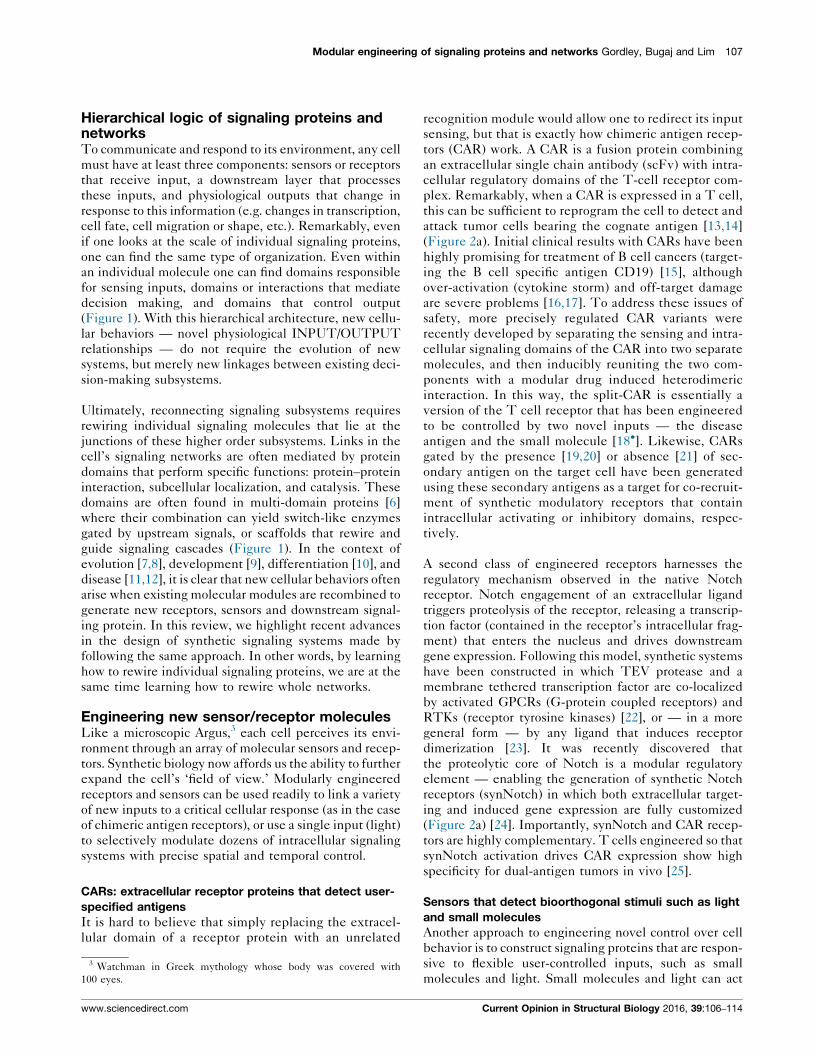
Figure 1
sensors informationprocessing responses
recognitiontransmission
effectors
RECEPTORS INTRACELLULARSIGNALING NODES
INPUT OUTPUT
INPUT OUTPUT writer reader
eraser
INPUT OUTPUT
motif
INPUT
OUTPUT
SIGNALINGNETWORKS
SIGNALINGPROTEINS
scaf
fold
Current Opinion in Structural Biology
Hierarchical organization of signaling systems: cells and individual proteins as input/output nodes. At any scale, a signaling system must have
three components — it must have sensors to receive INPUT, an information-processing layer that decides what to make of this information, and
an OUTPUT function. These components are found in individual signaling molecules, which detect and effect particular upstream and downstream
molecular partners. In receptors that span the cell’s membrane, ligand binding to extracellular domains (INPUT) rapidly regulates the activity of
intracellular effector domains (OUTPUT). Similarly, posttranslationally-regulated binding motifs link the activities of upstream enzymes that ‘write’
and ‘erase’ the posttranslational mark (INPUT; e.g. kinases and phosphatases) to recruitment of dedicated ‘reader’ domains (OUTPUT). The same
classes of components are found in signaling networks and whole cells, but in this case receptor molecules function as INPUT sensors, networks
of intracellular proteins function as the information processing layer, and various cellular response modules control OUTPUT.
within the cell and eliminate the need for transmembrane
receptors that are able to transmit signals across the
membrane. Inducible signaling proteins help us under-
stand and engineer signaling networks by enabling us to
observe network function in response to precisely defined
pathway inputs. The first generation of inducible signal-
ing systems relied on chemically induced dimerization
(CID) of modular binding domains that homo-dimerize or
hetero-dimerize upon binding of a small molecule ligand.
These have been previously reviewed [26�]. Below, we
focus on the next generation of tools that use light as an
inducer (broadly termed ‘optogenetics’), providing new
strategies for protein control with exquisite spatial and
temporal precision. Optogenetic proteins are engineered
from natural photoreceptor domains that coordinate with
light-sensing cofactors. Absorption of light by these cofac-
tors induces photoisomerization that drives a conforma-
tional change in the associated protein domain. This
conformational change is then coupled to larger allosteric
changes or enhanced protein–protein interactions. A
small set of photoreceptor modules that undergo light-
induced dimerization, oligomerization, or steric regula-
tion (Figure 2b) have been recurrently used to achieve
optogenetic control over diverse signaling proteins and
networks.
Light-induced dimerization
Optogenetic protein homo-dimerization and hetero-
dimerization has been used in multiple strategies to
Current Opinion in Structural Biology 2016, 39:106–114
regulate signaling nodes throughout the cell. Receptor
tyrosine kinases (RTKs) EGFR, FGFR, and Ret have
been endowed with blue light regulation through fusion
of their intracellular tails to a small, blue-light-sensing
homo-dimerization domain [27�]. Hetero-dimerization
has been used to regulate signaling through protein
recruitment to particular cellular compartments. Plasma
membrane recruitment of activating guanine nucleotide
exchange factors (GEFs) has been an effective strategy
for control of small GTPases Rac, Rho, Cdc42, and Ras
[28�,29], and, conversely, recruitment of an inhibitory
GTPase activating protein (GAP) was reported as a
method to inhibit G-protein coupled receptors [30].
Membrane recruitment was also used to regulate signal-
ing through Raf-1 [31] and PI3K [32]. Inducible nuclear
recruitment enabled optogenetic control of transcription
factor activity [33], and optogenetic recruitment to other
cellular compartments was reported as a general strategy
for titrating away signaling proteins [34,35]. Still other
dimerization-based approaches used homo-association of
Dronpa mutants to sterically or allosterically regulate
activity of a catalytically active GEF protein [36], and
optogenetic dissociation of a constitutive dimer enabled
optical control over protein trafficking and secretion [37].
Light-induced oligomerization
Multivalency and higher-order protein assembly play key
regulatory roles in many cellular signaling systems [38],
and recent work illustrates how protein clustering can be
www.sciencedirect.com

Modular engineering of signaling proteins and networks Gordley, Bugaj and Lim 109
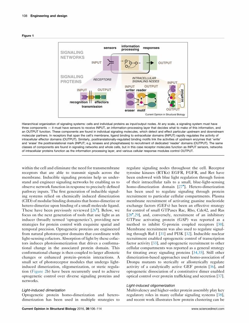
Figure 2
HETERO-DIMERIZATION OLIGOMERIZATION STERIC OCCLUSION
sequestration
degron
selectiveinhibitor
NLS
optogenetic control of signaling molecules - light controlled cells
chimeric receptors can redirect T cell activity towards novel cancer cell antigens
association
nuclearlocalization
membranelocalization
clustering
clusteringnuclearimport
CAR
cancercell
engineeredT cell
costimulatorydomain
scFv
CD3 zeta
proliferationcytokine productioncytotoxic function
antigen
native T cellbehaviors
synNotch
transcriptionfactor
cleavagesite
customized geneexpression
AND gate CAR AND-NOTgate CAR
small-molecule gated CAR
inhibitorydomain
dimerizingdomains
synNotch/CARAND gate
CAR
∗
EN
GIN
EE
RE
DR
EC
PE
TO
RS
HIG
HE
R-O
RD
ER
SE
NS
OR
S
(a)
(b)
Current Opinion in Structural Biology
Engineering new sensor/receptor systems. (a) The chimeric antigen receptor (CAR) was engineered to sense a tumor antigen and induce an
immunogenic response against tumor cells expressing that antigen. Modular recombination of the CAR domains with new sensor modules has enhanced
specificity of the CAR-T response either through logic gates requiring combinations of specific antigens or licensed by small molecule dimerization of
critical signaling domains [18�,19–21]. A second type of engineered receptor based on Notch (synNotch) allows both input (target antigen) and output
(gene expression) to be fully customized [24]. CAR and synNotch receptors can be combined synergistically, refining the specificity and scope of the
T cell response [25]. (b) Modular optogenetic tools for controlling receptors and signaling proteins. Protein domains from plants that undergo light-
induced dimerization, oligomerization, or steric regulation have been used to regulate signaling activities throughout the cell in a modular fashion.
www.sciencedirect.com Current Opinion in Structural Biology 2016, 39:106–114

110 Engineering and design
placed under optogenetic control. Blue light-induced
oligomerization of the Arabidopsis Cryptochrome2 pro-
tein has enabled activation of RTKs, both exogenous
[39,40,41] and endogenous [41], as well as the Orai1
calcium channel [42] and the canonical Wnt pathway
co-receptor LRP6 [43�]. Within the cytoplasm, cluster-
ing has been used to regulate Rho GTPase [43�] and
Raf1 kinase [44] activity. Inducible clustering has also
been used to regulate DNA damage signaling in the
absence of DNA damage through oligomerization of
TopBP1 [45].
Light-induced steric regulation
The blue light-induced conformational change of the
LOV2 domain from A. sativa phototropin has been suc-
cessfully used to sterically regulate small peptides con-
trolling multiple cellular functions in a modular fashion.
These functions include protein interaction [46,47], pro-
tein degradation [48,49], and nuclear translocation
[50,51]. Steric occlusion of the appropriate peptides has
also yielded optogenetic inhibition of specific kinases
[52�] and activation of calcium channels [53].
Engineering phosphorylation control:intracellular posttranslational circuitryOnce a cellular sensor is activated, the signal is often
relayed through a posttranslational regulatory network
that processes that information and directs the cell to
execute an appropriate response. Phosphorylation is the
most common posttranslational modification [54], and
Figure 3
enp
tran
(b)ENGINEERED SCAFFOLDS
MAPK
MAPKK
MAPKKK
synthetic scaffold
syntheticfeedback
PMAPK
MAPKK
MAPKKK
(a)
phos
Post-translational signaling: rewiring phosphorylation devices. Novel linkage
proteins and networks. (a) Synthetic scaffolding of the MAPK pathway was
scaffolding with negative regulatory effectors can tune the pathway respons
nuclear import and export sequences is a successful strategy to create dyn
Mutation of a phospho-site in a 3-pronged AND-gate for protein degradatio
Current Opinion in Structural Biology 2016, 39:106–114
efforts to engineer phospho-signaling proteins have shed
light on how posttranslational networks function and how
they can be rewired.
Engineered scaffolds for phospho-signaling
Proteins with multiple interaction domains can serve as
molecular scaffolds, organizing multiple proteins in a
signaling pathway into a complex (Figure 1). In yeast,
the scaffold protein Ste5 orchestrates the mating phero-
mone MAP kinase (MAPK) pathway. Ste5 co-localizes a
kinase cascade (MAPKKK ! MAPKK ! MAPK) and
serves as a platform for the spatial and temporal control
of these enzymes. To investigate how modular interac-
tions mediate kinase cascades, Ryu and Park designed
synthetic scaffolds using strings of repeated peptide
binding domains (PDZ) and fused complementary
PDZ ligands to each of the Ste5-associated kinases:
Ste11 (MAPKKK), Ste7 (MAPKK), and Fus3 (MAPK).
Synthetic scaffolds that co-localized two or more kinases
(MAPKKK AND [MAPKK OR MAPK]) at the cell
membrane were sufficient to functionally replace Ste5
[55] (Figure 3a). Moreover, these minimal scaffolds dem-
onstrated logic gate properties and could be tuned by the
co-recruitment of negative regulatory phosphatases. This
result matches findings with endogenous MAPK path-
ways, where recruitment of regulators to engineered
scaffolds reshapes the amplitude and timing of pathway
behavior [56]. This approach was recently extended by
the use of bacterial effectors [57]. The utility of this
approach is exemplified by OspF, a toxic protein that
P P P
123 input
Ub ligase
kinase
P
dockingmotif
NES plasm
PHOSPHO-REGULATED LINEAR MOTIFS
fluorescentprotein
gineeredhospho-
activatedslocation
P
NLS
nucleus
degradation
input
export
primingphorylation
cascade
Current Opinion in Structural Biology
cytop
scentfluoreseieieinnnproproprottete
s between signaling modules enable new functionality in signaling
sufficient to induce downstream signaling [55] (top left), while co-
e [57]. (b) Combining kinase docking motifs with phospho-regulated
amic fluorescent reporters of specific kinase activity [62��] (top right).
n generated a 2-pronged degradation AND-gate [69�].
www.sciencedirect.com

Modular engineering of signaling proteins and networks Gordley, Bugaj and Lim 111
irreversibly inactivates MAPKs by catalyzing a beta-elim-
ination reaction that removes the hydroxyl group of the
key phospho-threonine side chain, thereby preventing
MAPK phosphorylation and consequent activation. This
mechanism enables Shigella flexneri to disable human
epithelial and dendritic cells. Repurposed as a tool for
synthetic biology, this pathogenic inhibitor has been used
to engineer a negative feedback loop that reshapes the
dynamic response of the yeast osmolarity pathway
(Figure 3a) and a T-cell ‘pause switch’ for adoptive
immunotherapy [57].
Phospho-regulated linear motifs
Signaling proteins are enriched in unstructured regions
(linear motifs) where the ‘writers’, ‘readers’, and ‘erasers’
of phosphorylation (kinases, phospho-binding domains,
and phosphatases, respectively) collectively regulate pro-
tein binding, concentration, and localization [58,59]
(Figure 1). Phospho-regulated linear motifs have been
used to create dynamic reporters of intracellular signaling.
FRET reporters for specific kinases link a phospho-regu-
lated intramolecular binding event (phosphorylated sub-
strate peptide + phospho-binding domain) to fluorescent
protein co-localization [60]. More recently, a synthetic,
phospho-stabilized, version of the destabilizing PEST
domain was fused to a fluorescent protein to construct
a live cell reporter of Erk activity [61]. In a third example
of phospho-engineering, Regot et al. built a reporter that
translocates in response to c-Jun N-terminal kinase (JNK)
activation by combining a nuclear export signal (NES)
activated by JNK phosphorylation, a phospho-inhibited
nuclear import signal (NLS), and a fluorescent protein
[62��] (Figure 3b). Importantly, this design can be readily
adapted to multiple types of kinases by either substitut-
ing the docking site for JNK with that of another MAP
kinase (MAPK), or mutating a kinase’s naturally occurring
substrate to introduce the NLS and NES modules. This
work illustrates the potential for modular engineering
with linear motifs, and complements recent advances
in the computational design of posttranslational regula-
tion [63].
Scaling up phospho-circuit design
In proteins that contain multiple phosphorylation sites,
phospho-regulated linear motifs can collectively form
information processors that integrate inputs, set re-
sponse thresholds [64], tune binding affinities [65],
amplify weak signals, and serve as ‘conduits’ for se-
quential signal transduction [66]. Analogous synthetic
‘devices’, built from combinations of phospho-regula-
tory modules, may ultimately make it possible to endow
engineered signaling proteins with complex informa-
tion processing behaviors.
One of the best-studied examples of multisite phosphor-
ylation is Sic1, a critical cell cycle regulator whose phos-
pho-induced degradation triggers S phase in budding
www.sciencedirect.com
yeast [64,67]. Cyclin dependent kinase 1 (Cdk1), in
complex with a cyclin and the phospho-adaptor Cks1,
phosphorylates Sic1 in an ordered sequence at multiple
sites, culminating in the activation of phosphodegrons
within Sic1 that recruit the SCFCdc4 ubiquitin ligase
complex, thereby targeting Sic1 for rapid degradation.
Using synthetic Cdk1 substrates, Loog and colleagues
found that the rate at which Cdk1 phosphorylation pro-
pagates is determined by how well the fixed spatial
orientation of the three docking sites on the cyclin-
Cdk1–Cks1 complex fits with the linear pattern of phos-
phorylation sites along the length of substrates like Sic1
[68�]. These results suggest that a simple set of rules
might define the overall pattern and rates of phosphor-
ylation in a multisite cluster.
Eco1, a regulator of sister chromatid cohesion, is also
degraded after multisite phosphorylation. The timing of
Eco1 function is usually restricted to S phase by the
collective action of three different kinases (Cdk1,
Cdc7, Mck1). In healthy cells, sequential phosphorylation
by these kinases forms an SCFCdc4 phosphodegron, trig-
gering Eco1 destruction. However, inhibition of one
kinase (Cdc7) by the DNA damage response prevents
Eco1 destruction, allowing establishment of cohesion
after S phase. Lyons et al. characterized this system
and generated a mutant version of Eco1 in which Cdk1
directly primes Mck1 phosphorylation — bypassing
Cdc7’s phospho-regulation. This study revealed that a
single point deletion converts the naturally occurring 3-
pronged AND gate into a synthetic 2-pronged gate with
altered cell cycle sensitivity [69�] (Figure 3b).
Overall, these findings suggest that fairly simple rules
governing linear phospho-motifs are used by nature to
achieve quite sophisticated information processing. As we
learn to better manipulate these motifs, we may be able to
test and harness these emerging rules.
Forward evolution of signaling networksUltimately, to engineer cellular behavior, we want to
engineer cellular networks. But as described above, to
engineer cellular networks we need to learn how to
engineer individual signaling proteins and their connec-
tivity. We postulate that much of the functional innova-
tion in cellular signaling networks has evolved through
repeated duplication and recombination of modular
domains [7]. Researchers interested in posttranslational
engineering can now use bioinformatics tools [70,71,72] to
mine these naturally occurring circuits for domains that
are, in essence, ‘pre-validated’ for a high degree of
modularity (successfully functioning in many fusion con-
texts) and minimal crosstalk with other cellular compo-
nents. These parts complement the growing toolbox of
regulatory domains that have been validated (in an anal-
ogous manner) through repeated use in synthetic circuits
[5,73]. With these enriched building blocks, generating
Current Opinion in Structural Biology 2016, 39:106–114

112 Engineering and design
Figure 4
endogeneoussignaling systems
libraries of modular parts
screen + characterize
small librariesof circuits
functionally screen
designinterations
validatedparts computational
modeling
PIP2 PIP 3
PI3K
PTEN
PH PHAktPLCδ
kinasesphosphatases
phospho-binding
PIP3
PHPI3K
synthetic cellpolarization
modular parts
synthetic proteins
synthetic circuits
computationalmodeling
Current Opinion in Structural Biology
Combinatorial design for engineering signaling proteins and networks. We present here a conceptual workflow for engineering signaling networks
with desired properties. Small libraries of candidate circuits can be semi-rationally designed using a combination of validated signaling and
regulatory components together with computational models. These circuits can then be screened and optimized for the proper function. The
design of a network for cell polarization [74��] is provided as an example of this approach.
new signaling proteins could potentially be a straightfor-
ward matter of screening domain combinations (combi-
natorial libraries) and optimizing protein expression
[18�,28�,56]. The same approach can be iterated to gen-
erate posttranslational circuits of increasing complexity.
One successful example has been the construction of a
synthetic circuit for inducing artificial cell polarization in
yeast. Modular binding domains that recognize phospho-
inositide species were combined with modular catalytic
domains that modify these species, yielding a set of
proteins that form spatially localized positive and nega-
tive feedback loops. Together, this system of synthetic
proteins generates a self-organizing asymmetric pole of
the signaling molecule PIP3 [74��] (Figure 4). It is likely
that in the coming years, we will see more examples of the
construction of more complex synthetic signaling sys-
tems, enabled by a better understanding of modular
domains, but also by advances in computational design
and experimental combinatorial screening of libraries of
modular synthetic circuits. As the field of synthetic sig-
naling systems matures, a semi-predictive approach that
combines computational design with combinatorial
screening offers a pragmatic strategy for learning nature’s
design principles while tailoring cellular responses to
applications in medicine and biotechnology.
Conflict of interest statementWendell Lim is a Founder of Cell Design Labs and a
member of its scientific advisory board.
Current Opinion in Structural Biology 2016, 39:106–114
AcknowledgementsThe authors sincerely thank Dr. Nicholas Frankel, Dr. Ricardo Almeida,Dr. Scott Coyle, Dr. Kole Roybal and Dr. Kyle Daniels for comments on themanuscript, as well as other members of the Lim lab for helpful discussions.We apologize to authors whose relevant work was not cited in this reviewdue to space constraints. The authors are supported by the NationalInstitutes of Health (P50 GM081879, PN2 EY016546, R01 GM055040, R01GM096164, R01 CA196277), NSF Synthetic Biology and EngineeringResearch Center, and the Howard Hughes Medical Institute.
References and recommended readingPapers of particular interest, published within the period of review,have been highlighted as:
� of special interest�� of outstanding interest
1. Alon U: An introduction to systems biology: design principles ofbiological circuits. CRC Press; 2006:: 324.
2. Lim W, Mayer B, Pawson T: Cell signaling: principles andmechanisms. Taylor & Francis; 2014.
3. Lim WA: Designing customized cell signalling circuits. Nat RevMol Cell Biol 2010, 11:393-403.
4. Brophy JAN, Voigt CA: Principles of genetic circuit design. NatMethods 2014, 11:508-520.
5. Slusarczyk AL, Lin A, Weiss R: Foundations for the design andimplementation of synthetic genetic circuits. Nat Rev Genet2012, 13:406-420.
6. Tordai H, Nagy A, Farkas K, Banyai L, Patthy L: Modulesmultidomain proteins organismic complexity. FEBS J 2005,272:5064-5078.
7. Di Roberto RB, Peisajovich SG: The role of domain shuffling inthe evolution of signaling networks. J Exp Zool B Mol Dev Evol2014, 322:65-72.
www.sciencedirect.com

Modular engineering of signaling proteins and networks Gordley, Bugaj and Lim 113
8. Lim WA, Pawson T: Phosphotyrosine signaling: evolving a newcellular communication system. Cell 2010, 142:661-667.
9. Buljan M, Chalancon G, Eustermann S, Wagner GP, Fuxreiter M,Bateman A et al.: Tissue-specific splicing of disorderedsegments that embed binding motifs rewires proteininteraction networks. Mol Cell 2012, 46:871-883.
10. Tonegawa S: Somatic generation of antibody diversity. Nature1983, 302:575-581.
11. Aman P: Fusion oncogenes in tumor development. SeminCancer Biol 2005, 15:236-243.
12. Davey NE, Trave G, Gibson TJ: How viruses hijack cellregulation. Trends Biochem Sci 2011, 36:159-169.
13. Kalos M, June CH: Adoptive T cell transfer for cancerimmunotherapy in the era of synthetic biology. Immunity 2013,39:49-60.
14. Sadelain M, Brentjens R, Riviere I: The basic principles ofchimeric antigen receptor design. Cancer Discov 2013,3:388-398.
15. Kalos M, Levine BL, Porter DL, Katz S, Grupp SA, Bagg A et al.: Tcells with chimeric antigen receptors have potent antitumoreffects and can establish memory in patients with advancedleukemia. Sci Transl Med 2011, 3.
16. Morgan RA, Yang JC, Kitano M, Dudley ME, Laurencot CM,Rosenberg SA: Case report of a serious adverse eventfollowing the administration of T cells transduced with achimeric antigen receptor recognizing ERBB2. Mol Ther J AmSoc Gene Ther 2010, 18:843-851.
17. Brentjens RJ, Riviere I, Park JH, Davila ML, Wang X, Stefanski Jet al.: Safety and persistence of adoptively transferredautologous CD19-targeted T cells in patients with relapsed orchemotherapy refractory B-cell leukemias. Blood 2011,118:4817-4828.
18.�
Wu C-Y, Roybal KT, Puchner EM, Onuffer J, Lim WA: Remotecontrol of therapeutic T cells through a small molecule-gatedchimeric receptor. Science 2015, 350:aab4077.
Illustrates systematic combinatorial design – assembly and screening of asmall library of domain combinations yielded a split version of the CARwhose function is conditional upon drug-induced hetero-dimerization.
19. Kloss CC, Condomines M, Cartellieri M, Bachmann M, Sadelain M:Combinatorial antigen recognition with balanced signalingpromotes selective tumor eradication by engineered T cells.Nat Biotechnol 2013, 31:71-75.
20. Lanitis E, Poussin M, Klattenhoff AW, Song D, Sandaltzopoulos R,June CH et al.: Chimeric antigen receptor T cells withdissociated signaling domains exhibit focused antitumoractivity with reduced potential for toxicity in vivo. CancerImmunol Res 2013, 1:43-53.
21. Fedorov VD, Themeli M, Sadelain M: PD-1-based and CTLA-4-based inhibitory chimeric antigen receptors (iCARs) divert off-target immunotherapy responses. Sci Transl Med 2013,5:215ra172.
22. Barnea G, Strapps W, Herrada G, Berman Y, Ong J, Kloss B et al.:The genetic design of signaling cascades to record receptoractivation. Proc Natl Acad Sci U S A 2008, 105:64-69.
23. Daringer NM, Dudek RM, Schwarz KA, Leonard JN: Modularextracellular sensor architecture for engineering mammaliancell-based devices. ACS Synth Biol 2014, 3:892-902.
24. Morsut L, Roybal KT, Xiong X, Gordley RM, Coyle SM,Thomson M et al.: Engineering customized cell sensing andresponse behaviors using synthetic notch receptors. Cell2016, 164:780-791.
25. Roybal KT, Rupp LJ, Morsut L, Walker WJ, McNally KA, Park JSet al.: Precision tumor recognition by T cells withcombinatorial antigen-sensing circuits. Cell 2016, 164:770-779.
26.�
Voß S, Klewer L, Wu Y-W: Chemically induced dimerization:reversible and spatiotemporal control of protein function incells. Curr Opin Chem Biol 2015, 28:194-201.
A recent review of chemically induced dimerization, which has beenwidely used for small-molecule induction of signaling proteins.
www.sciencedirect.com
27.�
Grusch M, Schelch K, Riedler R, Reichhart E, Differ C, Berger Wet al.: Spatio-temporally precise activation of engineeredreceptor tyrosine kinases by light. EMBO J 2014, 33:1713-1726.
The authors use a small homodimerizing domain to regulate associationand signaling of receptor tyrosine kinase activation using blue light.
28.�
Toettcher JE, Weiner OD, Lim WA: Using optogenetics tointerrogate the dynamic control of signal transmission by theRas/Erk module. Cell 2013, 155:1422-1434.
The authors leverage rapid optogenetic switching to quantify temporalfiltering and frequency response of signal transduction through the Ras/Erk pathway. This demonstrates the power of optogenetics for a probingsignaling network function within living cells.
29. Levskaya A, Weiner OD, Lim WA, Voigt CA: Spatiotemporalcontrol of cell signalling using a light-switchable proteininteraction. Nature 2009, 461:997-1001.
30. O’Neill PR, Gautam N: Subcellular optogenetic inhibition of Gproteins generates signaling gradients and cell migration. MolBiol Cell 2014, 25:2305-2314.
31. Zhang K, Duan L, Ong Q, Lin Z, Varman PM, Sung K et al.: Light-mediated kinetic control reveals the temporal effect of theRaf/MEK/ERK pathway in PC12 cell neurite outgrowth. PLOSONE 2014, 9:e92917.
32. Toettcher JE, Gong D, Lim WA, Weiner OD: Light-basedfeedback for controlling intracellular signaling dynamics. NatMethods 2011, 8:837-839.
33. Beyer HM, Juillot S, Herbst K, Samodelov SL, Muller K,Schamel WW et al.: Red light-regulated reversible nuclearlocalization of proteins in mammalian cells and zebrafish. ACSSynth Biol 2015, 4:951-958.
34. Lee S, Park H, Kyung T, Kim NY, Kim S, Kim J et al.: Reversibleprotein inactivation by optogenetic trapping in cells. NatMethods 2014, 11:633-636.
35. Yang X, Jost AP-T, Weiner OD, Tang C: A light-inducibleorganelle-targeting system for dynamically activating andinactivating signaling in budding yeast. Mol Biol Cell 2013,24:2419-2430.
36. Zhou XX, Chung HK, Lam AJ, Lin MZ: Optical control ofprotein activity by fluorescent protein domains. Science2012, 338:810-814.
37. Chen D, Gibson ES, Kennedy MJ: A light-triggered proteinsecretion system. J Cell Biol 2013, 201:631-640.
38. Mammen M, Choi S-K, Whitesides GM: Polyvalent interactionsin biological systems: implications for design and use ofmultivalent ligands and inhibitors. Angew Chem Int Ed 1998,37:2754-2794.
39. Chang K-Y, Woo D, Jung H, Lee S, Kim S, Won J et al.: Light-inducible receptor tyrosine kinases that regulate neurotrophinsignalling. Nat Commun 2014, 5:4057.
40. Kim N, Kim JM, Lee M, Kim CY, Chang K-Y, Heo WD:Spatiotemporal control of fibroblast growth factor receptorsignals by blue light. Chem Biol 2014, 21:903-912.
41. Bugaj LJ, Spelke DP, Mesuda CK, Varedi M, Kane RS,Schaffer DV: Regulation of endogenous transmembranereceptors through optogenetic Cry2 clustering. Nat Commun2015, 6:6898.
42. Kyung T, Lee S, Kim JE, Cho T, Park H, Jeong Y-M et al.:Optogenetic control of endogenous Ca(2+) channels in vivo.Nat Biotechnol 2015, 33:1092-1096.
43.�
Bugaj LJ, Choksi AT, Mesuda CK, Kane RS, Schaffer DV:Optogenetic protein clustering and signaling activation inmammalian cells. Nat Methods 2013, 10:249-252.
The authors describe the natural light-induced oligomerizaton of Arabi-dopsis Cryptochrome 2 and demonstrate that this activity may be used ina modular fashion to aggregate and regulate multiple signaling effectors.
44. Wend S, Wagner HJ, Muller K, Zurbriggen MD, Weber W,Radziwill G: Optogenetic control of protein kinase activity inmammalian cells. ACS Synth Biol 2014, 3:280-285.
45. Ozkan-Dagliyan I, Chiou Y-Y, Ye R, Hassan BH, Ozturk N,Sancar A: Formation of Arabidopsis cryptochrome
Current Opinion in Structural Biology 2016, 39:106–114

114 Engineering and design
2 photobodies in mammalian nuclei: application as anoptogenetic DNA damage checkpoint switch. J Biol Chem2013, 288:23244-23251.
46. Strickland D, Lin Y, Wagner E, Hope CM, Zayner J, Antoniou Cet al.: TULIPs: tunable, light-controlled interacting protein tagsfor cell biology. Nat Methods 2012, 9:379-384.
47. Guntas G, Hallett RA, Zimmerman SP, Williams T, Yumerefendi H,Bear JE et al.: Engineering an improved light-induced dimer(iLID) for controlling the localization activity of signalingproteins. Proc Natl Acad Sci U S A 2015, 112:112-117.
48. Bonger KM, Rakhit R, Payumo AY, Chen JK, Wandless TJ:General method for regulating protein stability with light. ACSChem Biol 2014, 9:111-115.
49. Renicke C, Schuster D, Usherenko S, Essen L-O, Taxis C: A LOV2domain-based optogenetic tool to control protein degradationand cellular function. Chem Biol 2013, 20:619-626.
50. Yumerefendi H, Dickinson DJ, Wang H, Zimmerman SP, Bear JE,Goldstein B et al.: Control of protein activity and cell fatespecification via light-mediated nuclear translocation. PLOSONE 2015, 10:e0128443.
51. Niopek D, Benzinger D, Roensch J, Draebing T, Wehler P, Eils Ret al.: Engineering light-inducible nuclear localization signalsfor precise spatiotemporal control of protein dynamics inliving cells. Nat Commun 2014, 5:4404.
52.�
Yi JJ, Wang H, Vilela M, Danuser G, Hahn KM: Manipulation ofendogenous kinase activity in living cells usingphotoswitchable inhibitory peptides. ACS Synth Biol 2014,3:788-795.
The authors describe how the light-induced conformational change ofLOV2 might be engineered to sterically regulate inhibitors of endogenousproteins. This study both provides general guidelines for engineering theLOV2 photoswitch and provides methods for optogenetic signal controlwithout overexpression of the enzymes under control.
53. Pham E, Mills E, Truong K: A synthetic photoactivated proteinto generate local or global Ca(2+) signals. Chem Biol 2011,18:880-890.
54. Khoury GA, Baliban RC, Floudas CA: Proteome-wide post-translational modification statistics: frequency analysis andcuration of the Swiss-Prot database. Sci Rep 2011, 13:1.
55. Ryu J, Park S-H: Simple synthetic protein scaffolds can createadjustable artificial MAPK circuits in yeast and mammaliancells. Sci Signal 2015, 8:ra66.
56. Bashor CJ, Helman NC, Yan S, Lim WA: Using engineeredscaffold interactions to reshape MAP kinase pathwaysignaling dynamics. Science 2008, 319:1539-1543.
57. Wei P, Wong WW, Park JS, Corcoran EE, Peisajovich SG,Onuffer JJ et al.: Bacterial virulence proteins as tools to rewirekinase pathways in yeast and immune cells. Nature 2012,488:384-388.
58. Gsponer J, Babu MM: The rules of disorder or why disorderrules. Prog Biophys Mol Biol 2009, 99:94-103.
59. Van Roey K, Uyar B, Weatheritt RJ, Dinkel H, Seiler M, Budd Aet al.: Short linear motifs: ubiquitous and functionally diverseprotein interaction modules directing cell regulation. ChemRev 2014, 114:6733-6778.
60. Komatsu N, Aoki K, Yamada M, Yukinaga H, Fujita Y, Kamioka Yet al.: Development of an optimized backbone of FRETbiosensors for kinases and GTPases. Mol Biol Cell 2011,22:4647-4656.
Current Opinion in Structural Biology 2016, 39:106–114
61. Albeck JG, Mills GB, Brugge JS: Frequency-modulated pulsesof ERK activity transmit quantitative proliferation signals. MolCell 2013, 49:249-261.
62.��
Regot S, Hughey JJ, Bajar BT, Carrasco S, Covert MW: High-sensitivity measurements of multiple kinase activities in livesingle cells. Cell 2014, 157:1724-1734.
Concretely demonstrates how desired signaling behaviors can be cre-ated by combining or overlaying phospho-regulated linear motifs.
63. Strumillo M, Beltrao P: Towards the computational design ofprotein post-translational regulation. Bioorg Med Chem 2015,23:2877-2882.
64. Nash P, Tang X, Orlicky S, Chen Q, Gertler FB, Mendenhall MDet al.: Multisite phosphorylation of a CDK inhibitor sets athreshold for the onset of DNA replication. Nature 2001,414:514-521.
65. Lee CW, Ferreon JC, Ferreon ACM, Arai M, Wright PE: Gradedenhancement of p53 binding to CREB-binding protein (CBP)by multisite phosphorylation. Proc Natl Acad Sci U S A 2010,107:19290-19295.
66. Galea CA, Wang Y, Sivakolundu SG, Kriwacki RW: Regulation ofcell division by intrinsically unstructured proteins: intrinsicflexibility, modularity, and signaling conduits. Biochemistry(Mosc) 2008, 47:7598-7609.
67. Hao B, Oehlmann S, Sowa ME, Harper JW, Pavletich NP:Structure of a Fbw7-Skp1-cyclin E complex: multisite-phosphorylated substrate recognition by SCF ubiquitinligases. Mol Cell 2007, 26:131-143.
68.�
Koivomagi M, Ord M, Iofik A, Valk E, Venta R, Faustova I et al.:Multisite phosphorylation networks as signal processors forCdk1. Nat Struct Mol Biol 2013, 20:1415-1424.
These articles shed light on the structural basis for ordered multisitephosphorylation, suggesting a basis for rationale design of phospho-regulated systems that rapidly perform multistep computations.
69.�
Lyons NA, Fonslow BR, Diedrich JK, Yates JR, Morgan DO:Sequential primed kinases create a damage-responsivephosphodegron on Eco1. Nat Struct Mol Biol 2013, 20:194-201.
These articles shed light on the structural basis for ordered multisitephosphorylation, suggesting a basis for rationale design of phospho-regulated systems that rapidly perform multistep computations.
70. Letunic I, Doerks T, Bork P: SMART: recent updates, newdevelopments and status in 2015. Nucleic Acids Res 2015,43(Database issue):D257-D260.
71. Dinkel H, Van Roey K, Michael S, Davey NE, Weatheritt RJ,Born D et al.: The eukaryotic linear motif resource ELM:10 years and counting. Nucleic Acids Res 2014, 42(Databaseissue):D259-D266.
72. Van Roey K, Dinkel H, Weatheritt RJ, Gibson TJ, Davey NE: Theswitches.ELM resource: a compendium of conditionalregulatory interaction interfaces. Sci Signal 2013, 6:rs7.
73. Lienert F, Lohmueller JJ, Garg A, Silver PA: Synthetic biology inmammalian cells: next generation research tools andtherapeutics. Nat Rev Mol Cell Biol 2014, 15:95-107.
74.��
Chau AH, Walter JM, Gerardin J, Tang C, Lim WA: Designingsynthetic regulatory networks capable of self-organizing cellpolarization. Cell 2012, 151:320-332.
This article illustrates how modules derived from naturally occurringposttranslational networks can be extracted and used to build cells withcustomized signaling behaviors of increasing complexity.
www.sciencedirect.com