Embed Size (px)
Citation preview

miRNA-205 Suppresses Melanoma Cell Proliferation andInduces Senescence via Regulation of E2F1 Protein*□S
Received for publication, February 2, 2011, and in revised form, March 22, 2011 Published, JBC Papers in Press, March 24, 2011, DOI 10.1074/jbc.M111.227611
Altaf A. Dar‡, Shahana Majid§, David de Semir‡, Mehdi Nosrati‡, Vladimir Bezrookove‡,and Mohammed Kashani-Sabet‡1
From the ‡California Pacific Medical Center Research Institute, San Francisco, California 94107 and the §Department of Urology,Veterans Affairs Medical Center and University of California, San Francisco, California 94121
MicroRNAs (miRNAs) regulate gene expressionby repressingtranslation or directing sequence-specific degradation of com-plementary mRNA. Here, we report that expression of miR-205is significantly suppressed in melanoma specimens when com-pared with nevi and is correlated inversely with melanoma pro-gression. miRNA target databases predicted E2F1 and E2F5 asputative targets. The expression levels of E2F1 and E2F5 werecorrelated inversely with that of miR-205 in melanoma celllines. miR-205 significantly suppressed the luciferase activity ofreporter plasmids containing the 3�-UTR sequences comple-mentary to either E2F1 or E2F5. Overexpression of miR-205 inmelanoma cells reduced E2F1 and E2F5 protein levels. The pro-liferative capacity ofmelanomacellswas suppressedbymiR-205andmediated by E2F-regulatedAKTphosphorylation.miR-205overexpression resulted in induction of apoptosis, as evidencedby increased cleaved caspase-3, poly-(ADP-ribose) polymerase,and cytochrome c release. Stable overexpression of miR-205suppressed melanoma cell proliferation, colony formation, andtumor cell growth in vivo and induced a senescence phenotypeaccompanied by elevated expression of p16INK4A and othermarkers for senescence. E2F1 overexpression in miR-205-ex-pressing cells partially reversed the effects on melanoma cellgrowth and senescence. These results demonstrate a novel rolefor miR-205 as a tumor suppressor in melanoma.
MicroRNAs (miRNAs)2 are a class of short noncoding RNAsthat regulate gene expression by complementary base pairingwith the 3�-UTR of target mRNAs and causing their degrada-tion (1) or by directly mediating mRNA degradation (2). Inaddition, miRNAs can also inhibit translation in the event ofimperfect base pair matching with the target. miRNAs are
expressed in a tissue-specificmanner and are considered to playimportant roles in cell proliferation, apoptosis, and differentia-tion (3, 4).Deregulation of miRNA expression has been identified in a
number of cancers (5, 6), and accumulating data indicate thatsomemiRNAs can function as tumor suppressors or oncogenesand as such are important in cancer development. Specific sub-sets of miRNAs also have been shown to be dysregulated invarious solid tumors (7, 8). The discovery of miRNAs at previ-ously identified chromosomal breakpoints, as well as deletionand amplification sites in certain cancers, implies that theymaybe involved in disease initiation or progression.The human genome is predicted to encode as many as 1000
miRNAs (9). Although it is difficult to identify accurately indi-vidual miRNA-target interactions, computational predictionsof miRNA target genes indicate that as many as one-third of allhuman protein-encoding genes may be regulated by miRNAs(10). Due to their tremendous regulatory potential and tissue-anddisease-specific expression patterns, there is increasing evi-dence that miRNA expression profiles could be indicative ofdisease risk or burden. Thus, miRNAs are being assessed aspossible biomarkers to aid in the diagnosis and prognosis ofdifferent cancers, including melanoma (11, 12). However, todate, very few miRNA expression profiling analyses have beenperformed on human melanoma samples.E2F1, the best characterized member of the E2F family, is a
master regulator of the G1/S transition phase in the cell cyclethat is tightly regulated by the retinoblastoma protein (Rb).E2F1 transactivates a variety of genes involved in chromosomalDNA replication and cell cycle progression (13). Overexpres-sion of E2F1 is an oncogenic event that predisposes cells totransformation (14). E2F1 gene amplification and/or abnor-malities in its expression have been reported in various tumortypes, including melanoma (15, 16). E2F5, another E2F familymember, is an oncogenic cell cycle regulator demonstrated tobe amplified or overexpressed in various tumors (17, 18).In this study, we report that miR-205 is significantly down-
regulated in melanoma tumor samples and cell lines. In addi-tion, we examine the consequences of miR-205 overexpressionand identify E2F1 and E2F5 as downstream targets of miR-205action in melanoma.
MATERIALS AND METHODS
Cell Culture, Plasmids, and Transfection—The WM3211,DO4, WM278, and 1205-Lu (kindly provided by Dr. Boris Bas-tian) and Lox (kindly provided by Dr. Oystein Fodstad) mela-
* This work was supported, in whole or in part, by National Institutes ofHealth, United States Public Health Service Grants CA114337 andCA122947 (to M. K.-S.). This work was also supported by the Herschel andDiana Zackheim Endowment Fund.
□S The on-line version of this article (available at http://www.jbc.org) containssupplemental Fig. 1.
1 To whom correspondence should be addressed: Center for MelanomaResearch and Treatment, California Pacific Medical Center Research Insti-tute, 475 Brannan St., Ste. 220, San Francisco, CA 94107. Tel.: 415-600-3800;Fax: 415-600-1091; E-mail: [email protected].
2 The abbreviations used are: miRNA, microRNA; qRT-PCR, quantitative RT-PCR; PCNA, proliferating cell nuclear antigen; SA-�-gal, senescence-asso-ciated �-galactosidase; PARP, poly-(ADP-ribose) polymerase; Rb, retino-blastoma protein; AKT, v-akt murine thymoma viral oncogene homolog;HPRT, hypoxanthine phosphoribosyltransferase 1; MCM3, minichromo-some maintenance complex component 3.
THE JOURNAL OF BIOLOGICAL CHEMISTRY VOL. 286, NO. 19, pp. 16606 –16614, May 13, 2011Printed in the U.S.A.
16606 JOURNAL OF BIOLOGICAL CHEMISTRY VOLUME 286 • NUMBER 19 • MAY 13, 2011
by guest on May 13, 2018
http://ww
w.jbc.org/
Dow
nloaded from

noma cell lines were grown in RPMI with 10% fetal bovineserum. C8161.9 cells (obtained from Dr. Danny Welch) weregrown in DMEM/F12 with 5% fetal bovine serum (Invitro-gen) at 37 °C in an atmosphere containing 5% CO2. Normalhuman melanocytes were purchased from Lifeline CellTechnology and grown in LL-0027 medium (Lifeline CellTechnology, Walkersville, MD). Plasmids pMax-GFP(Lonza, Walkersville, MD), pMax-E2F1 (Addgene, Cam-bridge, MA), miRNASelectTM pEP-miR null control vector(pEP Null), and miRNASelectTM pEP-hsa-mir-205 expressionvector (pEP miR-205) (Cell Biolabs, Inc., San Diego, CA) werepurchased. TaqMan probes for hsa-miR-205 and negative con-trol pre-miR were purchased from Applied Biosystems (FosterCity, CA). Transient transfection was carried out by Lipo-fectamine 2000 (Invitrogen) according to the manufacturer’sprotocol.Quantitative Real-time PCR—Mature miRNAs and other
mRNAs were assayed using the TaqMan MicroRNA Assaysand Gene Expression Assays, respectively, in accordance withthe manufacturer’s instructions (Applied Biosystems). All RTreactions, including no-template controls and RT minus con-trols, were run in a 7500 Fast Real-time PCR System (AppliedBiosystems). RNA concentrations were determined with aNanoDrop (Thermo Scientific, Rockford, IL). Samples werenormalized to RNU48 or HPRT (Applied Biosystems), as indi-cated. Gene expression levels were quantified using 7500 FastReal-time Sequence detection system Software. Comparativereal-time PCR was performed in triplicate, including no-tem-plate controls. Relative expression was calculated using thecomparative Ct method.RNA and miRNA Extraction from Tissue Samples and Cell
Lines—Samples from patients with primary (n � 20), meta-static melanoma (n � 27), and benign nevi (n � 20) wereobtained under a protocol approved by the Institutional ReviewBoard. RNA was extracted by using RNeasy Mini Kit (Qiagen,Valencia, CA) following the manufacturer’s protocol. miRNAswere extracted by using the mirVana miRNA extraction kit(Applied Biosystems) from tissues and cell lines following themanufacturer’s instructions.Cell Viability, Colony Formation, and Cell Cycle Analysis—
Cells were plated in 96-well plates at a density of 3 � 103 cellsper well. Cell viability was assessed at 24, 48, 72, and 96 h post-transfection using Cell Counting Kit-8 (Dojindo, Rockville,MD) following themanufacturer’s protocol. For the colony for-mation assay, 500 cells were plated in a p100 plate and allowedto grow until visible colonies appeared. Colonies were stainedwith Giemsa and counted. Cell cycle analysis was performed asdescribed previously (19).Western Blot Analysis—Cell lysates were prepared in PBS
containing 1� Halt protease inhibitor mixture and 1� Haltphosphatase inhibitormixture (Pierce) centrifuged at 3500 rpmfor 10 min at 4 °C. Proteins (10–15 ug) from each sample weresubjected to SDS-PAGE and transferred onto a nitrocellulosemembrane. Target proteins were detected by using specificantibodies against E2F5, PARP, p16INK4a, and GAPDH (SantaCruz Biotechnology, Santa Cruz, CA), E2F1, AKT, pAKT (Ser-473), pRB (Ser-807/811), MCM3, caspase-9, BAD, pBAD,PCNA, cytochrome c, and cleaved caspase-3 (Cell Signaling
Technology, Danvers, MA) and p-caspase-9 (Abcam, Cam-bridge MA).Luciferase Assays—The 3�-UTR region of E2F1 and E2F5
containing target site sequences complementary to the seedsequence of miR-205 were cloned downstream of the luciferasegene in the pMIR-REPORT luciferase vector (Ambion, Cam-bridge, MA), and the resultant vectors were named E2F1–3�-UTR and E2F5–3�-UTR. Mutated 3�-UTR sequences of E2F1and E2F5 complementary to miR-205 were cloned in the samevector, and the resultant vectors were named E2F1-Mut3�-UTR and E2F5-Mut 3�-UTR, respectively. For reporterassays, cells were transiently transfected with wild-type ormutant reporter plasmid and miR-205. Firefly luciferase activ-ities were measured by using the Dual-Luciferase Assay (Pro-mega, Madison, WI) 48 h after transfection, and the resultswere normalized with Renilla luciferase. Each reporter plasmidwas transfected at least three times (on different days), and eachsample was assayed in triplicate.Stable Cell Generation and in Vivo Study—C8161.9 cells
were transfected with pEP Null and pEP miR-205 vectors (CellBiolabs, San Diego, CA) and selected with puromycin (1�g/ml). For in vivo studies as described previously (20), 1� 106cells were injected into nude mice subcutaneously, and tumorgrowth was followed for 28 days. All animal care was in accord-ance with the institutional guidelines.SA-�-gal Staining—Staining for senescence-associated�-ga-
lactosidase (SA-�-gal) was performed using the SenescenceCell Histochemical Staining Kit from Sigma as per the manu-facturer’s protocol. In brief, cells were fixed with formalin,washed with PBS, and incubated at 37° C with staining mixtureuntil the cells were stained blue. The percentage of SA-�-gal-positive cells was determined by counting the number ofstained cells with bright field illumination.Statistical Analysis—All quantified data represent an average
of at least triplicate samples or as indicated. Error bars repre-sent S.E. Statistical significance was determined by the Stu-dent’s t test, and two-tailed p values �0.05 were consideredsignificant.
RESULTS
miR-205 Is Down-regulated inMelanoma, and Its ExpressionIs Inversely Correlated with That of E2F1 and E2F5—To deter-mine the expression pattern of miRNAs in melanoma, we per-formed a miRNA microarray on a small number of nevi, pri-mary, andmetastatic tumor samples (n� 5 per group) using theAgilent platform. In this analysis, miR-205 emerged as themiRNA with the highest degree of down-regulation in mela-nomametastases (data not shown), and its expression showed asignificant decline from nevus to primary to metastatic tumorsamples (mean values of 8.2, 6.8, and 0.8 for nevus, primary, andmetastatic samples, respectively). We aimed to validate themicroarray data by miRNA quantitative RT-PCR (miR qRT-PCR) analysis on an independent cohort of nevus and mela-noma tissues. miR qRT-PCR of nevus (n � 20), primary (n �20), and metastatic (n � 27) samples indicated that miR-205expression is down-regulated significantly in primary andmet-astatic samples when compared with nevi (Fig. 1A). A similarpattern of reduced expression from primary to metastatic
miR-205 in Melanoma
MAY 13, 2011 • VOLUME 286 • NUMBER 19 JOURNAL OF BIOLOGICAL CHEMISTRY 16607
by guest on May 13, 2018
http://ww
w.jbc.org/
Dow
nloaded from

tumor samples was observed. Overall, this analysis demon-strated the utility of miR-205 as a tumor progression marker inmelanoma. We then determined the expression levels of miR-205 in a panel of human melanoma cell lines and normal mela-nocytes. Our results indicate a significant down-regulation inexpression of miR-205 in melanoma cells as compared withnormal melanocytes (Fig. 1B), thereby suggesting a potentialtumor suppressor role for miR-205 in melanoma. To identifythe effectors of miR-205, we used different algorithms that pre-dict the mRNA targets of a miRNA: miRanda (21), microRNAtarget predictions (22), TargetScan (10), and PicTar (23).Among the list of potential targets ofmiR-205were themRNAsencoding E2F1 and E2F5. The seed sequence of miR-205 wascomplementary to the 3�-UTR of these genes and was con-served highly among six different species (Fig. 1C). To investi-gate the correlation between expression of miR-205 and that ofE2F1 and E2F5, we determined expression of E2F1 and E2F5 atthemRNAand protein levels in the same panel of cell lines. Theexpression levels of E2F1 and E2F5 mRNA and protein werehigher when comparedwith the normalmelanocyte line (Fig. 1,D and E), although the absolute level of expression variedamong different melanoma cell lines. These data demonstratedan inverse correlation between the expression of miR-205 andthat of E2F1 and E2F5 inmelanoma, supporting a potential rolefor E2F1 and E2F5 as targets of miR-205.3�-UTR of E2F1 and E2F5 Are Direct Targets of miR-205—
Next,we investigatedwhether the 3�-UTRof E2F1 andE2F5 arefunctional targets of miR-205. We cloned the 3�-UTR of E2F1and E2F5 harboring the complementary sequence to the miR-205 seed sequence in a reporter plasmid vector. In a parallelexperiment, the 3�-UTR of E2F1 and E2F5 complementary to
themiR-205 seed sequencewasmutated and cloned in the samereporter plasmid (Fig. 2A). Transient transfection of humanC8161.9 melanoma cells with the E2F1-3�-UTR or E2F5-3�-UTR construct along withmiR-205 led to a significant decreasein reporter expression when compared with the control vector(Fig. 2, B and C). The luciferase activity of the reporter vectorcontaining a mutated 3�-UTR of E2F1 or E2F5 was unaffectedby a simultaneous transfection with miR-205 (Fig. 2, B and C).These results indicated that the conserved nucleotides in the3�-UTR of E2F1 and E2F5 were responsible for miR-205 target-ing in vitro. Taken together, these results demonstrate E2F1and E2F5 as targets of miR-205 action in melanoma. Transienttransfection of miR-205 had no significant effect on E2F1 andE2F5 mRNA levels (Fig. 2, D and E).miR-205 Suppresses E2F1 and E2F5 andNegatively Regulates
the AKT Pathway inMelanoma Cell Lines—We then sought todetermine whether the overexpression of miR-205 in mela-noma cell lines can regulate E2F1 and E2F5 protein levels andalter downstream signaling events. C8161.9 cells were trans-fected with miR-205, resulting in miR-205 overexpression asdetermined by miR qRT-PCR analysis (Fig. 3A). Western blotanalysis confirmed the down-regulation of E2F1 and E2F5 atthe protein level following miR-205 overexpression (Fig. 3B).These results support the notion that miR-205 binds to the3�-UTR of these genes and regulates their expression at theprotein level. Expression of E2F3, another putative target ofmiR-205, was unaffected by miR-205 overexpression (data notshown).As E2F1 is reported to be themost abundant E2F familymember in melanoma (24), we further analyzed its role in theresponse tomiR-205 overexpression. E2F1has been reported toactivate the AKT pathway and transduce a proliferative signal
FIGURE 1. miR-205 expression is down-regulated in melanoma and is inversely correlated with expression of E2F1 and E2F5. A, miRNA quantitativeRT-PCR analysis of miR-205 expression level in a cohort of nevus and melanoma tissues. B, miR-205 expression in a panel of human melanoma cells and normalmelanocytes (Mela). C, the miR-205 seed sequence is complementary to the 3�-UTR of E2F1 and E2F5 and is conserved in six different species. D and E, E2F1 andE2F5 expression at the mRNA and protein levels in different human melanoma cell lines and normal melanocytes. *, p � 0.05.
miR-205 in Melanoma
16608 JOURNAL OF BIOLOGICAL CHEMISTRY VOLUME 286 • NUMBER 19 • MAY 13, 2011
by guest on May 13, 2018
http://ww
w.jbc.org/
Dow
nloaded from

(25). To determine whether the AKT pathway is affected bymiR-205-mediated suppression of E2F1, C8161.9 cells weretransfected with miR-205 or negative control pre-miR. West-ern blot analysis showed reduced levels of phosphorylatedAKT(Ser-473) (Fig. 3B) in cells with suppressed E2F1 expressionfollowing miR-205 overexpression. AKT plays an important
role in cell proliferation and is known to inhibit apoptosis byphosphorylating and inactivating apoptotic factors such asBAD and caspase-9 (26). As shown in Fig. 3B, BAD phosphor-ylation at Ser-136 and caspase-9 phosphorylation at Ser-196 aredecreased significantly in miR-205-overexpressing cells inwhich both E2F1 protein levels and AKT phosphorylation are
FIGURE 2. E2F1 and E2F5 3�-UTRs are targets of miR-205. A, the 3�-UTR of E2F1 or E2F5 and mutant 3�-UTR sequences that abolished binding. B and C,luciferase assay showing decreases in reporter activity after co-transfection of either E2F1–3�-UTR or E2F5–3�-UTR with miR-205 in C8161.9 cells. The mutantUTRs of either E2F1 or E2F5 had no effect on reporter activity. D and E, relative mRNA expression levels of E2F1 and E2F5 after transfection with miR-205 ornegative control pre-miR. Each experiment was performed in triplicate. *, p � 0.05. Cont.miR, negative control pre-miR.
FIGURE 3. miR-205 suppresses E2F1 and E2F5 expression at the protein level and regulates the AKT pathway. A, relative miR-205 expression level inC8161.9 cells after transfection with miR-205 as determined by miR qRT-PCR. B, Western blot analysis showing decreases in the phosphorylation levels of AKT,BAD, and caspase-9 in C8161.9 cells after miR-205 transfection. C, Western blot analysis showing up-regulation in the phosphorylation levels of AKT, BAD, andcaspase-9 after E2F1 overexpression in C8161.9 cells. Cont.miR, negative control pre-miR.
miR-205 in Melanoma
MAY 13, 2011 • VOLUME 286 • NUMBER 19 JOURNAL OF BIOLOGICAL CHEMISTRY 16609
by guest on May 13, 2018
http://ww
w.jbc.org/
Dow
nloaded from

markedly reduced.We next overexpressed E2F1 inmelanomacell lines to determine whether its overexpression restoresthe down-regulation of phosphorylated AKT. Overexpres-sion of E2F1 rescued miR-205-induced down-regulation ofAKT, BAD, and caspase-9 (Fig. 3C). These data indicate thatmiR-205 targets E2F1, which in turn results in suppression ofthe AKT pathway and activation of a proapoptotic signalingcascade.miR-205 Inhibits Melanoma Cell Proliferation and Colony
Formation—To determine whether activation of the proapo-ptotic cascade induced by miR-205 overexpression affects cellproliferation, melanoma cells were transfected transiently withmiR-205. A significant decrease in cell proliferation wasobserved over time in C8161.9 cells expressing miR-205 (Fig.4A) as compared with cells expressing negative control pre-miR.We further examined the effects ofmiR-205 onmelanomacell viability using a colony formation assay. The miR-205-transfected C8161.9 cells showed low colony formation ability,as both the size and number of foci in miR-205-expressing cellswere suppressed when compared with negative control pre-miR expressing cells (Fig. 4B). These results indicate that sup-pression of the AKT pathway mediated by miR-205 is accom-panied by reduced melanoma cell proliferation and survival.E2F1 Suppression by miR-205 Induces Apoptosis in Mela-
noma Cell Lines—To determine whether miR-205 overexpres-sion causes apoptosis, melanoma cells were transfected withmiR-205 or negative control pre-miR. As shown in Fig. 4C, asignificant increase in the sub-G1 phase was observed inC8161.9 cells transfected with miR-205 after 72 h, suggestingactivation of apoptosis upon miR-205 overexpression. We alsoobserved cell cycle arrest at the G2-M phase in miR-205-trans-fected cells (Fig. 4C). These results are supported by our obser-vation of activation of BAD and caspase-9 following down-reg-
ulation of E2F1 bymiR-205. Activation of BADhas been shownto initiate apoptosis by causing cytochrome c release into thecytoplasm after binding to Bcl-xL and Bcl-2 (27). Accordingly,cytochrome c expression was up-regulated in C8161.9 cellsexpressingmiR-205 when compared with negative control pre-miR-expressing cells, accompanied by significant cleavage ofprocaspase-3. Finally, there was an increased proportion ofcleaved PARP in relation to caspase-3 cleavage, indicating thatthe observed miR-205-induced apoptosis is also dependent oncaspase-3 (Fig. 4D). Taken together, these data strongly suggestthat E2F1-mediated suppression of AKT by miR-205 plays animportant role in inducing melanoma cell apoptosis.To confirm the effect of miR-205 on melanoma cell prolifer-
ation and apoptosis, miR-205 was transfected into Lox humanmelanoma cells. As shown in supplemental Fig. 1, A–C, a sig-nificant decrease in cell proliferation and colony formation,along with an increase in the apoptotic index, was observed inLox cells transfected with miR-205. These results confirm thephenotypic effects of miR-205 overexpression in human mela-noma cells.Stable Overexpression of miR-205 Inhibits Cell Survival, Col-
ony Formation, and in Vivo Tumor Cell Growth—Next, weexamined the effects of stable expression of miR-205 in mela-noma. C8161.9 cells stably expressingmiR-205were generated,and overexpression of miR-205 was confirmed by miR qRT-PCRanalysis (Fig. 5A). C8161.9 cells expressingmiR-205 exhib-ited a significant suppression in cell proliferation as comparedwith control vector-expressing cells (Fig. 5B). We observed amarked reduction in the expression of E2F1 and in AKT phos-phorylation (Fig. 5C), as was observed with transient transfec-tion of miR-205. A significant decrease in the colony formationcapability of miR-205-overexpressing cells was observed ascompared with control vector-expressing cells (Fig. 5D). Stable
FIGURE 4. miR-205 inhibits melanoma cell proliferation and colony formation, and induces apoptosis. A, the proliferative ability of C8161.9 cells aftermiR-205 transfection is reduced significantly as compared with negative control pre-miR (Cont.miR). B, miR-205 overexpression significantly inhibits the colonyformation ability of melanoma cells. C, cell cycle analysis showing increase in the sub-G1 phase of C8161.9 cells overexpressing miR-205. D, Western blotanalysis showing increases in the cleavage of caspase-3 and PARP and increased release of cytochrome c in miR-205-transfected cells. *, p � 0.05 (experimentsdepicted in A–C performed in triplicate).
miR-205 in Melanoma
16610 JOURNAL OF BIOLOGICAL CHEMISTRY VOLUME 286 • NUMBER 19 • MAY 13, 2011
by guest on May 13, 2018
http://ww
w.jbc.org/
Dow
nloaded from

overexpression of miR-205 dramatically suppressed tumorgrowth in vivo upon subcutaneous injection into nude micewhen compared with cells expressing control vector (Fig. 5E).E2F1 andE2F5were suppressed at the protein level inmiR-205-overexpressing tumors when compared with control tumors(Fig. 5E).miR-205 Overexpression Induces Senescence—An unex-
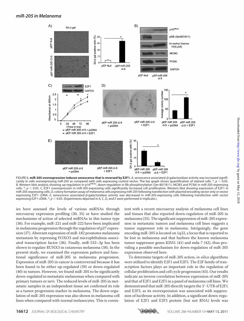
pected phenotypic change, namely cell enlargement and vacu-olization, observed in miR-205-expressing C8161.9 cells,prompted us to explore the possibility that overexpression ofmiR-205 can effectively activate senescence pathways. Asshown in Fig. 6, A and B, miR-205-expressing C8161.9 cellsexhibited significantly increased staining for SA-�-gal activitywhen compared with control vector-expressing cells. Thus,52% of C8161.9 cells stably expressing miR-205 expressedthis senescence marker, when compared with 8% of controlvector-expressing cells. Overexpression of miR-205 alsoinduced a marked increase in expression of p16INK4a andtrimethyl-histone H3 (Lys-9), suggesting activation of bothclassical senescence as well as alternate pathways thatinvolve heterochromatin-associated histone modification(Fig. 6B). Hypophosphorylation of Rb (Ser-807/811) anddown-regulation of key downstream targets of E2F1, MCM3and PCNA, further reinforces the activation of Rb-mediatedsenescence (28) upon overexpressing miR-205 in this mela-noma cell line (Fig. 6B).
E2F1OverexpressionRescues Phenotypes InducedbymiR-205—Overexpression of E2F1 in C8161.9 cells stably expressingmiR-205 resulted in significantly increased cell proliferation andcolony formation (Fig. 6, C and D), indicating reversal of theinhibitory effects of miR-205 on melanoma cell growth. E2F1overexpression also resulted in reversion of the senescenceinduced by miR-205 in melanoma cells (Fig. 6E).
DISCUSSION
As miRNAs modulate gene expression, it is not surprisingthat they have been implicated in regulating a wide variety ofbiological processes. Various studies have characterized themodulation of miRNA expression in cancers and identified anumber of miRNAs that are up- or down-regulated intumors (29, 30). An increasing number of oncogenes ortumor suppressor genes have now been identified as targetsof aberrantly expressed miRNAs (31, 32). For example,miR-21 has been shown to be involved in invasion andmetastasis in many cancer types by its action on numerousgenes involved in extracellular matrix modification (33). Inaddition, the miR-200 family, along with miR-205, has beenshown to regulate epithelial to mesenchymal transition, akey process for initiating metastasis (34).Despite these advances, little is known about the repertoire
and function of miRNAs in melanoma, and few targets ofmiRNAs in melanoma have been identified. A handful of stud-
FIGURE 5. Stable overexpression of miR-205 inhibits cell proliferation in vitro and in vivo. A, relative miR-205 expression levels in C8161.9 cells stablyexpressing miR-205 as determined by miR qRT-PCR. B, stable overexpression of miR-205 in melanoma cell lines significantly suppressed cell proliferation.C, Western blot analysis showing suppression in E2F1, E2F5, and AKT phosphorylation levels. D, colony formation ability is significantly reduced by miR-205.E, tumor volume following subcutaneous injection of C8161.9 cells expressing miR-205 was reduced significantly (n � 10 mice per group). Western blotshowing expression of E2F1 and E2F5 from tumors from pEP-Null and pEP-miR-205 mice.*, p � 0.05.
miR-205 in Melanoma
MAY 13, 2011 • VOLUME 286 • NUMBER 19 JOURNAL OF BIOLOGICAL CHEMISTRY 16611
by guest on May 13, 2018
http://ww
w.jbc.org/
Dow
nloaded from
ies have assessed the levels of various miRNAs throughmicroarray expression profiling (30, 35) or have studied themechanisms of action of selected miRNAs in this tumor type(36). For example, miR-221 and miR-222 have been implicatedinmelanoma progression through the regulation of p27 expres-sion (37). Aberrant expression ofmiR-182 promotesmelanomametastasis by repressing FOXO3 and microphthalmia-associ-ated transcription factor (36). Finally, miR-532–3p has beenshown to regulate RUNX3 in cutaneous melanoma (38). In thepresent study, we examined the expression pattern and func-tional significance of miR-205 in melanoma progression.Expression of miR-205 in cancer is controversial because it hasbeen found to be either up-regulated (39) or down-regulated(40) in tumors. However, we found miR-205 to be significantlydown-regulated inmetastaticmelanomaswhen comparedwithprimary tumors or nevi. The reduced levels of miR-205 in met-astatic samples in an independent tissue set confirmed its roleas a tumor progression marker in melanoma. The down-regu-lation of miR-205 expression was also shown in melanoma celllines when compared with normal melanocytes. This is consis-
tent with a recent microarray analysis of melanoma cell linesand tissues that also reported down-regulation of miR-205 inmelanoma (35). The significant suppression ofmiR-205 expres-sion in metastatic tumors and melanoma cell lines suggests atumor suppressor role in melanoma. Intriguingly, the geneencodingmiR-205 is located on 1q32, a locus that is reported tobe lost in melanoma and that harbors the known melanomatumor suppressor genes KISS1 (41) and mda-7 (42), thus pro-viding a possible mechanism for down-regulation of miR-205expression observed here.To determine targets of miR-205 action, in silico algorithms
were utilized to identify E2F1 and E2F5. The E2F family of tran-scription factors plays an important role in the regulation ofcellular proliferation and cell cycle progression (43).Our resultsindicate an inverse correlation between expression of miR-205and that of E2F1 and E2F5 in a panel ofmelanoma cell lines.Wedemonstrated thatmiR-205 directly targets the 3�-UTRof E2F1and E2F5, as its overexpression was associated with suppres-sion of luciferase activity. In addition, a significant down-regu-lation of E2F1 and E2F5 protein (but not RNA) levels was
FIGURE 6. miR-205 overexpression induces senescence that is reversed by E2F1. A, senescence associated-�-galactosidase activity was increased signifi-cantly in cells overexpressing miR-205 as compared with cells expressing control vector. The bar graph shows quantification of stained cells. *, p � 0.05.B, Western blot analysis showing up-regulation in p16INK4a, down-regulation in Rb phosphorylation (Ser-807/811), MCM3 and PCNA in miR-205-expressingcells. *, p � 0.05. C, E2F1 overexpression in miR-205-expressing cells significantly increased cell proliferation. Western blot showing expression of E2F1 inmiR-205-expressing cells. D, colony formation assay of melanoma cells expressing miR-205 following transfection with plasmid encoding vector only or vectorexpressing E2F1 cDNA. E, senescence associated-�-galactosidase activity was decreased in miR-205-expressing cells following transfection with vectorexpressing E2F1 cDNA. *, p � 0.05. (Experiments depicted in A, C, D, and E were performed in triplicate.).
miR-205 in Melanoma
16612 JOURNAL OF BIOLOGICAL CHEMISTRY VOLUME 286 • NUMBER 19 • MAY 13, 2011
by guest on May 13, 2018
http://ww
w.jbc.org/
Dow
nloaded from

observed after miR-205 overexpression, indicating the post-transcriptional regulation of E2F1 and E2F5 via targeting oftheir 3�-UTRs.Due to the demonstrated importance of E2F1 in melanoma
(24), we further characterized its role in response to miR-205.E2F1 has long been considered an oncoprotein, and shown topromote breast cancer cell and hepatocarcinoma cell prolifer-ation (44). However, E2F1 also has been reported to eitherinduce or inhibit apoptosis (45). E2F1-directed prosurvival sig-nals are mediated by the AKT pathway, which plays an impor-tant role in regulating cell growth, survival, and inhibition ofapoptosis (46). Recently, E2F1-epidermal growth factor recep-tor interaction was reported to play a significant role in mela-noma progression (47).We observed that miR-205 overexpres-sion reduced AKT phosphorylation, which was restored byE2F1 overexpression, confirming that the miR-205-mediateddown-regulation of AKT phosphorylation was due to E2F1.miR-205 overexpression suppressed the phosphorylation ofcaspase-9 and BAD, thus initiating a caspase cascade to induceapoptosis. Our results indicate that miR-205 inhibited cell pro-liferation and induced apoptosis in melanoma cells, an obser-vation that was confirmed in two different human melanomacell lines. miR-205-mediated induction of apoptosis was foundto be associated concomitantly with the cleavage of caspase-3and PARP and the release of cytochrome c. These antiprolifera-tive effects of miR-205, mediated, at least in part, by suppres-sion of E2F1 and AKT phosphorylation, were confirmed fol-lowing stable overexpression of miR-205 in C8161.9 cells. Inaddition, in vivo studies demonstrated a striking reduction insubcutaneous tumor cell growth in mice injected with C8161.9melanoma cells overexpressing miR-205.Furthermore, stable overexpression of miR-205 induced
senescence in melanoma cells. C8161.9 cells overexpressingmiR-205 had increased SA-�-gal activity, as well as cell enlarge-ment and vacuolization, which represent conventionalmarkersfor senescence (48). Trimethyl-histone H3 (Lys-9), anothermarker of senescence at the chromatin level, was up-regulatedfollowing miR-205 overexpression. The p16INK4a-Rb tumorsuppressor pathway plays an important role in the initiationandmaintenance of senescence (49). Up-regulation of p16INK4a
activates Rb by suppressing its phosphorylation through theinhibition of CDK4/6 kinase activities (50). Rb activationinduces senescence and suppresses the expression of E2F1 genetargets such as MCM3 and PCNA in senescent cells (28). Ourresults suggest that miR-205-induced senescence is mediated,at least in part, by this pathway, as evidenced by the up-regula-tion of p16INK4a and the suppression of Rb phosphorylation. Inaddition, bothMCM3 and PCNAwere down-regulated inmiR-205-overexpressing melanoma cells. Our study, for the firsttime, implicates miR-205 in the senescence of human mela-noma cells.Importantly, the significant effects induced bymiR-205 over-
expression on melanoma cell growth and senescence werereversed partially following restoration of E2F1 expression.These mechanistic analyses indicate that miR-205 mediates itseffects on melanoma cell proliferation and senescence via reg-ulation of E2F1 expression.
Finally, these results help explain previously described differ-ential patterns of Rb and E2F1 activity in melanoma progres-sion. Rb has been reported to be constitutively inactive in met-astatic melanoma cells (24), whereas E2F1 and its gene targetsare overexpressed in metastases when compared with primarymelanomas (51). Our data is consistent with these findings andsuggests that the down-regulation of miR-205 in metastaticmelanomas may be responsible for the previously observedactivation of E2F1 and inactivation of Rb.In conclusion, our study demonstrates a potent and impor-
tant tumor suppressor role for miR-205 in melanoma.
REFERENCES1. Ambros, V., and Chen, X. (2007) Development 134, 1635–16412. He, L., and Hannon, G. J. (2004) Nat. Rev. Genet. 5, 522–5313. Sempere, L. F., Freemantle, S., Pitha-Rowe, I., Moss, E., Dmitrovsky, E.,
and Ambros, V. (2004) Genome Biol. 5, R134. Bartel, D. P. (2004) Cell 116, 281–2975. Volinia, S., Calin, G. A., Liu, C. G., Ambs, S., Cimmino, A., Petrocca, F.,
Visone, R., Iorio, M., Roldo, C., Ferracin, M., Prueitt, R. L., Yanaihara, N.,Lanza, G., Scarpa, A., Vecchione, A., Negrini, M., Harris, C. C., and Croce,C. M. (2006) Proc. Natl. Acad. Sci. U.S.A. 103, 2257–2261
6. Porkka, K. P., Pfeiffer, M. J., Waltering, K. K., Vessella, R. L., Tammela,T. L., and Visakorpi, T. (2007) Cancer Res. 67, 6130–6135
7. Michael,M. Z., O’ Connor, S.M., vanHolst, Pellekaan, N. G., Young, G. P.,and James, R. J. (2003)Mol. Cancer Res. 1, 882–891
8. Yanaihara, N., Caplen, N., Bowman, E., Seike, M., Kumamoto, K., Yi, M.,Stephens, R. M., Okamoto, A., Yokota, J., Tanaka, T., Calin, G. A., Liu,C. G., Croce, C. M., and Harris, C. C. (2006) Cancer Cell 9, 189–198
9. Berezikov, E., Guryev, V., van de Belt, J.,Wienholds, E., Plasterk, R. H., andCuppen, E. (2005) Cell 120, 21–24
10. Lewis, B. P., Burge, C. B., and Bartel, D. P. (2005) Cell 120, 15–2011. Yi, R., and Fuchs, E. (2010) Cell Death Differ 17, 229–23512. Bartels, C. L., and Tsongalis, G. J. (2009) Clin. Chem. 55, 623–63113. DeGregori, J. (2002) Biochim. Biophys. Acta 1602, 131–15014. Pierce, A. M., Schneider-Broussard, R., Gimenez-Conti, I. B., Russell, J. L.,
Conti, C. J., and Johnson, D. G. (1999)Mol. Cell. Biol. 19, 6408–641415. Saito, M., Helin, K., Valentine, M. B., Griffith, B. B., Willman, C. L., Har-
low, E., and Look, A. T. (1995) Genomics 25, 130–13816. Nelson,M. A., Reynolds, S. H., Rao, U. N., Goulet, A. C., Feng, Y., Beas, A.,
Honchak, B., Averill, J., Lowry, D. T., Senft, J. R., Jefferson, A.M., Johnson,R. C., and Sargent, L. M. (2006) Cancer Biol. Ther. 5, 407–412
17. Kothandaraman, N., Bajic, V. B., Brendan, P. N., Huak, C. Y., Keow, P. B.,Razvi, K., Salto-Tellez, M., and Choolani, M. (2010) BMC Cancer 10, 64
18. Umemura, S., Shirane, M., Takekoshi, S., Kusakabe, T., Itoh, J., Egashira,N., Tokuda, Y., Mori, K., and Osamura, Y. R. (2009) Br. J. Cancer 100,764–771
19. Dar, A. A., Zaika, A., Piazuelo, M. B., Correa, P., Koyama, T., Belkhiri, A.,Washington, K., Castells, A., Pera,M., and El-Rifai,W. (2008)Cancer 112,1688–1698
20. Dar, A. A.,Majid, S., Nosrati, M., de Semir, D., Federman, S., and Kashani-Sabet, M. (2010) J. Invest. Dermatol. 130, 2071–2079
21. John, B., Enright, A. J., Aravin, A., Tuschl, T., Sander, C., and Marks, D. S.(2004) PLoS Biol. 2, e363
22. Betel, D., Wilson, M., Gabow, A., Marks, D. S., and Sander, C. (2008)Nucleic Acids Res. 36, D149–153
23. Krek, A., Grun, D., Poy, M. N., Wolf, R., Rosenberg, L., Epstein, E. J.,MacMenamin, P., da Piedade, I., Gunsalus, K. C., Stoffel, M., and Rajew-sky, N. (2005) Nat. Genet. 37, 495–500
24. Halaban, R., Cheng, E., Smicun, Y., and Germino, J. (2000) J. Exp. Med.191, 1005–1016
25. Ladu, S., Calvisi, D. F., Conner, E. A., Farina, M., Factor, V. M., and Thor-geirsson, S. S. (2008) Gastroenterology 135, 1322–1332
26. Datta, S. R., Dudek, H., Tao, X., Masters, S., Fu, H., Gotoh, Y., and Green-berg, M. E. (1997) Cell 91, 231–241
27. Guo, X., Chen, K. H., Guo, Y., Liao, H., Tang, J., and Xiao, R. P. (2007)Circ.
miR-205 in Melanoma
MAY 13, 2011 • VOLUME 286 • NUMBER 19 JOURNAL OF BIOLOGICAL CHEMISTRY 16613
by guest on May 13, 2018
http://ww
w.jbc.org/
Dow
nloaded from

Res. 101, 1113–112228. Narita, M., Nunez, S., Heard, E., Narita, M., Lin, A. W., Hearn, S. A.,
Spector, D. L., Hannon, G. J., and Lowe, S. W. (2003) Cell 113, 703–71629. Jiang, J., Lee, E. J., Gusev, Y., and Schmittgen, T. D. (2005) Nucleic Acids
Res. 33, 5394–540330. Gaur, A., Jewell, D. A., Liang, Y., Ridzon, D., Moore, J. H., Chen, C., Am-
bros, V. R., and Israel, M. A. (2007) Cancer Res. 67, 2456–246831. Harfe, B. D. (2005) Curr. Opin. Genet. Dev. 15, 410–41532. Tong, A. W., and Nemunaitis, J. (2008) Cancer Gene Ther. 15, 341–35533. Nicoloso, M. S., Spizzo, R., Shimizu, M., Rossi, S., and Calin, G. A. (2009)
Nat. Rev. Cancer 9, 293–30234. Gregory, P. A., Bracken, C. P., Bert, A. G., and Goodall, G. J. (2008) Cell
Cycle 7, 3112–311835. Philippidou, D., Schmitt, M., Moser, D., Margue, C., Nazarov, P. V.,
Muller, A., Vallar, L., Nashan,D., Behrmann, I., andKreis, S. (2010)CancerRes. 70, 4163–4173
36. Segura, M. F., Hanniford, D., Menendez, S., Reavie, L., Zou, X., Alvarez-Diaz, S., Zakrzewski, J., Blochin, E., Rose, A., Bogunovic, D., Polsky, D.,Wei, J., Lee, P., Belitskaya-Levy, I., Bhardwaj, N., Osman, I., andHernando,E. (2009) Proc. Natl. Acad. Sci. U.S.A. 106, 1814–1819
37. Felicetti, F., Errico, M. C., Bottero, L., Segnalini, P., Stoppacciaro, A., Bif-foni, M., Felli, N., Mattia, G., Petrini, M., Colombo, M. P., Peschle, C., andCare, A. (2008) Cancer Res. 68, 2745–2754
38. Kitago, M., Martinez, S. R., Nakamura, T., Sim, M. S., and Hoon, D. S.(2009) Clin. Cancer Res. 15, 2988–2994
39. Gottardo, F., Liu, C. G., Ferracin, M., Calin, G. A., Fassan, M., Bassi, P.,Sevignani, C., Byrne, D., Negrini, M., Pagano, F., Gomella, L. G., Croce,C. M., and Baffa, R. (2007) Urol. Oncol. 25, 387–392
40. Sempere, L. F., Christensen, M., Silahtaroglu, A., Bak, M., Heath, C. V.,
Schwartz, G.,Wells,W., Kauppinen, S., and Cole, C. N. (2007)Cancer Res.67, 11612–11620
41. Lee, J. H., Miele, M. E., Hicks, D. J., Phillips, K. K., Trent, J. M., Weissman,B. E., and Welch, D. R. (1996) J. Natl. Cancer Inst. 88, 1731–1737
42. Huang, E. Y., Madireddi, M. T., Gopalkrishnan, R. V., Leszczyniecka, M.,Su, Z., Lebedeva, I. V., Kang, D., Jiang, H., Lin, J. J., Alexandre, D., Chen, Y.,Vozhilla, N., Mei, M. X., Christiansen, K. A., Sivo, F., Goldstein, N. I.,Mhashilkar, A. B., Chada, S., Huberman, E., Pestka, S., and Fisher, P. B.(2001) Oncogene 20, 7051–7063
43. Attwooll, C., Lazzerini Denchi, E., and Helin, K. (2004) EMBO J. 23,4709–4716
44. Louie, M. C., Zou, J. X., Rabinovich, A., and Chen, H.W. (2004)Mol. Cell.Biol. 24, 5157–5171
45. Jiang, Y., Saavedra, H. I., Holloway, M. P., Leone, G., and Altura, R. A.(2004) J. Biol. Chem. 279, 40511–40520
46. Thomas, G. V., Horvath, S., Smith, B. L., Crosby, K., Lebel, L. A., Schrage,M., Said, J., De Kernion, J., Reiter, R. E., and Sawyers, C. L. (2004) Clin.Cancer Res. 10, 8351–8356
47. Alla, V., Engelmann, D., Niemetz, A., Pahnke, J., Schmidt, A., Kunz, M.,Emmrich, S., Steder, M., Koczan, D., and Putzer, B. M. (2010) J. Natl.Cancer Inst. 102, 127–133
48. Collado, M., Gil, J., Efeyan, A., Guerra, C., Schuhmacher, A. J., Barradas,M., Benguría, A., Zaballos, A., Flores, J. M., Barbacid, M., Beach, D., andSerrano, M. (2005) Nature 436, 642
49. Haferkamp, S., Becker, T. M., Scurr, L. L., Kefford, R. F., and Rizos, H.(2008) Aging Cell 7, 733–745
50. Serrano, M., Hannon, G. J., and Beach, D. (1993) Nature 366, 704–70751. Tuve, S.,Wagner, S. N., Schittek, B., and Putzer, B.M. (2004) Int. J. Cancer
108, 162–166
miR-205 in Melanoma
16614 JOURNAL OF BIOLOGICAL CHEMISTRY VOLUME 286 • NUMBER 19 • MAY 13, 2011
by guest on May 13, 2018
http://ww
w.jbc.org/
Dow
nloaded from

and Mohammed Kashani-SabetAltaf A. Dar, Shahana Majid, David de Semir, Mehdi Nosrati, Vladimir Bezrookove
Regulation of E2F1 ProteinmiRNA-205 Suppresses Melanoma Cell Proliferation and Induces Senescence via
doi: 10.1074/jbc.M111.227611 originally published online March 24, 20112011, 286:16606-16614.J. Biol. Chem.
10.1074/jbc.M111.227611Access the most updated version of this article at doi:
Alerts:
When a correction for this article is posted•
When this article is cited•
to choose from all of JBC's e-mail alertsClick here
Supplemental material:
http://www.jbc.org/content/suppl/2011/03/24/M111.227611.DC1
http://www.jbc.org/content/286/19/16606.full.html#ref-list-1
This article cites 51 references, 18 of which can be accessed free at
by guest on May 13, 2018
http://ww
w.jbc.org/
Dow
nloaded from