Embed Size (px)
Citation preview

Semmelweis University Department of Anatomy, Histology and Embryology
Faculty of Dentistry 2nd year 1st semester
September 2021
MICROSCOPIC ANATOMY AND EMBRYOLOGY II HANDBOOK
Dr. Andrea D. Székely Associate Professor
Course Director of the English Language Program
Dr. Gábor Gerber
Associate Professor Head of the Anatomy, Histology and Embryology subjects for the Faculty of Dentistry
Dean of the Faculty of Dentistry

Anatomy, Histology and Embryology for ED students
TEACHING DEPARTMENT: SEMMELWEIS UNIVERSITY Department of Anatomy, Histology and Embryology Budapest, Tűzoltó utca 58. H-1094 Budapest http://semmelweis.hu/anatomia
LEARNING OBJECTIVES
Demonstration of the fine structure of cells and tissues composing the organs of the human body specifically to provide the future doctors of dental medicine with a valid body of information describing the microscopical elements of clinically significant morphological structures (including cell biology, general histology and the histology of organs).
General embryology demonstrates the steps of the formation of a new human being together with the stages of intrauterine development, including the clinically relevant aspects of the development of organ systems. Teaching is done in the form of lectures and histology laboratory classes Competences acquired by completion of the course:
Understanding the microscopical composition of the human body together with the understanding of human development in order to draw parallels with macroscopical anatomy. Clear understanding of histological structure and function. Ability to identify basic structural elements within the tissue specimen. Identification of general directions/landmarks within digitized tissue slides.
LECTURES: First semester: 2 x 45 min; second semester: 2 x 45 min. PRACTICAL CLASSES: First semester: 2 x 45 min; second semester: 2 x 45 min. ECTS CREDITS: Altogether 8 (first semester: 4; second semester: 4). MIDTERM TESTS: Written (in the Moodle system)
ACCEPTENCE OF THE SEMESTER: Active participation in laboratory sessions is obligatory for every student. Students should attend at least 75% of the scheduled hours to gain a signature proving the validity of the semester. Absences are therefore limited in 25%. Attendance will be recorded in the classes.
TYPE OF EXAMS: oral and written The final examination consists of written and oral (practical and theoretical) parts 1. Written pretest (e-learning module – access to SeKA account is obligatory) 2. Oral examination (identification of structures on digitized histological slides) including relevant theoretical questions from the fields of Histology and Embryology
COURSE DESCRIPTION
Microscopic Anatomy and Embryology II. Lectures and histology classes Subject matter: Histology of the lymphatic system, together with the histology and developmental aspects of the central and peripheral nervous systems, endocrine organs and organs of special senses, including the skin. Credits: 4 Prerequisites: Macroscopic Anatomy II (successful final examination) Microscopic Anatomy and Embryology I (successful examination)

Academic Year 2021/2022 Faculty of Dentistry
ED II. Microscopic Anatomy and Embryology II.
Weeks Lectures
Mondays 10.00 - 11.40 Lecturer Histology laboratory
Tuesdys 10.00-11.30
Week 1 09. 6-10.
1. Cellular components of lymphatic tissue. Thymus, tonsils, MALT
2. Structure and circulation of lymph nodes and spleen
1 Puskár 2 Puskár
Thymus, tonsils
Week 2 09. 13-
17.
3. Nerve tissue: neurons and glial cells, synapses, receptors and effectors 4. Microscopy of the CNS – Fine structure of the spinal cord, spinal nerves
3 Pálfi 4 Gerber
Lymph node, spleen
Week 3 09. 20-
24.
5. Microscopy of the CNS – Spinal reflexes, receptors and effectors, proprioceptive, nociceptive (withdrawal) and autonomic reflex arcs
6. Microscopy of the CNS – Fine structure of the cerebral cortex. Cortical fields, Brodmann areas
5 Kozsurek 6 Vereczki
Histology of the peripheral nervous system
Week 4 09.27 - 10. 1.
7. Microscopy of the CNS – Microscopy of the cerebellum, pathways. Functional considerations
8. Microscopy of the CNS – Cranial nerve nuclei
7 Altdorfer 8 Shahbazi
Histology of the central nervous system
Week 5 10. 4-8.
9. Microscopy of the CNS – Thalamic nuclei. 10. Microscopy of the CNS – Sensory systems, epicritical and
protopathic pathways arising from the brain stem
9 Gallatz 10 Vereczki
Midterm test 1 (Histological slides of weeks 1-4)
Microscopy of the CNS – consultation Cross sections of the brainstem
Week 6 10. 11-
15.
11. Microscopy of the CNS – Motor systems, pyramidal tract. 12. Microscopy of the CNS – Extrapyramidal system: structure and connections of the basal ganglia. Brainstem monoaminergic system
11 Shahbazi 12 Kozsurek
Microscopy of the CNS – consultation Cross sections of the brainstem
Week 7 10. 18-
22.
13. Microscopy of the CNS – Limbic system 14. Microscopy of the CNS – Hypothalamus, the hypothalamo-hypophysial system
13 Gerber 14 Tóth Zs
Microscopy of the CNS - consultation
Week 8 10. 25-
29.
15. Histology of the endocrine organs: Thyroid, parathyroid, suprarenal glands, hypophysis, pineal body
16. Differentiation of the neural tube. Cranio-caudal and dorso-ventral differentiation. Differentiation of the brain vesicles
15 Durst 16 Kozsurek
Microscopy of the CNS - consultation
Week 9 11. 1-5.
Nov. 1
National Holiday
17. - Formation and derivatives of the neural crest and placode ectoderm 18. - Development of the skull.
ONLINE 17 Minkó 18 Gallatz
Midterm test 2 (Microscopy of the CNS. Development of the nervous system) Endocrine system I
Week 10 11. 8-12.
19. Development of the vertebral column, limb development 20. Skin and appendages. Mammary gland
19 Székely 20 Székely
Endocrine system II
Week 11 11.15-19.
21. Fibrous and vascular coats of the eyeball. Lens, chambers of the eye, vitreous body, accommodation 22. Inner coat of the eyeball, retina.
21 Barna 22 Gerber
Histology of palm skin, scalp skin. Mammary gland
Week 12 11. 22-26
23. Visual pathway, visual reflexes. Development of the eye 24. External ear, middle ear.
23 Lendvai 24 Gerber
Histology of the organ of vision
Week 13 11.29-12.3.
25. Bony and membranous labyrinth. Vestibular system 26. Spiral organ of Corti. Auditory pathway. development of the organ of hearing
25 Tóth Zs 26 Puskár
Histology of the organ of hearing
Week 14 12. 6-10.
27. Microscopy of the CNS – Olfactory and gustatory systems 28. Revision / Consultation
27 Gallatz 28 Gerber
Revision

ED II. Microscopic Anatomy and Embryology II. List of slides (FOK series)
Weeks Histology laboratory
Tuesdays 10.00-11.30
Week 1 09. 6-10.
Lymphatic system I. 47. Palatine tonsil (HE) DEM : ÁOK 42. Palatine tonsil (T/B cell IHC) 48. Lingual tonsil (HE) DEM: ÁOK 48. Pharyngeal tonsil (HE) 49. Thymus (HE)
Week 2 09. 13-17.
Lymphatic system II. 44. Lymph node (HE) 45. Spleen (HE) 46. Spleen (rinsed, HE) DEM: ÁOK 1.a, b Spleen (T/B cell IHC)
Week 3 09. 20-24.
Histology of the peripheral nervous system 36. Peripheral nerve (cross section, HE) DEM: Peripheral nerve (OsO4 impregnation), 6. nerves in the skin(HE) 37. Pseudounipolar neurones (DRG, HE) 38. Multipolar neurones (autonomic ggl, AgNO3 impregnation) DEM: Autonomic ggl in the intestinal wall (HE) 43. Motor end plate (striated muscle, ACh esterase histochemistry)
Week 4 09.27 - 10.
1.
Histology of the central nervous system 39. Spinal cord (multipolar neurones, Nissl) 40. Cerebral cortex (pyramidal neurones, Bielschowsky) 42. Cerebral cortex (pyramidal neurones, Golgi) 94. Hippocampus (HE) 95. Cerebellar cortex (HE) 41. Cerebellar cortex (GFAP ICC)
Week 5 10. 4-8.
Midterm test 1 Histological slides of weeks 1-4
Week 6 10. 11-15.
Microscopy of the CNS – cross sections of the brain stem 99. Mesencephalon (Luxol fast blue + Nissl) 100. medulla oblongata (Luxol fast blue + Nissl)
Week 7 10. 18-22.
Microscopy of the CNS - consultation
Week 8 10. 25-29.
Microscopy of the CNS - consultation
Week 9 11. 1-5.
Nov. 1 National Holiday
Midterm test 2 Microscopy of the CNS
Endocrine system I. 90. Epiphysis/ pineal body (HE) 86. Hypophysis/ pituitary gland (HE) 87. Hypophysis/ pituitary gland (chrom–hematoxyline-phloxin/Gömöri)
Week 10 11. 8-12.
Endocrine system II. DEM 74. Leydig cells, testicle (HE) 88. Thyroid gland (HE) 78. Ovarian follicles (HE) 89. Parathyroid gland (HE) 79. Corpus luteum (HE) 92. Adrenal/suprarenal gland (HE) 70. Islets of Langerhans, pancreas (HE)
Week 11 11.15-19.
Histology of palm skin, scalp skin. Mammary gland 6. Palm skin (HE) 11. Scalp/hairy skin (HE) 17. Scalp/hairy skin (AZAN) 18. Scalp/hairy skin (Hornowsky) 85. Mamma non lactans (HE) 93. Mamma lactans (HE)
Week 12 11. 22-26
Histology of the organ of vision 96. Eye bulb (HE) 97. Retina (semithin section, toluidine blue) 9. Pigment epithelium (unstained) 33. Lacrimal gland (HE)
Week 13 11.29-12.3.
Histology of the organ of hearing 98. Cochlea (semithin section, toluidine blue)
Week 14 12. 6-10.
Revision

ED II.
Subject matter of the present semester
I. Histology of lymphatic organs
II. Neurohistology a) Histology of neurons and supporting elements b) Fine structure of peripheral nerves c) Receptors and effectors, interneuronal synapses d) Histology of the brain and spinal cord
III. Development of the locomotor system a) Membranous and cartilaginous neurocranium and viscerocranium b) Development of the limbs and vertebral column c) Development of the muscular system
IV. Development of the nervous system and organs of special senses a) Development and primary differentiation of the neural tube b) Development of the peripheral nervous system (neural crest, placodes) c) Development of the organ of vision d) Development of the organ of hearing&equilibrium
V. Microscopy of the central nervous system a) Microscopic anatomy of brain and spinal cord b) Nuclei and tracts of brain and spinal cord c) Microscopy of the autonomic nervous system, tracts
VI. Organs of special senses (histology and embryology) a) Organ of vision, visual pathways b) Organ of hearing and equilibrium, auditory pathways, vestibular system c) Organ of smell, olfactory pathways d) Organ of taste, gustatory pathways e) Skin and appendages
VII. Endocrine organs (histology and embryology) a) Hypothalamo-hypophysial system b) Endocrine glands and cells
Midterm test I. Written midterm (Moodle) Topic: Histology of the lymphatic system, histology of the nervous system Date: 5th week Midterm test II. Written (Moodle) Topic: Microscopy and development of the central nervous system. Date: 9th week
Final examination Topics: Subject matter of the two semesters
1.Written pretest 2. Oral examination - identification of structures on 2 digitized tissue slides, as well as
1 theoretical question from the subject matter of the two semesters (see the Topic list).

ED II ANNOUNCEMENTS
Evaluation is made using a five-grade scale (1-5).
Signing of the lecture book: active participation in histology lab sessions is obligatory.
Students should attend at least 75% of the scheduled hours, including the obligatory
midterm examinations, to gain a signature proving the validity of the semester. Successful
passing of the 2nd midterm is required. Absences are limited in 25%.
Midterm examinations: During the semester, both practical and theoretical knowledge
will regularly be evaluated. There are two written (Moodle) midterm tests during the
semester.
Midterm 1 - obligatory to attend
Midterm 2 – obligatory to pass
The second (Microscopy of the nervous system) midterm should be successfully
passed with at least a mark 2,00 or the semester is not accepted.
Students being absent from the midterm and/or having an unsuccessful result from
the 2nd test should attend at one of the given retake dates during the last two weeks of
the semester (TBA) or their semester will not be accepted.
The final examination is composed of the following parts:
Topics: Subject matter of the two semesters
Written pretest
Oral examination (identification of structures on 2 digitized tissue slides, as well as
1 theoretical question from the subject matter of the two semesters (see the Topic list).
Please note: Students may register for, or deregister from, the examinations via the neptun
system. In case neither the first nor the repeated takes of a semifinal exam have been successful the exam has to be postponed to the following exam period as a ’CV’ exam (if there are possibilities left).
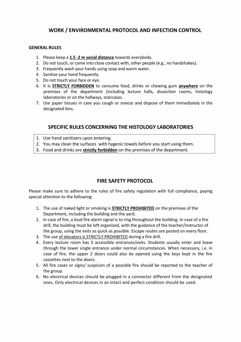
WORK / ENVIRONMENTAL PROTOCOL AND INFECTION CONTROL
GENERAL RULES
1. Please keep a 1.5 -2 m social distance towards everybody. 2. Do not touch, or come into close contact with, other people (e.g., no handshakes). 3. Frequently wash your hands using soap and warm water. 4. Sanitise your hand frequently. 5. Do not touch your face or eye. 6. It is STRICTLY FORBIDDEN to consume food, drinks or chewing gum anywhere on the
premises of the department (including lecture halls, dissection rooms, histology laboratories or on the hallways, staircases.
7. Use paper tissues in case you cough or sneeze and dispose of them immediately in the designated bins.
SPECIFIC RULES CONCERNING THE HISTOLOGY LABORATORIES
1. Use hand sanitizers upon entering. 2. You may clean the surfaces with hygenic towels before you start using them. 3. Food and drinks are strictly forbidden on the premises of the department.
FIRE SAFETY PROTOCOL
Please make sure to adhere to the rules of fire safety regulation with full compliance, paying special attention to the following:
1. The use of naked light or smoking is STRICTLY PROHIBITED on the premises of the Department, including the building and the yard.
2. In case of fire, a loud fire alarm signal is to ring throughout the building. In case of a fire drill, the building must be left organized, with the guidance of the teacher/instructor of the group, using the exits as quick as possible. Escape routes are posted on every floor.
3. The use of elevators is STRICTLY PROHIBITED during a fire drill. 4. Every lecture room has 3 accessible entrances/exits. Students usually enter and leave
through the lower single entrance under normal circumstances. When necessary, i.e. in case of fire, the upper 2 doors could also be opened using the keys kept in the fire cassettes next to the doors.
5. All fire cases or signs/ suspicion of a possible fire should be reported to the teacher of the group.
6. No electrical devices should be plugged in a connector different from the designated ones. Only electrical devices in an intact and perfect condition should be used.

List of textbooks
The Developing Human – Clinically Oriented Embryology, 10th ed. by KL Moore, TVN Persaud and M Torchia, Saunders, 2015; ISBN 9780323313384 Histology: A Text and Atlas: With Correlated Cell and Molecular Biology; 7th Edition by MH Ross and W Pawlina ; Wolters Kluwer 2015, ISBN 9781451187427 Wheater's Functional Histology, A Text and Colour Atlas, 6th Edition by B Young, G O'Dowd and P Woodford Churchill Livingstone, Edinburgh, 2013, ISBN 9780702047473 Oral Anatomy, Histology and Embryology, 4th Edition, by B. Berkovitz Paperback with STUDENT CONSULT Online Access and e-book ISBN: 9780723434115 Copyright: 2009 Functional Anatomy, Histology and Embryology for medical and dental students by M. Réthelyi and J. Szentágothai, Medicina, 2018.
Recommended textbooks
Langmann’s Medical Embryology, 13th Edition by TW Sadler, Wolters Kluwer, ISBN 9781469897806, 2014 Junqueira's Basic Histology: Text and Atlas; 13th Edition by Anthony Mescher, New York, McGraw-Hill Medical, 01/03/2013 ISBN13 978007178033 Wheater’s Functional Histology, A Text and Colour Atlas, 6th Edition by B Young, G O’Dowd and P Woodford ISBN 9780702047473, Churchill Livingstone, Edinburgh, 2013. Illustrated Dental Embryology, Histology, and Anatomy, 3rd Edition by Mary Bath-Balogh ISBN: 9781437717303, 2011. Further study aids: To be downloaded from the homepage of the Department of Anatomy, Histology and Embryology (http://semmelweis.hu/anatomia ) or from Knowledgebase on the Library homepage: (https://lib.semmelweis.hu/knowledge_base).

TOPICS OF THE FINAL EXAMINATION (topics of the two semesters)
General Histology Concept of basic tissues Definition and classification of epithelial tissue Simple epithelia Stratified epithelia Glandular epithelia Pigment epithelium, sensory neuroepithelium Cells of connective tissue Ground substance and fibres of connective tissue Types of connective tissue Blood and the corpuscular elements of blood Histology of the bone marrow, maturation of erythrocytes and platelets Differentiation of granulocytes, lymphocytes and monocytes Histology of cartilage and bone tissue Intramembranous ossification. Endochondral ossification. Growth and remodeling of bone Smooth muscle and myoepithelial cells Skeletal muscle tissue Cardiac muscle tissue Histology of arteries and arterioles Histology of veins and capillaries Histology of organs Wall structure of hollow organs General composition of parenchymal (solid/compact) organs Histology of the lip and tongue Histology of the respiratory tract. Larynx. Trachea. Lung Histology of the esophagus and stomach Histology of the small and large intestines. Fine structure of the intestinal vili,
enteroendocrine system Histology of the liver. Gall bladder, biliary ducts Histology of the pancreas Histology of kidney. Ureter. Urinary bladder Histology of the male and female gonads and genital organs/ducts Histology of the uterus (prolipherative, secretory phases) menstrual cycle, vagina General Embryology Spermatogenesis, spermiogenesis Oogenesis Fertilization, cleavage of the zygote Blastocyst formation; the bilaminar embryonic disc Implantation Formation of body axes Formation of the intraembryonic mesoderm; the notochord

Neurulation (neural tube and neural crest) Derivatives of ectoderm Derivatives endoderm Differentiation of the intraembryonic mesoderm Folding of the embryo Development of the primitive cardiovascular system The structure and function of the placenta Development of the fetal membranes (chorion and amnion) and the umbilical cord Development of internal organs Development of the heart, looping of the heart tube Formation of atria, development of the interatrial septum Formation of ventricles, development of the aorticopulmonary septum Development of arteries Development of the inferior vena cava Development of the portal vein Development of the suprior vena cava, azygos and hemiazygos veins Fetal circulation Development and differentiation of the midgut Development and differentiation of the hindgut Formation of the liver and pancreas Development of the lower airways including the lungs Kidney development Development of the urinary passages Gonadal development Development of the male genital tract Development of the female genital tract Development of the male/female external genitals Develeopment and divisioning of the body cavities Development of the peritoneum Maxillofacial Histology and Embryology Enamel Amelogenesis Dentin Dentinogenesis Structure of the dental papilla Cementum (two types) Parodontium Gingiva – subdivisions and histology Tooth development Tooth eruption Development of the mandible and maxilla Development of the face. Formation of the nasal cavity and paranasal sinuses Microscopic Anatomy and development of the primary and secondary palates Microscopic Anatomy and development of the tongue Microscopic Anatomy and development of salivary glands Derivatives of pharyngeal pouches and grooves Derivatives of pharyngeal arches

Lymphatic organs Histological structure of lymph nodes Spleen (fine structure and circulation) Thymus Tonsils, MALT Development of the nervous system and organs of special senses Development and primary differentiation of the neural tube Development of brain vesicles Development of the peropheral nervous system (neural crest, placodes) Development of the organ of vision Development of the organ of hearing&equilibrium Development of the locomotor system Membranous and cartilaginous neurocranium and viscerocranium Development of the limbs and vertebral column Development of the muscular system Histologyof the nervous system Histology of the neurons developing from the neural tube Glial cells Histology of the neurons and supporting cells developing from the neural crest Fine structure of peripheral nerves Receptors and effectors Interneuronal synapses Microscopy of the central nervous system Fine structure (microscopy) of the spinal cord Proprioceptive reflexes Nociceptive reflexes Autonomic reflexes Fine structure of the medulla oblongata Fine structure of the pons Fine structure of the midbrain Classification of cranial nerve nuclei Tracts of the brain stem Reticular formation, monoaminergic systems Fine structure of the cerebellum Cerebellar afferents and efferents Fine structure of the thalamus Hypothalamo-hypophyseal system Fine structure of the basal ganglia Fine structure of the cerebral cortex, cortical fields Tracts of the protopathic sensibility (anterolateral system) Tracts of the epicritic sensibility (posterior funiculus/medial lemniscus) Corticospinal tract (pyramidal tract) Extrapyramidal system Limbic system (nuclei and tracts)

Endocrine organs Microscopical anatomy of the pituitary gland; development of the posterior lobe Microscopical anatomy and development of the anterior and intermediate lobes of the pituitary gland Blood supply of the pituitary gland Microscopical anatomy of the pineal gland Microscopical anatomy and the development of the thyroid gland Microscopical anatomy and the development of the parathyroid gland Microscopical anatomy and the development of the suprarenal gland Histology of the islands of Langerhans Organs of special senses Microscopical structure of the skin (scalp and palm) Histology and development of skin appendages, mammary gland Coats of the eyeball Chambers of the eye, vitreous body Lens, accomodation Visual pathway, visual reflexes External ocular muscles, eye movements Accessory and protective apparatus of the eye (palpebrae, conjunctiva, fasciae, lacrimal apparatus) External ear, tympanic membrane. Tympanic cavity, auditory tube. Hearing ossicles (joints, muscles) Vestibulocochlear nerve. Organ of Corti. Cochlea, cochlear duct Auditory pathway. Vestibular system Bony and membranous labirynth, vestibulum Organ of olfaction, olfactory pathway, olfactory nerve Organ of taste, central processing of taste (tracts)