Embed Size (px)
Citation preview

Microbiology
Miscellaneous Gram-Positive Bacilli
Karen Honeycutt, M.Ed., MT(ASCP)SM
MT 418 Clinical Microbiology
Student Laboratory Session

Microbiology
Differentiation of Major GPR Genera
• Gram stain morphology and arrangement
• Formation of spores
• Catalase reaction

Microbiology
Gram-positive rods
• Large w/spores • Pleomorphic, palisades of parallel cells, “V” or “L” shapes

Microbiology
Gram-positive rods
• “regular-shaped” • Branching (specimen GS)

Microbiology
Spore-forming, Catalase +
• Bacillus species – only aerobic spore-forming GPR – Produced when organism stressed– Appear clear on Gram stains – In vitro induction of spore production
• Heat shock
Gram-positive rods

Microbiology
Bacillus species
• Widely distributed in nature
• In clinical specimens considered:– Environmental contaminants or – Normal flora
• Grow on SBA or CHOC, 24 hrs., ambient air or CO2
Gram-positive rods – spore-forming, catalase +

Microbiology
Bacillus species
• May stain gram-negative or gram-variable3% KOH Test: emulsify & raise loop
GNR = viscous thread
GPR = no thread
Gram-positive rods – spore-forming, catalase +
What can you see on a Gram stain to confirm an organism is a Bacillus sp.? Spores

Microbiology
Bacillus species
Gram-positive rods – spore-forming, catalase +
• Identification– Usually rule out Bacillus anthracis & report
as Bacillus sp. if:
**Beta-hemolytic
Motile

Microbiology
Bacillus anthracis
Gram-positive rods – spore-forming, catalase +
• Etiologic agent of anthrax
• Infections acquired by:– Contact w/infected animals or animal
products– Inhalation of spores (dangerous for lab)
• No documentation of human to human transfer

Microbiology
Bacillus anthracis
Gram-positive rods – spore-forming, catalase +
• Virulence: – Anti-phagocytic capsule – Exotoxins: cell & tissue destruction

Microbiology
Bacillus anthracis
Gram-positive rods – spore-forming, catalase +
• Cutaneous anthrax: site of spore penetration develops ulceration of black eschar
• May lead to fatal toxemia
• 20% mortality

Microbiology
Bacillus anthracis
Gram-positive rods – spore-forming, catalase +
• Pulmonary anthrax: inhalation of spores– Respiratory distress to cyanosis – 100% fatal if not treated early
• Gastrointestinal anthrax: ingestion of spores– Usually fatal due to toxemia & sepsis

Microbiology
Bacillus anthracis
Gram-positive rods – spore-forming, catalase +
• Identification: – Non-hemolytic– Non-motile– Penicillin “S”
• Refer – Report

Microbiology
Bacillus anthracis
Gram-positive rods – spore-forming, catalase +
• Prevention– Animal vaccines– Human vaccine available

Microbiology
GPR “regular shape”, No Spores, Catalase +
Gram-positive rods: nonspore-forming, catalase +
• Listeria monocytogenes – Bacteremia/meningitis immunosuppressed– In utero passed to fetus: systemic infection
& stillbirth
• Transmission– Ingest contaminated food: luncheon meats,
dairy products – Will grow at refrigerator temperatures

Microbiology
Listeria monocytogenes
Gram-positive rods: nonspore-forming, catalase +
• Identification – BAP: 24 hrs (growth is
small), ambient air or CO2;
– beta-hemolytic (small zone)
– Gram stain: small GPR – Catalase positive

Microbiology
Listeria monocytogenes
Gram-positive rods: nonspore-forming, catalase +
• Identification – Motility
• Tumbling (wet mount)• Inverted umbrella (motility
media)
– Esculin hydrolysis = positive– Sodium hippurate = positive

Microbiology
Listeria monocytogenes
Gram-positive rods: nonspore-forming, catalase +
• Identification – Can be confused w/Grp B
Streptococcus
What rapid biochemical test will differentiate Grp B Strep and Listeria monocytogenes? Catalase

Microbiology
Who Am I? Isolated from an arm wound
Catalase: bubbles produced when _________added to the organism (on a glass slide)
Hydrogen peroxide
Bacillus species

Microbiology
Corynebacterium species
GPR: irregular, nonspore-forming, catalase +
• Many species are normal flora of skin & mucous membranes
• Most species are nonpathogenic
• Referred to as “diphtheroids”– Palisading GPR– Catalase positive

Microbiology
GPR, irregular-shaped, catalase +, no spores
Gram-positive rods: nonspore-forming, catalase +
• Corynebacterium sp.
• Club-shaped, pleomorphic, irregularly staining due to metachromatic granules
• Can palisade (parallel, V, L-forms, “Chinese letters”, “picket fence”)

Microbiology
Corynebacterium species
GPR: irregular, nonspore-forming, catalase +
• Isolation – BAP: growth 24 hrs, ambient air
or CO2
– Various colony morphologies: dry, irregular shaped, white to buff colored
• Loeffler’s methylene blue stain (Albert’s stain) = positive

Microbiology
Clinically Significant Corynebacterium species
GPR: irregular, nonspore-forming, catalase +
• Corynebacterium diphtheriae – diphtheria
• Humans are only host– If cultured from a healthy person, considered
a carrier (not considered normal flora)
• Respiratory or cutaneous forms

Microbiology
Corynebacterium diphtheriae
GPR: irregular, nonspore-forming, catalase +
• Respiratory: • Organism in upper respiratory tract and forms a
pseudomembrane (WBCs & organism)– Pseudomembrane should be cultured– May cause respiratory obstruction
• Exotoxin produced, bloodstream, acts on cardiac tissue & peripheral nervous system
• Mortality 10-30% due to congestive heart failure

Microbiology
Corynebacterium diphtheriae
GPR: irregular, nonspore-forming, catalase +
Pseudomembrane (WBCs & organism)
should be cultured
Microbiology
Corynebacterium diphtheriae
GPR: irregular, nonspore-forming, catalase +
• Cutaneous – nonhealing ulcers
• Treatment – antitoxin given in form of toxoid (detoxified antitoxin)
• Prevention – DPT immunization

Microbiology
Corynebacterium diphtheriae
GPR: irregular, nonspore-forming, catalase +
• Culture & Isolation – Specific request– BAP: 24-48 hrs; small gray translucent to
medium, opaque white colonies– Loeffler’s media: stimulates growth &
formation of metachromatic granules– Cystine-tellurite: grayish-black colonies– Modified Tinsdale: black colonies w/dk brown
halo

Microbiology
Corynebacterium diphtheriae
GPR: irregular, nonspore-forming, catalase +
Cystine
tellurite:
grayish
black
colonies
ModifiedTinsdale:blackcoloniesw/dkbrown halo
Microbiology
Corynebacterium diphtheriae
GPR: irregular, nonspore-forming, catalase +
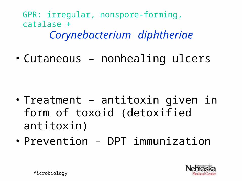
Gram stainCHO utilization
Glucose +Sucrose (-)
Urea (-)
NitrateReductionvariable

Microbiology
Corynebacterium diphtheriae
GPR: irregular, nonspore-forming, catalase +
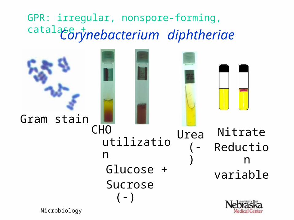
• If id as C. diphtheriae, then must determine if isolate is an exotoxin producer (i.e., can cause disease)– In vivo test (guinea pigs)– Immunodiffusion – ELEK– Tissue culture neutralization– PCR
Microbiology
Corynebacterium diphtheriae
GPR: irregular, nonspore-forming, catalase +
• Immunodiffusion – ELEK
Filter paper soaked with antitoxin
Bacterial isolate
Microbiology
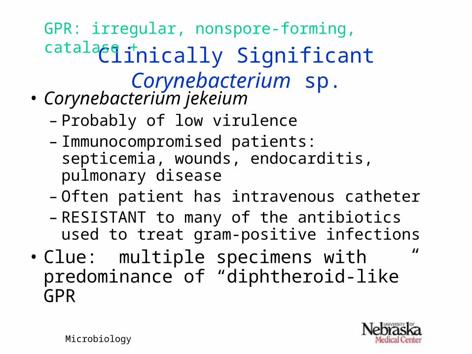
Clinically Significant Corynebacterium sp.
GPR: irregular, nonspore-forming, catalase +
• Corynebacterium jekeium– Probably of low virulence– Immunocompromised patients: septicemia,
wounds, endocarditis, pulmonary disease– Often patient has intravenous catheter– RESISTANT to many of the antibiotics used
to treat gram-positive infections
• Clue: multiple specimens with predominance of “diphtheroid-like” GPR

Microbiology
Corynebacterium jekeium
GPR: irregular, nonspore-forming, catalase +
• Isolation & Identification– BAP: nonhemolytic, small white to gray
colonies, may take 48-72 hrs for good growth– GS: like other diphtheroids– Very inert
• Lipophilic• Glucose “O”, Maltose is variable• Resistant to antibiotics used to treat GP
infections, Vancomycin susceptible

Microbiology
Review – Aerobic (Facultative) GPR
GPR
• Identification1. Gram stain & catalase reaction
2. Colony morphology & growth characteristics
3. Determine level of id needed (NF vs. pathogen) based on site and patient
4. Definitive biochemicals if needed
Microbiology
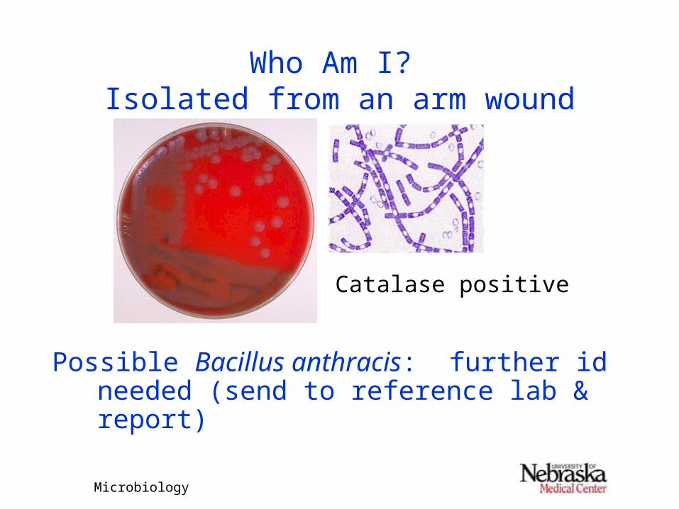
Who Am I? Isolated from an arm wound
Catalase positive
Possible Bacillus anthracis: further id needed (send to reference lab & report)