Embed Size (px)
Citation preview
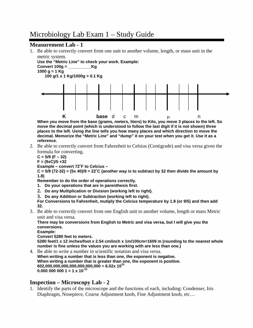
Microbiology Lab Exam 1 – Study Guide Measurement Lab - 1 1. Be able to correctly convert from one unit to another volume, length, or mass unit in the
metric system. Use the “Metric Line” to check your work. Example: Convert 100g = __________Kg 1000 g = 1 Kg
100 g/1 x 1 Kg/1000g = 0.1 Kg
K base d c m μ n When you move from the base (grams, meters, liters) to Kilo, you move 3 places to the left. So move the decimal point (which is understood to follow the last digit if it is not shown) three places to the left. Using the line tells you how many places and which direction to move the decimal. Memorize the “Metric Line” and “dump” it on your test when you get it. Use it as a reference.
2. Be able to correctly convert from Fahrenheit to Celsius (Centigrade) and visa versa given the formula for converting. C = 5/9 (F – 32) F = (9xC)/5 +32 Example – convert 72˚F to Celcius – C = 5/9 (72-32) = (5x 40)/9 = 22˚C (another way is to subtract by 32 then divide the amount by 1.8) Remember to do the order of operations correctly. 1. Do your operations that are in parenthesis first. 2. Do any Multiplication or Division (working left to right). 3. Do any Addition or Subtraction (working left to right). For Conversions to Fahrenheit, mutiply the Celcius temperature by 1.8 (or 9/5) and then add 32.
3. Be able to correctly convert from one English unit to another volume, length or mass Metric unit and visa versa. There may be conversions from English to Metric and visa versa, but I will give you the conversions. Example: Convert 5280 feet to meters. 5280 feet/1 x 12 inches/foot x 2.54 cm/inch x 1m/100cm=1609 m (rounding to the nearest whole number is fine unless the values you are working with are less than one.)
4. Be able to write a number in scientific notation and visa versa. When writing a number that is less than one, the exponent is negative. When writing a number that is greater than one, the exponent is positive. 602,000,000,000,000,000,000,000 = 6.02x 10²³
0.000 000 000 1 = 1 x 10⁻¹⁰ Inspection – Microscopy Lab - 2 1. Identify the parts of the microscope and the functions of each, including: Condenser, Iris
Diaphragm, Nosepiece, Coarse Adjustment knob, Fine Adjustment knob, etc…

The condenser – focuses the light going into the objective lens. The iris diaphragm – adjusts the amount of light going into the objective lens. Nosepiece – holds the objective lenses and rotates to change from one objective to another. Coarse adjustment knob- moves the stage larger distances to help get the object in focus. Fine adjustment knob – moves the stage smaller increments to help get the object in better focus. Objective lens – magnifies the object. It is located near the object. Occular lens- magnifies the image of the object from the objective lens. It is located near the eye.
2. Determine the total magnification of an object when using any combination of the various objective lenses (4x, 10x, 40x, 100x) and ocular lenses (10x, 15x, 20x). Total magnification= objective x ocular (e.g. If the total magnification is 200x, and the ocular lens is 5x, then the objective lens is __________ x magnification. 40x answer.)
3. Describe the process of getting the microscope in focus. List all of the steps in order. See lab manual. Write it in your own words. Use complete sentences. Include the stage, stage clip, coarse and fine adjustment knobs in your description.
4. Describe the purpose of the oil immersion lens. Oil immersion lens is used when oil is added to the slide. The oil must touch the object and the objective lens so that the light does not refract further.
5. Explain parfocal. Parfocal microscopes are able to move from one objective to the next and still be in focus due to the length of the objective lenses.
6. Explain resolution. The ability to distinguish between two objects that are very close together. Resolution is increased when you use oil, with the oil immersion lens, with a blue filter (so that only blue wavelength of light come through the filter.) The blue light is a smaller wavelength and makes the resolution greatest. Resolving power – the smallest distance between objects that can be resolved (smaller the distance, the better the resolving power, the better the resolution.)
7. What is resolving power and what are two ways to increase resolving power? What units are used to express resolving power? Is a larger or smaller resolving power better? Resolving Power is a measurement of resolution. It is increased by decreasing the wavelength of light (using a blue filter) and increasing the numerical aperture (higher magnifications have larger numerical apertures.) It is also increased by using oil immersion. The units used are nanometers (nm) because it is the smallest distance between two objects that can still be distinguished as two separate objects. The smaller the resolving power, the better. For example, electron microscopes have a resolving power of 0.2nm compared to light microscopes which have a resolving power of 200nm.
8. What is chromatic aberration? How are your microscopes corrected for chromatic aberration? The effect when many colors are seen toward the edge of the microscope field of vision (like a prism). This can be corrected with a special achromatic lens (a lens system corrected for red and blue light) or by using a blue filter. We have blue filters in our microscopes which both increase resolution and correct for chromatic aberration.
Eukaryotic Organisms - 3 1. Be able to identify the 20 eukaryotic eukaryotic organisms from pictures of the organism or when using the microscope. (see powerpoint on http://instructors.butlercc.edu/public/sforrest/mclabs.htm
2. Be able to match examples or the common name of organisms observed in this lab with the Kingdom, Phylum/Division or Classes: Kingdom Animalia Phylum Platyhelminthes - Flatworms Class Tremadoda – Flukes (non-segmented, flat worms) Fasciola hepatica, Shistosoma mansoni,
Clonorchis sinensis Class Cestoda – Tape worms (segmented, flat worms) Taenia solium, Echinococcus granulosis, Taenia pisiform Phylum Arthropoda – Jointed appendages Class Inesecta – 3 pairs of legs Mosquito, Flies, Fleas Class Arachnida Ticks, Mites Phylum Nemahelminthes – Round Worms Ascaris lumbricoides, Necator americanus Kingdom Protista Phylum Sarcomastigophora – Amoebas and Flagelletes Class Sarcodina – Amoebas Entameoba histolytica Class Mastigophora – Flagelletes
Trypanosoma bruceii, Trichomonas vaginalis, Giardia lamblia
Phylum Ciliophora - Ciliates Balantidium coli Phylum Acomplexian - Sporozoans Toxoplasma gondi Plasmodium malariae, Plasmodium falciparum Kingdom Fungi Division Zygomycota – Zygospore fungi Rhizopus stolinofer Division Ascomycota – Sac fungi Penicillin, Aspergillis Division Basidiomycota – Club fungi Mushrooms Wet Mount Technique - 4 1. Describe the process and purpose of making a wet-mount slide.
Write the process in your own words. Use your lab manual for help with this. The purpose is to be able to see living organisms under the microscope.
2. Discuss the advantages and disadvantages of using wet-mount slides to observe bacteria. Advantages – can see movement and determine if microbes possess their own method of motility. You can also see the actual shape and arrangement of microbes. Disadvantages – the movement is very fast (hard to slow them down), microbes are transparent (low contrast with background without the use of stains or a “darkfield stop”.
3. What is a producer? What structure is usually an indicator of a producer? Give examples of producers. Producesrs are autotrophic organisms that make their own food (photosynthetic.) Algae were an example of microbes that are producers in pond water. Chloroplasts (or chlorophyll – green color pigment) are an indicator that it is photosynthetic.
4. What is a consumer? Give examples of consumers.
Consumers are heterotrophic organisms that get their food from other sources (non-photosynthetic.) Protozoans such as paramecium and multicellular animals like rotifers or daphnia are examples.
5. What is osmosis? What organelle of protozoans responds to osmosis? Osmosis is the diffusion of water. The cell membrane is where diffusion occurs.
Inoculation – The Aseptic Technique - 5 1. Why do we use aseptic technique in transfer of bacteria?
We use this technique to obtain pure cultures and to prevent contamination of pure cultures. 2. When is it best to use a broth, agar slant, and agar deep?
Broth allows lots of bacteria to grow. Agar slant are used to subculture bacteria that does not need to be “isolated’ and to store the bacteria for several months. Deeps are used to determine motility and oxygen requirements of bacteria.
3. Describe the process of aseptic transfer from broth to agar slant, from broth to agar plate, from agar plate to slant, and agar plate to broth. (List all the steps.)
4. Why is the loop heated? The loop is heated to sterilize the loop so that no microbes that might be present (which would interfere with your isolated pure culture.)
5. Why is the top of the tube heated? The top of the tube is heated to heat the air at the top of the tube which would force the air out of the tube and keep contaminants (bacteria) from falling into the tube.
6. When is a needle used rather than a loop in transferring bacteria? The needle is used to pick up specific colonies without accidentally touching colonies of other species nearby. It is also used in staining to break up large chunks of bacteria that stick together. We use the needle to inoculate the agar deeps.
7. Be able to define and identify the following broth culture growth patterns – flocculant, pellicle, sediment, and turbidity. Flocculant – clumps of growth throughout Pellicle – A film of growth at the top of the broth Sediment – Precipitate that falls to the bottom of the tube Turbidity- Growth evenly distributed throughout.
8. Be able to define and identify the following agar slant growth patterns – arborescent, beaded, echinulate, filiform, rhizoid, and spreading. Arborescent- branched growth from the original streak. Beaded – Isolated colonies toward the top of the slant. Echinulate – pointed (spiny) edge of growth. Filiform – Evenly distributed on slant (only where streaked). Rhizoid – Root like distribution of growth (larger branches coming from the streak). Spreading – Growth has spread out beyond the original streak
Preparation of Smears and Simple Staining Lab - 6 1. Describe the purpose of staining.
To add contrast between the background and the microbe. 2. What are the advantages of using a simple stain? Disadvantages?
Advantages: Quick and easy to perform. Can see the shape and arrangement of the cells. Disadvantages: Cannot differentiate the Gram Positive and Gram Negative cell wall based on this .
3. Differentiate between acidic and basic dyes.
Acidic dyes have their color pigment on the negative ion and therefore are repelled by the cell, but stain the background. Examples: Congo Red, Nigrosin, and India Ink. Basic dyes have their color pigment on the positive ion and therefore are attracted to the cell and stain the cell. Examples: Methylene Blue, Crystal Violet, Safranin, Malachite Green, and Carbol Fuchsin. There are many more examples – most dyes are basic.
4. Which dye stains the cell? Which type of dye stains the background? Basic dyes stain the cell. Acidic dyes stain the background.
5. What is the purpose of “fixing” the bacteria? What are two different methods for fixing the bacteria? To attach the bacteria to the slide (so they won’t wash away with the rinse.) Heat fixing is what we do – running the slide through the Bunsen burner for a couple of seconds. Chemical fixing uses methyl alcohol to fix the bacteria. In addition to making it stay on the slide, it kills the cells.
Gram Stain Lab – 7 1. Describe the purpose of the gram stain.
To distinguish between cells that have a Gram Negative and Gram Positive Cell Wall. 2. Describe the process of the gram stain.
o Smear bacteria onto slide as normal. Let dry. o Heat fix. Let cool. o Crystal violet – primary dye – for 1 minute. Rinse with water. o Iodine – mordant – for 1 minute. Rinse with water. o Ethanol – decolorizer – for 30 seconds. Rinse with water (to stop decolorizing). o Gram’s Safranin – Secondary dye (counterstain) – 1-5 minutes (depending on cultures
and strength of dye.) 3. List the primary dye, counterstain, and decolorizer used in this stain
o Primary – Crystal Violet o Counterstain – Safranin o Decolorizer – Ethanol 95%
4. Discuss the purpose of a mordant and list the mordant used in the Gram Stain. Mordant chemically changes dye so that it is not able to leave the cell wall. It is trapped in the peptidoglycan layer.
5. Describe differences in staining as they pertain to the differences in the structure of the cell wall.
o Gram negative – has an outer membrane of phospholipid that surrounds the thin peptidoglycan layer. It is able to be dissolved with alcohol and therefore the dye is removed during the decolorizing step with alcohol.
o Gram positive – has a thick peptidoglycan layer and no outer membrane of phospholipids. Therefore, it is not decolorized by alcohol.
6. Identify cells as Gram positive or Gram negative according to the color of their gram stain. o Gram positive – purple o Gram negative – red/pink.
7. Explain why alcohol washing step is critical to the Gram staining process. If no alcohol is applied, then all the cells will remain purple and no distinction between Gram + and – will be seen.
Differential and Structural Stains – 8 Acid Fast Staining Lab 1. Describe the purpose of the Acid-Fast Stain.
o Purpose – to distinguish between cells that are acid-fast and those that are non-acid fast. To determine if bacteria in sputum is acid fast, indicating that it might be Mycobacterium tuberculosis.
2. Describe the process of the Acid-Fast Stain. o Smear bacteria on slide as normal (however, with Mycobacterium it is usually clumpy
and will require a needle to separate the clumps in the smear.) Air dry. o Heat Fix. Let cool. o Apply Carbol Fuchsin dye to bacteria for 15 minutes over boiling water to steam the
dye into the bacteria. Rinse with water. o Decolorize with Acid Alcohol for 30 seconds. Rinse with water. o Apply counter stain – Methylene blue for 1 minute. Rinse.
3. Identify the following: primary dye, counterstain, and decolorizing agent. o Primary Dye – Carbol Fuchsin o Counterstain – Methylene Blue o Decolorizer – Acid Alcohol (3% Hydrochloric Acid + 95% Alcohol)
4. Explain why cells that are acid fast appear red and non-acid fast appears blue. o Acid fast cells will be stained by the carbol fuchsin and will appear red because they
do not decolorize easily (not even with acid alcohol.) o Non-Acid Fast cells – will decolorize with acid alcohol and when counterstained will
appear blue (from the Methylene blue dye.) o If only alcohol was used for the decolorizer, some non-acid fast cells would not be
decolorized (Gram + cells that were non-acid fast would retain the dye because the 95% alcohol is not strong enough to decolorize them. To decolorize non-acid fast-gram positive cells, you must use acid alcohol.)
5. Describe differences in the cell wall of acid-fast and non-acid-fast cells o Acid fast cell walls have a waxy cell wall – due to the Mycolic acid in their cell wall o Non-acid fast cells do not have the waxy cell wall – no Mycolic acid.
6. Give examples of disease-causing acid-fast bacteria. o Tuberculosis and Leprosy are caused by bacteria in the Mycobacterium genus.
Capsule stain 1. Describe the purpose of a capsule stain.
o Purpose – to view the capsule or determine if a capsule is present. 2. Describe the process of the capsule stain.
o Smear as usual. o No Heat Fixing. Let it air dry. o Apply an acidic dye (like Nigrosin or India ink) in a thin smear – let air dry. Remove
excess dye. NO RINSE. o Apply a basic dye on the stained slide for 1 minute. Remove excess dye. NO RINSE. o Blot dry. View
3. List two functions of a capsule. o Anti-phagocytic factor – makes it difficult for a phagocyte to engulf and “digest” it. o Keeps the bacteria from drying out.
4. Identify the name and type of dye used to stain the cells in a capsule stain. o Basic dyes, like Maneval’s, stain the cell of the bacteria.
5. Identify the name and type of dye used to stain the background in a capsule stain. o Acidic dyes stain the background around the bacteria.
6. Describe the appearance of bacteria and capsules when using this stain technique. o Bacteria are surrounded by a halo of white (the capsule) with a dark background.
Spore Stain 1. What is the purpose of this stain?
o To view the spore or determine if the species of bacteria produce a spore. 2. What is a spore?
o A thick layer of polysaccharide that encloses the genetic information and enzymes needed to maintain life at minimal metabolic levels until conditions are right for normal

metabolism, growth and reproduction. It is a survival mechanism, not a reproductive structure. One spore produces one cell.
3. What is the primary dye?(name and what does it stain) o Malachite Green – it stains the spore
4. What is the counter stain? (name and what does it stain) o Safranin – it stains the vegetative cell around the spore.
5. What is the decolorizing agent? o Water
6. What are examples of disease causing genera and the diseases they cause.? o Clostridium and Bacillus o Tetanus, Botulism, Anthrax, food poisoning
Sterilization Techniques: Media Preparation – 9 1. What is an autoclave?
An autoclave sterilizes media and instruments (inanimate objects) by using pressurized steam.
2. How is heat generated in the autoclave? Water in the container is heated in a sealed compartment. The steam has no where to expand to and therefore pressure builds up, allowing the steam to reach 121˚C.
3. At what temperature, pressure, and time was required to sterilize our TSA medium using the autoclave? The autoclave will reach 121˚C at 15 pounds of pressure. 15 minutes is required to sterilize a normal load (however if larger amounts of liquid are being sterilized, more time may be required.)
4. Explain the use of the ampules (Bacillus stearothermophilis endospores) in autoclaving. The Bacillus stearothermophilis is a spore-forming thermophile. Therefore it is difficult to kill with boiling and the temperature must reach 120 or more to kill this bacteria. If this bacteria is killed, then all other non-spore-formers that are not thermophiles, should be killed as well. The ampoule contains bromthymol puple – pH indicator. If fermentation of the bacteria occurs (resulting from germination of the spore), it will produce an acidic environment and will change the pH indicator to yellow. This visible result will clearly indicate that fermentation has occurred and that sterilization was not achieved.
5. Define the term sterilization. Absence of all microorganisms (spore-forming and viruses). Mcrobes are no longer able to reproduce.
6. What are the dissolving and solidifying temperatures of agar? The dissolving temperature is between 97-100 ˚C. The solidifying temperature is 42 ˚C.
Isolation by Dilution – 10 1. Describe the purpose of the streak plate and pour plate. Streak plates allow isolation of colonies of a sample of bacteria on the surface of an agar plate from undiluted samples. The bacteria are diluted from one section of the plate to the next by flaming the loop and only bringing a small portion of bacteria to the next section of the plate (by streaking). Spread plates allow isolation of colonies of a sample of bacteria on the surface of an agar plate from diluted samples. Pour plates allow isolation by dilution of bacteria to levels that allow isolated colonies throughout agar. 2. Compare and contrast these three techniques by pattern of growth on the surface.
• Streak plates require dilution by flaming the loop after streaking each quadrant and only taking 3 streaks from the previous quadrant into the new quadrant. By doing this, each quadrant is more dilute than the previous quadrant. The 4th quadrant should always be dilute enough to contain isolated colonies.
• Pour plates have isolated colonies of bacteria evenly spread throughout the medium (not just on the surface, but all throughout the agar.). In the Pour Plate procedure, the bacteria is diluted in sterile “water blanks” prior to plating the bacteria and pouring agar over the bacteria. The bacteria sample that is dilute enough will produce isolated colonies throughout the medium (rather than just on the surface.)
• Spread Plate procedure is similar, to the pour plate in that the bacteria is diluted prior to inoculation on the plate. The bacteria sample is placed on an agar plate and then spread evenly across the surface of the agar (rather than throughout the medium).
3. Compare and contrast these three techniques by where bacteria grow in the media. • Streak Plate – bacteria grow only on the surface • Pour Plate – bacteria grow throughout the media • Spread Plate – bacteria grow only on the surface 4. Differentiate between a pure culture, mixed culture, and contaminated culture. • Pure – only one type of bacteria is present in a culture. • Mixed – two or more types of bacteria are contained within a culture (intentionally.) • Contaminated – two or more types of bacteria are found in a culture (unintentionally.) 5. Explain the purpose of inverting plates when incubating.
Inverting plates makes sure that condensed water does not fall on the surface of agar plates. Water that falls on the plate will mix different colonies together and not allow isolated colonies to appear on the surface.
6. What is the purpose of the Standard Plate Count? To determine how many Colony Forming Units/ml are present.
7. What is the range CFUs/plate that are countable? 30-300 CFUs per plate are statistically significant.
8. What are two assumptions that are made in the SPC? 1) That all bacteria will grow in the media and under conditions provided. 2) That one bacteria results in one colony forming unit.
9. Describe how to make a specific dilution (like 1:10 or 1:100). To make a 1:10 dilution, 1 ml of the sample is added to 9 ml of sterile water. This gives a 1:10 dilution because the dilution is calculated by amount added divided by the final volume. (1/1+9 = 1/10)
10. Describe how to calculate the dilution, total dilution, and number of CFU’s/ml of the original sample. • Calculate dilution by taking the amount added divided by the final volume (ie. If you added
1 ml to 9 ml, then 1/1+9= 1/10 (1 ml added, 10 ml was the final volume). • Calculate total dilution by multiplying the dilutions – for example, if we diluted a sample 4
times – 1ml added to 9ml each time – then the dilution is a 1:10 of a 1:10 of a 1:10 of a 1:10. In mathematical sentences, “of” means multiply. So, 1/10 x 1/10 x 1/10 x 1/10 = 1/10,000. (notice that there are 4 dilutions of 1/10 and the final dilution has 4 zeros. Count the zeros in your problem and in your answer to check yourself.)
• CFU’s/ml – The CFU’s are colony forming units. Since we assume that one colony is the result of 1 bacteria cell which originally was inoculated on the plate (not two or more that were very close together), we call them colony forming units.
The formula is: CFUs/ml = Total CFUs on the whole plate / amount plated x dilution 1. Determine the amount of CFUs on the whole plate. Many questions will give the amount on a 1/4th or ½ of the plate. That is not the whole plate. To estimate the amount on the whole, multiply by 4 (if 1/4th is given) or 2 (if ½ is given). 2. Determine the total dilution.
One thing to remember – if you plate 0.1ml instead of a full 1.0ml, your total dilution on the plate is 1/10 of your dilution. Unless otherwise stated, assume that 1ml of your diluted sample, was plated. Multiply the total CFUs/plate x the inverse of the total dilution. (The dilution is a fraction. When dividing by a fraction, you are essentially multiplying by the inverse of the fraction. Therefore, CFUs x inverse of dilution is another way to look at it.)
Growth Factors: Nutritional Requirements - 11 1. Define chemically defined and complex media. Give an example of each type of media.
Chemically defined media is composed of exact amounts of chemically pure, specifically identified organic or inorganic components. Examples include glucose salt broth or inorganic synthetic broth. Complex media is composed of organic materials that are not chemically pure and not specifically identified chemical components. Examples include Nutrient Broth/Agar, Tryptic Soy Broth/Agar, and Blood agar.
2. What is enriched media and give an example. Why is it necessary for growing fastidious bacteria? Enriched media is composed of general purpose media that has something added to help fastidious bacteria (those that require specific nutrients in order to grow – “Picky Eaters”) to grow better. Examples include Blood Agar, Nutrient Broth with Yeast Extract.
3. What is the relationship between the amount of bacteria in a culture and the absorbance? Directly Related. As the amount of bacteria increases, the amount of turbidity increases, therefore the absorbance is increased. Transmittance? Indirectly related to the amount of bacteria. 100% transmittance would be your reading with 0 cells.
4. Would heterotrophic organisms grow well in inorganic salt media? Why or why not? Heterotrophic organisms require organic compounds for their energy source and their carbon source. Without any organic molecules in the media, the bacteria will have nothing for energy or a carbon source. Therefore, the bacteria will not grow in this media.
5. Why is complex media generally used to cultivate microorganisms? Complex media has lots of nutrients (amino acids, sugars, vitamins and minerals) which would be required for growth of most microorganisms. For most microbes, this is enough for growth.
Growth Factors: Environmental Conditions – 12 Temperature 1. Describe the minimum, maximum, and optimal growth temperatures for: psychrophiles,
mesophiles, and thermophiles. • Minimum temperature – the lowest temperature at which a bacteria population can survive • Maximum temperature - the highest temperature at which a bacteria population can
bacteria population can survive • Optimal temperature – the temperature at which bacteria thrive (greatest rate of
metabolism and reproduction) • Psychrophiles – 0-15°C ( with a range of -15to20) • Mesophiles – 20-40°C (with a range of 10-50) • Thermophiles – 45- 80°C (with a range of 45-80) • Hyperthermophiles – 80°C+
Atmospheric Oxygen Requirements 1. Differentiate between aerobes, anaerobes, facultatives, and microaerophiles.
o Obligate aerobes – have the ability to live in oxygen and require oxygen for metabolism
o Obligate anaerobes – are not able to live in oxygen and do not require oxygen for metabolism
o Microaerophiles – can grow in oxygen, but only require a small amount for metabolism o Facultative – have not preference – growth is seen in both aerobic and anaerobic
environments 2. Describe how the anaerobe chamber achieves an anaerobic environment.
o Anaerobe chambers take oxygen out of the air, combine it with hydrogen, and produce water.
3. Describe the indicator that an anaerobic environment is present. o Indicator tablets are purple in oxygen and red in anaerobic environments.
Electromagnetic Radiation 1. Compare ionizing and non-ionizing radiation.
Ionizing radiation is so great that it can eject an electron from the atom. Ionizing radiation produces free radicals which damage DNA and completely break down DNA (rather than mutating). Non-ionizing radiation has less energy and is unable to eject an electron from the atom. It can cause abnormal bonding (such as thymine (pyrimadine) dimers in DNA that can eventually lead to death of the cell.
2. Give examples of ionizing and non-ionizing radiation. Ionizing – Gamma and X-rays Non-Ionizing – UV radiation
3. What is the germicidal range for electromagnetic radiation? <300 nm (less than 300nm) 4. What is the germicidal range for Ultraviolet Radiation? 210-300nm 5. What the effect of UV light on DNA?
UV light causes Thymine Dimers to form (covalent bonds connecting the Thymine next to each other on a strand of DNA. This inhibits the replication and transcription of that strand of DNA.
6. What is the appearance of Serratia marcescens that has been mutated by UV light? S. marcescens that has been mutated by UV light does not produce the red pigment and therefore would have a cream color.