Embed Size (px)
Citation preview
Microbiological Risk Assessment of Raw
Goat Milk
Risk Assessment Microbiology Section December 2009

RAW GOAT MILK RISK ASSESSMENT
[THIS PAGE IS INTENTIONALLY BLANK]
RAW GOAT MILK RISK ASSESSMENT i
TABLE OF CONTENTS
Acknowledgements .................................................................................................................. iii Abbreviations ........................................................................................................................ iv 1 Executive summary ...................................................................................................... 1 2 Background ................................................................................................................... 5 3 Purpose and scope ........................................................................................................ 6
3.1 Definition of raw milk ........................................................................................... 6 3.2 Approach ............................................................................................................... 7
4 Australian risk assessments ......................................................................................... 9 5 Goat dairy farming in Australia ............................................................................... 11 6 Consumption of raw goat milk in Australia ............................................................ 12
6.1 Goat milk production statistics ............................................................................ 12 6.2 Consumption of raw goat milk ............................................................................ 12
7 Microbiological hazards associated with raw goat milk ......................................... 14 8 Occurrence of microbiological hazards associated with raw goat milk ................ 16
8.1 Australian data ..................................................................................................... 16 8.2 International data ................................................................................................. 17 8.3 Summary ............................................................................................................. 18
9 Foodborne illness associated with raw goat milk .................................................... 19 9.1 Australia .............................................................................................................. 19 9.2 International data ................................................................................................. 19 9.3 Attribution of foodborne illness .......................................................................... 20
10 Primary production factors impacting on raw goat milk safety ............................ 21 10.1 Animal health/husbandry .................................................................................... 21 10.2 Environmental factors ......................................................................................... 27 10.3 Milking practices ................................................................................................. 28 10.4 Milk storage ......................................................................................................... 30 10.5 Milk delivery ....................................................................................................... 30
11 Summary of major primary production risk factors for raw goat milk production in Australia .................................................................................................................. 31
12 Assessing the safety of raw goat milk in Australia .................................................. 33 12.1 Qualitative risk rating .......................................................................................... 33 12.2 Comparison with previous risk assessments ....................................................... 35 12.3 Uncertainty and variability .................................................................................. 36
13 Discussion and summary ........................................................................................... 38 14 Data gaps and areas for further research ................................................................ 41 15 Conclusion ................................................................................................................... 43 APPENDICES ....................................................................................................................... 45 Appendix 1: Dairy Goat Industry .................................................................................... 46
1. Production statistics ............................................................................................. 46 Appendix 2: Hazard identification / hazard characterisation of pathogens ................ 48
1. Bacillus cereus .................................................................................................... 48 2. Brucella melitensis .............................................................................................. 54 3. Burkholderia pseudomallei ................................................................................. 59 4. Campylobacter spp. ............................................................................................. 62 5. Clostridium perfringens ...................................................................................... 67 6. Coxiella burnettii ................................................................................................. 73 7. Cryptosporidium spp. .......................................................................................... 77 8. Escherichia coli (pathogenic) .............................................................................. 81
RAW GOAT MILK RISK ASSESSMENT ii
9. Leptospira interrogans ........................................................................................ 91 10. Listeria monocytogenes ....................................................................................... 94 11. Mycobacterium avium subsp. paratuberculosis ................................................ 100 12. Salmonella spp. ................................................................................................. 106 13. Staphylococcus aureus ...................................................................................... 113 14. Streptococcus spp. ............................................................................................. 119 15. Toxoplasma gondii ............................................................................................ 123 16. Yersinia enterocolitica ...................................................................................... 126 17. Yersinia pseudotuberculosis .............................................................................. 130
Appendix 3: Occurrence of microbiological hazards associated with raw goat milk 132 1. Australian data ................................................................................................... 132 2. International data ............................................................................................... 133
Appendix 4: Foodborne illness associated with consumption of raw goat milk ........ 136 1. Australian data ................................................................................................... 136 2. International data ............................................................................................... 136
Appendix 5: Qualitative framework for categorising hazards ................................... 138 Appendix 6: Qualitative framework inputs .................................................................. 140 Appendix 7: Outcomes of State risk assessments ......................................................... 143
1. Overview ........................................................................................................... 143 2. Uncertainty and assumptions ............................................................................ 143 3. Conclusions ....................................................................................................... 144
Appendix 8: Regulation of dairy products in Australia .............................................. 146 1. Regulations for unpasteurised goat milk ........................................................... 146
Appendix 9: Testing requirements for unpasteurised goat milk ................................ 148 Appendix 10: References .................................................................................................. 150

RAW GOAT MILK RISK ASSESSMENT iii
Acknowledgements Food Standards Australia New Zealand gratefully acknowledges the support and assistance of many goat milk producers, industry organisations and individuals in undertaking this risk assessment. Special thanks are extended to members of the Dairy Scientific Advisory Panel for their commitment, guidance and advice throughout the risk assessment process, to Australian dairy goat industry personnel who graciously authorised access to premises and industry information and to State authorities for the provision of microbiological data and risk assessments.
RAW GOAT MILK RISK ASSESSMENT iv
Abbreviations AIDS Acquired immune deficiency syndrome CDC Centres for Disease Control and Prevention CDT Cytolethal distending toxin CJT Campylobacter jejuni toxin Codex Codex Alimentarius Commission EAEC Enteroaggregative E. Coli EHEC Enterohaemorrhagic E. Coli EIEC Eenteroinvasive E. Coli EPEC Enteropathogenic E. coli (EPEC) ETEC Enterotoxigenic E. Coli FAO Food and Agriculture Organization of the United Nations FDA United States Food and Drug Administration FSANZ Food Standards Australia New Zealand FSIS Food Safety and Inspection Service HACCP Hazard analysis critical control point HTST High-temperature-short-time HUS Haemolytic ureamic syndrome ICMSF International Commission on Microbiological Specifications for Foods MAP Mycobacterium avium subsp. paratuberculosis MMWR Morbidity and Mortality Weekly Report NEPPS National Enteric Pathogen Surveillance Scheme NSW New South Wales OIE World Organisation for animal health pers. comm. Personal communication PCR Polymerase chain reaction QDPI Queensland Department of Primary Industries SCC Somatic cell count SPC Standard plate count STEC Shiga toxin-producing E. coli The Profile A Risk Profile of Dairy Products in Australia WHO World Health Organization
RAW GOAT MILK RISK ASSESSMENT 1
1 Executive summary The risk assessment of raw goat milk describes information on microbiological risks which may be associated with raw goat milk. The purpose of the risk assessment is to provide an objective interpretation of the available scientific data on the public health risks associated with the consumption of raw goat milk. The risk assessment was undertaken within the existing framework of Australian raw goat milk regulations and risk management practices where they exist, and will support the development of regulatory and/or non-regulatory risk management measures as appropriate The risk assessment was undertaken to address the following overarching questions: • What are the risks to public health and safety posed by the consumption, in Australia,
of raw goat milk? • What are the factors that would have the greatest impact on public health and safety
along the production chain for raw goat milk for direct consumption? The key findings of the risk assessment can be summarised as: • A range of pathogenic microorganisms may contaminate raw goat milk • Enterohaemorrhagic Escherichia coli poses a high risk to the general population • Enterohaemorrhagic E. coli, Toxoplasma gondii and Listeria monocytogenes pose a
high risk and Salmonella spp. pose a moderate risk to susceptible populations • The key risk factors during primary production and processing affecting the
microbiological status of raw goat milk are: o Disease status of the animal o External contamination from the farm and processing environment
• The relative contribution of each risk factor to the overall risk to public health and safety will differ for each pathogen
Raw goat milk has a mixed microflora which is not dissimilar to that found in raw cow milk, with the microbial diversity the result of multiple factors. However, there is little published information available on the incidence and prevalence of pathogens in raw goat milk in Australia. Where pathogens have been detected in raw goat milk in Australia, they are similar to those reported internationally and reflect those generally found in cow milk. Organisms include Staphylococcus aureus, Campylobacter spp., E. coli, Salmonella spp., Streptococcus spp., Bacillus cereus, L. monocytogenes and Yersinia enterocolitica. Brucella spp. have been reported internationally but have not been reported in Australia. Coxiella burnetti and Mycobacterium avium subsp. paratuberculosis have also been reported internationally in raw milk although foodborne transmission of these agents is the subject of ongoing debate. The available microbiological data shows a very low level of hazards of public health significance in Australian raw goat milk, however the level and frequency of testing is limited. It is however important to note that shiga-like toxin producing E. coli was detected in raw goat milk destined for retail sale during routine sampling in Western Australia. The low level of reported foodborne illness associated with raw goat milk may give the impression this is a safe product, although this may simply reflect the generally low level of consumption in Australia and the overall under-reporting of foodborne illness. The impression of safety must
RAW GOAT MILK RISK ASSESSMENT 2
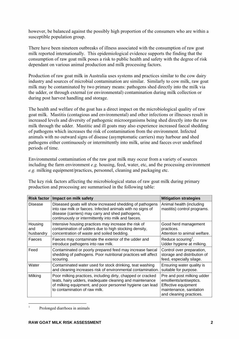
however, be balanced against the possibly high proportion of the consumers who are within a susceptible population group. There have been nineteen outbreaks of illness associated with the consumption of raw goat milk reported internationally. This epidemiological evidence supports the finding that the consumption of raw goat milk poses a risk to public health and safety with the degree of risk dependant on various animal production and milk processing factors. Production of raw goat milk in Australia uses systems and practices similar to the cow dairy industry and sources of microbial contamination are similar. Similarly to cow milk, raw goat milk may be contaminated by two primary means: pathogens shed directly into the milk via the udder, or through external (or environmental) contamination during milk collection or during post harvest handling and storage. The health and welfare of the goat has a direct impact on the microbiological quality of raw goat milk. Mastitis (contagious and environmental) and other infections or illnesses result in increased levels and diversity of pathogenic microorganisms being shed directly into the raw milk through the udder. Mastitic and ill goats may also experience increased faecal shedding of pathogens which increases the risk of contamination from the environment. Infected animals with no outward signs of disease (asymptomatic carriers) may harbour and shed pathogens either continuously or intermittently into milk, urine and faeces over undefined periods of time. Environmental contamination of the raw goat milk may occur from a variety of sources including the farm environment e.g. housing, feed, water, etc, and the processing environment e.g. milking equipment/practices, personnel, cleaning and packaging etc. The key risk factors affecting the microbiological status of raw goat milk during primary production and processing are summarised in the following table: Risk factor Impact on milk safety Mitigation strategies Disease Diseased goats will show increased shedding of pathogens
into raw milk or faeces. Infected animals with no signs of disease (carriers) may carry and shed pathogens, continuously or intermittently into milk and faeces.
Animal health (including mastitis) control programs.
Housing and husbandry
Intensive housing practices may increase the risk of contamination of udders due to high stocking density, concentration of waste and soiled bedding.
Good herd management practices. Attention to animal welfare.
Faeces Faeces may contaminate the exterior of the udder and introduce pathogens into raw milk.
Reduce scouring1. Udder hygiene at milking.
Feed Contaminated or poorly prepared feed may increase faecal shedding of pathogens. Poor nutritional practices will affect scouring.
Control over preparation, storage and distribution of feed, especially silage.
Water Contaminated water used for stock drinking, teat washing and cleaning increases risk of environmental contamination.
Ensuring water quality is suitable for purpose.
Milking Poor milking practices, including dirty, chapped or cracked teats, hairy udders, inadequate cleaning and maintenance of milking equipment, and poor personnel hygiene can lead to contamination of raw milk.
Pre and post milking udder emollients/antiseptics. Effective equipment maintenance, sanitation and cleaning practices.
1 Prolonged diarrhoea in animals
RAW GOAT MILK RISK ASSESSMENT 3
Risk factor Impact on milk safety Mitigation strategies Storage Inappropriate temperature control of raw goat milk after
milking can lead to growth of pathogens. Rapid cooling and holding of milk.
Packaging/ Delivery
Packaging and poor hygiene may contribute to cross-contamination of raw milk. Inappropriate temperature control of milk during delivery can lead to proliferation of pathogens.
Correct sanitising and packaging procedures. Effective cold chain management.
The relative contribution that each of these risk factors has on the overall risk will differ for each pathogen and could not be determined without quantitative through chain data. However it should be highlighted that the goat itself is the primary source of contamination on-farm. Raw goat milk does not undergo any pathogen elimination or reduction step. The safety of raw goat milk is therefore primarily dependent upon the control of risk factors on-farm to minimize the opportunity for microbiological hazards to contaminate raw milk. If raw milk does become contaminated, failure to maintain appropriate temperature control throughout storage, distribution and consumer handling may allow the growth of pathogens and increase risk. Other dairy products are rendered safe principally through the pasteurisation process. FSANZ employed a qualitative framework based on Codex principles to assess the risk from foodborne hazards associated with the consumption of raw goat milk by Australian consumers. Using the qualitative framework, the principal microbiological risks to public health and safety from the consumption of raw goat milk are:
Organism Risk rating (Total population unless otherwise stated)
Bacillus cereus Low Burkholderia pseudomallei Negligible (general population)
Very Low (susceptible population) Brucella melitensis* Moderate (if introduced to Australia) Campylobacter jejuni/coli Low Clostridium perfringens Low Cryptosporidium parvum Low Enterohaemorrhagic Escherichia coli High Leptospira interrogans Negligible Listeria monocytogenes Very low (general population)
High (susceptible population) Salmonella spp. Low (general population)
Moderate (susceptible population) Staphylococcus aureus Low Streptococcus spp. Low Toxoplasma gondii Low (general population)
High (susceptible population) Yersinia enterocolitica Very low Yersinia pseudotuberculosis Very low * Currently exotic to Australia. Risk rating applies if introduced into Australia either through imported raw goat milk
product or its introduction into domestic herds.
RAW GOAT MILK RISK ASSESSMENT 4
Various data gaps were identified during the course of this risk assessment including: • The prevalence and concentration of pathogens in the domestic raw goat milk supply • The virulence and infectivity of some pathogens • The frequency and amount of consumption • The demographics of the consuming population
Further research in these areas may assist to more accurately estimate the impact and magnitude of any illness resulting from consumption of raw goat milk in Australia, as well as to assess the impact of any control measures put in place. While the consumption of raw goat milk is considered to be very low among the general population, there is a group of consumers who have very strong beliefs in the health benefits attributed to raw goat milk and subsequently choose this as their milk of choice. Unpublished research conducted in New South Wales indicated that raw goat milk was marketed with a “health food image”, with purchasers of the product including people with serious illnesses such as cancer patients and mothers intending to feed the product to their infants (AgriQ, 2000). Similarly, work undertaken during the South Australian raw goat milk risk assessment identified consumers as including those with allergies to cow milk and children with digestive problems. This suggests that a higher than normal proportion of raw goat milk consumers may have lowered or less developed immunity and may therefore be more susceptible to foodborne pathogens than the general population. While the volume of raw goat milk consumed in Australia is very low there are risks for both general and susceptible populations consuming this product. Raw goat milk is frequently provided to members of the population who are more susceptible to infection by L. monocytogenes, enterohaemorrhagic E. coli and Salmonella spp. Raw goat milk is often provided to very young children, children with special dietary needs, older people and people convalescing. These sub-populations are at-risk, and exposure to even low levels of these microbial pathogens may result in serious illness.
RAW GOAT MILK RISK ASSESSMENT 5
2 Background Food Standards Australia New Zealand (FSANZ) has responsibility for protecting the health and safety of consumers through the development of food standards. A comprehensive evaluation to identify and examine microbiological hazards along the entire dairy supply chain has previously been conducted by FSANZ entitled A Risk Profile of Dairy Products in Australia (the Profile) (FSANZ, 2006).2. A key finding of the Profile was that Australian dairy products have an excellent reputation for food safety. This is because dairy products in Australia are pasteurised, and pasteurisation represents the principal process for rendering dairy products safe for consumption. This finding was supported by a lack of evidence attributing foodborne illness to dairy products. The Profile confirmed that unpasteurised dairy products are the most common cause of dairy associated foodborne illness. However, the Profile did not specifically examine the risks to public health and safety from the consumption of raw goat milk. This document seeks to assess the risks to public health and safety resulting from consumption of raw goat milk. It utilises available scientific data and addresses the uncertainty and variability in the conclusions drawn from the data e.g. consideration of the relevance and quality of data and the veracity of its source. The output of this risk assessment provides an estimate of risk following the consumption of raw goat milk in Australia. It also identifies hazard control measures along the production chain that have the greatest impact on minimising risk, thereby informing risk managers where intervention will be most effective. The outputs of the assessment will be used by FSANZ to develop regulatory and/or non-regulatory measures as appropriate.
2 http://www.foodstandards.gov.au/_srcfiles/DAR_P296_Dairy_PPPS_Attach2%20Parts%20A-
B.pdf#search=%22Risk%20Profile%22
RAW GOAT MILK RISK ASSESSMENT 6
3 Purpose and scope The purpose of this microbiological risk assessment is to provide an objective analysis of available scientific data and information to identify the public health and safety risks associated with the consumption of raw goat milk, and to identify the factors along the production chain that have the greatest impact on public health and safety for the consumption of raw goat milk. The assessment of the public health and safety risks posed by consumption of raw goat milk in Australia was undertaken to address the following overarching questions:
Specific questions in relation to raw goat milk for human consumption are: • What are the microbial hazards of public health significance in raw goat milk? What
are the prevalence and levels of identified hazards in raw goat milk? • Do these levels pose a risk if the raw goat milk is directly consumed? • What are the factors during primary production that impact on the level of these
hazards? What practices/controls have the greatest impact on the level of hazard? • What is the impact of retail and consumer handling on the level of risk to public health
and safety on these hazards? 3.1 Definition of raw milk The Codex definition of raw milk3 is “milk4 which has not been heated beyond 40°C or undergone any treatment that has an equivalent effect”. The European Union Directives define raw milk as “milk produced by secretion of the mammary glands of one or more cows, ewes, goats, or buffaloes from a single holding that has not been heated beyond 40°C or undergone any treatment having a similar effect”. The Food Standards Code5 specifies processing temperatures for pasteurisation and thermisation in relation to milk and therefore “raw milk” for the purposes of this assessment, is defined as “milk which has not been heat treated in accordance with the Food Standards Code”.
3 Code of Hygienic Practice for Milk and Milk Products (CAC/RCP 57-2004) 4 Defined in Codex General Standard for the Use of Dairy Terms (CODEX STAN 206-1999) 5 The Australia New Zealand Food Standards Code - Standard 1.6.2 – Processing Requirements
1. What are the risks to public health and safety posed by the consumption, in Australia, of raw goat milk?
2. What are the factors that would have the greatest impact on public health and safety
along the production chain for raw goat milk for direct consumption?
RAW GOAT MILK RISK ASSESSMENT 7
3.2 Approach The risk assessment qualitatively identifies hazards, epidemiological data and other information to determine whether these hazards have presented, or are likely to present a public health risk, and to identify where in the raw goat milk supply chain these hazards may be introduced. Animal health issues were considered only in the context of those that differ from cow milk production and which specifically impact upon human health via foodborne transmission. The assessment draws upon the Risk Profile of Dairy Products in Australia (FSANZ, 2006) and utilises available information including current scientific and epidemiological data, surveillance data from enforcement agencies and existing published and unpublished Australian risk assessments on the safety of raw goat milk. The Codex Alimentarius Commission, the Food and Agriculture Organization of the United Nations (FAO) and the World Health Organization (WHO) have established an internationally recognised framework for undertaking a microbiological risk assessment6. The risk assessment process used by FSANZ is consistent with international protocols and involves four distinct steps: hazard identification, hazard characterisation, exposure assessment and risk characterisation. There is no internationally agreed framework for undertaking a qualitative risk assessment for microbiological hazards. Codex7 and FSANZ8 have guidelines for conducting microbiological risk assessments but they do not provide actual tools that can be used to objectively assess or rank the risk to public health and safety. In the absence of an internationally agreed method to qualitatively assess the risk of foodborne hazards associated with the consumption of raw goat milk, FSANZ has used a tool developed by Food Science Australia (Appendix 5) for the assessment of microbiological hazards in a raw milk cheese9. The approach utilises a qualitative framework based on Codex principles and employs elements of Risk Ranger (Ross and Sumner, 2002), a widely accepted semi-quantitative tool. 3.2.1 A Risk Profile of Dairy Products in Australia The Profile was undertaken within the framework of existing management and regulations in Australia. It identified and examined hazards along the entire dairy supply chain from milk production through to consumption of dairy products and considered relevant inputs e.g. feed, water, etc along the dairy primary production and processing chain. The primary focus of the Profile was the production of cow milk, however, the report also incorporated information on milk from non-bovine species. Information identified as being relevant to the dairy goat industry has been utilised in this assessment.
6 Risk assessment is a scientific process undertaken to characterise the risk to public health and safety
posed by foodborne hazards associated with a food commodity. Codex Alimentarius Commission (1999). Principles and guidelines for the conduct of microbiological risk assessment. ALINORM, 99/13, Appendix IV, pp. 58-64
7 CODEX (CAC/GL 30, 1999) Principles and Guidelines for the Conduct of Microbiological Risk Assessment http://www.codexalimentarius.net/download/standards/357/CXG_030e.pdf
8 FSANZ (2009) The Analysis of Food-Related Health Risks. http://www.foodstandards.gov.au/_srcfiles/Food%20Related%20Health%20Risks%20WEB_FA.pdf
9 Application A499 To permit the sale of Roquefort cheese http://www.foodstandards.gov.au/_srcfiles/A499_Roquefort_FAR_FINALv2.pdf#search=%22A499%22
RAW GOAT MILK RISK ASSESSMENT 8
3.2.2 Australian Risk Assessments In undertaking the scientific assessment FSANZ has, with permission, drawn upon the findings of unpublished risk assessments conducted for New South Wales (AgriQ, 2002), South Australia (Pointon et al., 2004) and Queensland (QDPI, 2004). 3.2.3 Qualitative framework The qualitative framework considers the characteristics of identified hazards (hazard identification and characterisation) and an assessment of the likely exposure to these hazards (exposure assessment) to arrive at a final estimate of risk (risk characterisation). The hazard characterisation module categorises each identified hazard based on the probability of disease (infective dose) and the severity of the disease. The exposure module considers the likelihood of the hazard being present in the raw product and the effect of processing on the hazard. This assumes no change in the hazard over time in the product. The risk characterisation combines the hazard characterisation and exposure modules to give an overall categorisation of risk. Essentially the framework categorises the risk for each hazard by combining information about the hazard (severity and infective dose) with exposure information (prevalence in raw materials and effect of processing). A detailed example of the risk characterisation for EHEC in the general population is given in Appendix 5. Briefly, the hazard characterisation for EHEC is high due to the low infective dose (conservatively estimated to be <10 organisms) and serious consequence of exposure in the general population. The exposure assessment was rated as low due to the infrequent product contamination combined with no effect of processing. Combining the hazard characterisation and exposure assessment results gives EHEC in raw goat milk a risk characterisation of high for the general population. Assumptions used for assigning risk categories for all hazards under consideration for both the general and susceptible population groups are given in Appendix 6. Information used to derive these assumptions included scientific data, published literature, professional judgement and expert elicitation. Susceptible populations have been described as individuals who may be more susceptible to infection from specific microbiological hazards due to an impaired immune system and includes the very young and old, the immunocompromised and pregnant women and their unborn children. This assessment uses the term susceptible populations to include all susceptible individuals.
RAW GOAT MILK RISK ASSESSMENT 9
4 Australian risk assessments In recent years, authorities in South Australia, Queensland and New South Wales have commissioned risk assessments of the raw goat milk industry. Some differences exist between organisms considered in each state’s risk assessment, e.g. Burkholderia pseudomallei is an organism limited to tropical regions of Australia such as Queensland and the Northern Territory and was only considered in the Queensland and New South Wales risk assessments. Details of each risk assessment are included in Appendix 7. The South Australian study was undertaken following a risk profile of the primary industry sector and aimed to identify appropriate food safety risk management options, both policy and regulatory for the dairy goat milk industry. The study adopted a qualitative risk ranking approach, based on International Commission on Microbiological Specifications for Foods principles and considered hazard severity; occurrence of the hazard in foods; potential for growth; effects of production, processing and handling (including a consumer terminal step); and epidemiological data. Findings from the South Australian study identified Cryptosporidium parvum, Enterohaemorrhagic Escherichia coli (EHEC), Listeria monocytogenes, Salmonella spp. and Toxoplasma gondii as high risk for susceptible populations, whilst Campylobacter jejuni/coli, Salmonella and EHEC were rated as medium risk for the general population. C. parvum, L. monocytogenes, Staphylococcus aureus and T. gondii were all rated low risk for the general population. The risk assessment undertaken in Queensland utilised methodology based on Codex Alimentarius Commission principles (CAC/GL-30 1999) to rank food safety hazards identified during an extensive literature search. A semi-quantitative approach assigned risk scores (maximum of 100) to hazards and determined total assessed risk scores for each of four population segments based on exposure and severity of consequence. Queensland’s risk assessment concluded that for the general population where goat milk is typically not consumed, there was an overall low risk. E. coli O157:H7 and other pathogenic E. coli were a medium risk to babies and infants, and L. monocytogenes was a medium risk to babies/infants and the immunocompromised. For the niche market where raw goat milk is consumed as the milk of choice, there was an overall increase in risk compared to the normal market population. S. aureus toxins and B. pseudomallei were considered a medium risk to all populations segments, while E. coli O157:H7 was a medium risk to the general population and immunocompromised and a high risk to babies/infants. L. monocytogenes, while considered a low risk to the general population, was a high risk to both babies/infants and the immunocompromised. The New South Wales (NSW) risk assessment was conducted as two separate parts: a qualitative analysis of risk for each hazard identified in a previous hazard analysis, and a stochastic semi-quantitative model (Excel-@Risk) to scope the public health significance of Salmonella spp., S. aureus and L. monocytogenes. The NSW risk assessment declined to make any determination, qualitative or quantitative, on the risks associated with microbial hazards identified in the hazard analysis.
RAW GOAT MILK RISK ASSESSMENT 10
Modelling of the public health significance for Salmonella spp., S. aureus and L. monocytogenes indicates that a single contamination event resulting from contamination and subsequent abuse of the product could lead to severe public health consequences.
RAW GOAT MILK RISK ASSESSMENT 11
5 Goat dairy farming in Australia The main breeds of dairy goats in Australia are the three Swiss breeds (Saanen, British Alpine and Toggenburg), crosses of the Swiss breeds, Anglo-Nubians and crosses of the Swiss breeds with Anglo-Nubians. The Saanen generally produces a greater volume of milk over a longer lactation period than the other dairy goat breeds. Toggenburgs are the second highest producers, with British Alpine and then Anglo-Nubians next in line. The Anglo-Nubians’ milk has the highest percentage of butterfat, which is coveted by cheese makers. Typical lactations last for 300 days or greater and herd production ranges from 2 - 3 litres of milk per doe per day. At the peak of lactation, average production may reach 3.5 - 4 litres of milk per doe per day, with a good doe producing milk for ten years (McGregor, 1997). The dairy goat industry in Australia has expanded in recent years driven primarily by specialty cheese production (Appendix 1). There are an estimated 65 commercial dairy goat farms in Australia carrying almost 11,000 goats, producing approximately 5.4 million litres of goat milk annually. Currently it is estimated that only 300,000 litres is sold as raw goat milk. Four states currently permit the sale of raw goat milk: NSW, South Australia, Western Australia and Queensland. In NSW, raw goat milk is regulated by the NSW Food Authority using Regulations under the Food Production (Safety) Act 1998. In 2000, there were 17 hobby/small permit holding farms and an estimated 50 unlicensed dairy goat units (AgriQ, 2000). South Australia has experienced rapid development in the dairy goat industry, with five commercial goat milk producers licensed by the Dairy Authority of South Australia in 2004. Three farmers sold raw goat milk direct to the public, and operated under the Authority’s Code of Practice for Dairy Food Safety and the Guidelines for Raw or Unpasteurised Goat Milk (Dairy Authority of South Australia, 2005). Recently the number of accredited (previously licensed) goat milk producers selling raw milk has decreased to a single supplier. In 2004 there were three operating and licensed raw goat milk dairies in Queensland. These operations comprised approximately 700 goats, including 300 milking animals (QDPI, 2004). Safe Food Queensland regulates raw goat milk under the Food Production (Safety) Act 2000, with Part 3 of the Food Production (Safety) Regulation 2002 stating requirements for production, testing and labelling. At the time of writing, Western Australia had an estimated three unlicensed goat dairies supplying raw goat milk to the public. The Health Department of Western Australia regulates dairy products under the Health Act 1911 and Health (Food Hygiene) Regulations 1993 with regulations specific for raw goat milk defined under Part VIIA, Division 4 of the Health Act. Codes of Practice for the Goat Dairy Industry outline requirements for Building and Facilities (Part 1) and Hygiene (Part 2). These Codes of Practice are currently not enforceable under any legislation but are used by local government enforcement officers who may be required to approve a goat dairy if a development application is received. Regulations applicable to the production of raw goat milk are contained within Appendix 8. State testing specifications for raw goat milk are outlined in Appendix 9.
RAW GOAT MILK RISK ASSESSMENT 12
6 Consumption of raw goat milk in Australia Food consumption data can be derived from total production statistics or food consumption surveys. Food production statistics provide an estimate of the amount of a specific food commodity that is available to the total population. Consumption surveys (such as national nutrition surveys, independent single source surveys, etc) provide detailed information on the types and amount of food consumed by individuals or households and sometimes the frequency with which these foods are consumed. 6.1 Goat milk production statistics Total sales of goat milk in Australia are steadily increasing with the majority of supermarket sales occurring in NSW, followed by Queensland, Victoria, Western Australia and South Australia. The majority of the raw goat milk sold in Australia is distributed through health food shops or farm gate sales. Accurate information on the volumes of goat milk and in particular raw goat milk produced and sold in Australia is difficult to obtain. The most recent production information indicates around 5.4 million litres of goat milk were produced in Australia during 2003/04. It is estimated that approximately 300,000 litres of raw goat milk was marketed in Australia during 2003 (Abud, 2005). However, observations by industry organisations suggest that the amount of raw goat milk entering the market from unlicensed sources could be double the estimated volume from licensed premises (pers. comm. Riches, 2006). NSW markets around two-thirds of its whole goat milk production as raw milk (approximately 270,000 litres). South Australia had an estimated 32,000 litres of raw milk sales in 2003 (Pointon et al., 2004), however this is expected to decrease in the future as a consequence of a reduction in the number of accredited producers from three to one. Queensland and Western Australia are responsible for small volumes of raw goat milk although no actual figures could be obtained. 6.2 Consumption of raw goat milk Data from the Australian National Nutrition Survey provides information regarding the types and amounts of dairy foods consumed by Australians. The most recent national survey was conducted during the period February 1995 to March 1996 using the 24-hour recall method. Approximately 13,800 people aged two years or over from urban and rural areas in all States and Territories participated in the survey. Only 0.08% (11/12,858 respondents) consumed goat milk, with an average consumption of 248 grams per day. Of the eleven people consuming goat milk, one was a child aged 2 - 3 years and two were females aged 65+ years. The data did not permit differentiation between pasteurised or raw goat milk, as there was no specific information available on the consumption of raw goat milk. The very low numbers reported in this survey for consumption of goat milk does not enable an accurate determination of population consumption patterns to be made. In a recent consumer survey in Australia, less than 1% (7/1000) of those surveyed either consumed or knew of consumers of raw goat milk (Colmar Brunton Social Research, 2008, unpublished). Pointon et al. (2004) notes that people owning their own goats or people who
RAW GOAT MILK RISK ASSESSMENT 13
have an allergy to cow milk are the primary consumers of goat milk. There is also a group of consumers who have very strong beliefs regarding the health benefits attributed to raw goat milk and subsequently choose this as their milk of choice. Unpublished research conducted in NSW indicated that raw goat milk was marketed with a “health food image”, with purchasers of the product including people with serious illnesses such as cancer patients and mothers intending to feed the product to their infants (AgriQ, 2002). Goat milk is widely promoted throughout the industry as an infant milk replacement, particularly where infants are intolerant to cow milk (AgriQ, 2000). The population drinking goat milk is therefore assumed to include a high number of children (Pointon et al., 2004). The consumption of raw goat milk is a contentious issue with the niche market of consumers of this product having strong beliefs in its health benefits. There is also a prevalent belief among its consumers that the product has properties that limit the survival of human pathogens. In many cases these consumers have an equally strong opposition to the commercial processing of foods, in particular pasteurisation and homogenisation (QDPI, 2004).
RAW GOAT MILK RISK ASSESSMENT 14
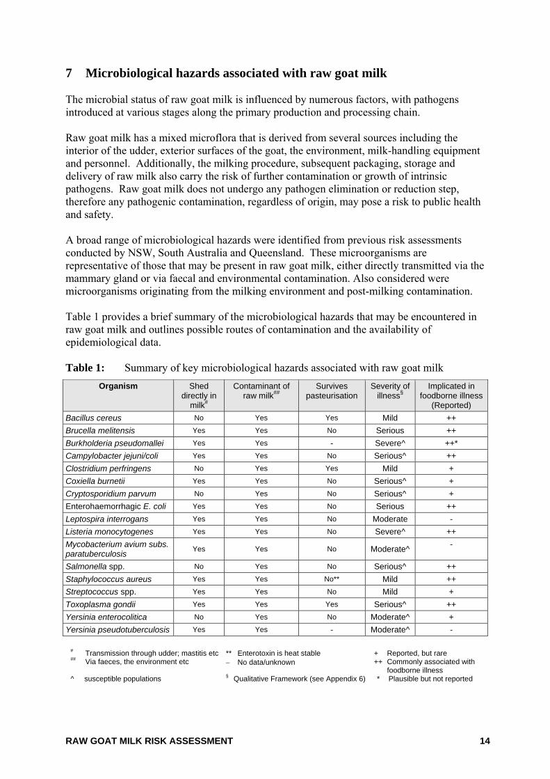
7 Microbiological hazards associated with raw goat milk The microbial status of raw goat milk is influenced by numerous factors, with pathogens introduced at various stages along the primary production and processing chain. Raw goat milk has a mixed microflora that is derived from several sources including the interior of the udder, exterior surfaces of the goat, the environment, milk-handling equipment and personnel. Additionally, the milking procedure, subsequent packaging, storage and delivery of raw milk also carry the risk of further contamination or growth of intrinsic pathogens. Raw goat milk does not undergo any pathogen elimination or reduction step, therefore any pathogenic contamination, regardless of origin, may pose a risk to public health and safety. A broad range of microbiological hazards were identified from previous risk assessments conducted by NSW, South Australia and Queensland. These microorganisms are representative of those that may be present in raw goat milk, either directly transmitted via the mammary gland or via faecal and environmental contamination. Also considered were microorganisms originating from the milking environment and post-milking contamination. Table 1 provides a brief summary of the microbiological hazards that may be encountered in raw goat milk and outlines possible routes of contamination and the availability of epidemiological data. Table 1: Summary of key microbiological hazards associated with raw goat milk
Organism Shed directly in
milk#
Contaminant of raw milk##
Survives pasteurisation
Severity of illness§
Implicated in foodborne illness
(Reported) Bacillus cereus No Yes Yes Mild ++ Brucella melitensis Yes Yes No Serious ++ Burkholderia pseudomallei Yes Yes - Severe^ ++* Campylobacter jejuni/coli Yes Yes No Serious^ ++ Clostridium perfringens No Yes Yes Mild + Coxiella burnetii Yes Yes No Serious^ + Cryptosporidium parvum No Yes No Serious^ + Enterohaemorrhagic E. coli Yes Yes No Serious ++ Leptospira interrogans Yes Yes No Moderate - Listeria monocytogenes Yes Yes No Severe^ ++ Mycobacterium avium subs. paratuberculosis Yes Yes No Moderate^ -
Salmonella spp. No Yes No Serious^ ++ Staphylococcus aureus Yes Yes No** Mild ++ Streptococcus spp. Yes Yes No Mild + Toxoplasma gondii Yes Yes Yes Serious^ ++ Yersinia enterocolitica No Yes No Moderate^ + Yersinia pseudotuberculosis Yes Yes - Moderate^ -
# Transmission through udder; mastitis etc ** Enterotoxin is heat stable + Reported, but rare
## Via faeces, the environment etc − No data/unknown ++ Commonly associated with foodborne illness
^ susceptible populations § Qualitative Framework (see Appendix 6) * Plausible but not reported
RAW GOAT MILK RISK ASSESSMENT 15
While many of the organisms listed in Table 1 are commonly implicated in foodborne illness, the following organisms are not proven pathogens via ingestion or have not been found in Australian goats: • Mycobacterium avium subsp. paratuberculosis is the causative agent for Johne’s
disease in ruminants. A statistical association between Johne’s disease in animals and Crohn’s disease in humans has been reported but there is insufficient evidence presently available to either prove or disprove a causal link (Anon, 2004a; Feller et al., 2007).
• Although Coxiella burnettii infection has been associated with consumption of raw goat milk (Rampling, 1998), ingestion is considered a minor route for human infection (Maurin and Raoult, 1999). Consequently little information exists regarding ingestion mediated illness.
• Brucella melitensis is an important infectious organism within dairy goats although it has not been reported in Australian herds.
These organisms have been included within Table 1 and Appendix 2 as potential pathogens associated with raw goat milk, however the risk characterisation for raw goat milk has been treated separately. Detailed descriptions of the organisms identified in Table 1 are contained within Appendix 2. Severity rankings are discussed within Appendix 6.
RAW GOAT MILK RISK ASSESSMENT 16
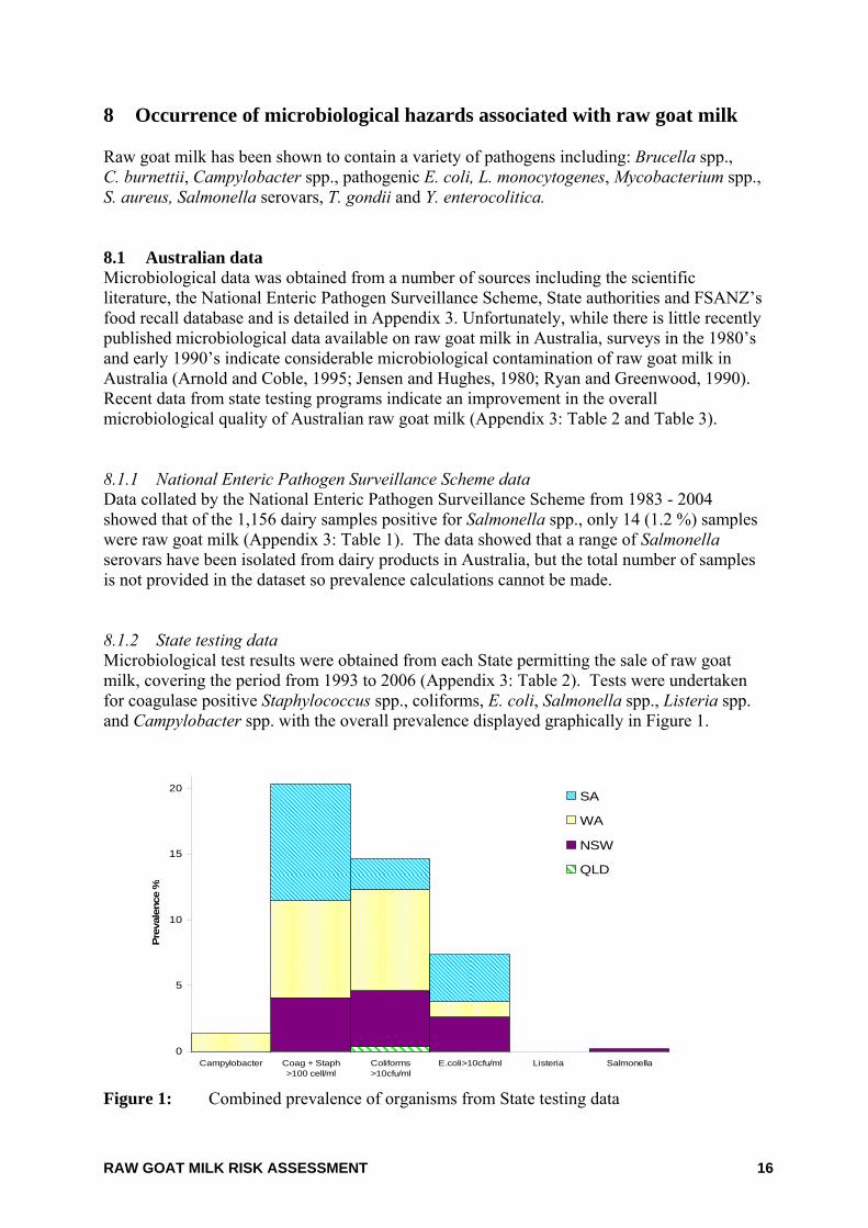
8 Occurrence of microbiological hazards associated with raw goat milk Raw goat milk has been shown to contain a variety of pathogens including: Brucella spp., C. burnettii, Campylobacter spp., pathogenic E. coli, L. monocytogenes, Mycobacterium spp., S. aureus, Salmonella serovars, T. gondii and Y. enterocolitica. 8.1 Australian data Microbiological data was obtained from a number of sources including the scientific literature, the National Enteric Pathogen Surveillance Scheme, State authorities and FSANZ’s food recall database and is detailed in Appendix 3. Unfortunately, while there is little recently published microbiological data available on raw goat milk in Australia, surveys in the 1980’s and early 1990’s indicate considerable microbiological contamination of raw goat milk in Australia (Arnold and Coble, 1995; Jensen and Hughes, 1980; Ryan and Greenwood, 1990). Recent data from state testing programs indicate an improvement in the overall microbiological quality of Australian raw goat milk (Appendix 3: Table 2 and Table 3). 8.1.1 National Enteric Pathogen Surveillance Scheme data Data collated by the National Enteric Pathogen Surveillance Scheme from 1983 - 2004 showed that of the 1,156 dairy samples positive for Salmonella spp., only 14 (1.2 %) samples were raw goat milk (Appendix 3: Table 1). The data showed that a range of Salmonella serovars have been isolated from dairy products in Australia, but the total number of samples is not provided in the dataset so prevalence calculations cannot be made. 8.1.2 State testing data Microbiological test results were obtained from each State permitting the sale of raw goat milk, covering the period from 1993 to 2006 (Appendix 3: Table 2). Tests were undertaken for coagulase positive Staphylococcus spp., coliforms, E. coli, Salmonella spp., Listeria spp. and Campylobacter spp. with the overall prevalence displayed graphically in Figure 1.
0
5
10
15
20
Campylobacter Coag + Staph>100 cell/ml
Coliforms>10cfu/ml
E.coli>10cfu/ml Listeria Salmonella
Prev
alen
ce %
SA
WA
NSW
QLD
Figure 1: Combined prevalence of organisms from State testing data
RAW GOAT MILK RISK ASSESSMENT 17
Coagulase positive Staphylococcus spp., coliforms and E. coli were regularly detected, whilst Campylobacter spp. had a very low prevalence and Salmonella spp. and Listeria spp. were generally not detected (Salmonella spp. was detected in 1 out of 511 samples) (Appendix 3: Table 2). A sample of raw goat milk was recently reported as testing positive for Shiga-like toxin producing E. coli during routine testing in Western Australia (pers. comm. Calder, 2008). Direct comparison of results was difficult due to differences between each State in relation to the types of organisms tested for, the frequency of testing and the manner of reporting results e.g. pass/fail or detected/not detected. The effectiveness of State sampling plans to detect pathogens in raw goat milk has been queried. In assessing the South Australian regulations Pointon et al. (2004) determined that monthly sampling for indicators of hygiene and quarterly sampling for some pathogens provides minimal confidence that contaminated milk is not entering the marketplace. 8.1.3 Summary of data from other programs Aside from routine testing programs, raw goat milk has been analysed during pilot studies and during data collection for risk assessments (Appendix 3: Table 3). There is some overlap between data provided by the South Australia Risk Assessment (Pointon et al., 2004) and the data obtained directly from the Dairy Authority of South Australia. Survey data results cited in the risk assessment undertaken for NSW (AgriQ, 2002) have also been included where not recorded elsewhere in this report. Generally, coagulase positive Staphylococcus spp., coliforms and E. coli have been frequently detected whilst Salmonella spp., Campylobacter spp. and Yersinia spp. are rarely detected. Listeria monocytogenes was not detected but the non-pathogenic, non-haemolytic Listeria innocua was detected at a very low incidence. 8.1.4 Food recalls There were 43 recalls for dairy products due to microbiological concerns during the period 1990-2005, out of a total of 716 food recalls. Of these 43 dairy recalls, only three were from products made from goat milk (0.42%) and only one was positively identified as being from raw goat milk (Appendix 3: Table 5). Frozen raw goat milk was also recalled in Queensland in early 2008 due to Salmonella Zanzibar contamination. 8.2 International data While limited data is published on Australian raw goat milk, the international literature indicates a range of microorganisms can contaminate raw goat milk (Appendix 3: Section 2). It is difficult to directly compare results between individual studies due to differences in the type and number of samples taken, the point in production from where the sample was taken and the methodology used to isolate and/or enumerate the various organisms. In general, the reported prevalence of microbiological hazards in raw goat milk is highly variable and influenced by local factors.
RAW GOAT MILK RISK ASSESSMENT 18
Pathogens detected in raw goat milk internationally include Brucella spp., C. burnettii, Campylobacter spp., Listeria spp., pathogenic E. coli including E. coli O157:H7, Mycobacterium spp., S. aureus, Streptococcus spp., T. gondii and Y. enterocolitica. 8.3 Summary There is little published information available on the incidence and prevalence of pathogens in raw goat milk. Information which is available indicates a variety of pathogens may be isolated from raw goat milk both in Australia and internationally, although greater diversity is reported internationally. This may be an artefact of the level of microbiological testing undertaken in those countries which permit raw goat milk and raw goat milk products. Similarities exist between the pathogens detected internationally and in Australia. Coagulase positive Staphylococcus spp. and E. coli are more commonly detected, while Campylobacter spp., Salmonella spp. and Yersinia spp. detections are generally low. Contamination with Listeria spp. in Australia is very low whereas it appears to be more problematic internationally (Appendix 3: Table 10). Particular mention should be made of the prevalence of Brucella spp. internationally. Although Brucella spp. have been isolated from raw goat milk (Appendix 3: Table 6), it is important to note that Australia has been free from B. abortus since 1989 and B. melitensis has never been reported in Australian livestock (Australian Quarantine and Inspection Service, 1999).
RAW GOAT MILK RISK ASSESSMENT 19
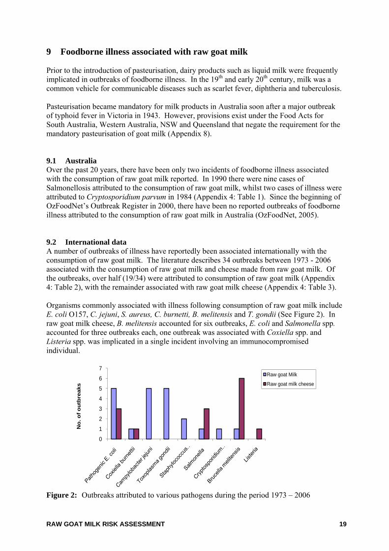
9 Foodborne illness associated with raw goat milk Prior to the introduction of pasteurisation, dairy products such as liquid milk were frequently implicated in outbreaks of foodborne illness. In the 19th and early 20th century, milk was a common vehicle for communicable diseases such as scarlet fever, diphtheria and tuberculosis. Pasteurisation became mandatory for milk products in Australia soon after a major outbreak of typhoid fever in Victoria in 1943. However, provisions exist under the Food Acts for South Australia, Western Australia, NSW and Queensland that negate the requirement for the mandatory pasteurisation of goat milk (Appendix 8). 9.1 Australia Over the past 20 years, there have been only two incidents of foodborne illness associated with the consumption of raw goat milk reported. In 1990 there were nine cases of Salmonellosis attributed to the consumption of raw goat milk, whilst two cases of illness were attributed to Cryptosporidium parvum in 1984 (Appendix 4: Table 1). Since the beginning of OzFoodNet’s Outbreak Register in 2000, there have been no reported outbreaks of foodborne illness attributed to the consumption of raw goat milk in Australia (OzFoodNet, 2005). 9.2 International data A number of outbreaks of illness have reportedly been associated internationally with the consumption of raw goat milk. The literature describes 34 outbreaks between 1973 - 2006 associated with the consumption of raw goat milk and cheese made from raw goat milk. Of the outbreaks, over half (19/34) were attributed to consumption of raw goat milk (Appendix 4: Table 2), with the remainder associated with raw goat milk cheese (Appendix 4: Table 3). Organisms commonly associated with illness following consumption of raw goat milk include E. coli O157, C. jejuni, S. aureus, C. burnetti, B. melitensis and T. gondii (See Figure 2). In raw goat milk cheese, B. melitensis accounted for six outbreaks, E. coli and Salmonella spp. accounted for three outbreaks each, one outbreak was associated with Coxiella spp. and Listeria spp. was implicated in a single incident involving an immunocompromised individual.
0
1
2
3
4
5
6
7
No.
of o
utbr
eaks
Raw goat Milk
Raw goat milk cheese
Figure 2: Outbreaks attributed to various pathogens during the period 1973 – 2006
RAW GOAT MILK RISK ASSESSMENT 20
9.3 Attribution of foodborne illness Over the last 35 years, raw goat milk has only been associated with two reported outbreaks of illness in Australia and 19 reported outbreaks internationally. The extent to which illness can be attributed to raw goat milk does not enable risk assessors to clearly determine the relative risk that consumption of raw goat milk poses to consumers. Sources of foodborne illness are generally determined through epidemiological and/or microbiological associations in outbreak investigations. Critical in this process is the ability to identify an outbreak through the existing surveillance system to enable an investigation to then proceed. Difficulties exist in identifying and attributing illness to a particular food and include: • Food recall biases when gathering food consumption histories • Time delays in recognition or notification of an outbreak • Inability to trace food products to their source • Reluctance of individuals to participate in investigations, particularly when they have
purchased foods that are not permitted to be sold legally • Long exposure windows for specific pathogens (e.g. L. monocytogenes) • Inability to obtain representative food samples for analysis • A lack of precision in or suitable methods for sample analysis and pathogen
identification It is important to recognise that outbreak data only represents a small proportion of actual cases of foodborne illness, as many outbreaks go unrecognised and/or unreported to health authorities. People do not always seek medical attention for mild forms of gastroenteritis, medical practitioners do not always collect specimens for analysis and not all foodborne illnesses require notification to health authorities. Pointon et al. (2004) notes the likelihood of significant under-reporting of illness associated with the consumption of raw goat milk in Australia. A contributing factor is the overall under-reporting of gastrointestinal illness combined with the low frequency of consumption of unpasteurised goat milk among the population (only 32,000 litres were sold in SA in 2002). This means the sensitivity of the surveillance system to detect outbreaks and sporadic illness associated with raw goat milk is low (Pointon et al., 2004). This is evidenced by the fact that only two incidents of illness have been reported in Australia over the last 20 years and none since the inception of OzFoodNet’s Outbreak Register in 2000.
RAW GOAT MILK RISK ASSESSMENT 21
10 Primary production factors impacting on raw goat milk safety Raw goat milk has a mixed microflora which is a result of multiple factors. Contamination may occur when microorganisms are shed directly into the milk from the goat udder, through environmental contamination, and via contamination from the milking environment or personnel. The microflora encountered is not dissimilar to that found in raw cow milk. Primary production factors that impact on these routes of contamination and the microbiological quality of the raw goat milk include: • Animal-related factors e.g. animal health10 and husbandry • Environment-related factors e.g. housing, faeces, feed, soil, and water • Milking related practices e.g. milking methods, personnel, equipment, storage,
packaging and delivery In Australia, successful goat dairy farms are operating on systems developed for cow dairying. As indicated in Section 3.2.1 only those factors which differ significantly to those depicted for cow milk production have been discussed. 10.1 Animal health/husbandry Generally goats are considered clean animals as they produce pelletised faeces and do not like to walk in water or mud (QDPI, 2004). Although goats are generally thought to be naturally healthy animals they succumb quickly when they do become ill, hence veterinary treatment and vaccination of goats may involve the off-label use of veterinary medicines registered for use in other species for other conditions. Therapies developed for dairy cows may be used in goats under veterinary prescription in certain circumstances. Common diseases of goats include mastitis, toxoplasmosis, leptospirosis, viral infections (including caprine retrovirus) and Johne’s disease. Goat health problems may impact on the microbiological quality of raw milk. Diseased11 goats will show increased shedding of pathogens directly into raw milk through udder infections or into faeces which may contaminate the production and milking environment. Infected12 animals with no signs of disease (asymptomatic carriers) may harbour and shed pathogens, often intermittently, into milk and faeces. 10.1.1 Carrier status The retention of a disease agent in a group of animals frequently depends on the presence of an individual animal which carries the organism without showing the disease. These are difficult to detect and frequently require repeated laboratory tests to confirm their carrier status. Carriers may be animals which have recovered from the clinical disease or animals which have never had the disease. Their presence confounds conventional disease diagnosis
10 Animal health is defined as incorporating both disease (the clinical and/or pathological manifestation of
infection), infection and carrier status of the animal. 11 Disease is defined in the OIE Terrestrial Animal Health Code (2007) as the clinical and/or pathological
manifestation of infection (http://www.oie.int/eng/normes/mcode/en_chapitre_1.1.1.htm) 12 Infection is defined in the OIE Terristrial Animal Health Code (2007) as the presence of the pathogenic
agent in the host (http://www.oie.int/eng/normes/mcode/en_chapitre_1.1.1.htm)
RAW GOAT MILK RISK ASSESSMENT 22
and herd treatments and may result in the recrudescence of a disease in a previously negatively tested group. Some carriers may be masked and not release organisms unless stressed or immunocompromised. In these cases the isolation of microorganisms may be negative until the infection re-activates. The specificity and sensitivity of the laboratory testing will also limit the ability to detect carriers. Where detection is difficult, it is often the reappearance of disease in susceptible animals which is the first indication that carrier animals exist in a group. Destocking and complete replacement with disease free animals may be the only way of removing a disease carrier. Many human pathogens co-exist in their animal host with little or no apparent ill-effect. For example E. coli O157:H7 asymptomatically colonises the terminal rectum of cattle, and a vaccine is being tested to reduce secretion levels by carrier animals (Peterson et al., 2007). This vaccine may potentially also be used in the treatment of goats. Research by Brownlie and Grau in the 1960’s demonstrated the effects that stress and starvation have on the shedding of enteric pathogens such as E. coli and Salmonella spp. in cattle and sheep (Grau et al., 1968; Grau et al., 1969). The frequency and amount of pathogen excreted by a carrier varies with the organism, the animal, its husbandry and immune status, and the natural history of the disease in that animal species. In some diseases, carriers continue to be infected for many years while in others it can be a matter of a few months. Good husbandry will reduce stress but will not necessarily relieve certain types of production stresses such as pregnancy, parturition and lactation. These are significant stresses which do modulate the immune system and can precipitate the excretion of organisms in a carrier animal. 10.1.2 Mastitis Mastitis, both clinical (actual signs of infection) and subclinical (no outward signs of infection) can be caused by the same organisms which can damage the udder, reduce production and adversely affect the quality and quantity of milk produced. Bacteria which infect the mammary gland are classified into two major categories, contagious or environmental pathogens (Tomita and Hart, 2000). The most prevalent contagious pathogens associated with mastitis in goats are Streptococcus agalactiae and S. aureus. Causal pathogens of environmental mastitis13 are present in urine, faeces, soil and bedding. Transmission mainly occurs between milking, but can also occur during milking. Environmental pathogens commonly isolated from infected udders are coliform bacteria, Streptococcus spp. other than St. Agalactiae, and Staphylococcus spp. other than S. aureus (Tomita and Hart, 2000). Staphylococci have frequently been reported as the most prevalent organism in clinical and subclinical mastitis in goats. S. aureus is the most significant pathogen associated with clinical mastitis and has been reported at prevalences around 13% (Deinhofer and Pernthaner, 1995; Kalogridou-Vassiliadou, 1991; White and Hinckley, 1999), although one study in Norway reported prevalence of 96.2% in bulk tank milk (Jorgensen et al., 2005).
13 Environmental mastitis occurs as a result of an ascending infection through the teat canal.
RAW GOAT MILK RISK ASSESSMENT 23
International prevalences of approximately 13% are consistent with those reported in Australia (Appendix 3: Table 2). Other organisms which have been associated with mastitis in goats include E. coli, Streptococcus spp., Pseudomonas spp., Corynebacteria spp. and Bacillus spp. (Al-Graibawi et al., 1986; Bergoinier et al., 2003; Deinhofer and Pernthaner, 1995; Jorgensen et al., 2005; Kalogridou-Vassiliadou, 1991; Ryan and Greenwood, 1990; White and Hinckley, 1999). A case of caprine mastitis associated with Yersinia pseudotuberculosis (synonym, Pasteurella pseudotuberculosis) was documented in California in 1972. Raw or inadequately pasteurized milk contaminated with Y. pseudotuberculosis, regardless from which animal species, may be a possible source of Yersinia infections in man (Cappucci et al., 1978). Somatic cell counts (SCC) are used as a method to determine levels of mastitis infection in individual goats, or in bulk milk samples. Some studies have indicated that an increase in SCC alone is not an accurate indicator of mastitic infection in goats and suggest that the establishment of bacteriological examinations (particularly of mastitis-related pathogens) would help establish a SCC threshold (Wilson et al., 1995; Zeng et al., 1997). Healthy goat udders can have high SCC levels normally, with stage of lactation influencing the actual counts (Haenlein, 2002). SSC levels in milk from goats are higher than from cows and sheep, with the standard SCC in goat milk at 1 x 10⁶ cells/ml (Olechnowicz and Jaskowski, 2004). This limit is imposed in the USA and France, although levels in the range of 300,000 - 400,000 cells/ml have often been achieved (Stubbs and Abud, 2002). An acceptable level of SCC in cow milk is less than 400,000 cells/ml with counts above 200,000 indicating that either clinical or subclinical mastitis is present to a significant degree (Stubbs and Abud, 2002). Hence it is inappropriate to attempt to correlate SCC results between cow and goat species. Importantly, goats with mastitis are much more likely than cows to develop lumps, abscesses and fibrosis in the udder. 10.1.3 Other zoonotic diseases/infections Goat health issues other than mastitis may also influence the microbiological quality of the raw milk. There may be increased shedding of pathogens either directly into the milk or into the faeces or urine from sick and diseased animals. Common zoonotic diseases, other than mastitis, affecting goats are discussed below. 10.1.3.1 Leptospirosis Leptospirosis, also known as Weil’s or Canecutter’s Disease, is caused by Leptospira interrogans (Appendix 2). L. interrogans can be spread by contact directly between infected animals and humans, by ingestion of contaminated water or food, through aerosolised urine particles, animal foetal fluids or through direct contact with skin (Baranton and Postic, 2006). Similarities exist between the serovars of L. interrogans found in cows and goats. Serovars Hardjo, Pomona and Grippotyphosa are common to both cattle and goats, while Canicola, Australis and Icterohaemorrhagiae are further associated with cattle (Anon, 2004b).
RAW GOAT MILK RISK ASSESSMENT 24
Humans are susceptible to all pathogenic serovars found in domestic animals with between 100 - 200 humans cases of leptospirosis reported each year in the US (CDC, 2005). Leptospirosis is a notifiable disease in Australia and had an annual notification rate of 1.3 cases per 100,000 population in 2000 (Anon, 2002). Notifications have been declining in recent years from 243 cases reported in 2000 to 177 in 2004. Leptospirosis occurs across Australia, although the majority of cases are reported in Queensland. The most common serovars are generally Zanoni, Hardjo and Australis (QHSS, 2004). Primarily excreted via the urine of infected animals, shedding of viable leptospires has been recorded in mastitic cow milk (Bolin and Koellner, 1988). Leptospirosis is problematic for cow dairies and can occur in goats but the extent is unknown. Vaccination programs available to control leptospirosis in cows are unavailable for goats. Urine splashing is less common in goat dairies than cow dairies, although the potential for contamination of raw goat milk does exist. There is a lack of data on raw goat milk mediated foodborne illness from L. interrogans. 10.1.3.2 Melioidosis Burkholderia pseudomallei (previously Pseudomonas pseudomallei) (Appendix 2) is the aetiologic agent of melioidosis (also called Whitmore's disease). Melioidosis is generally a disease only seen in tropical and sub-tropical regions; predominately during the wet season. Goats and sheep are particularly susceptible with cases of infection often eventuating in the death of the animal (Choy et al., 2000). B. pseudomallei are limited to tropical regions of Australia such as Queensland and the Northern Territory and are hence exotic to southern regions. It has been suggested that there is a possible public health risk from drinking contaminated milk from an animal infected with the disease melioidosis (Thomas et al., 1988; Choy et al., 2000). B. pseudomallei has been isolated from infected goat’s udders (Van der Lugt and Henton, 1995), mastitic goat milk (Choy et al., 2000) and is excreted in goat faeces (Dance, 2000). In the Northern Territory, raw goat milk has been banned because of the high incidence of asymptomatic mastitis in dairy goats caused by B. pseudomallei (pers. comm. Currie, 2006). A Darwin study undertaken by Choy et al., (2000) found 15/43 (35%) of goats had evidence of mastitis from B. pseudomallei, although no information is available on the prevalence of B. pseudomallei in raw goat milk. Melioidosis is known to be a major cause of human morbidity and mortality in the Australian tropics (Dance, 2000). Twelve human deaths out of 33 infections occurred during one outbreak in the Northern Territory during 1990 and 1991 (AgriQ, 2000). B. pseudomallei can survive the low pH of the stomach indicating infection by ingestion is possible. A small number of cases in a Darwin prospective study are thought to have resulted from ingestion rather than percutaneous or inhalation routes (Ralph et al., 2004). There is a lack of data on virulence and infectivity for B. pseudomallei obtained via ingestion and no information available on the dose-response relationship for B. pseudomallei in human infections. While there is limited information implicating ingestion of B. pseudomallei from raw goat milk, it is plausible that foodborne illness could result from consumption of contaminated raw goat milk.
RAW GOAT MILK RISK ASSESSMENT 25
10.1.3.3 Johne’s disease Mycobacterium avium subsp. paratuberculosis (MAP) (Appendix 2) is the organism responsible for Johne’s disease in many ruminant species, including goats. Goats are susceptible to both cattle and sheep strains of Johne’s disease. MAP is excreted primarily in the faeces of infected animals and is excreted during both the sub-clinical and clinical stages of disease. In dairy animals, MAP can be transmitted both vertically through the placenta to the foetus in advanced infection and also through the young animal ingesting colostrum, milk or faeces from an infected animal. MAP is also transmitted horizontally through the faecal-oral route (Streeter et al., 1995; Sweeney et al., 1992, Scientific Committee on Animal Health and Animal Welfare, 2000; Anon, 2004b). A statistical association has been reported between MAP and Crohn’s disease, a chronic intestinal enteritis in humans. While such an association is reported, whether it is causal is a matter for debate. The debate is characterised by firmly entrenched opinions on either side, and the subject has been comprehensively reviewed several times (Chiodini, 1989; Thompson, 1994; Anon, 1998; Harris and Lammerding, 2001; Lipiec, 2003; Chacon et al., 2004; Feller et al, 2007). Presently there is insufficient evidence to prove or disprove a causal association link between Johne’s Disease in ruminants and Crohn’s disease in humans (Anon, 2004a; Feller et al., 2007). 10.1.3.4 Q fever Coxiella burnettii (Appendix 2) causes the zoonotic illness Q fever and is commonly found in cattle, sheep and goats. C. burnettii has been associated with consumption of raw goats milk and cheese in Europe, Canada and the USA (Rampling, 1998), although this is considered a minor route for human infection (Vanderlinde, 2004; Maurin and Raoult, 1999). 10.1.3.5 Brucellosis Brucella spp. (Appendix 2) are pathogenic for both humans and a wide range of animals. B. melitensis is a major cause of brucellosis in sheep and goats and is more pathogenic to humans than other Brucella spp. B. melitensis is widespread in southern Europe, west and central Asia, Mexico, South America and Africa but has never been reported in sheep or goats in Australia. Zoonotic transmission from infected animals to humans may occur either via direct or indirect transmission (Kasimoglu, 2002). Brucella is most commonly transmitted via raw milk or raw milk products, such as cheeses (Kasimoglu, 2002). Ewes and goats milk has been found to be a more significant source of Brucella spp. than cow’s milk. 10.1.3.6 Enterotoxaemia Clostridium perfringens (Appendix 2) is a zoonotic organism producing disease in goats which is generically called enterotoxaemia (Uzal, 2004). The infection is characterised by profuse diarrhoea which may last for days or weeks.
RAW GOAT MILK RISK ASSESSMENT 26
10.1.3.7 Toxoplasmosis The protozoan parasite Toxoplasma gondii (Appendix 2) is the cause of the potentially severe disease toxoplasmosis. It can infect a wide range of animals with the primary host belonging to the cat family (Felidae) and secondary hosts including all warm blooded animals (e.g. mammals and birds) (Tenter et al., 2000). T. gondii causes great losses in sheep and goats, however the disease is more severe in goats (Hill and Dubey, 2003). Transmission of T. gondii occurs via the faecal-oral route, transplacental transfer between mother and foetus, and through the consumption of infected meat and/or milk containing tachyzoites or other forms of the infective parasite from the secondary host (Chiari and Neves, 1984; Skinner et al., 1990; Smith, 1993b; Tenter et al., 2000). Raw goat milk has been linked as a probable route of infection in outbreaks of T. gondii (Smith, 1993b). One study suggests that T. gondii has the ability to survive in refrigerated raw goat milk (Walsh et al., 1999). Exposure to 50oC renders tachyzoites non-infectious and therefore pasteurisation will eliminate tachyzoites (Smith, 1993b). Toxoplasmosis is widespread in humans, being one of the most common parasitic zoonoses worldwide (Tenter et al., 2000). T. gondii infection is very serious in cases where the secondary host is pregnant as this organism has the ability to cause spontaneous abortion or severe congenital defects in the off-spring of the host (Tenter et al., 2000). 10.1.3.8 Cryptosporidiosis Cryptosporidium parvum is a common aetiological agent of diarrhoea in goat kids (de Graaf et al., 1999), with large numbers of oocytes being shed in the faeces during infection. Cryptosporidium spp. can also cause illness in humans (Appendix 2). 10.1.3.9 Caprine retrovirus Caprine retrovirus,formerly known as caprine arthritis encephalitis or “Big Knee” is of major concern for the goat industry. The signs of caprine retrovirus in affected goats are age dependent with kids under 6 months normally developing encephalitis, whilst in older goats the virus primarily affects joints. Chronic progressive pneumonia and the condition known as “hard udder” may also be associated with caprine retrovirus. The disease occurs mainly in dairy goats and has been reported in Australia, New Zealand, UK, USA and other countries (Stubbs and Abud, 2002). Infection with caprine retrovirus usually lasts for the lifetime of the animal with the transmission of the virus primarily being through the colostrum and milk. The virus does not survive for long in the general environment, such as soil and sheds, and is destroyed by heating such as pasteurisation but not refrigeration (Stubbs and Abud, 2002). There is no information on the incidence and effect, if any, of caprine retrovirus in humans. Caprine retrovirus, although identified as being of significant concern within dairy goats in Australia, and milk being identified as a transmission vehicle, was not considered in the risk assessment as there is no documented association with human illness.
RAW GOAT MILK RISK ASSESSMENT 27
10.2 Environmental factors Pathogens may originate from the dairy goat environment such as housing, urine and faeces, feed, soil and water. Environmental contaminants may therefore contaminate raw milk or contribute to mastitic or systemic infection in the animal. 10.2.1 Housing Goats are kept in situations which vary from extensive grazing to close confinement and housing (Bureau of Animal Welfare, 2001). Mixed housing systems are common in Australia i.e. some grazing and some intensive housed herds (Stubbs and Abud, 2002). Intensive housing has been associated with a higher risk of udder contamination, which may lead to mastitic infection as a result of increased contact with urine and faeces, wet bedding, and contact with other animals, as well as other environmental contaminants. Pathogens that have been associated with intensive housing for cattle include L. monocytogenes, E. coli, B. cereus and Salmonella spp. It is assumed that for goats managed under similar intensive housing systems, the same pathogens may also be of concern. 10.2.2 Faeces Unlike the moist faeces produced by cows, goats produce drier pelletised faeces. Pelletised faeces may reduce the amount of direct faecal contamination on the udder; however there is a greater risk of dust being formed. The pelletised faeces can become soft and liquid under circumstances of disease, stress and unsuitable feeding regimes. Risk of contamination on the udder is then enhanced. The goat’s hairy udder may more readily become contaminated and may be more difficult to clean than that of a cow udder (QDPI, 2004). Milking animals may shed a variety of enteric pathogens in the faeces as a result of infection or from ingestion of the organisms from feed or water. Pathogens associated with dairy cattle include Listeria spp., Salmonella spp., E. coli, Bacillus spp., Campylobacter spp. and pathogenic E. coli. There is limited information available regarding the prevalence of pathogens in goat faeces; however a strain of E. coli O157:H7 was detected in goat faeces during a study undertaken in Greece. The testing of 351 faecal samples from goat, sheep and cattle found an E. coli O157:H7 prevalence of 0.2% in goat faeces, indicating that goats can be a reservoir of E. coli O157:H7 and goat milk, dairy products and meat may serve as a vehicle for the transmission of this pathogen to humans (Dontorou et al., 2004). Faecal carriage of Campylobacter spp. is also common in both sheep and goats. Campylobacter spp. have been isolated in goat faeces from 0 - 2.7% of healthy goats and 3.7% of diarrheic goats (Kaneene and Potter, 2003). The clinical manifestations of listeriosis in goats closely resemble those in sheep: encephalitis, septicaemia and abortion. Goats may be asymptomatic carriers, shedding L. monocytogenes in the faeces and milk, which may result in environmental contamination (Schukken et al., 2003).
RAW GOAT MILK RISK ASSESSMENT 28
10.2.3 Feeding practices Feeding regimes for goats vary greatly and include irrigated pasture, scrub, hay, grains, silage, bread and supplements. In Australia, most farms graze goats on some pasture but the degree of pasture management is variable. Some is simply rangeland, much is set stocking and some is highly managed rotational grazing. Hay and grains are commonly used, while silage with minerals and other additives is often fed to goats (Abud and Stubbs, 2005). Feed can play an important role as a primary vehicle for animal contamination and also an environmental contaminant of raw milk. Contamination of feed may originate from storage of the feedstuff on farm or from the source of the feed (including contamination during feed manufacture). Animal feed can be contaminated with pathogens of faecal, plant and soil origin (Desmarchelier, 2001). A broad range of pathogens have been associated with dairy cattle feed and feeding practices and may be extrapolated to goats where practices are similar. These pathogens include; Listeria spp. and C. jejuni in pasture, Enterobacteriacea, Listeria spp., Clostridia, Bacillus spp. and E. coli in silage and Salmonella spp., E. coli, Campylobacter spp., B. cereus and Listeria spp. in feed concentrates. 10.2.4 Soil and water Soil represents an important source of pathogens for grazing animals with a wide variety of organisms, including pathogens, often found. B. cereus spores have been found internationally to vary at levels between <50 - 380,000 cfu/g (Christiansson et al., 1999). Australian studies have found Bacillus spores in soil at levels between 5.6 x 10² - 1.8 x 10³ cfu/g (Cook and Sandeman, 2000). Listeria spp., Salmonella spp. and enteropathogenic E. coli have also been reported as existing and surviving in soil (Desmarchelier, 2001; Fenlon et al., 1996). Water can be a primary source of contamination and is used extensively on goat dairy farms for cleaning, cooling, stock drinking and irrigation. Various pathogenic bacteria have been reported in water including E. coli O157:H7 and other pathogenic E. coli, Campylobacter spp. and Salmonella spp. (Lejeune et al., 2001; Rice and Johnson, 2000; Stanley et al., 1998; Wallace, 1999). Cryptosporidium parvum oocysts have been shown to be able to survive up to 176 days in drinking water or river water stored at 4oC (Robertson et al., 1992). 10.3 Milking practices 10.3.1 Milking systems Goats are milked by hand or by machine (bucket system or pipeline) with milking methods dependent upon the specific dairy’s herd management practices. The most popular milking parlours for goats are herringbone types and side-by-side parlours with two platforms (Billon, 2002). In Australia, most systems are of a herringbone design with rotary systems only used for very large herds. Unlike milking systems for cows, a kick rail is not needed for goats (Stubbs and Abud, 2002). Because goats tend not to kick off cups during milking, the likelihood of faecal contamination being sucked into the milk line is reduced.
RAW GOAT MILK RISK ASSESSMENT 29
The type of milking system employed may influence bacterial contamination of the raw milk. A South African study found that raw milk obtained by the bucket system milking machine had the lowest total bacterial count (16,450 cfu/ml), as compared to that by pipeline milking machine (36,300 cfu/ml) or hand milking (48,000 cfu/ml) (Kyozaire et al., 2005). Filtration of milk before it enters the bulk milk tank provides a safeguard to ensure sediment or other extraneous matter is removed from the milk prior to storage. Although filtration removes most of the soil and other particles, it does not remove all the bacteria adhering to these particles, nor does it remove any dissolved matter from the milk. Sediment that has been trapped by the filter continues to be washed by the milk flowing through. This dislodges bacteria adhering to the particles and thus contributes to an increased bacterial count in the bulk milk. If filters are not adequately cleaned, this process may be a source of cross-contamination of the milk. 10.3.2 Milking practices Poor milking practices may lead to contamination of raw milk. The teat surface is the major avenue of entry for microorganisms into raw milk and the goat’s hairy udder may be a greater source of contamination than a cow udder. Pre-milking udder hygiene e.g. washing with clean water and drying using hand towels reduces milk contamination by transient bacteria located on the udder. This practice has been advocated for all goat dairies producing liquid milk to be consumed raw (Ryan and Greenwood, 1990). Post-milking teat disinfection reduces the resident teat skin bacterial population, which is the main source of infection for the mammary gland. In dairy cattle, the rate of new intramammary infection due to S. aureus and St. agalactiae is reduced by approximately 50% when post-milking teat disinfection is practiced (Sheldrake and Hoare, 1980). A comparable effect on infection rate in goats would also be expected. 10.3.3 Cleaning and sanitation There are various methods for cleaning dairy goat milking parlours and yards. Cleaning and sanitation procedures applicable to the cow dairy industry may also be applied to the goat dairy industry, however due to goats’ drier pelletised faeces; a water washout may not be undertaken after each milking. The milking bays and floors of the milking area may only undergo sweeping out of solids. When a water washout is carried out, a source of cross contamination is the cleaning water contacting the milking equipment and the creation of aerosols. Wastewater generated by cleaning operations may contaminate pasture and transmit pathogens to grazing animals. Cleaning of milk handling equipment involves a combination of chemical, thermal and physical processes. The key principles of a good cleaning system involves sufficient hot water (temperature and volume), correct wash solutions (detergent, acid/alkali), adequate contact time and sufficient turbulence to prevent build up of milk residues and bacteria in the equipment.
RAW GOAT MILK RISK ASSESSMENT 30
10.4 Milk storage The composition of milk makes it an excellent growth medium for many microorganisms unless it is frozen or further processed to kill or prevent their growth. As the temperature of raw milk is over 30ºC as it leaves the udder, pathogenic bacteria if present, will grow rapidly. At temperatures between 0 and 5ºC, the growth of most pathogenic and spoilage mesophilic bacteria is slowed. Cooling of milk to less than 5ºC in 3½ hours or less from the start of milking minimises the likelihood of bacterial growth. Goat milk requires rapid cooling to lower temperatures to reduce bacterial activity because of infrequent milk pickup/delivery (Stubbs and Abud, 2002). In Australia, milk is usually stored in cooled vats of sufficient size to cope with up to a week’s production. Cooling systems employed range from artisanal to sophisticated, and include pre-cooling in-line, ripple coolers, plate exchangers, direct expansion vat, cool rooms and buckets in ice (Stubbs and Abud, 2003). Any breakdown in the refrigeration system or failure to properly cool milk prior to collection or delivery may adversely impact on the microbial load in raw milk. 10.5 Milk delivery Most producers of raw goat milk for direct human consumption bottle their own milk on site. Consequently correct sanitising procedures for packaging, aseptic packaging, and effective cold chain management practices for the raw milk are important steps for minimising cross-contamination and growth of any microorganism present in the raw milk. Because of the size of the industry, the goat farmer will typically need to deliver raw milk to the processor if milk is not sold directly off-farm. An Australia-wide survey undertaken by the Rural Industries Research and Development Corporation indicated that self-delivery in food grade plastic containers, twice a week, was the most common method of milk transport (Stubbs and Abud, 2003). A further study conducted in NSW (Miles and Van Den Hout, 2000) indicated that deliveries do not always occur in refrigerated vehicles, but use alternatives such as Styrofoam boxes and packing products in ice covered with a wet blanket. Information pertaining to the time and temperature conditions which raw goat milk is subject to post milking is scarce. Furthermore, limited information is available on the integrity of cold chain management throughout distribution, retail storage and consumer handling practices. However it is generally accepted that retail and domestic refrigeration units can sometimes be a weak link in cold chain management. Time and temperature conditions post milking, i.e. through storage and distribution, have an important influence on the concentration of any contaminating pathogens. Even assuming the integrity of the cold chain is maintained, growth of L. monocytogenes and Y. enterocolitica can still occur at refrigeration temperatures if organisms are present in the milk. Other pathogenic microorganisms, if present, will also grow if the temperature increases by only a few degrees, i.e. E. coli, Salmonella and S. aureus may all grow at temperatures between 7 - 8oC (ICMSF, 1996).
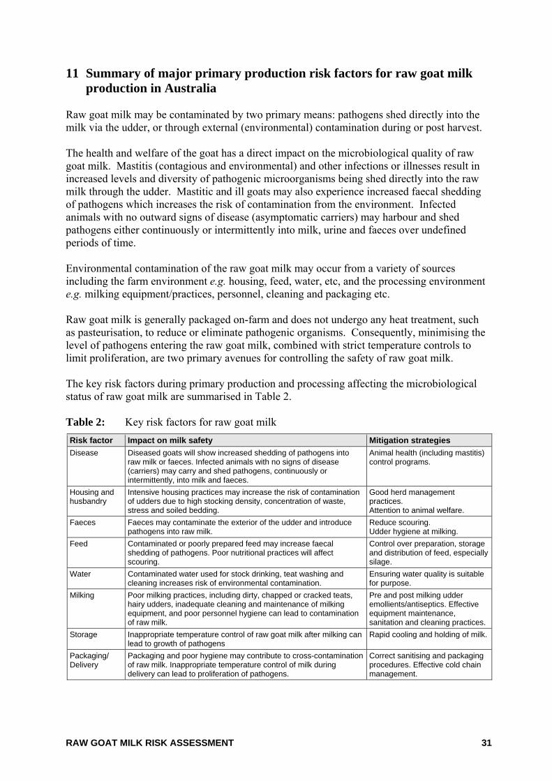
RAW GOAT MILK RISK ASSESSMENT 31
11 Summary of major primary production risk factors for raw goat milk production in Australia
Raw goat milk may be contaminated by two primary means: pathogens shed directly into the milk via the udder, or through external (environmental) contamination during or post harvest. The health and welfare of the goat has a direct impact on the microbiological quality of raw goat milk. Mastitis (contagious and environmental) and other infections or illnesses result in increased levels and diversity of pathogenic microorganisms being shed directly into the raw milk through the udder. Mastitic and ill goats may also experience increased faecal shedding of pathogens which increases the risk of contamination from the environment. Infected animals with no outward signs of disease (asymptomatic carriers) may harbour and shed pathogens either continuously or intermittently into milk, urine and faeces over undefined periods of time. Environmental contamination of the raw goat milk may occur from a variety of sources including the farm environment e.g. housing, feed, water, etc, and the processing environment e.g. milking equipment/practices, personnel, cleaning and packaging etc. Raw goat milk is generally packaged on-farm and does not undergo any heat treatment, such as pasteurisation, to reduce or eliminate pathogenic organisms. Consequently, minimising the level of pathogens entering the raw goat milk, combined with strict temperature controls to limit proliferation, are two primary avenues for controlling the safety of raw goat milk. The key risk factors during primary production and processing affecting the microbiological status of raw goat milk are summarised in Table 2. Table 2: Key risk factors for raw goat milk Risk factor Impact on milk safety Mitigation strategies Disease Diseased goats will show increased shedding of pathogens into
raw milk or faeces. Infected animals with no signs of disease (carriers) may carry and shed pathogens, continuously or intermittently, into milk and faeces.
Animal health (including mastitis) control programs.
Housing and husbandry
Intensive housing practices may increase the risk of contamination of udders due to high stocking density, concentration of waste, stress and soiled bedding.
Good herd management practices. Attention to animal welfare.
Faeces Faeces may contaminate the exterior of the udder and introduce pathogens into raw milk.
Reduce scouring. Udder hygiene at milking.
Feed Contaminated or poorly prepared feed may increase faecal shedding of pathogens. Poor nutritional practices will affect scouring.
Control over preparation, storage and distribution of feed, especially silage.
Water Contaminated water used for stock drinking, teat washing and cleaning increases risk of environmental contamination.
Ensuring water quality is suitable for purpose.
Milking Poor milking practices, including dirty, chapped or cracked teats, hairy udders, inadequate cleaning and maintenance of milking equipment, and poor personnel hygiene can lead to contamination of raw milk.
Pre and post milking udder emollients/antiseptics. Effective equipment maintenance, sanitation and cleaning practices.
Storage Inappropriate temperature control of raw goat milk after milking can lead to growth of pathogens
Rapid cooling and holding of milk.
Packaging/ Delivery
Packaging and poor hygiene may contribute to cross-contamination of raw milk. Inappropriate temperature control of milk during delivery can lead to proliferation of pathogens.
Correct sanitising and packaging procedures. Effective cold chain management.

RAW GOAT MILK RISK ASSESSMENT 32
The extent to which one risk factor is more important will be hazard specific, and could not be determined in this risk assessment due to a lack of quantitative through chain data.
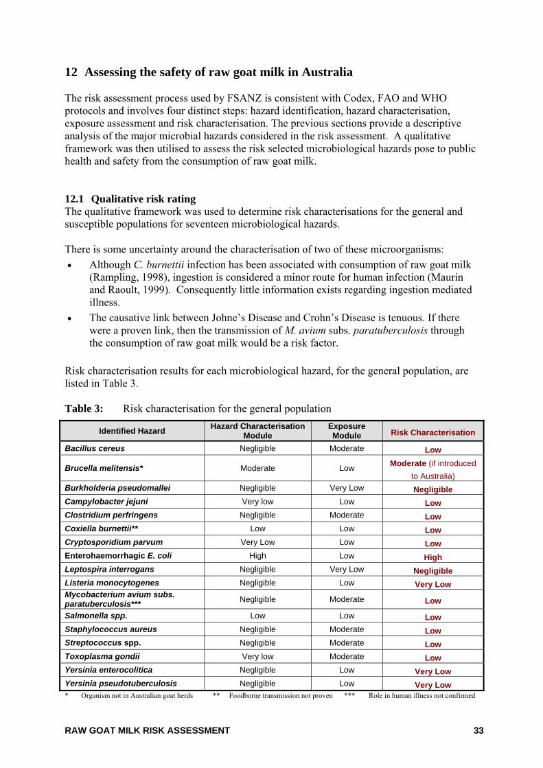
RAW GOAT MILK RISK ASSESSMENT 33
12 Assessing the safety of raw goat milk in Australia The risk assessment process used by FSANZ is consistent with Codex, FAO and WHO protocols and involves four distinct steps: hazard identification, hazard characterisation, exposure assessment and risk characterisation. The previous sections provide a descriptive analysis of the major microbial hazards considered in the risk assessment. A qualitative framework was then utilised to assess the risk selected microbiological hazards pose to public health and safety from the consumption of raw goat milk. 12.1 Qualitative risk rating The qualitative framework was used to determine risk characterisations for the general and susceptible populations for seventeen microbiological hazards. There is some uncertainty around the characterisation of two of these microorganisms: • Although C. burnettii infection has been associated with consumption of raw goat milk
(Rampling, 1998), ingestion is considered a minor route for human infection (Maurin and Raoult, 1999). Consequently little information exists regarding ingestion mediated illness.
• The causative link between Johne’s Disease and Crohn’s Disease is tenuous. If there were a proven link, then the transmission of M. avium subs. paratuberculosis through the consumption of raw goat milk would be a risk factor.
Risk characterisation results for each microbiological hazard, for the general population, are listed in Table 3. Table 3: Risk characterisation for the general population
Identified Hazard Hazard Characterisation Module
Exposure Module Risk Characterisation
Bacillus cereus Negligible Moderate Low
Brucella melitensis* Moderate Low Moderate (if introduced to Australia)
Burkholderia pseudomallei Negligible Very Low Negligible Campylobacter jejuni Very low Low Low Clostridium perfringens Negligible Moderate Low Coxiella burnettii** Low Low Low Cryptosporidium parvum Very Low Low Low Enterohaemorrhagic E. coli High Low High Leptospira interrogans Negligible Very Low Negligible Listeria monocytogenes Negligible Low Very Low Mycobacterium avium subs. paratuberculosis*** Negligible Moderate Low
Salmonella spp. Low Low Low Staphylococcus aureus Negligible Moderate Low Streptococcus spp. Negligible Moderate Low Toxoplasma gondii Very low Moderate Low Yersinia enterocolitica Negligible Low Very Low Yersinia pseudotuberculosis Negligible Low Very Low * Organism not in Australian goat herds ** Foodborne transmission not proven *** Role in human illness not confirmed
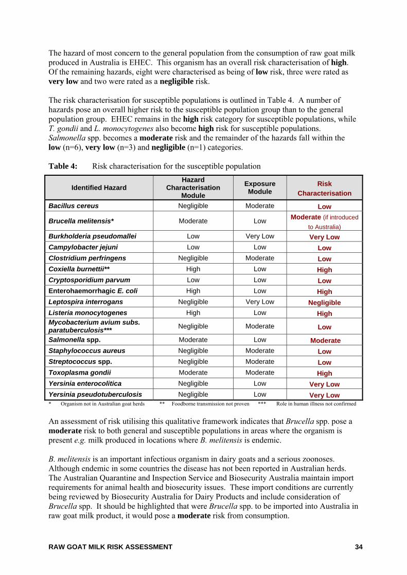
RAW GOAT MILK RISK ASSESSMENT 34
The hazard of most concern to the general population from the consumption of raw goat milk produced in Australia is EHEC. This organism has an overall risk characterisation of high. Of the remaining hazards, eight were characterised as being of low risk, three were rated as very low and two were rated as a negligible risk. The risk characterisation for susceptible populations is outlined in Table 4. A number of hazards pose an overall higher risk to the susceptible population group than to the general population group. EHEC remains in the high risk category for susceptible populations, while T. gondii and L. monocytogenes also become high risk for susceptible populations. Salmonella spp. becomes a moderate risk and the remainder of the hazards fall within the low (n=6), very low (n=3) and negligible (n=1) categories. Table 4: Risk characterisation for the susceptible population
Identified Hazard Hazard
Characterisation Module
Exposure Module
Risk Characterisation
Bacillus cereus Negligible Moderate Low
Brucella melitensis* Moderate Low Moderate (if introduced to Australia)
Burkholderia pseudomallei Low Very Low Very Low Campylobacter jejuni Low Low Low Clostridium perfringens Negligible Moderate Low Coxiella burnettii** High Low High Cryptosporidium parvum Low Low Low Enterohaemorrhagic E. coli High Low High Leptospira interrogans Negligible Very Low Negligible Listeria monocytogenes High Low High Mycobacterium avium subs. paratuberculosis*** Negligible Moderate Low
Salmonella spp. Moderate Low Moderate Staphylococcus aureus Negligible Moderate Low Streptococcus spp. Negligible Moderate Low Toxoplasma gondii Moderate Moderate High Yersinia enterocolitica Negligible Low Very Low Yersinia pseudotuberculosis Negligible Low Very Low * Organism not in Australian goat herds ** Foodborne transmission not proven *** Role in human illness not confirmed An assessment of risk utilising this qualitative framework indicates that Brucella spp. pose a moderate risk to both general and susceptible populations in areas where the organism is present e.g. milk produced in locations where B. melitensis is endemic. B. melitensis is an important infectious organism in dairy goats and a serious zoonoses. Although endemic in some countries the disease has not been reported in Australian herds. The Australian Quarantine and Inspection Service and Biosecurity Australia maintain import requirements for animal health and biosecurity issues. These import conditions are currently being reviewed by Biosecurity Australia for Dairy Products and include consideration of Brucella spp. It should be highlighted that were Brucella spp. to be imported into Australia in raw goat milk product, it would pose a moderate risk from consumption.
RAW GOAT MILK RISK ASSESSMENT 35
Coxiella spp. may pose a low risk to the general population and a high risk for susceptible populations if a definitive link was established for ingestion as a transmission route. M. avium subs. paratuberculosis would pose a low risk to both the general and susceptible population groups if there was a proven link between Johne’s disease and Crohn’s disease. 12.2 Comparison with previous risk assessments As outlined in Section 4 and detailed in Appendix 7, risk assessments on raw goat milk were undertaken in Queensland, NSW and South Australia. These assessments were largely state specific although the South Australian assessment expanded on the other two risk assessments, by evaluating existing controls including the efficacy of microbiological testing programs and provided risk management options. Some differences exist in organisms considered between each risk assessment, e.g. Burkholderia pseudomallei is an organism limited to tropical regions of Australia and was only considered in the Queensland and NSW risk assessments. The South Australian and Queensland risk assessments concluded that consumption of raw goat milk represents a medium to high risk for certain hazards for the general and susceptible populations. C. parvum, EHEC, L. monocytogenes, Salmonella spp. and T. gondii were identified in the South Australian risk assessment as posing a high risk for susceptible populations, whilst C. jejuni/coli, Salmonella spp. and EHEC were all rated as medium risk for the general population. The risk assessment for Queensland determined E. coli O157 and L. monocytogenes posed a medium risk to members of the general population. E. coli O157 and L. monocytogenes were a high risk and S. aureus toxins and B. pseudomallei were a medium risk to certain members of the susceptible population. These conclusions are consistent with the results of this assessment, although the risk characterisation for EHEC in the general population is higher in this assessment. The lower risk characterisation of B. pseudomallei in this assessment would be a result of limited information available on infective dose and product contamination levels which feed into the qualitative framework. The New South Wales risk assessment declined to make any determination, qualitative or quantitative, on the risks associated with microbial hazards, although it did predict the possible magnitude of foodborne illness outbreaks for Salmonella spp., S. aureus and L. monocytogenes. A direct comparison of the assessments is difficult as approaches varied from qualitative, through semi-quantitative to fully quantitative (Appendix 7). There is a large degree of uncertainty in all assessments due to assumptions made, particularly in relation to consumption of raw goat milk. The NSW and Queensland assessments quantitatively estimated consumption based on sales/production estimates and population data. This produced a very conservative but definitive outcome which differs to both this assessment and the South Australian assessments. South Australia and FSANZ characterised the risk of raw goat milk on a “per-serve” basis, whereas New South Wales and Queensland quantified risk as “individual risk” e.g. risk to the consumer. For all assessments, risk was characterised for both the general and susceptible populations. Some assessments divided susceptible populations into subgroups e.g. infants,
RAW GOAT MILK RISK ASSESSMENT 36
immunocompromised and pregnant. Regardless of the degree of grouping, the overall characterisation of risk is comparable between assessments. Consistent to all risk assessments was the lack of information available on raw goat milk within Australia. Particularly deficient is information pertaining to the incidence and level of pathogens in Australian goat milk, the amount of goat milk consumed, the identity of the consuming population and the incidence of illness associated with raw goat milk. More data in these areas would reduce the level of uncertainty and produce more accurate risk estimates. 12.3 Uncertainty and variability In characterising the risk associated with consuming raw goat milk in Australia, the level of confidence in the final estimate of risk depends on the adequacy and quality of the available data. Variability is associated with biological systems, food processing technologies, food preservation methods and human behaviour and is therefore inherent. Uncertainty relates to assumptions which had to be made due to a lack of information. Details of the assumptions used in the qualitative framework are contained in Appendix 6. There was a degree of uncertainty in each component of the qualitative framework due to limited data being available. Particularly in relation to: • The prevalence and levels of pathogens detected in Australian raw goat milk • The levels likely to cause illness in consumers (infective dose) e.g. B. pseudomallei • The severity of illness within certain population groups • Mode of transmission for some organisms e.g. Coxiella spp. • The effect of processing
Where data was not available, assumptions derived following expert consultations were used to determine inputs to populate the framework. Consultations were primarily sought for infective dose/dose response information, severity of illness in general and susceptible populations and contamination levels for the raw goat milk. The qualitative framework inputs are detailed in Appendix 6: Table 1. Similarly, justifications for assigning raw product contamination levels are outlined in Appendix 6: Table 4. The level of uncertainty in the model, due to the assumptions used gives an overall conservative estimate of risk. More data, particularly on the incidence and prevalence of pathogens in Australian raw goat milk would reduce this uncertainty and improve the level of confidence. The exposure assessment module characterises exposure to the hazard based on the likely level of the hazard in the initial raw product and the effect of processing on the hazard. Variability exists within the processes used for the production of raw goat milk. The type of milking system used e.g. hand milking or by various mechanised methods, may influence the level of bacterial contamination in the milk. The efficacy of different types of in-line cooling systems is also not considered in the model. For example, the open design of ripple coolers may not be as efficient at cooling milk as in-line plate heat exchangers and may also allow a greater likelihood of environmental contamination. No data was available on the efficacy of different milking or cooling processes used for Australian goat milk.
RAW GOAT MILK RISK ASSESSMENT 37
Uncertainty exists not only in the degree of contamination of the milk, but also within the effect of processing. Raw goat milk does not undergo any pathogen kill step, either at the packaging stage or the consumer end. For the purposes of the framework, it was assumed that there was no effect of processing. However, this assumes good agricultural, veterinary and processing practices and does not take into account any post-milking contamination, variability in contamination rates due to different milking systems or growth of pathogens which may occur due to poor hygiene practices and ineffective temperature control. Nor does it take into account the growth of any contaminating psychrotrophic pathogens. Quantitative data was also not available on cold chain integrity from packaging, through storage and transport processes. Failure to maintain appropriate temperature control may allow the growth of pathogens. Even if correct temperature control is maintained, the framework does not take into account the ability of some pathogens to grow at refrigeration temperatures regardless of how and when they enter the raw milk. The majority of assumptions used in the model introduce conservative estimates of risk to account for worst-case scenarios. The assumptions and variability pertaining to the effect of processing accounts for best-case scenarios and goes some way to offset the conservativeness of the earlier assumptions. More data for all components would assist to reduce the uncertainty and give greater confidence in the estimates of risk.
RAW GOAT MILK RISK ASSESSMENT 38
13 Discussion and summary The production of raw goat milk in Australia is very small. Only 300,000 litres of an estimated 5.4 million litres of goat milk produced in Australia annually is marketed as raw milk. However, the volume of raw goat milk entering the market from unlicensed sources is unknown. There is little information available on the consumption of goat milk and in particular raw goat milk. The 1995 National Nutrition Survey indicated 0.08% of the surveyed population consumed goat milk, although it is unknown what proportion of this, if any, was raw goat milk. A recent survey indicated less than 1% of the population consumes raw goat milk. Therefore, it can be assumed that consumption of raw goat milk is generally very low among the general population. Raw goat milk is sold primarily through health food shops or farm gate sales and it appears there is a niche group of consumers who consume raw goat milk as their milk of choice. This niche group of consumers generally has strong beliefs in the perceived health benefits of raw milk and widely promote it as having restorative powers, especially as a cow milk replacement for babies. It has further been suggested that the niche consumer group has a high proportion of people with a lowered or less developed immunity to infection. In Australia, illness arising from the consumption of raw goat milk appears to be rare with only two incidents of foodborne illness reported over the past 20 years. Internationally there have been 19 outbreaks of illness associated with the consumption of raw goat milk and 14 with raw goat milk cheese reported. The low level of reported foodborne illness associated with raw goat milk may give the impression this is a safe product, although it may simply reflect the generally low consumption in Australia and the overall underreporting of foodborne illness. The impression of safety must however, be balanced against the possibly high proportion of the consuming population who are within a susceptible population group. Typical production of raw goat milk in Australia is undertaken using systems and practices similar to the cow dairy industry. Similarly, contamination of raw milk during primary production and along the processing chain is primarily by two means: pathogens shed directly into the milk via the udder, or via external contamination of the milk during or post harvest. The key risk factors affecting the microbiological quality of raw goat milk are summarised in Table 2. The extent to which one risk factor is more important will be hazard specific, and could not be determined in this assessment due to a lack of quantitative through chain data. Raw goat milk does not undergo any pathogen elimination or reduction step. Whereas pasteurisation represents the principal processing intervention to render other dairy products safe for consumption, the safety of raw goat milk is primarily dependent upon the control of risk factors to minimize the opportunity for microbiological hazards to contaminate raw milk. If raw milk does become contaminated, failure to maintain appropriate temperature control throughout storage, distribution and consumer handling may allow the growth of pathogens and increase risk. However, it should also be noted that some pathogens, including L. monocytogenes and Y. enterocolitica are able to grow at refrigeration temperatures. Australian microbiological survey data show a very low incidence of hazards of public health significance in raw goat milk, although pathogenic bacteria including coagulase positive
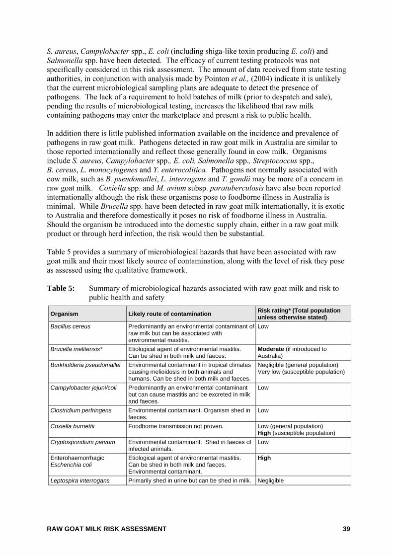
RAW GOAT MILK RISK ASSESSMENT 39
S. aureus, Campylobacter spp., E. coli (including shiga-like toxin producing E. coli) and Salmonella spp. have been detected. The efficacy of current testing protocols was not specifically considered in this risk assessment. The amount of data received from state testing authorities, in conjunction with analysis made by Pointon et al., (2004) indicate it is unlikely that the current microbiological sampling plans are adequate to detect the presence of pathogens. The lack of a requirement to hold batches of milk (prior to despatch and sale), pending the results of microbiological testing, increases the likelihood that raw milk containing pathogens may enter the marketplace and present a risk to public health. In addition there is little published information available on the incidence and prevalence of pathogens in raw goat milk. Pathogens detected in raw goat milk in Australia are similar to those reported internationally and reflect those generally found in cow milk. Organisms include S. aureus, Campylobacter spp., E. coli, Salmonella spp., Streptococcus spp., B. cereus, L. monocytogenes and Y. enterocolitica. Pathogens not normally associated with cow milk, such as B. pseudomallei, L. interrogans and T. gondii may be more of a concern in raw goat milk. Coxiella spp. and M. avium subsp. paratuberculosis have also been reported internationally although the risk these organisms pose to foodborne illness in Australia is minimal. While Brucella spp. have been detected in raw goat milk internationally, it is exotic to Australia and therefore domestically it poses no risk of foodborne illness in Australia. Should the organism be introduced into the domestic supply chain, either in a raw goat milk product or through herd infection, the risk would then be substantial. Table 5 provides a summary of microbiological hazards that have been associated with raw goat milk and their most likely source of contamination, along with the level of risk they pose as assessed using the qualitative framework. Table 5: Summary of microbiological hazards associated with raw goat milk and risk to
public health and safety
Organism Likely route of contamination Risk rating* (Total population unless otherwise stated)
Bacillus cereus Predominantly an environmental contaminant of raw milk but can be associated with environmental mastitis.
Low
Brucella melitensis* Etiological agent of environmental mastitis. Can be shed in both milk and faeces.
Moderate (if introduced to Australia)
Burkholderia pseudomallei Environmental contaminant in tropical climates causing melioidosis in both animals and humans. Can be shed in both milk and faeces.
Negligible (general population) Very low (susceptible population)
Campylobacter jejuni/coli Predominantly an environmental contaminant but can cause mastitis and be excreted in milk and faeces.
Low
Clostridium perfringens Environmental contaminant. Organism shed in faeces.
Low
Coxiella burnettii Foodborne transmission not proven. Low (general population) High (susceptible population)
Cryptosporidium parvum Environmental contaminant. Shed in faeces of infected animals.
Low
Enterohaemorrhagic Escherichia coli
Etiological agent of environmental mastitis. Can be shed in both milk and faeces. Environmental contaminant.
High
Leptospira interrogans Primarily shed in urine but can be shed in milk. Negligible
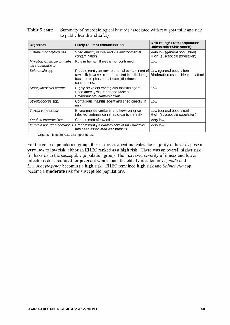
RAW GOAT MILK RISK ASSESSMENT 40
Table 5 cont: Summary of microbiological hazards associated with raw goat milk and risk to public health and safety
Organism Likely route of contamination Risk rating* (Total population unless otherwise stated)
Listeria monocytogenes Shed directly in milk and via environmental contamination.
Very low (general population) High (susceptible population)
Mycobacterium avium subs. paratuberculosis
Role in human illness is not confirmed. Low
Salmonella spp. Predominantly an environmental contaminant of raw milk however can be present in milk during bacteremic phase and before diarrhoea commences.
Low (general population) Moderate (susceptible population)
Staphylococcus aureus Highly prevalent contagious mastitis agent. Shed directly via udder and faeces. Environmental contamination.
Low
Streptococcus spp. Contagious mastitis agent and shed directly in milk.
Low
Toxoplasma gondii Environmental contaminant, however once infected, animals can shed organism in milk.
Low (general population) High (susceptible population)
Yersinia enterocolitica Contaminant of raw milk. Very low Yersinia pseudotuberculosis Predominantly a contaminant of milk however
has been associated with mastitis. Very low
* Organism is not in Australian goat herds. For the general population group, this risk assessment indicates the majority of hazards pose a very low to low risk, although EHEC ranked as a high risk. There was an overall higher risk for hazards to the susceptible population group. The increased severity of illness and lower infectious dose required for pregnant women and the elderly resulted in T. gondii and L. monocytogenes becoming a high risk. EHEC remained high risk and Salmonella spp. became a moderate risk for susceptible populations.
RAW GOAT MILK RISK ASSESSMENT 41
14 Data gaps and areas for further research During the preparation of the risk assessment, a number of data gaps were identified along the raw goat milk supply chain. Research into these data gaps may better assist in describing factors along the farm-to-table continuum that impact on the likelihood and magnitude of any illness resulting from consumption of raw goat milk in Australia. Significant data gaps exist in the following areas: • Data on the relative contribution of on-farm risk factors towards contamination of raw
goat milk o Numerous on-farm factors have been identified as potential contamination
sources of raw goat milk; however their relative impact, either singularly or in combination, has not been determined due to a lack of data. Examples of these factors include: animal health and welfare, housing, faeces, feed, water etc.
• Details on animal health and on-farm hygiene control measures o There is a lack of qualitative and/or quantitative data on the effect of on-farm
control measures on pathogens (either individually or collectively) such as: animal health and mastitis programs, herd management practices, animal welfare, feed preparation, storage and distribution, water quality etc.
• The relative contribution of processing risk factors upon contamination of raw goat milk
o The extent to which cross-contamination from the farm environment, the milking environment, personnel, storage and packaging etc impacts on the microbial status of raw goat milk.
• Processing control measures o Limited data exists on the effect of pre and post milking antisepsis, cleaning
and maintenance protocols etc. and the contribution these have on the contamination of raw goat milk.
• The efficacy of different milking or cooling processes used for Australian raw goat milk
• Cold-chain management through packaging, storage and transport processes • The prevalence of pathogens detected in Australian raw goat milk
o Minimal surveillance of raw goat milk in Australia, as few States allow its production and sale, and also as a result of the limited testing schedules. Consequently, for those pathogens monitored, there is a low prevalence recorded for raw goat milk in Australia, whilst no domestic data exists for other pathogens.
• The level of pathogens in raw goat milk o Very few studies have quantified the contamination level in raw goat milk
either domestically or internationally. Under the current testing protocol there is no requirement to quantify any positive detection of pathogens in raw goat milk. Levels of pathogens in raw goat milk will be affected by the source and quality of milk, method of milking, any cross-contamination and the time and temperature conditions during distribution and storage (potential for growth).
RAW GOAT MILK RISK ASSESSMENT 42
• The virulence and infectivity of some organisms as well as the zoonotic potential of certain organisms is not well documented
o To populate the qualitative framework with input variables for these pathogens, assumptions were made based on expert consultations. Data on the infective dose and severity of illness was either limited or not available for a number of organisms including; B. pseudomallei, L. interrogans, M. avium subs. paratuberculosis, Streptococcus spp., Y. pseudotuberculosis and T. gondii. Further information is required on the dose-response information for these organisms. Plus information on the milkborne transmission of Coxiella and any causal link association between Johne’s disease and Crohn’s disease was also not available.
• Limited data currently exists on the frequency and amount of raw goat milk consumed as well as the demographics of the consuming population
o Discussions with jurisdictional representatives and the industry suggests that raw goat milk is often consumed by individuals with lowered or less-developed immune responses, so further quantitative and/or qualitative data would assist in contextualising the impact of any illness resulting from consumption of raw goat milk.
• Extent and cause of sporadic human cases of raw goat milk associated foodborne illness
o Outbreak data is not necessarily indicative of the true incidence and causes of sporadic raw goat milk associated foodborne illness. Attribution of sporadic cases is difficult due to factors such as the general under-reporting of foodborne illness, retrospective nature of foodborne illness investigation, the often non-point source nature of exposure and the low frequency of consumption.
Availability of the following data would also assist in more accurately estimating the impact and magnitude of any illness resulting following the consumption of raw goat milk in Australia per year: • Levels of pathogens present in Australian raw goat milk both at the farm level and
along the supply chain, including at retail sale • Time and temperature conditions which raw goat milk is subject to throughout storage,
distribution and retail sale • The frequency and amount of consumption of raw goat milk, as well as the
demographics of the consuming population
RAW GOAT MILK RISK ASSESSMENT 43
15 Conclusion The assessment of risk associated with raw goat milk demonstrates that raw goat milk may be contaminated with a range of pathogenic microorganisms and pose a public health risk from consumption. The safety of raw goat milk is primarily dependent upon: ensuring good animal health and welfare; preventing environmental contamination; strict adherence to good milking practices, including cleaning and drying of teats; rapid cooling of milk; strict control over milking parlour hygiene; and correct temperature conditions maintained throughout storage and distribution. This minimizes the opportunity for microbiological hazards to contaminate raw milk and reduces the likelihood that these hazards will proliferate if milk is contaminated. A failure to maintain strict control over these risk factors would be likely to significantly increase the risk to public health and safety from consumption of raw goat milk. As raw goat milk does not undergo a pathogen elimination step, such as pasteurisation, the microbiological quality of raw goat milk will reflect the stringency of control exercised over these risk factors. The consumption of raw goat milk in Australia is low, and reflecting this is the low number of reported foodborne outbreaks associated with its consumption. However, raw goat milk has been responsible for a number of outbreaks of foodborne illness overseas. Using a qualitative framework, the principal risks to public health and safety from the consumption of raw goat milk are: Organism Risk rating using qualitative model Enterohaemorrhagic E. coli High (all populations)Salmonella spp. Moderate (susceptible population only) Toxoplasma gondii High (susceptible population only) Listeria monocytogenes High (susceptible population only)
The principal identified risk to public health and safety, for both general and susceptible populations from the consumption of raw goat milk is pathogenic E. coli. This estimation of risk is supported epidemiologically, as there have been five reported outbreaks of E. coli O157:H7 associated with the consumption of raw goat milk overseas. The risk of Salmonella spp. is estimated to be moderate for the susceptible population and there has been one outbreak in Australia attributed to the consumption of raw goat milk. For susceptible populations, T. gondii and L. monocytogenes are estimated to be high risk. Again, T. gondii has been epidemiologically linked with foodborne illness in a number of outbreaks overseas. The risk rating for L. monocytogenes is still considered conservative as the qualitative framework does not account for possible growth of the organism at refrigeration temperature. Although ranked as a low risk, C. parvum has been linked to one foodborne illness outbreak in Australia. It should also be highlighted that although Brucella spp. is presently exotic to Australia, were it to be introduced either in raw goat milk product or into domestic herds, it would pose a moderate risk following the consumption of raw goat milk. While the volume of raw goat milk consumed in Australia is very low, raw goat milk poses a risk to both general and susceptible population groups who consume this product. The information available suggests raw goat milk is often provided to very young children,

RAW GOAT MILK RISK ASSESSMENT 44
children with special dietary needs, older people and people convalescing. These sub-populations are at-risk, and exposure to even low levels of microbial pathogens could result in serious illness.

RAW GOAT MILK RISK ASSESSMENT 45
APPENDICES
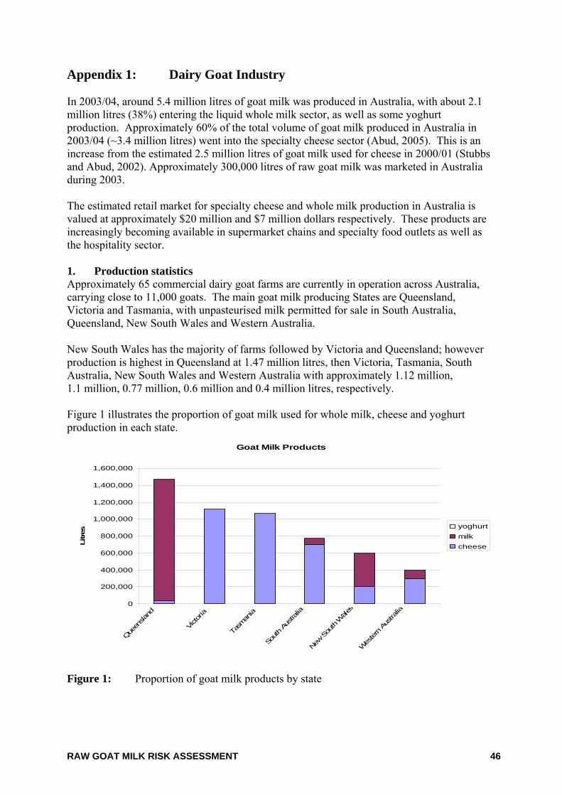
RAW GOAT MILK RISK ASSESSMENT 46
Appendix 1: Dairy Goat Industry In 2003/04, around 5.4 million litres of goat milk was produced in Australia, with about 2.1 million litres (38%) entering the liquid whole milk sector, as well as some yoghurt production. Approximately 60% of the total volume of goat milk produced in Australia in 2003/04 (~3.4 million litres) went into the specialty cheese sector (Abud, 2005). This is an increase from the estimated 2.5 million litres of goat milk used for cheese in 2000/01 (Stubbs and Abud, 2002). Approximately 300,000 litres of raw goat milk was marketed in Australia during 2003. The estimated retail market for specialty cheese and whole milk production in Australia is valued at approximately $20 million and $7 million dollars respectively. These products are increasingly becoming available in supermarket chains and specialty food outlets as well as the hospitality sector. 1. Production statistics Approximately 65 commercial dairy goat farms are currently in operation across Australia, carrying close to 11,000 goats. The main goat milk producing States are Queensland, Victoria and Tasmania, with unpasteurised milk permitted for sale in South Australia, Queensland, New South Wales and Western Australia. New South Wales has the majority of farms followed by Victoria and Queensland; however production is highest in Queensland at 1.47 million litres, then Victoria, Tasmania, South Australia, New South Wales and Western Australia with approximately 1.12 million, 1.1 million, 0.77 million, 0.6 million and 0.4 million litres, respectively. Figure 1 illustrates the proportion of goat milk used for whole milk, cheese and yoghurt production in each state.
Goat Milk Products
0
200,000
400,000
600,000
800,000
1,000,000
1,200,000
1,400,000
1,600,000
Queen
sland
Victo
ria
Tasm
ania
South Au
strali
a
New Sou
th W
ales
Wes
tern Aus
tralia
Litres
yoghurtmilkcheese
Figure 1: Proportion of goat milk products by state
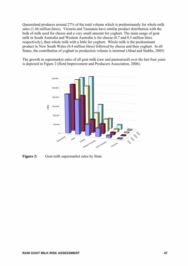
RAW GOAT MILK RISK ASSESSMENT 47
Queensland produces around 27% of the total volume which is predominantly for whole milk sales (1.44 million litres). Victoria and Tasmania have similar product distribution with the bulk of milk used for cheese and a very small amount for yoghurt. The main usage of goat milk in South Australia and Western Australia is for cheese (0.7 and 0.3 million litres respectively), then whole milk with a little for yoghurt. Whole milk is the predominant product in New South Wales (0.4 million litres) followed by cheese and then yoghurt. In all States, the contribution of yoghurt to production volume is minimal (Abud and Stubbs, 2005) The growth in supermarket sales of all goat milk (raw and pasteurised) over the last four years is depicted in Figure 2 (Herd Improvement and Producers Association, 2006).
QueenslandNSW
Victoria
Western Australia
South Australia
Tasmania01/02
02/03
03/04
04/050
100,000
200,000
300,000
400,000
500,000
600,000
Litr
es
Figure 2: Goat milk supermarket sales by State
RAW GOAT MILK RISK ASSESSMENT 48
Appendix 2: Hazard identification / hazard characterisation of pathogens 1. Bacillus cereus The genus Bacillus encompasses a great diversity of species and strains. Bacillus cereus are Gram-positive, facultatively aerobic spore-forming bacterium shaped as large rods and are motile by means of peritrichous flagella. B. cereus is widely distributed in the environment and is readily isolated from soil, dust, cereal crops, vegetation, animal hair, fresh water and sediments (ICMSF, 1996). Growth characteristics Strains of B. cereus vary widely in their growth and survival characteristics. Psychrotrophic strains are able to grow at 4 – 5ºC but not at 30 – 35ºC, whilst mesophilic strains grow between 15 - 55ºC. The optimum growth temperature ranges from 30 – 40ºC. The pH range at which growth will occur is 5.0 - 8.8 with an optimum of 6.0 - 7.0. The minimum water activity for survival and growth for B. cereus is 0.93 (ICMSF, 1996). The maximum salt concentration tolerated by B. cereus is 7% at pH 6 - 7 and 30 - 35ºC (Jenson and Moir, 2003). Growth is optimal in the presence of oxygen but can occur under anaerobic conditions. Toxin production is reduced under anaerobic conditions (ESR, 2001). Vegetative cells are relatively sensitive to environmental stress such as heat, chemicals, preservatives and radiation. However, B. cereus spores are more resistant due to their metabolic dormancy and tough physical nature (Jenson and Moir, 2003). Spores are more resistant to dry heat than moist heat. Spores can survive for long periods in dried foods. The heat resistance of B. cereus spores has been reported as D85ºC = 33.8 - 106 minutes in phosphate buffer; D95ºC = 1.5 - 36.2 minutes in distilled water and 1.8 - 19.1 minutes in milk. Thus, there is considerable strain variability, with D-values for spores of some B. cereus strains up to 15 to 20 times greater than for the more heat sensitive strains (ICMSF, 1996). Preservatives such as 0.26% sorbic acid at pH 5.5 and 0.39% potassium sorbate at pH 6.6 can inhibit growth. Nisin is inhibitory to B. cereus. Other antimicrobials which have an effect on B. cereus include benzoate, ethylenediaminetetraacetic acid and polyphosphates (Jenson and Moir, 2003). Spores are also more resistant to radiation than vegetative cells (Farkas, 1994). Pathology of illness There are two types of B. cereus-mediated intoxications. The two forms of illness are caused by significantly different toxins; diarrhoeal toxins (enterotoxins) and emetic toxins. Diarrhoeagenic enterotoxins are formed in the small intestine following consumption of a large number of cells, which then results in illness. These toxins are heat labile, being inactivated in 5 minutes at 56ºC (but not 45ºC for 30 minutes). Four enterotoxins have been identified and characterised: two three-component enterotoxins (haemolysin BL and non-haemolytic); enterotoxin T; and a cytotoxin. The toxins are unstable at pH values outside the range 4 to 11 and sensitive to proteolytic enzymes (Jenson and Moir, 2003). Toxin activity is reduced after 1 to 2 days at 32ºC, one week at 4ºC and several weeks at –20ºC (Andersson et al., 1995). The incubation period for the diarrhoeal type of food poisoning is usually 10 - 13 hours post ingestion, although incubation periods from 8 – 16 hours have been reported. Gastroenteritis is usually mild, with abdominal cramps, profuse watery diarrhoea,
RAW GOAT MILK RISK ASSESSMENT 49
rectal spasms and moderate nausea, usually without vomiting. Recovery typically occurs within 24 hours. The emetic toxin is preformed during B. cereus growth in foods, survives the gut environment and causes illness. It has been identified as a small ring form peptide of 1.2 kDa, called cereulide (Hui et al., 2001), and is thought to be an enzymatically synthesised peptide (Granum and Lund, 1997). The emetic toxin is extremely resistant to heat and can survive 90 minutes at 126ºC (ESR 2001). It is also very resistant to pH and proteolysis, but is not antigenic. Illness caused by the ingestion of emetic toxin generally has a short incubation period. Acute nausea and vomiting often occurs 1 - 5 hours post ingestion, with recovery within 12 - 24 hours. Diarrhoeal symptoms are not normally associated with the emetic illness. Neither form of illness is considered life-threatening to normal healthy individuals; with very few fatal cases have being reported (Jenson and Moir, 2003). Humans may vary in their susceptibility to B. cereus illness. Since most strains of B. cereus have the potential to produce toxins, the severity of illness is dependent on the quantity and type of toxins produced (Notermans and Batt, 1998). In a small number of cases, both types of symptoms (diarrhoeal and vomiting) have been recorded, and this is probably due to the production of both types of toxin. Characteristics of the two types of illness caused by B. cereus are summarised in Table 1. Table 1: Characteristics of the two types of illness caused by B. cereus (Granum and Lund
1997) Diarrhoeal syndrome Emetic syndrome Infective dose 105 - 107 (total) 105 - 108 (cells/g) Toxin produced In the small intestine of the host Preformed in foods Type of toxin Protein Cyclic peptide Incubation period 8 - 16 h (occasionally >24 h) 0.5 - 5 h Duration of illness
12 - 24 h (occasionally several days) 6 - 24 h
Symptoms Abdominal pain, watery diarrhoea and occasionally nausea
Nausea, vomiting and malaise (sometimes followed by diarrhoea, potentially due to additional enterotoxin production)
Foods most frequently implicated
Meat products, soups, vegetables, puddings/sauces and milk/milk products
Fried and cooked rice, pasta, pastry and noodles
B. cereus has also been associated with non-foodborne non-gastrointestinal infections such as ocular and wound infections, bacteraemia, central nervous system infections, respiratory tract infections and endocarditis. Individuals who are immunocompromised, either by illness or medication, are more susceptible to all infections and illnesses caused by this organism (Hui et al., 2001). Mode of transmission The enterotoxin (diarrhoeal syndrome) form of B. cereus poisonings is caused by the ingestion of a large number of cells and the subsequent production of the toxin in the small intestine. The emetic syndrome of B. cereus food poisoning occurs after the ingestion of food in which the organism has grown and formed toxin(s). Most documented reports of B. cereus
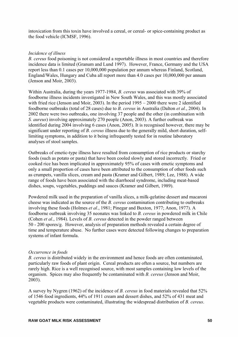
RAW GOAT MILK RISK ASSESSMENT 50
intoxication from this toxin have involved a cereal, or cereal- or spice-containing product as the food vehicle (ICMSF, 1996). Incidence of illness B. cereus food poisoning is not considered a reportable illness in most countries and therefore incidence data is limited (Granum and Lund 1997). However, France, Germany and the USA report less than 0.1 cases per 10,000,000 population per annum whereas Finland, Scotland, England/Wales, Hungary and Cuba all report more than 4.0 cases per 10,000,000 per annum (Jenson and Moir, 2003). Within Australia, during the years 1977-1984, B. cereus was associated with 39% of foodborne illness incidents investigated in New South Wales, and this was mostly associated with fried rice (Jenson and Moir, 2003). In the period 1995 – 2000 there were 2 identified foodborne outbreaks (total of 28 cases) due to B. cereus in Australia (Dalton et al., 2004). In 2002 there were two outbreaks, one involving 37 people and the other (in combination with S. aureus) involving approximately 270 people (Anon, 2003). A further outbreak was identified during 2004 involving 6 cases (Anon, 2005). It is recognised however, there may be significant under reporting of B. cereus illness due to the generally mild, short duration, self-limiting symptoms, in addition to it being infrequently tested for in routine laboratory analyses of stool samples. Outbreaks of emetic-type illness have resulted from consumption of rice products or starchy foods (such as potato or pasta) that have been cooled slowly and stored incorrectly. Fried or cooked rice has been implicated in approximately 95% of cases with emetic symptoms and only a small proportion of cases have been attributed to the consumption of other foods such as crumpets, vanilla slices, cream and pasta (Kramer and Gilbert, 1989; Lee, 1988). A wide range of foods have been associated with the diarrhoeal syndrome, including meat-based dishes, soups, vegetables, puddings and sauces (Kramer and Gilbert, 1989). Powdered milk used in the preparation of vanilla slices, a milk-gelatine dessert and macaroni cheese was indicated as the source of the B. cereus contamination contributing to outbreaks involving these foods (Holmes et al., 1981; Pinegar and Buxton, 1977; Anon, 1977). A foodborne outbreak involving 35 neonates was linked to B. cereus in powdered milk in Chile (Cohen et al., 1984). Levels of B. cereus detected in the powder ranged between 50 - 200 spores/g. However, analysis of preparation methods revealed a certain degree of time and temperature abuse. No further cases were detected following changes to preparation systems of infant formula. Occurrence in foods B. cereus is distributed widely in the environment and hence foods are often contaminated, particularly raw foods of plant origin. Cereal products are often a source, but numbers are rarely high. Rice is a well recognised source, with most samples containing low levels of the organism. Spices may also frequently be contaminated with B. cereus (Jenson and Moir, 2003). A survey by Nygren (1962) of the incidence of B. cereus in food materials revealed that 52% of 1546 food ingredients, 44% of 1911 cream and dessert dishes, and 52% of 431 meat and vegetable products were contaminated, illustrating the widespread distribution of B. cereus.
RAW GOAT MILK RISK ASSESSMENT 51
A study of milk and dairy products showed contamination rates of 9 - 48% and UHT-treated milk was contaminated in approximately 50% of samples (ICMSF, 1996). The available data indicates that under normal circumstances, B. cereus is found in food at concentrations <103 cfu/g and mostly <102 cfu/g (ICMSF, 1996). The presence of B. cereus in processed foods results from contamination of raw materials and the subsequent resistance of spores to heat treatment processes undergone during manufacture. Virulence and infectivity of B. cereus The pathogenic mechanism for the emetic toxin has been elucidated. The emetic toxin is a dodecadepsipeptide named cereulide, and causes vacuole formation in Hep-2 cells and emesis. The pathogenic mechanism for the diarrhoeal form of illness has not been clearly elucidated, although it is known that at least four different enterotoxins are involved (Jenson and Moir, 2003). One of these enterotoxins, Haemolysin BL, consists of three protein components (L2, L1, and B), and causes the destruction of red blood cells. The second enterotoxin, non-haemolytic enterotoxin, also consists of 3 protein moieties (B, L1 and L2) and all components are needed for maximum cytotoxicity. Both of these enterotoxins have been responsible for outbreaks of diarrhoeal food poisoning. The third enterotoxin, Enterotoxin T, consists of a single protein that is cytotoxin positive in the mouse ileal loop assay and possesses vascular permeability activity but does not appear to be involved in food poisoning (Hui et al., 2001). The role of enterotoxin T is unclear (Jenson and Moir, 2003). Lund et al.(2000) identified the fourth enterotoxin which is a single cytotoxin protein (CytK). CytK is necrotic and haemolytic. This toxin was implicated in a severe food poisoning outbreak in France resulting in three deaths (Lund et al., 2000). Since diarrhoeal enterotoxins are unstable and are inactivated by low pH and digestive enzymes, any preformed toxins should be destroyed during passage through the stomach and are not likely to cause illness (Notermans and Batt 1998; Granum and Lund 1997). Other potential virulence factors associated with diarrhoeal illness that have been identified include sphingomyelinase, phosphatidylinositol- and phosphatidylcholine-specific phospholipases and haemolysins I and II (Jenson and Moir, 2003). The involvement of intestinal receptor site(s) for the tripartite enterotoxins in diarrhoeal symptoms has not been fully elucidated. It has been postulated that the enterotoxins disrupt the membrane of epithelial cells (Notermans and Batt 1998). The mechanisms for cereulide synthesis are also unclear, but data suggest the peptide is enzymatically produced (Hui et al., 2001). Dose response Kramer and Gilbert (1989) have summarised a large number of outbreaks caused by B. cereus. The concentration of B. cereus in foods implicated in diarrhoeal illness ranged from 1.2 x 10³ – 108 cfu/g. It has also been reported that 10% of outbreaks have been associated with foods containing less than 105 cfu/g (Kramer and Gilbert, 1989). It has been found that concentrations of B. cereus of 103 - 105 cfu/g can result in illness in infants or aged and infirm individuals, although such illness was rare (Becker et al., 1994).
RAW GOAT MILK RISK ASSESSMENT 52
A study reported that concentrations ranging from 200 to 109cfu/g (or /ml) of B. cereus have been reported in foods implicated in food poisoning, giving total infective doses ranging from about 5 x 10⁴ to 1011organsims (Granum and Lund 1997). Partly due to the large differences in the amount and type of enterotoxin produced by different strains, the total infective dose seems to vary between about 105 and 108 viable cells or spores. Thus it was suggested that an average serving of food containing more than 103 B. cereus/g cannot be considered completely safe for consumption. It has also been suggested that the infectious dose for B. cereus may vary from about 1 x 10⁵ to 1 x 10⁸ viable cells or spores (Rowan et al., 1997; Notermans and Batt 1998). Notermans and Batt (1998) also suggest food servings containing greater than 1 x 10⁴ B. cereus/g may not be safe for consumption. From the available data it is estimated that the minimum total infectious dose is 105 viable cells or spores. Immune status All people are believed to be susceptible to B. cereus food poisoning. B. cereus has the potential to cause mild food-poisoning which does not, as a rule, last more than 12 - 24 hours. However, some individuals, especially young children, are particularly susceptible and may be more severely affected (ICMSF, 1996). Infants, therefore, may be susceptible to illness from a lower infectious dose, but there is no available data to support this. Food matrix The impact of the food matrix on the heat resistance of spores has been investigated. B. cereus spores are moderately heat resistant, however resistance is increased in high-fat and oily foods (e.g. for soybean oil, D121ºC = 30 minutes) and in foods with lower water activity (Jenson and Moir, 2003). References Andersson, A., Ronner, U. and Granum, P.E. (1995) What problems does the food industry have with the spore-forming pathogens Bacillus cereus and Clostridium perfringens? Int.J Food Microbiol. 28(2):145-155.
Anon. (1977) Communicable Disease Report. Public Health Lab.Service, London 77(44): 3.
Anon. (2003) OzFoodNet: Foodborne disease in Australia: incidence, notifications and outbreaks. Annual report of the OzFoodNet network, 2002. Commun Dis Intell. 27(2):209-243.
Anon. (2005) OzFoodNet: Foodborne disease in Australia: incidence, notifications and outbreaks. Annual report of the OzFoodNet network, 2004. CDI 29(2):164-190.
Becker, H., Schaller, G., Wiese, W.v. and Terplan, G. (1994) Bacillus cereus in infant foods and dried milk products. International Journal of Food Microbiology.1994 23(1):1-15.
Cohen, J., Marambio, E., Lynch, B. and Moreno, A. (1984) Bacillus cereus infection in newborn infants. Rev Chil.Pediatr 55(1):20-25.
Dalton, C.B., Gregory, J., Kirk, M.D., Stafford, R.J., Givney, R., Kraa, E. and Gould, D. (2004) Foodborne disease outbreaks in Australia, 1995 to 2000. Commun Dis Intell. 28(2):211-224.
ESR (2001) Microbial Pathogen Data Sheet - Bacillus cereus. http://www.nzfsa.govt.nz/science-technology/data-sheets/bacillus-cereus.pdf.
RAW GOAT MILK RISK ASSESSMENT 53
Farkas, J. (1994) Tolerance of spores to ionizing radiation: mechanisms of inactivation, injury and repair. J Appl Bacteriol. Symp Supp(76):815-905.
Granum, P.E. and Lund, T. (1997) Bacillus cereus and its food poisoning toxins. FEMS Microbiol Lett 157(2):223-228.
Holmes, J.R., Plunkett, T., Pate, P., Roper, W.L. and Alexander, W.J. (1981) Emetic food poisoning caused by Bacillus cereus. Arch.Intern.Med 141(6):766-767.
Hui, Y.H., Pierson, M.D. and Gorham, J.R. (2001) Food-borne disease handbook. Second edition ed, Marcel Dekker, New York, USA.
ICMSF. (1996) Microorganisms in Food 5: Microbiological Specifications of Food Pathgens. Blackie Academic and Professional, London.
Jenson, I. and Moir, C.J. (2003) Bacillus cereus and other Bacillus species. In: Foodborne Microorganisms of Public Health Significance. 6th ed, Australian Institute of Food Science and Technology Inc., New South Wales Branch Food Microbiology Group, Sydney, pp445-478.
Kramer, J.M. and Gilbert, R.J. (1989) Bacillus cereus and other Bacillus species. In: Doyle, M.P. eds. Foodborne bacterial pathogens. Marcel Dekker, New York, USA, pp21-70.
Lee, W.H. (1988) Lee, W.H. (1988) Bacillus cereus food poisoning associated with crumpets. Australian Food Microbiology Conference, Surfers Paradise, Queensland. Australian Institute Food Science and Technology, Abstracts.
Lund, T., De Buyser, M.L. and Granum, P.E. (2000) A new cytotoxin from Bacillus cereus that may cause necrotic enteritis. Mol.Microbiol 38(2):254-261.
Notermans, S. and Batt, C.A. (1998) A risk assessment approach for food-borne Bacillus cereus and its toxins. Symp.Ser.Soc Appl Microbiol 27:51S-61S.
Nygren, B. (1962) Phospholipase C-producing bacteria and food poisoning. An experimental study on Clostridium perfringens and Bacillus cereus. Acta Paediatr Scand Suppl(160):1-88.
Pinegar, J.A. and Buxton, J.D. (1977) An investigation of the bacteriological quality of retail vanilla slices. J Hyg.(Lond) 78(3):387-394.
Rowan, N.J., Anderson, J.G. and Anderton, A. (1997) Bacteriological quality of infant milk formulae examined under a variety of preparation and storage conditions. Journal of Food Protection.1997 60(9):1089-1094.
RAW GOAT MILK RISK ASSESSMENT 54
2. Brucella melitensis Brucella spp. are non-motile, short, Gram-negative coccoid to short rod-shaped cells which grow aerobically. They are catalase-negative and usually oxidase-negative (ICMSF, 1996). Brucella spp. are pathogenic for both humans and a wide range of animals and are often located intracellularly in infected animals (Tantillo et al., 2001). Until recently bovine brucellosis was present throughout the world with areas of high incidence including the Mediterranean , Middle East, South America and some areas of Asia (Corbel, 1997). However, a number of countries have now succeeded in eradicating this disease. Australia declared freedom from bovine brucellosis (Brucella abortus) in 1989 and there have been no recurrences of the disease since that time (Whittem, 1978; Mylrea, 1991; Cousins and Roberts, 2001). Brucella melitensis (along with Brucella abortus, Brucella suis and Brucella ovis) is a major cause of brucellosis in sheep and goats. The disease affects mainly adult female animals, causing abortion and udder infections. It is a serious zoonosis and is more pathogenic to human than other Brucella spp. B. melitensis infection has never been reported in sheep or goats in Australia (Animal Health Australia, 2005). However, overseas travellers occasionally arrive in Australia suffering from B. melitensis infection and since the organism is excreted in the urine of infected humans, infection of sheep and goats from this source is a possibly, although highly unlikely. Growth and survival characteristics Whilst the optimum temperature for growth on artificial media is 37ºC, Brucella spp. can grow at temperatures between 20 - 42ºC (ICMSF, 1996). There is some discrepancy regarding what time and temperatures are adequate to kill these bacteria. A time/temperature combination of 75 minutes at 85ºC was necessary to kill all 40 tested strains of B. abortus (Swann et al., 1981). Survival of Brucella spp. in milk and milk products declines with increasing storage temperatures. Brucellae at a concentration of 8 × 10⁹ cfu/ml survived for 800 days at –40ºC, but were eliminated within 2 days at 25ºC (Kuzdas and Morse, 1954). An increase in storage temperature from 2 - 4ºC to 18 - 22ºC reduced survival time by approximately 50% for Brucella spp. in Egyptian white cheese of the Domiati and Tallaga variety (Salem et al., 1977). In addition, the high fat content of products may have a protective effect. A sodium chloride content of 4% will prevent growth of B. melitensis on liver agar (Lerche et al., 1960). The survival rate of Brucella spp. appears to decrease with increased sodium chloride in milk products. A survival time of 6 months was reported for salted butter (2.3% NaCl), whereas in unsalted butter Brucella spp. remained viable for 13 months (ICMSF, 1996). However, Brucella spp. may resist high salt concentrations at lower temperatures. A survival time of 45 days was reported for Brucella spp. in a sheep cheese brine containing 27% salt and stored at a temperature of between 11 - 14ºC (ICMSF, 1996). There is a positive correlation between the survival of Brucella spp. in different cheeses and the water content of the cheeses. Brucella spp. survived six days in hard cheese (Emmentaler and Gruyer: water content of 35 - 36%), 15 days in Tilsit cheese (water content of 39 - 41%), 20 days in ‘quarterfat’ round cheese (water content 41 - 45%) and 57 days in soft cheeses (Munster and Camembert: water content 50%).
RAW GOAT MILK RISK ASSESSMENT 55
The optimum pH for growth in artificial media for all Brucella spp. is between 6.6 - 7.4 at 37ºC (Gerhardt, 1958; Corbel and Morgan, 1982). The upper growth limit is between pH 8.4 (Zobell and Meyer, 1932) and 8.7. Huddleson(1954) reported a lower growth limit of between 5.8 - 6.8, whilst Lerche and Entel(1959) reported a lower growth limit of between pH 4.1 - 4.5. Pathology of illness Brucellosis is a significant public health problem in endemic areas such as the Mediterranean region, western Asia, parts of Africa, the Indian subcontinent and Latin America (Kasimoglu, 2002). The signs and symptoms of foodborne illness associated with B. melitensis include fever, chills, sweating, weakness, headache, muscle and joint pain, diarrhoea and bloody stools during the acute phase (CDC, 2003; CDC, 2004). The incubation period ranges from 7 - 21 days, with the duration of illness being in the order of weeks. Treatment is usually with a combination of antibiotics such as tetracyclines, streptomycin and sulphonamides/trimethoprim (ICMSF, 1996). Mode of transmission Zoonotic transmission from infected animals to humans may be either via direct or indirect transmission (Kasimoglu, 2002). Direct transmission occurs via close contact with infected animals and involves the respiratory, conjunctival and cutaneous routes. Airborne transmission of Brucella spp. is often associated with occupational exposure to infected animals. Indirect transmission to humans is generally foodborne, and is often associated with consumption of raw milk and raw milk products (Lin et al., 2002). Incidence of illness There have been several outbreaks of brucellosis in humans in various parts of the world, these were often caused by the consumption of contaminated raw milk or raw milk products (Eckman, 1975; Young and Suvannoparrat, 1975; Acedo et al., 1997; Wallach et al., 1997; Altekruse et al., 1998; Mendez et al., 2003; Arimi et al., 2005). During the years 2000 - 2004 there were 139 cases of human brucellosis notified in Australia. The primary source of these infections, in Australia, is thought to be due to overseas infections (often from the consumption of unpasteurised dairy products) or may be related to the feral pig population in northern Queensland (B. suis) (Lin et al., 2002; Blumer et al., 2003; Yohannes et al., 2004; Miller et al., 2005; Yohannes et al., 2006). Occurrence in foods Brucella spp. are most commonly transmitted via raw milk or raw milk products, such as cheeses (Kasimoglu, 2002). Ewes and goats milk has been found to be a more significant source of Brucella spp. than cow’s milk. Raw milk, goat cheese made from unpasteurised milk, and contaminated meats are the foods most commonly associated with foodborne transmission.
RAW GOAT MILK RISK ASSESSMENT 56
Virulence and infectivity Brucella spp. can infect and multiply in both phagocytic and non-phagocytic cells (Sarinas and Chitkara, 2003). The exact mechanism of Brucella spp. pathogenesis is not fully understood, as no cell components specifically promoting cell adhesion and invasion have been characterised (Corbel and Morgan, 1982). Dose response There is no quantitative data on the infective dose (ICMSF, 1996). Precise information is lacking on the minimal effective oral dose of Brucella spp., but it is estimated that inhalation of 10 - 100 bacteria is sufficient to cause disease in humans (Kasimoglu, 2002). Host factors Individuals who are generally at greater risk of infection and/or risk of developing more severe outcomes from exposure to Brucella spp. include the very young, the elderly, pregnant women and the immunocompromised (organ transplant patients, cancer patients, AIDS patients). Food matrix Brucella spp. are unlikely to multiply in food kept under hygienic conditions, and are controlled most effectively by eliminating infected subjects from the animal stock. Pasteurisation or sterilisation of milk pre-market is sufficient to prevent milk-borne brucellosis (ICMSF, 1996). The combined effect of reduced water activity and pH has been found to reduce and/or eliminate Brucella spp. during the production of hard cheeses; however, Brucella spp. may survive conditions during the production of other types of cheeses such as soft cheeses. References Acedo, E., Diaz, M.E. and Leon, A.B. (1997) Incidence of Brucella sp. in raw milk and fresh regional cheese. Alimentaria.1997 (281):57-60. Altekruse, S.F., Timbo, B.B., Mowbray, J.C., Bean, N.H. and Potter, M.E. (1998) Cheese-associated outbreaks of human illness in the United States, 1973 to 1992: sanitary manufacturing practices protect consumers. J Food Prot. 61(10):1405-1407. Animal Health Australia (2005) National Animal Health Information System (NAHIS) - Bovine brucellosis. pp1-4. http://www.aahc.com.au/nahis/disease/BRA.htm. Accessed on 15 January 2006. Arimi, S.M., Koroti, E., Kang'ethe, E.K., Omore, A.O. and McDermott, J.J. (2005) Risk of infection with Brucella abortus and Escherichia coli O157:H7 associated with marketing of unpasteurized milk in Kenya. Acta Trop. 96(1):1-8. Blumer, C., Roche, P., Spencer, J., Lin, M., Milton, A., Bunn, C., Gidding, H., Kaldor, J., Kirk, M., Hall, R., Della-Porta, T., Leader, R. and Wright, P. (2003) Australia's notifiable diseases status, 2001: Annual report of the National Notifiable Diseases Surveillance System. Commun Dis Intell. 27(1):1-78. CDC (2003) U.S. Foodborne Disease Outbreaks. http://www.cdc.gov/foodborneoutbreaks/us_outb.htm. Accessed on 16 March 2005.
RAW GOAT MILK RISK ASSESSMENT 57
CDC (2004) Diagnosis and Management of Foodborne Illness - A Primer for Physicians and other Health Care Professionals. (April 16, 2004/53(RR04)). Department of Health and Human Services, pp1-33. http://www.cdc.gov/mmwr/preview/mmwrhtml/rr5304a1.htm. Accessed on 16 February 2005. Corbel, M.J. and Morgan, W.J.B. (1982) Classification of Brucella. In: Blobel, H. and Schliesser, T. eds. Handbuch der baketeriellen Infektionnend bei Tieren. Chapter 4. Stuggart: Verlag Gustav Fischer, pp17-52. Corbel, M.J. (1997) Brucellosis: an overview. Emerg.Infect Dis 3(2):213-221. Cousins, D.V. and Roberts, J.L. (2001) Australia's campaign to eradicate bovine tuberculosis: the battle for freedom and beyond. Tuberculosis.(Edinb.) 81(1-2):5-15. Eckman, M.R. (1975) Brucellosis linked to Mexican cheese. JAMA 232(6):636-637. Gerhardt, P. (1958) The nutrition of Brucella. Bacteriological Reviews. Bacteriological Reviews 22:81-98. Huddleson, I.F. (1954) A study of factors that influence the isolation and growth of Brucella in or on culture mediums. Proceedings of the 58th Annual Meeting of the US Livestock Sanitation Association. Proceedings of the 58th Annual Meeting of the US Livestock Sanitation Association, pp123-134. ICMSF. (1996) Microorganisms in Food 5: Microbiological Specifications of Food Pathgens. Blackie Academic and Professional, London. Kasimoglu, A. (2002) Determination of Brucella spp. in raw milk and Turkish white cheese in Kinkkale. Dtsch Tierarztl Wochenschr. 109(7):324-326. Kuzdas, C.D. and Morse, E.V. (1954) The survival of Brucella abortus, U.S.D.A. strain 2308, under controlled conditions in nature. Cornell Vet. 44(2):216-228. Lerche, M. and Entel, H.J. (1959) Uber die Saureresistenz von Brucella-Bakterien und ihr Vehalten während der Fleischreifung. Archiv für Lebensmittelhygiene 10:194-198. Lerche, M., Enterl, H.J. and Mossdorft, R. (1960) Die Widerstandskraft von Brucella-Bakterien gegenüber Kochsalz und Nitritpökelsalz sowie ihr Verhalten Bie der Pökelung infizierten Fleisches. Archiv fur Lebensmittelhygiene 11:271-276. Lin, M., Roche, P., Spencer, J., Milton, A., Wright, P., Witteveen, D., Leader, R., Merianos, A., Bunn, C., Gidding, H., Kaldor, J., Kirk, M., Hall, R. and Della-Porta, T. (2002) Australia's notifiable diseases status, 2000. Annual report of the National Notifiable Diseases Surveillance System. Commun Dis Intell. 26(2):118-203. Mendez, M.C., Paez, J.A., Cortes-Blanco, M., Salmoral, C.E., Mohedano, M.E., Plata, C., Varo, B.A. and Martiinez, N.F. (2003) Brucellosis outbreak due to unpasteurized raw goat cheese in Andalucia (Spain), January - March 2002. Euro.Surveill 8(7):164-168. Miller, M., Roche, P., Yohannes, K., Spencer, J., Bartlett, M., Brotherton, J., Hutchinson, J., Kirk, M., McDonald, A. and Vadjic, C. (2005) Australia's notifiable diseases status, 2003 annual report of the National Notifiable Diseases Surveillance System. Commun.Dis.Intell. 29(1):1-61. Mylrea, P.J. (1991) The origins of bovine brucellosis in New South Wales and its eradication. Aust.Vet.J. 68(6):189-192. Salem, A.A., Nour, M.A., Shawkat, M.E. and Fayed, A.A. (1977) Viability of Brucella abortus in white cheese. Veterinary Medical Journal Cairo(25):215-220. Sarinas, P.S. and Chitkara, R.K. (2003) Brucellosis. Semin.Respir.Infect 18(3):168-182. Swann, A.I., Garby, C.L., Schnurrenberger, P.R. and Brown, R.R. (1981) Safety aspects in preparing suspensions of field strains of Brucella abortus for serological identification. Vet.Rec. 109(12):254-255.
RAW GOAT MILK RISK ASSESSMENT 58
Tantillo, G., Pinto, A.d., Vergara, A. and Buonavoglia, C. (2001) Polymerase chain reaction for the direct detection of Brucella spp. in milk and cheese. Journal of Food Protection.2001 64(2):164-167. Wallach, J.C., Samartino, L.E., Efron, A. and Baldi, P.C. (1997) Human infection by Brucella melitensis: an outbreak attributed to contact with infected goats. FEMS Immunol.Med.Microbiol. 19(4):315-321. Whittem, J.H. (1978) Bovine brucellosis eradication Australia. Proc.Annu.Meet.U.S.Anim Health Assoc. (82):139-141. Yohannes, K., Roche, P., Blumer, C., Spencer, J., Milton, A., Bunn, C., Gidding, H., Kirk, M. and Della-Porta, T. (2004) Australia's notifiable diseases status, 2002: Annual report of the National Notifiable Diseases Surveillance System. Commun Dis Intell. 28(1):6-68. Yohannes, K., Roche, P.W., Roberts, A., Liu, C., Firestone, S.M., Bartlett, M., East, I., Hull, B.P., Kirk, M.D., Lawrence, G.L., McDonald, A., McIntyre, P.B., Menzies, R.I., Quinn, H.E. and Vadjic, C. (2006) Australia's notifiable diseases status, 2004, annual report of the National Notifiable Diseases Surveillance System. Commun.Dis.Intell. 30(1):1-79. Young, E.J. and Suvannoparrat, U. (1975) Brucellosis outbreak attributed to ingestion of unpasteurised goat cheese. Arch.Intern.Med. 135(2):240-243. Zobell, C.E. and Meyer, K.F. (1932) Metabolism studies on the Brucella group. IX. Physiochemical requirements in synthetic mediums. J Infect Dis 51:361-381.
RAW GOAT MILK RISK ASSESSMENT 59
3. Burkholderia pseudomallei Burkholderia pseudomallei (previously named Pseudomonas pseudomallei) are Gram-negative bacteria often present in soil and surface water of tropical regions. This organism is the aetiologic agent of melioidosis (also called Whitmore's disease); an animal and human disease predominantly found in tropical climates. It has been identified as being endemic in northern Australia, the South Pacific and South East Asia. It is however generally only found within 20° either side of the equator (Munckhof et al., 2001; Howe et al., 1971). Infection occurs in a wide range of animal species, with sheep and goats being particularly susceptible (Choy et al., 2000). Growth characteristics B. pseudomallei is an environmental saprophyte which is a oxidase-positive, bacillus. It is a strict aerobe, motile, and can break down arginine. It is easily cultured on blood or nutrient agar incubated at a temperature of 37 - 42°C and shows corrugated, wrinkled, and dry looking colonies in around 1 - 2 days. It may also develop an orange pigmentation on prolonged incubation. B. pseudomallei are resistant to aminoglycosides and polymyxins (Thomas et al., 1988a). B. pseudomallei is unable to grow at temperatures below 21°C and requires high moisture levels, however it has been known to survive in soil and water samples for up to 36 months (Dance, 2000). Pathology of illness B. pseudomallei have the ability to cause disease, melioidosis, in humans as well as in a wide range of animal species. Melioidosis is generally a disease only seen in tropical and sub-tropical regions, predominately during the wet season. The most commonly affected animals in Australia include goats, sheep, pigs, alpacas, camels, horse and deer, with goats and sheep being particularly susceptible with cases of infection often eventuating in the death of the animal (Choy et al., 2000). Wildlife can also be affected with species such as birds, crocodiles and kangaroos having presented with signs of melioidosis disease previously (Choy et al., 2000). Infections can manifest in a wide variety of clinical symptoms and may involve a latency period. Depending on the mode of transmission melioidosis may manifest as mastitis (especially in goats), pulmonary infection or lung lesions, septicaemia, chronic suppurative infection or localised skin infections. These manifestations may range from acute to chronic and may be also associated with sub-clinical infections in both animals and humans (Choy et al., 2000). Mode of transmission Infection may occur through inhalation or ingestion of the organism, or through contamination of penetrating injuries or skin wounds with dust particles or water containing viable B. pseudomallei organisms (Choy et al., 2000). It is assumed that the majority of infections occur through direct contact of skin wounds and abrasions to contaminated soil and/or water (Dance 2000). Zoonotic transmission is also believed to be a possible means of transmitting B. pseudomallei, with evidence that viable
RAW GOAT MILK RISK ASSESSMENT 60
organisms are found in infected individual’s (animal and human) pus, sputum, urine and faeces, possibly leading to contamination of the individual’s surroundings. It has been suggested that there is a possible public health risk from drinking contaminated milk from an animal infected with the disease melioidosis (Thomas et al., 1988a; Thomas et al., 1988b; Choy et al., 2000). Rarely has person to person transmission (Dance 2000) or transmission in human breast milk been shown to occur (Ralph et al., 2004). Incidence of illness The average annual human incidence of melioidosis in Australia is estimated at 5.8 cases per 100,000 population per annum depending on the rainfall during the year (Cheng et al., 2003), however in the Northern Territory the incidence is estimated at around 16.5 cases per 100,000 population per annum, with one particularly prolonged rainy season in 1997-1998 seeing 34.5 cases per 100,000 (Currie et al., 2000). These cases are thought to be caused by skin contact with contaminated soil and/or water with the risk of infection greatly increasing with rainfall. Melioidosis is a notifiable disease in the Northern Territory. Two outbreaks in Northern Australia have been attributed to ingestion of contaminated drinking water sourced from the community water supply (Inglis et al., 1999; Cheng et al., 2003). Only three possible zoonotic cases (direct transmission from animal to human) of melioidosis have been thought to have occurred in Australia, however these manifested as skin infections and were not due to ingestion of the organism (Choy et al., 2000). Occurrence in foods B. pseudomallei can be an environmental contaminant and thus may be present on the surface of water-contaminated fruit and vegetables. B. pseudomallei has also been isolated from infected goat’s udders (Van der Lugt and Henton, 1995) and is excreted in goat faeces (Dance 2000); which suggests that contamination of raw goat’s milk is a possibility. However no information is available on the occurrence of Burkholderia pseudomallei in raw goat’s milk or in any other food source. Virulence and infectivity Melioidosis is known to be a major cause of human morbidity and mortality in the Australian tropics (Dance 2000). There were 12 human deaths out of 33 infections during one outbreak in the Northern Territory during 1990 and 1991. It is however unknown how these infections were acquired and are not thought to be via ingestion (AgriQ, 2000). There is a complete lack of data on virulence and infectivity for B. pseudomallei obtained via ingestion. Dose response There is no information available on the dose-response relationship for B. pseudomallei in human or animal infections.
RAW GOAT MILK RISK ASSESSMENT 61
Host factors Risk factors for human melioidosis include diabetes, high alcohol intake and renal disease (Inglis et al., 1999; Cheng et al., 2003). References AgriQ. (2000) Hazard Analysis of Sheep and Goat Milk Products in New South Wales for SafeFood (Production) NSW. (Unpublished Work).
Cheng, A.C., Hanna, J.N., Norton, R., Hills, S.L., Davis, J., Krause, V.L., Dowse, G., Inglis, T.J. and Currie, B.J. (2003) Melioidosis in northern Australia, 2001-02. Commun Dis Intell 27(2):272-277.
Choy, J.L., Mayo, M., Janmaat, A. and Currie, B.J. (2000) Animal melioidosis in Australia. Acta Trop 74(2-3):153-158.
Currie, B.J., Fisher, D.A., Howard, D.M., Burrow, J.N., Selvanayagam, S., Snelling, P.L., Anstey, N.M. and Mayo, M.J. (2000) The epidemiology of melioidosis in Australia and Papua New Guinea. Acta Trop 74(2-3):121-127.
Dance, D.A.B. (2000) Ecology of Burkholderia pseudomallei and the interactions between environmental Burkholderia spp. and human-animal hosts. Acta Tropica 74(2-3):159-168.
Howe, C., Sampath, A. and Spotnitz, M. (1971) The pseudomallei group: a review. J Infect Dis 124(6):598-606.
Inglis, T.J., Garrow, S.C., Adams, C., Henderson, M., Mayo, M. and Currie, B.J. (1999) Acute melioidosis outbreak in Western Australia. Epidemiol Infect 123(3):437-443.
Munckhof, W.J., Mayo, M.J., Scott, I. and Currie, B.J. (2001) Fatal human melioidosis acquired in a subtropical Australian city. Am J Trop Med Hyg 65(4):325-328.
Ralph, A., McBride, J. and Currie, B.J. (2004) Transmission of Burkholderia pseudomallei via breast milk in northern Australia. Pediatr Infect Dis J 23(12):1169-1171.
Thomas, A.D., Forbes-Faulkner, J.C., Norton, J.H. and Trueman, K.F. (1988a) Clinical and pathological observations on goats experimentally infected with Pseudomonas pseudomallei. Aust Vet J 65(2):43-46.
Thomas, A.D., Spinks, G.A., D'Arcy, T.L., Norton, J.H. and Trueman, K.F. (1988b) Evaluation of four serological tests for the diagnosis of caprine melioidosis. Aust Vet J 65(9):261-264.
Van der Lugt, J.J. and Henton, M.M. (1995) Melioidosis in a goat. J S Afr.Vet Assoc 66(2):71-73.
RAW GOAT MILK RISK ASSESSMENT 62
4. Campylobacter spp. Campylobacter spp. are Gram-negative non-spore forming bacteria. Their cells are 0.2 - 0.8 μm wide and 0.5 - 5 μm long. They are mostly slender, spiral, curved rods, with a single polar flagellum at one or both ends of the cell. They are typically motile with a characteristic rapid darting corkscrew-like mobility (Smibert, 1984; Vandamme, 2000). Campylobacter spp. are classified under Campylobacteraceae, a bacterial family comprised of genera Campylobacter, Arcobacter and Sulfurospirillum. Among the 16 species and six subspecies of Campylobacter, two are most commonly isolated from stool samples of human gastroenteritis (Vandamme, 2000). They are Campylobacter jejuni subspecies jejuni and Campylobacter coli. C. jejuni accounts for approximately 95% of Campylobacter spp. caused human gastroenteritis, and C. coli are responsible for approximately 3 - 4% of the human illness. Campylobacter spp. are often a normal part of the intestinal flora of young cattle, sheep, goats, dogs, rabbits, monkeys, cats, chickens, turkeys, ducks, seagulls, pigeons, blackbirds, starlings and sparrows pigs (Smibert, 1984; Nielsen et al., 1997), and in blood and faecal material from humans with Campylobacter enteritis. They have also been found in the reproductive organs and oral cavity of humans and animals. Healthy puppies and kittens, rodents, beetles and houseflies have also been shown to carry Campylobacter spp. (Hartnett et al., 2002). Growth characteristics Campylobacter spp. require microaerophilic conditions for growth and have varying degrees of oxygen tolerance (3 - 5%) between species (Forsythe, 2000). Optimal growth occurs under conditions of 5% oxygen and 2 - 10% carbon dioxide (Park, 2002). Most strains do not grow in the presence of air, other than a few that may grow slightly under aerobic conditions. Some species can grow under anaerobic conditions with fumarate, formate and fumarate, or fumarate and hydrogen in the medium (Smibert, 1984; Vandamme, 2000). Campylobacter spp. grow optimally at 42 - 43oC. C. jejuni can grow in the temperature range of 30 - 45oC, pH of 4.9 - 9.5 and water activity above 0.99. At 32oC, C. jejuni may double its biomass in approximately 6 hours (Forsythe, 2000). Campylobacter spp. do not multiply at temperatures below 30oC, which means that the numbers of Campylobacter in foods will not increase at normal room temperatures (20 – 25oC). Although unable to grow below 30oC, Campylobacter remain metabolically active, are able to generate ATP, and are motile at temperatures as low as 4oC (Park 2002). Although Campylobacter spp. are considered thermotolerant, they are sensitive to heat and are readily inactivated by pasteurisation treatment or domestic cooking processes. Cooking at 55 - 60oC for several minutes readily destroys Campylobacter spp. The D value for C. jejuni at 50oC is 0.88 - 1.63 minutes (Forsythe, 2000). Campylobacter spp. are also sensitive to freezing and/or freeze thawing (Chan et al., 2001). Other than temperature, a range of other environmental factors including desiccation, oxidation and osmotic stress influences the survival of Campylobacter spp. Campylobacter spp. are highly sensitive to desiccation and do not survive well on dry surfaces (Fernandez, 1985).
RAW GOAT MILK RISK ASSESSMENT 63
The microaerophilic nature of Campylobacter spp. means that these organisms are inherently sensitive to oxygen and its reduction substances (Park 2002). Campylobacter spp. are much less tolerant to osmotic stress than a number of other foodborne pathogenic bacteria. For example, they are not capable of multiplication in an environment where sodium chloride concentration is 2% or higher (Doyle and Roman, 1982) Due to its sensitivity to environmental conditions and inability of growth at temperatures below 30oC or under aerobic conditions, the ability of Campylobacter spp. to multiply outside of an animal host is severely restricted. Although not capable of multiplication in food during processing or storage, Campylobacter spp. may have the ability to survive outside their optimal growth conditions (Park 2002). Pathology of illness C. jejuni causes fever and enteritis in human, resulting in acute inflammatory diarrhoea with clinical signs similar to those of other acute bacterial infections of the intestinal tract, such as salmonellosis. Principal symptoms are diarrhoea, nausea, abdominal pain, fever, myalgia, headache, vomiting and blood in faeces (Lastovica and Skirrow, 2000). The onset of symptoms is often abrupt with cramping abdominal pains quickly followed by diarrhoea. The mean incubation period is approximately 3 days with a range of 18 hours to 8 days. A particular feature of infection is abdominal pain, which may become continuous and sufficiently intense to mimic acute appendicitis. This is the most frequent reason for admission of Campylobacter enteritis patients to hospital (Skirrow and Blaser, 2000). Although incidents are rare, Campylobacter spp. have been implicated in causing a range of extra-intestinal infections including appendicitis, haemolytic ureamic syndrome, abortion, hepatitis, cholecystitis, pancreatitis, nephritis and others (Skirrow and Blaser, 2000). C. jejuni may cause septicaemia, meningitis and serious neurological disorders such as Guillain-Barré syndrome, an acute neuromuscular paralysis, and reactive arthritis such as Reiter syndrome (Lastovica and Skirrow, 2000). Mode of transmission Friedmann et al. (2000) examined data from 111 food and waterborne outbreaks of campylobacteriosis reported in the US between 1978 - 1996. Other than unknown foods, milk and water were the most common food vehicles associated with transmission of Campylobacter spp. Raw (unpasteurised) milk is largely responsible for dairy-related transmission. Of four milk-borne outbreaks in the period of 1990 - 1992, three were linked to raw cow’s milk and raw goat’s milk (CDC, 2003). Surveys in other developed countries, including the United Kingdom, Sweden, Germany, New Zealand, Denmark, US and Norway, indicate milk is the most frequent cause of foodborne Campylobacter spp. infection (Friedman et al., 2000). Outbreak data of foodborne campylobacteriosis recorded in Australia between 1992 - 2001 present a similar picture to the above, where approximately 42% of recorded outbreaks were the result of consumption of milk, and among this, raw milk accounted for approximately 80% of milk-borne Campylobacter spp. outbreaks.
RAW GOAT MILK RISK ASSESSMENT 64
Published information by Eberhart-Phillips et al. (1997), Friedman et al. (2000), WHO (2000) and Vellinga and Loock, (2002) suggests that major routes of Campylobacter spp. transmission to humans are: • Consumption of food contaminated with Campylobacter spp., including consumption
of raw and unpasteurised milk and milk products, consumption of undercooked meat such as poultry meat, and consumption of raw seafood
• Consumption of water contaminated with Campylobacter spp. • Bathing or swimming in a Campylobacter spp. contaminated lake or pool • Direct contact with infected farm animals, such as cattle, sheep, chicken, etc • Contact with infected domestic animals, such as pet dogs, cattle and bird
Incidence of illness C. jejuni is one of the most commonly reported aetiological agents of foodborne illness in developed countries, including Australia, NZ, UK and US (Mead et al., 1999; Park 2002). In the US, approximately 80% of all the cases of human campylobacteriosis are foodborne (Mead et al., 1999). In the period of 1998 – 2004, the notification rate of campylobacteriosis in Australia has been 100 – 120 cases per 100,000 population per annum. Notification rates were highest in the 0 – 4 year age group (Anon 2005). Occurrence in foods Foods potentially contaminated with Campylobacter spp. include raw and unpasteurised milk and milk products, raw poultry, raw beef, raw pork and raw shellfish, as well as foods that may have been exposed to water contaminated with Campylobacter spp. (Institute of Food Technologists, 2002). Virulence and infectivity of campylobacter Although not fully understood, Campylobacter spp. virulence is thought to involve production of microbial toxins. An enterotoxin Wassenaar (1997) abbreviated as CJT for C. jejuni toxin, is immunologically similar to the Vibrio cholerae toxin and the E. coli heat-liable toxin. At least six cytotoxins have been observed in Campylobacter spp., these being a 70-kDa cytotoxin, a Vero/HeLa cell cytotoxin, a cytolethal distending toxin (CDT), a shiga-like toxin, a haemolytic cytotoxin and a hepatotoxin. The CDT toxin has been shown to cause dramatic distension of human tumour epithelial cells, which leads to cell disintegration (Pickett et al., 1996). Active CDT toxin has been found in roughly 40% of the over 700 Campylobacter strains tested (Johnson and Lior, 1988). However, the role of enterotoxin and the cytotoxins in Campylobacter pathogenesis has not been fully identified. Dose response Dose-response relationships have been developed based on results from human feeding studies, whereby human volunteers were fed known numbers of Campylobacter spp. cells and then monitored for their response (Black et al., 1988). These models make the assumption that (1) a single cell has the ability to initiate an infection and (2) the probability of causing infection increases as the level of the pathogen increases. Data from human trial experiments indicates that Campylobacter spp. infection correlates proportionally to the dose ingested and gradually reaches saturation. Despite a direct dose-response relationship being observed for
RAW GOAT MILK RISK ASSESSMENT 65
the probability of infection, the probability of illness following from infection was independent of the dose ingested. The FAO/WHO Joint Expert Meetings on Microbiological Risk Assessment proposed a conditional probability of illness based on the probability of infection. Beta distribution of this conditional probability Hartnett et al. (2002) suggests the probability of illness is 20 - 50% after the establishment of an infection by Campylobacter spp. For the human feeding trials 50% of individuals who ingested the minimum dose of 800 cells became infected (Black et al., 1988). Taking into consideration the limited size of the study, it has been proposed that the lowest infective dose would be somewhere close to 100 cells, which is comparable with epidemiological data (Prendergast et al., 2004) Immune status People with existing diseases are considered to have a higher susceptibility to campylobacteriosis than the general population (Pigrau et al., 1997). The incidence of Campylobacter spp. infection in patients with AIDS has been calculated to be 40-fold higher than that in the general population (Sorvillo et al., 1991). In addition, 16% of Campylobacter spp. infections resulted in bacteraemia in these immunocompromised patients, a rate much higher than those occurring in the general population. References Anon. (2005) OzFoodNet: Foodborne disease in Australia: incidence, notifications and outbreaks. Annual report of the OzFoodNet network, 2004. CDI 29(2):164-190.
Black, R.E., Levine, M.M., Clements, M.L., Hughes, T.P. and Blaser, M.J. (1988) Experimental Campylobacter jejuni infection in humans 1. J Infect Dis 157(3):472-479.
CDC (2003) U.S. Foodborne Disease Outbreaks. http://www.cdc.gov/foodborneoutbreaks/us_outb.htm. Accessed on 16 March 2005.
Chan, K.F., Tran, H.L., Kanenaka, R.Y. and Kathariou, S. (2001) Survival of clinical and poultry-derived isolates of Campylobacter jejuni at a low temperature (4 degrees C). Applied and Environmental Microbiology 67(9):4186-4191.
Doyle, M.P. and Roman, D.J. (1982) Prevalence and survival of Campylobacter jejuni in unpasteurized milk. Appl Environ Microbiol 44(5):1154-1158.
Eberhart-Phillips, J., Walker, N., Garrett, N., Bell, D., Sinclair, D., Rainger, W., B, a, t, e, s, , M and . (1997) Campylobacteriosis in New Zealand: results of a case-control study. J Epidemiol Comm Health 51(6):686-691.
Fernandez, H. (1985) Desiccation resistance in thermotolerant Campylobacter species. Infection 13:197.
Forsythe, S.T. (2000) The microbiology of safe food. Blackwell Science, Oxford.
Friedman, C.R., Neimann, J., Webner, H.C. and Tauxe, R.V. (2000) Epidemiology of Campylobacter jejuni infections in teh United States and otehr industralized nations. In: Nachamkin, I. and Blaser, M.J. eds. Campylobacter. 2nd ed, American Society of Microbiology, Washington D.C., pp121-138.
Hartnett, E., Paoli, G., Fazil, A., Lammerding, A., Anderson, A., Rosenquist, H., Christensen, B.B. and Nauta, M. (2002) Hazard identification, hazard characterization and exposure assessment of Campylobacter spp. in broiler chickens - Preliminary Report.
RAW GOAT MILK RISK ASSESSMENT 66
Institute of Food Technologists (2002) Expert report on emerging microbiological food safety issues: implications for control in teh 21st enctry. http://www.ift.org/cms/?pid=1000379.
Johnson, W.M. and Lior, H. (1988) A new heat-labile cytolethal distending toxin (CLDT) produced by Campylobacter spp. Microb.Pathog. 4(2):115-126.
Lastovica, A.J. and Skirrow, M.B. (2000) Clinical significance of Campylobacter and related species other than Campylobacter jejuni and C. coli. In: Nachamkin, I. and Blaser, M.J. eds. Campylobacter. 2nd ed, American Society of Microbiology, Washington D.C., pp89-120.
Mead, P.S., Slutsker, L., Dietz, V., McCaig, L.F., Bresee, J.S., Shapiro, C., Griffin, P.M. and Tauxe, R.V. (1999) Food-related illness and death in the United States. Emerg.Infect Dis 5(5):607-625.
Nielsen, E.M., Engberg, J. and Madsen, M. (1997) Distribution of serotypes of Campylobacter jejuni and C. coli from Danish patients, poultry, cattle and swine. FEMS Immunology and Medical Microbiology 19:47-56.
Park, S.F. (2002) The physiology of Campylobacter species and its relevance to their role as foodborne pathogens. International Journal of Food Microbiology 74:177-188.
Pickett, C.L., Pesci, E.C., Cottle, D.L., Russell, G., Erdem, A.N. and Zeytin, H. (1996) Prevalence of cytolethal distending toxin production in Campylobacter jejuni and relatedness of Campylobacter sp. cdtB gene. Infect.Immun. 64(6):2070-2078.
Pigrau, C., Bartolome, R., Almirante, B., Planes, A.M., Gavalda, J. and Pahissa, A. (1997) Bacteremia due to Campylobacter species: clinical findings and antimicrobial susceptibility patterns. Clin.Infect.Dis. 25(6):1414-1420.
Prendergast, M.M., Tribble, D.R., Baqar, S., Scott, D.A., Ferris, J.A., Walker, R.I. and Moran, A.P. (2004) In vivo phase variation and serologic response to lipooligosaccharide of Campylobacter jejuni in experimental human infection. Infect.Immun. 72(2):916-922.
Skirrow, M.B. and Blaser, M.J. (2000) Clinical aspects of Campylobacter infection. In: Nachamkin, I. and Blaser, M.J. eds. Campylobacter. 2nd ed, American Society of Microbiology, Washington D.C., pp69-88.
Smibert, R.M. (1984) Genus Campylobacter. In: Krieg, N.R. eds. Bergey's Manual of Systematic Bacteriology. Williams & Wilkins, Baltimore, pp111-117.
Sorvillo, F.J., Lieb, L.E. and Waterman, S.H. (1991) Incidence of campylobacteriosis among patients with AIDS in Los Angeles County. J.Acquir.Immune.Defic.Syndr. 4(6):598-602.
Vandamme, P. (2000) Taxonomy of the family Campylobacteraceae. In: Nachamkin, I. and Blaster, M.J. eds. Campylobacter. 2nd ed, ASM Press, Washington, DC, pp3-26.
Vellinga, A. and Loock, F.V. (2002) The dioxin crisis as experiment to determine poultry-related Campylobacter enteritis. Emerg.Infect Dis 8:19-22.
Wassenaar, T.M. (1997) Toxin production by Campylobacter spp. Clin.Microbiol.Rev. 10(3):466-476.
WHO (2000) Campylobacter (Fact Sheet No 255). http://www.who.int/mediacentre/factsheets/fs255/en/.
RAW GOAT MILK RISK ASSESSMENT 67
5. Clostridium perfringens Clostridia are generally anaerobic, Gram-positive, spore forming bacteria that are considered to be saprophytes. They are widely distributed in the environment and only a few species are known to be pathogenic to humans. Clostridium perfringens and Clostridium botulinum are thought to be the principal species likely to be transmitted via the foodborne route. C. perfringens is considered to be microaerophilic, widely distributed in soils and vegetation and is part of the normal intestinal flora of humans and animals. C. perfringens are grouped into five types (A - E) according to the particular soluble antigens (exotoxins) produced (Labbe, 1989). Only types A, C and D are pathogenic to humans, and only types A and C have been associated with foodborne illness (Bates and Bodnaruk, 2003). C. perfringens types A and C also produce an enterotoxin (Clostridium perfringens enterotoxin) which is associated with the acute abdominal pain, nausea and diarrhoea of C. perfringens foodborne illness. Growth characteristics C. perfringens requires anaerobic or microaerophilic conditions for growth. Cells of C. perfringens will grow between 12 - 50°C, with an optimum temperature of 43 - 45°C (Solberg and Elkind, 1970; Labbe, 1989). This organism is capable of rapid growth. Generation times as short as 7.1 minutes at 41°C were reported in a study of a number of strains, with an average generation time of 13 minutes at 40°C (Willardsen et al., 1978). Vegetative cells die rapidly below 10°C. In experiments in laboratory media it has been shown that the thermal resistance of vegetative cells increases as the growth temperature increases (Roy et al., 1981). It has also been suggested that temperature stability is enhanced in foods, perhaps due to a protective effect of fats (Bradshaw et al., 1977; Labbe, 1989). Optimum pH for growth is in the range 6.0 - 7.0, with growth inhibited below pH 5.5 and cell death occurring slowly below pH 5.0. Growth is also inhibited below a water activity of 0.93 (Bates and Bodnaruk, 2003). In general, conditions for sporulation are more limited than for growth. The optimal temperature range is between 35 - 40°C, and a pH between 6.0 - 8.0 (Labbe and Duncan, 1974). The water activity must be above 0.98 in order for sporulation to occur (Labbe, 1989). A large amount of enterotoxin formation accompanies sporulation, so the optimal conditions for sporulation and enterotoxin formation are similar. In foodborne outbreaks, sporulation occurs primarily in the small intestine (Labbe, 1989). There is a wide range of thermal resistance in spores of C. perfringens strains. In water, D90°C can be as long as 27.5 minutes (Adams, 1973), and thermal stability is greater in cooked meats than in water (Collee et al., 1961). Germination in some strains of C. perfringens is improved by a moderate heat shock, in the range of 65 - 80°C, usually for up to ten minutes (Labbe, 1989). Strains implicated in food poisoning are more likely to require heat-activation of germination. Pathology of illness Symptoms of C. perfringens food poisoning include diarrhoea and abdominal cramps (sometimes severe), typically without fever being present. There is normally no vomiting, shivering, headache or nausea. Onset of symptoms is usually within 8 - 24 hours after ingestion, and full recovery occurs within 24 - 48 hours. Unlike other toxin-mediated
RAW GOAT MILK RISK ASSESSMENT 68
foodborne pathogens, toxin production occurs after the organism has been ingested, and is excreted during the process of sporulation (Bates and Bodnaruk, 2003). Symptoms of enteritis necroticans, caused by C. perfringens, include abdominal pain and swelling, vomiting, profuse and often bloody diarrhoea, and patchy necrosis of the upper small intestine that can lead to obstruction requiring surgical intervention. This illness can be fatal (Millar, 1989). Mode of transmission C. perfringens is transmitted by the faecal-oral route and by contamination of food from the environment. C. perfringens produces spores which vary in their heat resistance. Those spores which are highly heat resistant will be more likely to cause food poisoning due to survival and subsequent outgrowth during and after cooking. The food vehicles are usually cooked meat and poultry dishes stored for long periods of time at ambient temperature after cooking (Millar et al., 1985; Millar 1989). Spores may survive normal cooking procedures, with germination being triggered by the heat shock received during cooking. Slow cooling and non-refrigerated storage can permit growth of vegetative cells to high numbers, particularly in anaerobic environments within food such as in cooked meat and poultry dishes. The high number of vegetative cells produced under these conditions allows some to survive through the acidic environment of the stomach to reach the intestine, where sporulation is accompanied by production of the enterotoxin (Foster, 1978; Brynestad and Granum, 2002). Incidence of illness Outbreaks of C. perfringens food poisoning are usually associated with inadequately heated or reheated meats, pot pies, stews or gravies. Spores become activated by the temperature shock of cooking, and if the food is not cooled to below 15°C rapidly enough, vegetative cells are able to rapidly multiply to high levels as competing bacteria are greatly reduced in numbers by the cooking process (Brynestad and Granum, 2002). A summary of the epidemiology of foodborne disease outbreaks in Australia from 1995 - 2000 reported that C. perfringens was the responsible agent in 30 outbreaks (14% of 214 identified outbreaks) involving 787 cases (10% of the total reported foodborne illness cases) and 1 death (Dalton et al., 2004). The median number of cases per outbreak was 25, with a range from 2 - 171. Meats were the food vehicles in 60% (18/30) of those outbreaks. The outbreak settings were approximately equally split between restaurants, commercial caterers, institutional and ‘other’ settings. Dairy products were implicated in one outbreak (27 cases). In 2001 - 2005 OzFoodNet, Australia’s enhanced foodborne disease surveillance network, catalogued a further 20 outbreaks of C. perfringens food poisoning involving 424 cases. Dairy products were not implicated in any of these outbreaks (Anon, 2002; Anon, 2003; Anon, 2004b; Anon, 2005). The US Centres for Disease Control and Prevention (CDC) listings of foodborne disease outbreaks for 1990 - 2002 (CDC, 2003), as reported to CDC through the Foodborne Disease Outbreak Surveillance System, demonstrate that C. perfringens was responsible for about 6% of outbreaks (10% of cases) of foodborne illness of confirmed aetiology during that period. The number of outbreaks due to C. perfringens ranged from 10 - 30 each year.
RAW GOAT MILK RISK ASSESSMENT 69
Approximately 70% of the C. perfringens outbreaks were attributable to meat products or dishes. One outbreak (1995: 9 cases) was due to hard cheese and one was due to white sauce (1997: 7 cases). Vegetable dishes are only rarely implicated in outbreaks of C. perfringens poisoning. In an analysis of several databases, only one outbreak due to C. perfringens relating to a vegetable product was identified in the period 1969 - 1998 (Roach and Sienko, 1992; Carlin et al., 2000). Outbreaks are often in institutional or mass-catering settings, where the large volumes of food prepared and/or inherent difficulties in maintaining appropriate standards of hygiene and sanitation may lead to improper cooking, cooling, holding and handling of potentially hazardous food. Because of the specific conditions leading to sporulation and growth of C. perfringens to high levels, it is believed that relatively few sporadic cases occur. There is some data on the incidence of enteritis necroticans (also known as pigbel or darmbrand) due to C. perfringens. The disease is most commonly encountered in developing countries and is associated with poor nutrition and protein-poor and/or trypsin-inhibitor rich diets. These conditions allow for survival of the β-toxin of type C strains, a protein which is usually rapidly proteolysed in healthy and well-nourished individuals (Millar 1989). Occurrence in foods C. perfringens spores and vegetative cells are likely to be present in uncooked foods of animal origin, vegetables exposed to soil, dust or faecal material, and in some dried spices (ICMSF, 1996). During the mid-1990s, the Food Safety and Inspection Service of the US Department of Agriculture conducted a number of surveys of the microbiological status of raw meat products. The results for C. perfringens showed a high prevalence of contamination in poultry meat products, at relatively low levels, while for pork and beef the prevalence was lower but the level of contamination was generally higher (Anon, 2004a). C. perfringens contamination has been found at relatively high prevalence, but usually at low levels, in some dried spices (ICMSF, 1998; Banerjee and Sarkar, 2003). A review of the scientific literature on the incidence of pathogenic spore-forming bacteria (including C. perfringens) in vegetables, spices and foods containing vegetables found that of 4040 samples, 3998 had <2 log cfu/g C. perfringens, and the remaining 42 samples had less than 5 log cfu/g (Carlin et al., 2000). Virulence and infectivity There are four major C. perfringens exotoxins, α, β, ε and ι (iota), and eight minor ones. All strains produce the α-toxin, a phospholipase C (lecithinase C) which causes enzymatic degradation of bilayer phospholipids (Bernheimer and Rudy, 1986) leading to disruption of cell membranes and cell lysis of erythrocytes, leukocytes, platelets, fibroblasts, and muscle cells (Titball, 1993). Several of the other toxins possess enzymatic activities, including a protease (λ-toxin), a deoxyribonuclease (υ-toxin) and a collagenase (κ-toxin). The β-toxin is implicated as the necrotic factor in enteritis necroticans (‘pigbel’). Dose response Ingestion of a large number of vegetative cells is required to cause C. perfringens food poisoning. From outbreak investigations, it has been estimated that levels of around
RAW GOAT MILK RISK ASSESSMENT 70
106 to 108 cfu/g in implicated foods will cause illness (Bates and Bodnaruk, 2003). Volunteer feeding studies have suggested a total dose of 5 x 10⁹ cells is required to cause illness (Hauschild and Thatcher, 1967). Ingestion of 8 - 10 mg of purified enterotoxin induces symptoms of gastroenteritis (Skjelkvale and Uemura, 1977a; Skjelkvale and Uemura, 1977b). However, food poisoning usually occurs from production of the enterotoxin in the gut, rather than ingestion of preformed toxin, so those levels may not represent a toxic dose under normal conditions of food poisoning. Host factors C. perfringens food poisoning may be more serious in the elderly and debilitated, but fatal cases are rare (Bates and Bodnaruk, 2003). Food matrix Germination and outgrowth of C. perfringens is enabled by the generation of microaerophilic environments in foods cooked for long periods of time with poor heat penetration and inadequate aeration, and/or prolonged holding of food at insufficient temperatures to prevent growth and/or toxin production (Bates and Bodnaruk, 2003). It has been suggested that the temperature stability of C. perfringens vegetative cells is enhanced in foods, perhaps due to a protective effect of fats (Bradshaw et al., 1977; Labbe, 1989). References Adams, D.M. (1973) Inactivation of Clostridium perfringens type A spores at ultrahigh temperatures. Appl Microbiol 26(3):282-287.
Anon. (2002) OzFoodNet: Foodborne disease in Australia: incidence, notifications and outbreaks. Annual report of the OzFoodNet network, 2001. CDI 26(3):
Anon. (2003) OzFoodNet: Foodborne disease in Australia: incidence, notifications and outbreaks. Annual report of the OzFoodNet network, 2002. Commun Dis Intell. 27(2):209-243.
Anon (2004a) Baseline Data. U.S. Department of Agriculture - Food Safety and Inspection Service. http://www.fsis.usda.gov/Science/Baseline_Data/index.asp. Accessed on 31 August 2004a.
Anon. (2004b) OzFoodNet: Foodborne disease in Australia: incidence, notifications and outbreaks. Annual report of the OzFoodNet network, 2003. CDI 28(3):359-389.
Anon. (2005) OzFoodNet: Foodborne disease in Australia: incidence, notifications and outbreaks. Annual report of the OzFoodNet network, 2004. CDI 29(2):164-190.
Banerjee, M. and Sarkar, P.K. (2003) Microbiological quality of some retail spices in India. Food Research International 36(5):469-474.
Bates, J.R. and Bodnaruk, P.W. (2003) Clostridium perfringens. In: Foodborne Microorganisms of Public Health Significance. 6th ed, Australian Institute of Food Science and Technology (New South Wales Branch) Food Microbiology Group, Sydney, pp492-504.
Bernheimer, A.W. and Rudy, B. (1986) Interactions between membranes and cytolytic peptides. Biochim.Biophys.Acta 864(1):123-141.
RAW GOAT MILK RISK ASSESSMENT 71
Bradshaw, J.G., Peeler, J.T. and Twedt, R.M. (1977) Thermal inactivation of ileal loop-reactive Clostridium perfringens type A strains in phosphate buffer and beef gravy. Appl Environ Microbiol 34(3):280-284.
Brynestad, S. and Granum, P.E. (2002) Clostridium perfringens and foodborne infections. Int.J.Food Microbiol. 74(3):195-202.
Carlin, F., Girardin, H., Peck, M.W., Stringer, S.C., Barker, G.C., Martinez, A., Fernandez, A., Fernandez, P., Waites, W.M., Movahedi, S., van Leusden, F., Nauta, M., Moezelaar, R., Torre, M.D. and Litman, S. (2000) Research on factors allowing a risk assessment of spore-forming pathogenic bacteria in cooked chilled foods containing vegetables: a FAIR collaborative project. Int J Food Microbiol 60(2-3):117-135.
CDC (2003) U.S. Foodborne Disease Outbreaks. http://www.cdc.gov/foodborneoutbreaks/us_outb.htm. Accessed on 16 March 2005.
Collee, J.G., Knowlden, J.A. and Hobbs, B.C. (1961) Studies on the growth, sporulation and carriage of Clostridium welchii with special reference to food poisoning traits. Journal of Applied Bacteriology 24:326-339.
Dalton, C.B., Gregory, J., Kirk, M.D., Stafford, R.J., Givney, R., Kraa, E. and Gould, D. (2004) Foodborne disease outbreaks in Australia, 1995 to 2000. Commun Dis Intell 28(2):211-224.
Foster, E.M. (1978) Foodborne hazards of microbial origin. Fed.Proc. 37(12):2577-2581.
Hauschild, A.H. and Thatcher, F.S. (1967) Experimental food poisoning with heat-susceptible Clostridium perfringens. Journal of Food Science 32:467-471.
ICMSF. (1996) Microorganisms in Food 5: Characteristics of Microbial Pathogens. Blackie Academic & Professional, London.
ICMSF. (1998) Microorganisms in Food 6: Microbial Ecology of Food Commodities. Blackie Academic & Professional, London.
Labbe, R. (1989) Clostridium perfringens. In: Doyle, M.P. eds. Foodborne Bacterial Pathogens. Marcel Dekker Inc., New York, pp191-234.
Labbe, R.G. and Duncan, C.L. (1974) Sporulation and enterotoxin production by Clostridium perfringens type A under conditions of controlled pH and temperature. Can.J Microbiol 20(11):1493-1501.
Millar, J.S. (1989) Enteritis necroticans: a review. Trop.Gastroenterol. 10(1):3-8.
Millar, J.S., Smellie, S. and Coldman, A.J. (1985) Meat consumption as a risk factor in enteritis necroticans. Int.J.Epidemiol. 14(2):318-321.
Roach, R.L. and Sienko, D.G. (1992) Clostridium perfringens outbreak associated with minestrone soup. Am J Epidemiol 136(10):1288-1291.
Roy, R.J., Busta, F.F. and Thompson, D.R. (1981) Thermal inactivation of Clostridium perfringens after growth at several constant and linearly rising temperatures. Journal of Food Science 46(5):1586-1591.
Skjelkvale, R. and Uemura, T. (1977a) Detection of enterotoxin in faeces and anti-enterotoxin in serum after Clostridium perfringens food-poisoning. J Appl Bacteriol. 42(3):355-363.
Skjelkvale, R. and Uemura, T. (1977b) Experimental Diarrhoea in human volunteers following oral administration of Clostridium perfringens enterotoxin. J Appl Bacteriol. 43(2):281-286.
Solberg, M. and Elkind, B. (1970) Effect of processing and storage conditions on the microflora of Clostridium perfringens-inoculated frankfurters. Journal of Food Science 35(2):126-129.
Titball, R.W. (1993) Bacterial phospholipases C. Microbiol Rev 57(2):347-366.
RAW GOAT MILK RISK ASSESSMENT 72
Willardsen, R.R., Busta, F.F., Allen, C.E. and Smith, L.B. (1978) Growth and survival of Clostridium perfringens during constantly rising temperatures. Journal of Food Science 43(2):470-475.
RAW GOAT MILK RISK ASSESSMENT 73
6. Coxiella burnettii Coxiella burnettii is a Gram-negative-like (non-staining) species of rickettsia. The organisms are variously described as coccobacillus or rod-like and are of the size 0.2 - 0.4μm by 0.4 - 1.0μm (Weiss and Moulder, 1984). C. burnettii is distributed globally and is the causative agent of the zoonotic illness ‘Q fever’ (Vanderlinde, 2004a). The usual animal reservoirs of C. burnettii are cattle, sheep and goats. Coxiella burnettii is also carried by ticks (Weiss and Moulder, 1984), with transmission to animal hosts occurring through contact, blood sucking and contaminated tick faeces (Hilbink et al., 1993). Infection in animals is usually subclinical but infected animals can shed large quantities of bacteria into the environment. Infected females can shed very large quantities during parturition and the bacteria can survive harsh environmental conditions. Growth characteristics C. burnettii is an obligate intracellular microorganism - it will not grow in foods or outside host cells. It is however able to survive in a desiccated form in soil and the environment for several months (Hilbink et al., 1993). This may be due its ability to form spore-like structures (Marrie, 2003). C. burnettii has a high resistance to drying, elevated temperatures and chemical agents including many common disinfectants . Complete inactivation may not be attained at 63ºC for 30 minutes, or at 85 - 90ºC for a few seconds (Weiss and Moulder, 1984; Vanderlinde, 2004a). Studies conducted with milk containing 100,000 guinea pig units (10 times that considered the maximum possible in cow’s milk) became non-infectious when held at 62.7ºC for 30 minutes, but holding milk at 61.6ºC for the same period of time was insufficient to inactivate the organism. It is strongly recommended that products undergo pasteurisation at 72ºC for 15 seconds to be sure of complete elimination of viable C. burnettii from whole raw milk (Enright et al., 1957). C. burnettii is also able to retain viability at 4ºC for one or more years in dried fomites such as tick faeces or in wool. Pathology of illness Of those people infected with C. burnettii, only about half develop clinical signs of illness (Kazar, 2005; Parker et al., 2006). Symptoms of acute infection may include the sudden onset of one or more of the following: high fever, severe headache, general malaise, myalgia, confusions, sore throat, chills, sweats, non-productive cough, nausea, vomiting, diarrhoea, abdominal pain and chest pain (Vanderlinde, 2004a). If a fever is present, it may last 1 - 2 weeks. Longer term symptoms include persistent weight loss, pneumonia (30 - 50% of cases), abnormal liver function tests and hepatitis. The majority of patients will make a full recovery without any treatment. Tetracycline compounds are the antibiotics of choice for treatment if required (Weiss and Moulder, 1984). The mortality rate in patients with acute Q fever is 1 - 2%. Although uncommon, Q fever infection may persist beyond the acute phase of six months and develop into the more serious situation of a chronic illness. This may develop as soon as a year after initial infection, or may occur as long as 20 years later. The chronic form may manifest as endocarditis. Those at risk of developing chronic Q fever are those with a pre-existing valvular heart disease, vascular graft, other transplant patients, patients with cancer and those with chronic kidney disease. The mortality rate for patients with chronic Q fever is as high at 65% (Vanderlinde, 2004a).
RAW GOAT MILK RISK ASSESSMENT 74
Mode of transmission Infection in humans usually occurs via inhalation of the organisms from air containing dust contaminated by dried biological fluids from infected herd animals. Ingestion of contaminated raw milk or raw milk products is also suggested as a route of transmission although this is considered a minor route for human infection (Maurin and Raoult 1999; Vanderlinde, 2004a). Incidence of illness Reliable estimates of the number of cases of Q fever worldwide are unavailable. This is due to the illness being rare and possibly under reported, with many human infections being subclinical (Vanderlinde, 2004a). Infected herd animals do not usually exhibit clinical disease. Abortion in goats and sheep may occur. Organisms are excreted in milk, urine and faeces. Additionally, high numbers of the organism are present in amniotic fluids and placenta during birthing (Maurin and Raoult 1999). The incidence rate of Q fever in France is estimated at 50 cases per 100,000 population per annum (Maurin and Raoult 1999). The number of clinical cases of disease increased from one reported case in France in 1982, to 107 reported in 1990 (Tissot et al., 1992). The majority (61%) of these cases presented with hepatitis, which is linked with oral exposure rather than aerosol exposure (Vanderlinde, 2004b). In 1985, five cases of hepatitis were reported from workers at a meat packing plant in California. Further investigation of the workforce found that 31 of the 42 persons tested were positive via serological testing for Q fever rickettsiae, with eight of these having recently experienced clinical symptoms of Q fever (MMWR, 1983). Exposure was concluded to be due to the handling of sheep carcasses. The notification rate for Q fever in Australia 1999 - 2002 was between 2.7 - 3.9 cases per 100,000 population (Australian Institute of Health and Welfare, 2004). In Australia, the incidence rate was estimated to be between 3.11 - 4.99 cases per 100,000 inhabitants for the period 1991 - 1994, whilst the hospital morbidity data for 2001 - 2002 indicates a case rate of 1.3 cases per 100,000 (Australian Institute of Health and Welfare, 2002). Despite the close proximity with Australia, New Zealand is generally believed to be free of Q fever, with the disease not being established in the ruminant population (Hilbink et al., 1993). Occurrence in foods C. burnettii has been associated with consumption of unpasteurised goats milk and cheese in Europe, Canada and the USA (Rampling, 1998). On average, 5% of sheep in France tested positive for C. burnettii in seroprevalence studies (Rousset et al., 2001), with C. burnettii recovered from 50% of milk samples collected from infected ewes (Berri et al., 2000). Infected animals may not show overt signs of clinical infection (Vanderlinde, 2004b). Of 147 goats within a cooperative of eight goat farms in Newfoundland, 82 (55.8%) tested seropositive with antibody titers ranging from 1:8 to > 1:4,096 (Hatchette et al., 2001). Virulence and infectivity The incubation period for Q fever is dependent upon the number of organisms that initially infected the patient, with greater numbers of organisms resulting in a shorter incubation period (Maurin and Raoult 1999). On average, most patients will exhibit symptoms within 2 - 3 weeks of exposure. Lifelong immunity against re-infection may be attained should a person fully recover from the infection (Vanderlinde, 2004a).
RAW GOAT MILK RISK ASSESSMENT 75
Dose response As humans are often very susceptible to the disease, very few organisms may be required to cause infections. Vanderlinde (2004a) reports that inhalation of as few as 10 organisms may result in disease in humans. MMWR (1983) and MMWR(1986) indicate a single inhaled organism is sufficient to initiate infection. No information is available on the number of organisms required to cause infection via ingestion. Host factors Persons at greatest risk of exposure to C. burnettii fever include those occupationally exposed such as farmers, veterinarians, livestock transport workers, abattoir workers, those in contact with dairy products, laboratory personnel performing C. burnettii culture and others working with C. burnettii-infected animals. References Australian Institute of Health and Welfare (2002) Australia's Health 2002. AIHW.
Australian Institute of Health and Welfare (2004) Australia's Health 2004. AIHW.
Berri, M., Laroucau, K. and Rodolakis, A. (2000) The detection of Coxiella burnetii from ovine gential swabs, milk and faecal samples by the use of single touchdown polymerase chain reaction. Vet Microbiology 72:285-293.
Enright, J.B., Sadler, W.W. and Thomas, R.C. (1957) Pasteurization of milk containing the organism of Q Fever. American Journal of Public Health 47:695-700.
Hatchette, T.F., Hudson, R.C., Schlech, W.F., Campbell, N.A., Hatchette, J.E., Ratnam, S., Raoult, D., Donovan, C. and Marrie, T.J. (2001) Goat-associated Q fever: a new disease in Newfoundland. Emerg.Infect Dis 7(3):413-419.
Hilbink, F., Penrose, M., Kovacova, E. and Kazar, J. (1993) Q fever is absent from New Zealand. Int.J.Epidemiol. 22(5):945-949.
Kazar, J. (2005) Coxiella burnetii Infection. Ann.N.Y.Acad.Sci. 1063:105-114.
Marrie, T.J. (2003) Coxiella burnetii pneumonia. Eur.Respir.J. 21(4):713-719.
Maurin, M. and Raoult, D. (1999) Q fever. Clin.Microbiol.Rev. 12(4):518-553.
MMWR. (1983) Leads from the MMWR. Gastrointestinal illness associated with brie cheese. JAMA 250(18):2430.
MMWR (1986) Q fever among slaughterhouse workers - California. 35, April 11 (14), pp223-226. http://www/cdc/gov/mmwr/preview/mmwrhtml/00000714.htm.
Parker, N.R., Barralet, J.H. and Bell, A.M. (2006) Q fever. Lancet 367(9511):679-688.
Rampling, A. (1998) The microbiology of milk and milk products. In: Topley and Wilson's microbiology and microbial infections, 9th edn, Vol. 2, Chapter 16. London: Arnold. In: Topley and Wilson's microbiology and microbial infections. 9th Vol 2 ed, Chapter 16. Arnold, London.
Rousset, E., Russo, P., Pepin, M. and Raoult, D. (2001) Epidemiologie de la fiever Q animale. Situation en France. Medicine et Maladies Infectieuses 31(2):233-246.

RAW GOAT MILK RISK ASSESSMENT 76
Tissot, D.H., Raoult, D., Brouqui, P., Janbon, F., Peyramond, D., Weiller, P.J., Chicheportiche, C., Nezri, M. and Poirier, R. (1992) Epidemiologic features and clinical presentation of acute Q fever in hospitalized patients: 323 French cases. Am.J.Med. 93(4):427-434.
Vanderlinde, P. (2004a) Qualitative Risk Assessment of Raw Milk Roquefort Cheese. Final Report, Food Science Australia, Prepared for Food Standards Australia New Zealand, October 2004.
Vanderlinde, P. (2004b) Scientific Evaluation of the Roquefort Cheese Application and HACCP. Final Report. Prepared for Food Standards Australia New Zealand, March 2004.
Weiss, E. and Moulder, J.W. (1984) Genus III. Coxiella. In: Kreig, N.R. and Hold, J.G. eds. Bergey's Manula of Systematic Bacteriology. 1 ed, pp701-704.
RAW GOAT MILK RISK ASSESSMENT 77
7. Cryptosporidium spp. Cryptosporidium spp. are an intestinal protozoan parasite that induce gastrointestinal symptoms when ingested by humans. Being an obligate parasite, the organism requires a host to reproduce, and is transmitted to humans via ingestion of the environmental stage of its life cycle, the oocyst. The oocysts are approximately 4 – 6 μm in diameter and are shed in the faeces of infected hosts in large numbers. Cryptosporidium spp. were discovered in 1907 but the first recognised case of human Cryptosporidium spp. infection was in 1976 (Berkelman, 1994). Many species of Cryptosporidium have been identified. Some strains appear to be adapted to certain hosts but cross-strain infectivity occurs and may or may not be associated with illness. The most important species in relation to human illness is Cryptosporidium parvum; however this species also infects and causes disease in a range of mammals, particularly cattle and sheep (Dawson, 2005). Growth and survival characteristics Cryptosporidium spp. will not grow outside an animal host. Cryptosporidium oocysts appear to be sensitive to heat, loosing infectivity rapidly at >60°C (Rose and Slifko, 1999). Standard high-temperature-short-time (HTST; 72°C/15 seconds) pasteurisation has been demonstrated to be sufficient to destroy the infectivity of C. parvum in milk and water (Harp et al., 1996). Low temperatures have also been shown to reduce oocyst infectivity. Fayer and Nerad (1996) investigated the infectivity of C. parvum oocysts stored at low temperatures (suspended in deionised water) in mice. Oocysts stored at 5°C and -10°C remained infective for seven days (the duration of study). At temperatures below -15°C, infectivity reduced after 1 day and no infection was noted by 7 days. Oocysts will survive and remain infective in moist conditions for long periods of time. C. parvum oocysts have been shown to be able to survive up to 176 days in drinking water or river water stored at 4°C, with inactivation between 89 99% of the population (Robertson et al., 1992). Desiccation is detrimental to oocyst survival and low water activity has been reported to result in reduced viability (Rose and Slifko, 1999). A study by Robertson et al (1992) showed air-drying at room temperature resulted in 97% inactivation within 2 hours and 100% inactivation within 4 hours. Studies have demonstrated survival of C. parvum oocysts in different medias (such as yoghurt) down to an acidity level of pH 4.0 (Deng and Cliver, 1999; Dawson et al., 2004). Pathology of illness Symptomatic cryptosporidiosis is usually characterised by profuse watery diarrhoea, often leading to rapid weight loss and dehydration. Other symptoms can include abdominal cramping, nausea, vomiting, low grade fever and headache (Smith, 1993). The disease is usually self-limiting, with symptoms normally lasting from 2 - 4 days (FDA, 2003). Severity and duration of symptoms is considerably greater for immunocompromised individuals. In these susceptible populations, infection may extend to other organs including the lungs and the bile duct and may be considered life threatening (Dawson, 2005).
RAW GOAT MILK RISK ASSESSMENT 78
Mode of transmission Cryptosporidium spp. are transmitted via the faecal-oral route. Person-to-person contact with oocysts is of particular concern in settings such as childcare centres (Berkelman, 1994). The majority of documented cryptosporidiosis outbreaks have been associated with waterborne transmission. Epidemiological data Cryptosporidiosis became a notifiable disease in Australia in 2001. A total of 3,255 cases (16.6 cases per 100,000 population) were notified to health authorities during 2002 (Yohannes et al., 2004). Children under the age of four have the highest cryptosporidiosis notification rate (129 cases per 100,000 population per annum). This may reflect an increased susceptibility of children to Cryptosporidium spp. and/or increased likelihood of exposure. The most prominent waterborne outbreak occurred in Milwaukee in 1993 and resulted in an estimated 403,000 cases of illness (Mac Kenzie et al., 1994). Cryptosporidium spp. oocysts are resistant to many disinfection techniques (Korich et al., 1990). It is for this reason that conventional water treatment plants are not always effective in removing the oocysts. Although the majority of reported cryptosporidiosis outbreaks are waterborne, a number of foodborne outbreaks have occurred. For example an outbreak was observed in Maine, US that was associated with consumption of fresh-pressed apple cider (Millard et al., 1994). Cryptosporidium spp. oocysts were detected in the apple cider, on the cider press and in the stool specimen of a calf on the farm that supplied the apples. The secondary transmission rate to other household members was 15%. Outbreaks have also been linked to consumption of unwashed green onions (MMWR, 1998). Two outbreaks of cryptosporidiosis occurred in Australia during 2001 which were associated with the consumption of unpasteurised cow’s milk (Ashbolt et al., 2002). One outbreak consisted of 8 children developing cryptosporidiosis following consumption of milk labelled as “unpasteurised pet milk” (Harper et al., 2002). For the other outbreak, it was suspected that consumption of unpasteurised milk during school camp was the cause of infection. A cryptosporidiosis outbreak (48 cases) occurred at a school in the UK during 1995 that was associated with consumption of pasteurised milk (Gelletlie et al., 1997). It was suggested that the milk may have been inadequately pasteurised to inactivate the Cryptosporidium spp. oocysts. Occurrence in foods Food may be contaminated via a number of sources such as direct contact with faecal material during production (e.g. slaughtering or during milking), exposure to contaminated water or exposure via infected food handlers. Once contaminated, C. parvum oocysts may survive in wet/moist foods, however they are not able to grow. Very few studies have been undertaken to determine the prevalence of C. parvum oocysts in food. Of the data that is available, it is hampered by the lack of consistent methodologies used to isolate oocysts from samples, methods of detection and viability assays.
RAW GOAT MILK RISK ASSESSMENT 79
Virulence and infectivity Cryptosporidium spp. are considered highly infective. Once ingested oocysts excysts in the small intestine and release sporozoites that attach to the gut epithelium. The sporozoites undergo several asexual and sexual reproduction cycles within the epithelium, resulting in the formation of both thick- and thin-walled oocysts. Thin-walled oocysts re-infect the same host and start a new life cycle, which can lead to severe tissue damage and changes to the absorptive properties of the small intestine. Thick-walled oocysts are excreted in the faeces. Dose-response DuPont et al. (1995) developed an exponential dose-response relationship for Cryptosporidium infection based on data from a feeding study using healthy adult volunteers. The median infectious dose was determined mathematically to be 132 oocysts. At the lowest dose of 30 oocysts, a probability of infection of 20% was observed. Host factors Severity and duration of cryptosporidiosis is generally more severe in immunocompromised individuals, including children aged under five years. It is estimated that approximately 1% of the immunocompetent population may be hospitalised with very little risk of mortality, Cryptosporidium spp. infections are associated with high rates of mortality, however, in the immunocompromised population (Rose and Slifko, 1999). Food matrix Survival data for Cryptosporidium spp. in different food and beverages is limited. Water activity and temperature appear to be major factors that determine oocyst survival (Rose and Slifko, 1999). Studies have shown that Cryptosporidium spp. oocysts are not able to survive the ice-cream making processes, largely due to sensitivity to low temperature. Ooycsts inoculated into milk have been found to survive the yoghurt-making process (Deng and Cliver, 1999). References Ashbolt, R., Givney, R., Gregory, J.E., Hall, G., Hundy, R., Kirk, M., McKay, I., Meuleners, L., Millard, G., Raupach, J., Roche, P., Prasopa-Plaizier, N., Sama, M.K., Stafford, R., Tomaska, N., Unicomb, L. and Williams, C. (2002) Enhancing foodborne disease surveillance across Australia in 2001: the OzFoodNet Working Group. Commun Dis Intell. 26(3):375-406.
Berkelman, R.L. (1994) Emerging infectious diseases in the United States, 1993. J Infect Dis 170(2):272-277.
Dawson, D. (2005) Foodborne protozoan parasites. International Journal of Food Microbiology 103(2):207-227.
Dawson, D.J., Samuel, C.M., Scrannage, V. and Atherton, C.J. (2004) Survival of Cryptosporidium species in environments relevant to foods and beverages. J.Appl.Microbiol. 96(6):1222-1229.
Deng, M.Q. and Cliver, D.O. (1999) Cryptosporidium parvum studies with dairy products. Int.J Food Microbiol 46(2):113-121.
DuPont, H.L., Chappell, C.L., Sterling, C.R., Okhuysen, P.C., Rose, J.B. and Jakubowski, W. (1995) The infectivity of Cryptosporidium parvum in healthy volunteers. N Engl.J Med 332(13):855-859.
RAW GOAT MILK RISK ASSESSMENT 80
Fayer, R. and Nerad, T. (1996) Effects of low temperatures on viability of Cryptosporidium parvum oocysts. Appl Environ Microbiol 62(4):1431-1433.
FDA (2003) The bad bug book (Foodborne Pathogenic Microorganisms and Natural Toxins Handbook ). US Food and Drug Administration. http://www.cfsan.fda.gov/~mow/intro.html. Accessed on 8 August 2004.
Gelletlie, R., Stuart, J., Soltanpoor, N., Armstrong, R. and Nichols, G. (1997) Cryptosporidiosis associated with school milk. Lancet 350(9083):1005-1006.
Harp, J.A., Fayer, R., Pesch, B.A. and Jackson, G.L. (1996) Effect of pasteurization on infectivity of Cryptosporidium parvum oocysts in water and milk. Appl Environ Microbiol 62(8):2866-2868.
Harper, C.M., Cowell, N.A., Adams, B.C., Langley, A.J. and Wohlsen, T.D. (2002) Outbreak of Cryptosporidium linked to drinking unpasteurised milk. Commun Dis Intell 26(3):449-450.
Korich, D.G., Mead, J.R., Madore, M.S., Sinclair, N.A. and Sterling, C.R. (1990) Effects of ozone, chlorine dioxide, chlorine, and monochloramine on Cryptosporidium parvum oocyst viability. Appl Environ Microbiol 56(5):1423-1428.
Mac Kenzie, W.R., Hoxie, N.J., Proctor, M.E., Gradus, M.S., Blair, K.A., Peterson, D.E., Kazmierczak, J.J., Addiss, D.G., Fox, K.R., Rose, J.B. and . (1994) A massive outbreak in Milwaukee of cryptosporidium infection transmitted through the public water supply. N.Engl.J Med. 331(3):161-167.
Millard, P.S., Gensheimer, K.F., Addiss, D.G., Sosin, D.M., Beckett, G.A., Houck-Jankoski, A. and Hudson, A. (1994) An outbreak of cryptosporidiosis from fresh-pressed apple cider. JAMA 272(20):1592-1596.
MMWR. (1998) Foodborne outbreak of cryptosporidiosis--Spokane, Washington, 1997. MMWR Morb.Mortal.Wkly.Rep 47(27):565-567.
Robertson, L.J., Campbell, A.T. and Smith, H.V. (1992) Survival of Cryptosporidium parvum oocysts under various environmental pressures. Appl Environ Microbiol 58(11):3494-3500.
Rose, J.B. and Slifko, T.R. (1999) Giardia, Cryptosporidium, and Cyclospora and their impact on foods: A review. J Food Prot 62(9):1059-1070.
Smith, J.L. (1993) Cryptosporidium and Giardia as agents of foodborne disease. Journal of Food Protection 56(5):451-461.
Yohannes, K., Roche, P., Blumer, C., Spencer, J., Milton, A., Bunn, C., Gidding, H., Kirk, M. and Della-Porta, T. (2004) Australia's notifiable diseases status, 2002: Annual report of the National Notifiable Diseases Surveillance System. Commun Dis Intell. 28(1):6-68.
RAW GOAT MILK RISK ASSESSMENT 81
8. Escherichia coli (pathogenic) Escherichia coli are members of the family Enterobacteriaceae and are a common part of the normal intestinal flora of humans and other warm-blooded animals. The organisms are described as gram-negative, facultative anaerobic rod shaped bacteria (Desmarchelier and Fegan, 2003). Although most strains of E. coli are considered harmless, the species does contain certain strains that can cause severe illness in humans (Bell and Kyriakides, 1998). Strains of E. coli are differentiated serologically, based on O (somatic) and H (flagella) antigens (Lake et al., 2003). Pathogenic E. coli are characterised into specific groups based on virulence properties, mechanisms of pathogenicity and clinical syndromes (Doyle et al., 1997). These groups include enteropathogenic E. coli (EPEC), enterotoxigenic E. coli (ETEC), enteroinvasive E. coli (EIEC), enteroaggregative E. coli (EAEC) and enterohaemorrhagic E. coli (EHEC). Many synonyms are used to describe EHEC, including Shiga toxin-producing E. coli (STEC), Shiga-like toxin-producing E. coli, and verocytotoxin-producing E. coli. E. coli O157:H7 is the best known and most widely studied serotype of E. coli. One of its natural habitats is the intestines of cattle, which creates the potential for contamination of milk and dairy products. In spite of this risk, milk and dairy products have only occasionally been implicated in outbreaks of E. coli O157:H7 food poisoning, and even more rarely does an outbreak involve a pasteurised product (Kirk and Rowe, 1999). Growth characteristics Growth and survival of pathogenic E. coli is dependent on the simultaneous effect of a number of environmental factors such as temperature, pH and water activity. In general, pathogenic E. coli strains behave similarly to non-pathogenic strains, however certain EHEC strains have been found to have a higher tolerance to acidic conditions than other groups of E. coli (Desmarchelier and Fegan, 2003). The optimum temperature for growth of E. coli is 37°C, and it can grow within the range of 7 - 8°C to 46°C (ICMSF, 1996). Heat sensitivity of pathogenic E. coli is similar to that of other Gram-negative bacteria and is dependent on the pH, water activity and composition of the food (Bell and Kyriakides, 1998). Due largely to its importance as a cause of foodborne illness in the US, most studies on the growth and/or survival of pathogenic E. coli have been undertaken with E. coli O157:H7 (an EHEC organism). Studies on the thermal sensitivity of E. coli O157:H7 have revealed that it is no more heat sensitive than Salmonella spp. (Doyle and Schoeni, 1984). Therefore, heating a product to kill typical strains of Salmonella will also kill E. coli O157:H7. Studies have demonstrated that some EHEC strains are acid-tolerant and can survive for at least five hours at pH 3.0 - 2.5 at 37°C (Benjamin and Datta, 1995). Stationary phase and starved pathogenic E. coli have been found to have an increased acid tolerance compared with exponential growth phase organisms (Arnold and Kaspar, 1995). Pathogenic E. coli may therefore be able to survive and/or grow in food products previously considered too acidic to support the survival of other foodborne pathogens. The effect of pH on E. coli survival is, however, dependent on the type of acid present. For example, E. coli O157:H7 can survive in a medium adjusted to pH 4.5 with hydrochloric acid but not when adjusted to the same pH with lactic acid (ICMSF, 1996).
RAW GOAT MILK RISK ASSESSMENT 82
The minimum water activity required for growth of pathogenic E. coli is 0.95, or approximately 8% sodium chloride (ICMSF, 1996). In sub-optimal temperature or pH conditions, the water activity required for growth increases (Desmarchelier and Fegan, 2003). Pathology of illness EPEC causes illness primarily in infants and young children in developing countries. Symptoms include watery diarrhoea, with fever, vomiting and abdominal pain. The diarrhoea is usually self-limiting and of short duration, but can become chronic (more than 14 days). EPEC is also recognised as a foodborne and waterborne pathogen of adults, where it causes severe watery diarrhoea (with mucus, but no blood) along with nausea, vomiting, abdominal cramps, fever, headache and chills. Duration of illness is typically less than three days (Doyle and Padhye, 1989; Dalton et al., 2004) ETEC is another major cause of diarrhoea in infants and children in developing countries, as well as being recognised as the main cause of ‘travellers diarrhoea’ (Doyle and Padhye, 1989). Symptoms include watery diarrhoea, low-grade fever, abdominal cramps, malaise and nausea. In severe cases the illness resembles cholera, with severe ‘rice-water’ diarrhoea and associated dehydration. Duration of illness is 3 - 21 days (Doyle and Padhye, 1989). EIEC cause a dysenteric illness similar to shigellosis. Along with profuse diarrhoea, symptoms include chills, fever, headache, muscle pain and abdominal cramps. Onset of symptoms is usually rapid (<24 hours) and may last several weeks (Doyle and Padhye, 1989). EHEC infection normally results in diarrhoea-like symptoms. Haemorrhagic colitis, an acute illness caused by EHEC organisms, is characterised by severe abdominal pain and diarrhoea. This diarrhoea is initially watery but becomes grossly bloody. Symptoms such as vomiting and low-grade fever may be experienced. The illness is usually self-limiting and lasts for an average of 8 days. The duration of the excretion of EHEC is about one week or less in adults, but it can be longer in children (ICMSF, 1996). Complications resulting from EHEC infections vary. About 5% of haemorrhagic colitis victims may develop haemolytic ureamic syndrome (HUS) (European Commission, 2000). This involves the rupture of red blood cells (haemolysis), subsequent anaemia, low platelet count and kidney failure. The case-fatality rate of HUS has been reported to be 3 – 7% (Codex, 2002). Shiga toxins produced by EHEC attack the lining of the blood vessels throughout the body, predominantly affecting the kidney. However other organs such as the brain, pancreas, gut, liver and heart are also affected and may result in further complications such as thrombotic thrombocytopenic purpura.
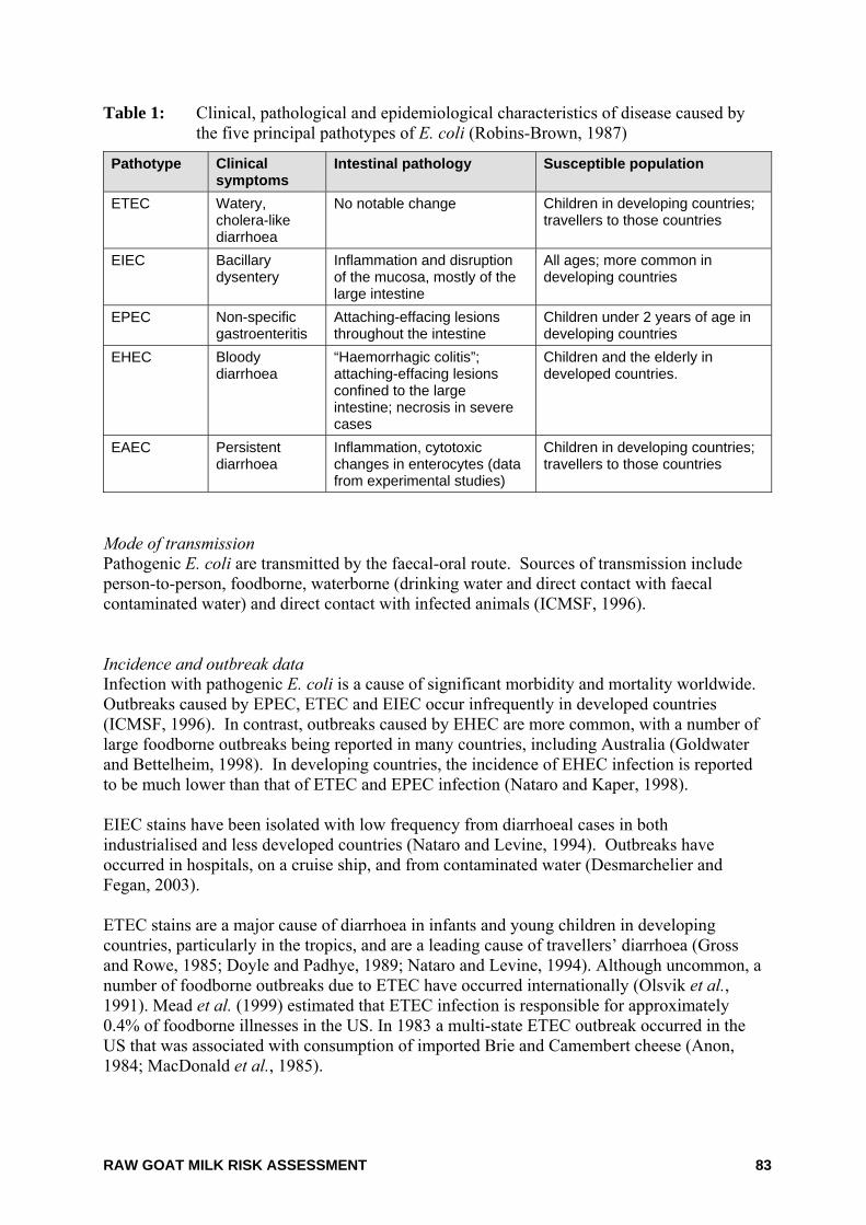
RAW GOAT MILK RISK ASSESSMENT 83
Table 1: Clinical, pathological and epidemiological characteristics of disease caused by the five principal pathotypes of E. coli (Robins-Brown, 1987)
Pathotype Clinical symptoms
Intestinal pathology Susceptible population
ETEC Watery, cholera-like diarrhoea
No notable change Children in developing countries; travellers to those countries
EIEC Bacillary dysentery
Inflammation and disruption of the mucosa, mostly of the large intestine
All ages; more common in developing countries
EPEC Non-specific gastroenteritis
Attaching-effacing lesions throughout the intestine
Children under 2 years of age in developing countries
EHEC Bloody diarrhoea
“Haemorrhagic colitis”; attaching-effacing lesions confined to the large intestine; necrosis in severe cases
Children and the elderly in developed countries.
EAEC Persistent diarrhoea
Inflammation, cytotoxic changes in enterocytes (data from experimental studies)
Children in developing countries; travellers to those countries
Mode of transmission Pathogenic E. coli are transmitted by the faecal-oral route. Sources of transmission include person-to-person, foodborne, waterborne (drinking water and direct contact with faecal contaminated water) and direct contact with infected animals (ICMSF, 1996). Incidence and outbreak data Infection with pathogenic E. coli is a cause of significant morbidity and mortality worldwide. Outbreaks caused by EPEC, ETEC and EIEC occur infrequently in developed countries (ICMSF, 1996). In contrast, outbreaks caused by EHEC are more common, with a number of large foodborne outbreaks being reported in many countries, including Australia (Goldwater and Bettelheim, 1998). In developing countries, the incidence of EHEC infection is reported to be much lower than that of ETEC and EPEC infection (Nataro and Kaper, 1998). EIEC stains have been isolated with low frequency from diarrhoeal cases in both industrialised and less developed countries (Nataro and Levine, 1994). Outbreaks have occurred in hospitals, on a cruise ship, and from contaminated water (Desmarchelier and Fegan, 2003). ETEC stains are a major cause of diarrhoea in infants and young children in developing countries, particularly in the tropics, and are a leading cause of travellers’ diarrhoea (Gross and Rowe, 1985; Doyle and Padhye, 1989; Nataro and Levine, 1994). Although uncommon, a number of foodborne outbreaks due to ETEC have occurred internationally (Olsvik et al., 1991). Mead et al. (1999) estimated that ETEC infection is responsible for approximately 0.4% of foodborne illnesses in the US. In 1983 a multi-state ETEC outbreak occurred in the US that was associated with consumption of imported Brie and Camembert cheese (Anon, 1984; MacDonald et al., 1985).
RAW GOAT MILK RISK ASSESSMENT 84
EPEC stains have caused infantile diarrhoea in hospitals and nurseries in the UK and the US (Robins-Brown 1987; Nataro and Levine, 1994). In developing countries, EPEC stains are still responsible for a high incidence of sporadic infant diarrhoea. Limited information is available on foodborne outbreaks associated with EPEC. An outbreak of EPEC (serotype O111) occurred amongst people on a coach trip to France, although no specific food was identified. The infection was believed to have been the result of consuming food at a restaurant in northern France (Wight et al., 1997). In the US, consumption of undercooked hamburger meat has been an important cause of EHEC outbreaks (Nataro and Kaper 1998). Since its identification as a human pathogen in 1982, and implication in a number of outbreaks in the US, E. coli O157:H7 has become identified as the most predominant cause of EHEC related disease (FAO/WHO, 2000). It is estimated that 85% of EHEC infections in the US are foodborne (Mead et al., 1999). A large multi-state E. coli O157:H7 outbreak involving consumption of contaminated hamburgers occurred in December 1992 – January 1993 with 732 cases identified, of which 195 were hospitalised and 4 died (Nataro and Kaper 1998). Foodborne outbreaks of E. coli O157:H7 have also been associated with consumption of contaminated fresh produce. In the US, outbreaks occurred in 1995 and 1996 (70 and 49 cases respectively), which were traced to consumption of lettuce (Tauxe, 1997). Studies have shown that E. coli O157:H7 can be transmitted to lettuce plant tissue from soil contaminated with manure and contaminated irrigation water (Solomon et al., 2002). Another large E. coli O157:H7 outbreak occurred in the US in 1996 which was linked to apple juice. Although the low pH of fruit juices will generally not allow the survival and growth of many Enterobacteriaceae, some strains of E. coli O157:H7 may survive due to their high acid-tolerance. In 2002, an outbreak of E. coli O157:H7 in Canada was attributed to the consumption of unpasteurised Gouda cheese (Honish et al., 2005). Over 200 non-O157 STEC serotypes have been isolated from humans, with the World Health Organisation identifying O26, O103, O111 and O145 as the most important foodborne non-O157 serogroups worldwide (WHO, 1998). STEC has been a notifiable disease in most Australia States and Territories since August 1998 (Roche et al., 2001). During the period of 2001 – 2005, the notification rate for STEC (excluding HUS cases) in Australia has been 0.2 – 0.3 cases per 100,000 population per annum (Ashbolt et al., 2002; OzFoodNet, 2003; OzFoodNet, 2004; OzFoodNet, 2005). E. coli O157 has been the most commonly reported serotype. Significant variations in notifications exist between states and territories, and part of this variation is likely to be a result of different practices employed by pathology laboratories when screening faecal samples for toxin producing E. coli (OzFoodNet, 2003). A large EHEC outbreak occurred in South Australia during 1995, which resulted in approximately 200 cases of illness. Twenty-two people aged between 4 months and 12 years developed HUS and were hospitalised and a 4 year old child died. Investigations of the outbreak identified EHEC strain O111:NM (or strain O111:H-, NM for non-motile) as the principal cause of the outbreak. A locally produced uncooked, fermented mettwurst was identified as the vehicle for the pathogen. The product was found to contain a variety of EHEC strains in addition to O111 (Paton and Paton, 1998).
RAW GOAT MILK RISK ASSESSMENT 85
Occurrence in food Humans appear to be the primary reservoir of EIEC, ETEC and EPEC organisms (Desmarchelier and Fegan, 2003). Therefore, contamination of food with these organisms is often due to human faecal contamination, either directly from an infected food handler or indirectly via contaminated water. Very little information is available on the occurrence of these organisms in food. The detection of these organisms in food is difficult, requiring sophisticated methodology and therefore food is not routinely screened for these organisms. In general, EPEC and ETEC organisms are more commonly isolated in foods from developing countries and their presence is associated with poor hygiene (Desmarchelier and Fegan, 2003). EPEC has been isolated from milk products in Iraq as well as from a variety of raw and cooked food in Malaysia (Abbar and Kaddar, 1991; Norazah et al., 1998). In Brazil, EPEC has been isolated from 21.1% of soft cheeses sampled (n=45) and has frequently been isolated from pasteurised milk (da Silva et al., 2001; Araújo et al., 2002). EIEC has only sporadically been isolated from foods (Olsvik et al., 1991). In addition to being a major cause of infantile diarrhoea in developing countries, ETEC organisms are a leading cause of traveller’s diarrhoea, which has been linked to the consumption of contaminated food and water (Nataro and Kaper 1998). ETEC has been isolated from Brazilian fish and shrimp which were harvested from waters contaminated with raw sewage (Teophilo et al., 2002). ETEC has also been detected in sauces at Mexican-style restaurants, and in chilli sauce sold by street vendors in Mexico (Adachi et al., 2002; Estrada-Garcia et al., 2002). In general, these sauces had been prepared and handled under poor hygienic conditions. The major reservoir of EHEC organisms appears to be the intestinal tract of ruminants, in particular cattle and sheep (Desmarchelier and Fegan, 2003). E. coli O157:H7 and other EHEC species have been isolated from both healthy and diarrhoeic animals, and individual animals can carry more than one serotype (Handysides and Cowden, 1998). Foods derived from these animals may become contaminated via exposure to faecal material during processing. Prevalence of STEC in raw milk has been determined in a limited number of studies. Caution must be exercised when comparing results between independent studies due to differences in sample size, stage of production where the samples were taken and different methodologies used to isolate the organisms. E. coli O157:H7 is the most widely studied EHEC serovar due to it being associated with a large number of outbreaks worldwide. In general, prevalence of STEC in raw milk is low. Adequate pasteurisation will ensure that STEC is inactivated. Very little information is available of the prevalence of EHEC organisms in food in Australia. Of the limited studies undertaken, the prevalence of E. coli O157:H7 in beef and sheep meat appears to be low, however, the prevalence of non-O157:H7 EHEC serotypes is unknown (Vanderlinde et al., 1998; Vanderlinde et al., 1999; Phillips et al., 2001a; Phillips et al., 2001b). Virulence and infectivity Clinical, pathological and epidemiological characteristics of disease caused by pathogenic E. coli vary between pathotypes and are discussed below. EPEC have technically been defined as “diarrhoeagenic E. coli belonging to serogroups epidemiologically incriminated as pathogens but whose pathogenic mechanisms have not been proven to be related either to heat-labile enterotoxins or heat-stable enterotoxins or to
RAW GOAT MILK RISK ASSESSMENT 86
Shigella-like invasiveness” (Edelman and Levine, 1983). EPEC cause characteristic attaching and effacing lesions in the intestine, similar to those produced by EHEC, but do not produce Shiga toxins. Attachment to the intestinal wall is mediated by a plasmid-encoded outer membrane protein called the EPEC Adherence Factor in type I EPEC. However, pathogenicity is not strictly correlated to the presence of the EPEC Adherence Factor, indicating that other virulence factors are involved (ICMSF, 1996). ETEC that survive passage through the stomach adhere to mucosal cells of the proximal small intestine and produce a heat-labile toxin and/or a heat-stable toxin. The heat-labile toxins are similar in structure and mode of action to cholera toxin, interfering with water and electrolyte movement across the intestinal epithelium (Desmarchelier and Fegan, 2003). If the volume of accumulated fluid exceeds the normal absorptive capacity of the large intestine, the excess is evacuated as watery diarrhoea. EAEC strains are defined as E. coli strains that do not secrete heat-labile or heat-stable toxins. These strains adhere to cultured human epithelial cells in a characteristic aggregative or “stacked-brick” pattern (Yatsuyanagi et al., 2002). The mechanisms causing enteric disease are not fully understood, however EAEC have been associated with persistent diarrhoea, primarily in infants and children (Desmarchelier and Fegan, 2003). Following ingestion, EIEC invade epithelial cells of the distal ileum and colon. The bacteria multiply within the cytoplasm of the cells, causing cell destruction and ulceration. Pathogenicity is associated with a plasmid-encoded type III secretory apparatus and other plasmid-encoded virulence factors (Desmarchelier and Fegan, 2003). The Shiga toxins (Stx1 and Stx2) of EHEC are closely related, or identical, to the toxins produced by Shigella dysenteriae. Additional virulence factors allow the organism to attach tightly to intestinal epithelial cells, causing what is commonly referred to as attaching-and-effacing lesions. Dose response EPEC: It is thought that only a few EPEC cells are necessary to cause illness in children (FDA 2003). Volunteer studies in adults demonstrated that illness could be caused by ingesting 106 – 1010 cells with sodium bicarbonate to neutralise stomach acidity (Doyle and Padhye, 1989). ETEC: Volunteer studies have shown that 108 – 1010 cells of ETEC are necessary for illness in adults (DuPont et al., 1971) although the infective dose is probably less for infants and children (FDA, 2003). EIEC: Volunteer studies have shown that 108 EIEC cells are necessary to cause illness in adults, with the infectious dose reduced to 106 when ingested with sodium bicarbonate (DuPont et al., 1971). However, the US Food and Drug Administration suggest that as few as 10 cells may be needed to cause illness in adults, based on the organisms similarity with Shigella (FDA 2003). The dose-response relationship for EHEC is complicated by the large number of serotypes and the association of EHEC with a variety of foods. Haas et al. (2000) developed a dose-response relationship for E. coli O157:H7 based on data from a prior animal study undertaken
RAW GOAT MILK RISK ASSESSMENT 87
by Pai et al. (1997) which involved oral administration of bacterial suspension to infant rabbits. The model was validated by comparison with two well-documented human outbreaks, one foodborne and the other waterborne. The model estimated that the dose required to result in 50% of the exposed population to become ill was 5 × 10⁵ organisms. The corresponding probability of illness for the ingestion of 100 organisms was 2.6 × 10⁻⁴. Dose-response relationships for E. coli O111 and O55 have been developed from human feeding trial data (Haas et al., 2000). The relationship estimated a dose required for 50% of the exposed population to become ill was 2.55 × 10⁶ and the probability of illness for ingestion of 100 organisms was 3.5 × 10⁻⁴. Investigations of other known outbreaks of foodborne illness due to E. coli O157:H7 and systematic studies aimed at quantifying the dose–response relationship suggest as few as 1 – 700 EHEC organisms can cause human illness (FDA 2003). Host susceptibility A variety of host factors may be important in the pathogenesis of specific E. coli serotypes. In general, the young and the elderly appear to be more susceptible to pathogenic E. coli infection. Epidemiological studies have identified that children are at higher risk of developing post-diarrhoeal HUS than other age groups (Cummings et al., 2002). References Abbar, F. and Kaddar, H.K. (1991) Bacteriological studies on Iraqi milk products. J Appl Bacteriol. 71(6):497-500.
Adachi, J.A., Mathewson, J.J., Jiang, Z.D., Ericsson, C.D. and DuPont, H.L. (2002) Enteric pathogens in Mexican sauces of popular restaurants in Guadalajara, Mexico, and Houston, Texas. Ann.Intern.Med 136(12):884-887.
Anon. (1984) Update: gastrointestinal illness associated with imported semi-soft cheese. Morbidity and Mortality Weekly Review 33(2):22.
Anon (1998) Zoonotic non-O157 shiga toxin-producion Escherichia coli (STEC). Report of a WHO Scientific Working Group meeting. World Health Organization, Geneva, Switzerland.
Araújo, V.S., Pagliares, V.A., Queiroz, M.L.P. and Freitas-Almeida, A.C. (2002) Occurrence of Staphylococcus and enteropathogens in soft cheese commercialized in the city of Rio de Janeiro, Brazil. Journal of Applied Microbiology 92(6):1172-1177.
Arnold, K.W. and Kaspar, C.W. (1995) Starvation- and stationary-phase-induced acid tolerance in Escherichia coli O157:H7. Appl Environ.Microbiol 61(5):2037-2039.
Ashbolt, R., Givney, R., Gregory, J.E., Hall, G., Hundy, R., Kirk, M., McKay, I., Meuleners, L., Millard, G., Raupach, J., Roche, P., Prasopa-Plaizier, N., Sama, M.K., Stafford, R., Tomaska, N., Unicomb, L. and Williams, C. (2002) Enhancing foodborne disease surveillance across Australia in 2001: the OzFoodNet Working Group. Commun Dis Intell. 26(3):375-406.
Bell, C. and Kyriakides, A. (1998) E. coli: a practical approach to the organism and its control in foods. Blackwell Science, Oxford, UK.
Benjamin, M.M. and Datta, A.R. (1995) Acid tolerance of enterohemorrhagic Escherichia coli. Applied and Environmental Microbiology 61(4):1669-1672.

RAW GOAT MILK RISK ASSESSMENT 88
Codex (2002) Risk profile for enterohemorragic E. coli including the identification of commodities of concern, including sprouts, ground beef and pork (CX/FH 03/5-Add .4). Codex Alimentarius Commission, Rome, Italy. ftp://ftp.fao.org/codex/ccfh35/fh0305de.pdf.
Cummings, K.C., Mohle-Boetani, J.C., Werner, S.B. and Vugia, D.J. (2002) Population-based trends in pediatric hemolytic uremic syndrome in California, 1994-1999: substantial underreporting and public health implications. Am J Epidemiol 155(10):941-948.
da Silva, Z.N., Cunha, A.S., Lins, M.C., de AM, C.L., Almeida, A.C. and Queiroz, M.L. (2001) Isolation and serological identification of enteropathogenic Escherichia coli in pasteurized milk in Brazil. Rev Saude Publica 35(4):375-379.
Dalton, C.B., Gregory, J., Kirk, M.D., Stafford, R.J., Givney, R., Kraa, E. and Gould, D. (2004) Foodborne disease outbreaks in Australia, 1995 to 2000. Commun Dis Intell. 28(2):211-224.
Desmarchelier, P.M. and Fegan, N. (2003) Enteropathogenic Escherichia coli. In: Hocking, A.D. eds. Foodborne microorganisms of public health significance. Sixth ed, Australian Institute of Food Science and Technology (NSW Branch), Sydney, Australia, pp267-310.
Doyle, M.P. and Padhye, V.V. (1989) Escherichia coli. In: Doyle, M.P. eds. Foodborne bacterial pathogens. Marcel Dekker Inc, New York, USA, pp235-281.
Doyle, M.P. and Schoeni, J.L. (1984) Survival and growth characteristics of Escherichia coli associated with hemorrhagic colitis. Appl Environ.Microbiol 48(4):855-856.
Doyle, M.P., Zhao, T., Meng, J. and Zhao, S. (1997) Escherichia coli O157H:7. In: Doyle, M.P., Beuchat, L.R., and Montville, T.J. eds. Food Microbiology: Fundamentals and frontiers. ASM Press, Washington, USA, pp171-191.
DuPont, H.L., Formal, S.B., Hornick, R.B., Snyder, M.J., Libonati, J.P., Sheahan, D.G., LaBrec, E.H. and Kalas, J.P. (1971) Pathogenesis of Escherichia coli diarrhea. N Engl.J Med 285(1):1-9.
Edelman, R. and Levine, M.M. (1983) From the National Institute of Allergy and Infectious Diseases. Summary of a workshop on enteropathogenic Escherichia coli. J.Infect.Dis. 147(6):1108-1118.
Estrada-Garcia, T., Cerna, J.F., Thompson, M.R. and Lopez-Saucedo, C. (2002) Faecal contamination and enterotoxigenic Escherichia coli in street-vended chili sauces in Mexico and its public health relevance. Epidemiol Infect 129(1):223-226.
European Commission (2000) Opinion of the scientific committee on veterinary measures relating to public health on food-borne zoonoses. Health and Consumer Protection Directorate-General, European Commission, Brussels, Belgium.
FAO/WHO (2000) Joint FAO/WHO expert consultation on risk assessment of microbiological hazards in foods.
FDA (2003) The bad bug book (Foodborne Pathogenic Microorganisms and Natural Toxins Handbook ). US Food and Drug Administration. http://www.cfsan.fda.gov/~mow/intro.html. Accessed on 8 August 2004.
Goldwater, P.N. and Bettelheim, K.A. (1998) New perspectives on the role of Escherichia coli O157:H7 and other enterohaemorrhagic E. coli serotypes in human disease. J.Med.Microbiol. 47(12):1039-1045.
Gross, R.J. and Rowe, B. (1985) Escherichia coli diarrhoea. J Hyg.(Lond) 95(3):531-550.
Haas, C.N., Thayyar-Madabusi, A., Rose, J.B. and Gerba, C.P. (2000) Development of a dose-response relationship for Escherichia coli O157:H7. Int J Food Microbiol 56(2-3):153-159.
Honish, L., Predy, G., Hislop, N., Chui, L., Kowalewska-Grochowska, K., Trottier, L., Kreplin, C. and Zazulak, I. (2005) An outbreak of E. coli O157:H7 hemorrhagic colitis associated with unpasteurized gouda cheese. Can.J Public Health 96(3):182-184.
RAW GOAT MILK RISK ASSESSMENT 89
ICMSF. (1996) Intestinally pathogenic Escherichia coli. In: Roberts, T.A., Baird-Parker, A.C., and Tompkin, R.B. eds. Microorganisms in foods 5: Microbiological specifications of food pathogens. First ed, Blackie Academic & Professional, London, UK, pp217-264.
Kirk, R. and Rowe, M. (1999) Pathogenic E. coli and milk. Milk Industry Int 101((Technical and Research Supplement MII)):4.
Lake, R., Hudson, A. and Cressey, P. (2003) Risk Profile: Shiga-like toxin producing Escherichia coli in uncooked comminuted fermented meat products. Institute of Environmental Science and Research Limited, Christchurch, New Zealand.
MacDonald, K.L., Eidson, M., Strohmeyer, C., Levy, M.E., Wells, J.G., Puhr, N.D., Wachsmuth, K., Hargrett, N.T. and Cohen, M.L. (1985) A multistate outbreak of gastrointestinal illness caused by enterotoxigenic Escherichia coli in imported semisoft cheese. J Infect Dis 151(4):716-720.
Mead, P.S., Slutsker, L., Dietz, V., McCaig, L.F., Bresee, J.S., Shapiro, C., Griffin, P.M. and Tauxe, R.V. (1999) Food-related illness and death in the United States. Emerg.Infect Dis 5(5):607-625.
Nataro, J.P. and Kaper, J.B. (1998) Diarrheagenic Escherichia coli. Clin Microbiol Rev 11(1):142-201.
Nataro, J.P. and Levine, M.M. (1994) Escherichia coli diseases in humans. In: Gyles, C.L. eds. Escherichia coli in domestic animals and humans. CAB International, Wallingford, UK, pp285-333.
Norazah, A., Rahizan, I., Zainuldin, T., Rohani, M.Y. and Kamel, A.G. (1998) Enteropathogenic Escherichia coli in raw and cooked food. Southeast Asian J Trop.Med Public Health 29(1):91-93.
Olsvik, Ø., Wasteson, Y., Lund, A. and Hornes, E. (1991) Pathogenic Escherichia coli found in food. Int J Food Microbiol 12(1):103-113.
OzFoodNet (2003) Foodborne disease in Australia: incidence, notification and outbreaks. Annual Report of the OzFoodNet Network, 2002. Communicable Diseases Intelligence 27(2):http://www.cda.gov.au/pubs/cdi/2003/cdi2702/htm/cdi2702g.htm.
OzFoodNet (2004) Foodborne disease investigations across Australia: Annual report of the OzFoodNet network, 2003. Commun Dis Intell. 28(3):359-389.
OzFoodNet (2005) Foodborne outbreaks associated with dairy products — Analysis of OzFoodNet data, 1995 – June 2004. Unpublished.
Pai, M., Kang, G., Ramakrishna, B.S., Venkataraman, A. and Muliyil, J. (1997) An epidemic of diarrhoea in south India caused by enteroaggregative Escherichia coli. Indian J.Med.Res. 106:7-12.
Paton, J.C. and Paton, A.W. (1998) Pathogenesis and diagnosis of Shiga toxin-producing Escherichia coli infections. Clin Microbiol Rev 11(3):450-479.
Phillips, D., Sumner, J., Alexander, J.F. and Dutton, K.M. (2001a) Microbiological quality of Australian beef. J Food Prot. 64(5):692-696.
Phillips, D., Sumner, J., Alexander, J.F. and Dutton, K.M. (2001b) Microbiological quality of Australian sheep meat. J Food Prot. 64(5):697-700.
Robins-Brown, R.M. (1987) Traditional enteropahogenic Escherichia coli of infantile diarrhea. Rev.Infect.Dis. 9:28-53.
Roche, P., Spencer, J., Lin, M., Gidding, H., Kirk, M., Eyeson-Annan, M., Milton, A., Witteveen, D. and Merianos, A. (2001) Australia's notifiable diseases status, 1999: annual report of the National Notifiable Diseases Surveillance System. Commun Dis Intell. 25(4):190-245.
Solomon, E.B., Potenski, C.J. and Matthews, K.R. (2002) Effect of irrigation method on transmission to and persistence of Escherichia coli O157:H7 on lettuce. J Food Prot. 65(4):673-676.
RAW GOAT MILK RISK ASSESSMENT 90
Tauxe, R.V. (1997) Emerging foodborne diseases: an evolving public health challenge. Emerg.Infect Dis 3(4):425-434.
Teophilo, G.N., dos, F., V, dos Prazeres, R.D. and Menezes, F.G. (2002) Escherichia coli isolated from seafood: toxicity and plasmid profiles. Int Microbiol 5(1):11-14.
Vanderlinde, P.B., Shay, B. and Murray, J. (1998) Microbiological quality of Australian beef carcass meat and frozen bulk packed beef. Journal of Food Protection 61(4):437-443.
Vanderlinde, P.B., Shay, B. and Murray, J. (1999) Microbiological status of Australian sheep meat. Journal of Food Protection 62(4):380-385.
WHO (1998) Zoonotic non-O157 shiga toxin-producing Escherichia coli (STEC). Preport of a WHO Scientific Working Group Meeting. Berlin, Germany, 26-26 June 1998. World Health Organization, Geneva.
Wight, J.P., Rhodes, P., Chapman, P.A., Lee, S.M. and Finner, P. (1997) Outbreaks of food poisoning in adults due to Escherichia coli O111 and campylobacter associated with coach trips to northern France. Epidemiol Infect 119(1):9-14.
Yatsuyanagi, J., Saito, S., Sato, H., Miyajima, Y., Amano, K. and Enomoto, K. (2002) Characterization of enteropathogenic and enteroaggregative Escherichia coli isolated from diarrheal outbreaks. J Clin Microbiol 40(1):294-297.
RAW GOAT MILK RISK ASSESSMENT 91
9. Leptospira interrogans Leptospires are aerobic spirochetes whose cells are flexulous, motile, tightly coiled and have a single axial filament. They are generally about 0.1 µm in diameter and 5 - 25µm in length. They are Gram-negative, although they share features of both Gram-positive and Gram-negative cellular walls (Bharti et al., 2003). The genus Leptospira is an incredibly varied group of organisms, containing hundreds of serovars and genetic types. There are over 200 known serovars with pathogenic bacteria almost entirely within the Leptospira interrogans genomospecies. Approximately 24 serovars are currently identified within the L. interrogans genomospecies with L. interrogans serovar Icterohaemorrhagiae best known for human infection (Anon, 2004). Leptospira spp. are ubiquitous environmental bacteria found around the world (Zhou et al., 2004). Infections due to the organism L. interrogans occur in both animals and humans, however they are more prevalent in developing and tropical areas (Baranton and Postic, 2006). Serovars Hardjo, Pomona and Grippotyphosa are common in cattle, sheep and goats, while Canicola and Icterohaemorrhagiae are further associated with cattle and Ballum associated with sheep and goats. Growth characteristics This organism requires high humidity for survival and is killed by dehydration or temperatures greater than 50°C, however they can remain viable for many weeks or months in contaminated soil and several weeks in cattle slurry. This organism is also easily destroyed be pasteurisation (Anon, 2005). Optimum growth temperatures are between 28 - 30°C with pH levels between 6.8 - 7.4 being required for growth (Bharti et al., 2003). Pathology of illness Leptospirosis (also called “Weil’s syndrome” or “Cane cutters disease - in its more extreme forms of infection) is the disease caused by the bacterium L. interrogans and other species of the genus Leptospira including L. noguchii, L. santarosai, L. meyeri, L. borgpetersenii, L. kirschneri, L. weilii, L .inadai, L .fainei and L. alexanderi (Anon, 2005). The symptoms of the infection may include fever, weakness, rash and headaches and possibly lead to more severe complications such as liver and kidney malfunction, febrile illness and jaundice (Brito et al., 2006). Leptospirosis can occur as a biphasic illness where sudden onset of febrile illness is reported with a typical duration of one week. This may then be followed by aseptic meningitis which occurs in up to 25% of all cases. Alternatively, Weil’s syndrome will progress and is distinguished by development of renal failure and jaundice. It is only in the most severe cases of Leptospirosis that Weil’s disease will develop without prior onset of febrile illness (Bharti et al., 2003). Infection of this organism in cattle, sheep or goats can lead to decreased milk productivity and cause abortions (Faine, 2000; Anon, 2000).
RAW GOAT MILK RISK ASSESSMENT 92
Mode of transmission L. interrogans is a very common zoonotic pathogen of cattle, sheep, goats, domestic and feral pigs, dogs, cats, rabbits, horses, deer, possums, and various rodents including rats and mice (Anon, 2000). L. interrogans can be spread by contact directly between infected animals and humans; by ingestion or absorption of contaminated water, soil or food; through aerosolized urine particles, animal foetal fluids or through direct contact with skin (Anon 2000; Baranton and Postic, 2006). This organism is excreted via the urine and other body fluids of acutely infected animals and can enter its host through mucosa or broken skin (Zhou et al., 2004). This bacterium is frequently found as the cause of complications following surgical operations, recreational water exposure and rural occupational injuries (Anon 2000; Zhou et al., 2004). Incidence of illness It has been estimated that in the US between 100 - 200 human cases of leptospirosis are reported each year (CDC, 2005). The incidence of infection seems to increase during warmer periods (e.g. summer) and during the rainy season (Baranton and Postic, 2006). In Australia prevalence of Leptospirosis is much higher in the tropical and wetter areas of the country, such as Far North Queensland and the Northern Territory (Anon, 2000). Incidence of L. interrogans due to foodborne contamination is unknown due to a lack of available data. Cases of infection often go unreported if symptoms are only mildly exhibited. This was illustrated in 1995 when an outbreak of Leptospirosis was recorded in Nicaragua. Investigations found that of the tested population, only 25 of a possible 85 serotype positive inhabitants reported febrile illness in the 2 months prior to the study. It has been shown in other studies that symptom-less infections are common in endemic areas (Bharti et al., 2003). In the case of acute infections, fatality rates are estimated at between 5 - 15%. Acute renal failure is reported in 16 - 40% of cases whilst pulmonary involvement is estimated to occur within 20 - 70% of cases (Bharti et al., 2003). Occurrence in foods No evidence in the literature can be identified that indicates this organism is shed in the milk of infected animals, or if it occurs naturally in any food source, other than undercooked infected kidney (Anon 2000). However cross-contamination of foods with infected urine or water is a possible mechanism for foodstuffs to be contaminated with L. interrogans and thus have the ability to cause infection upon ingestion (Bharti et al., 2003; Baranton and Postic, 2006). Virulence and infectivity Motility is thought to be one of the most important virulence factors for this organism. Additionally, studies have shown evidence of other virulence factors including phospholipase, sphingomyelinase and haemytic activities in vitro and pore forming proteins have also been identified when involved with a mammalian cells. Some virulent serovars have also been found to express fibronectin-binding proteins on the cellular surface and this is thought to aid in adhesion and invasion of host organisms (Bharti et al., 2003).
RAW GOAT MILK RISK ASSESSMENT 93
Dose response There is a lack of information on the dose-response relationship for L. interrogans for humans, however it has been suggested that invasion of some highly susceptible animals with less than 10 leptospires is sufficient to cause infection (Anon, 2000). References Anon (2000) A Scientific Review of Leptospirosis and Implication for Quarantine Policy. Biosecurity Austra, Canberra, Australia. http://www.affa.gov.au/corporate_docs/publications/pdf/market_access/biosecurity/animal/leptoreviewfinal.pdf. Accessed on 22 June 2006.
Anon (2004) Leptospirosis - Bacteriological Information. The Leptospirosis Information Centre. http://www.leptospirosis.org/bacteria. Accessed on 21 June 2006.
Anon (2005) Leptospirosis. OIE, Institute for International Cooperation in Animal Biologics. http://www.cfsph.iastate.edu/Factsheets/pdfs/leptospirosis.pdf. Accessed on 20 June 2006.
Baranton, G. and Postic, D. (2006) Trends in leptospirosis epidemiology in France. Sixty-six years of passive serological surveillance from 1920 to 2003. International Journal of Infectious Diseases 10(2):162-170.
Bharti, A.R., Nally, J.E., Ricaldi, J.N., Matthias, M.A., Diaz, M.M., Lovett, M.A., Levett, P.N., Gilman, R.H., Willig, M.R., Gotuzzo, E. and Vinetz, J.M. (2003) Leptospirosis: a zoonotic disease of global importance. Lancet Infect.Dis. 3(12):757-771.
Brito, T., Menezes, L., Lima, D., Lourenco, S., Silva, A. and Alves, V. (2006) Immunohistochemical and in situ hybridization studies of the liver and kidney in human leptospirosis. Virchows Archiv :
CDC (2005) Centres for Disease Control and Prevension – Leptospirosis. http://www.cdc.gov/ncidod/dbmd/diseaseinfo/leptospirosis_t.htm
Faine, S. (2000) Leptospira species. Antimicrobial :272-275.
Zhou, Z., Song, X., Li, Y. and Gong, W. (2004) Unique structural characteristics of peptide deformylase from pathogenic bacterium Leptospira interrogans. J.Mol.Biol. 339(1):207-215.
RAW GOAT MILK RISK ASSESSMENT 94
10. Listeria monocytogenes Listeria monocytogenes is a Gram-positive, non-spore forming rod-shaped bacteria that may be isolated from a variety of sources including soil, silage, sewage, food-processing environments, raw meats and the faeces of healthy humans and animals (FDA, 2003). L. monocytogenes belongs to the genus Listeria along with L. innocua, L. welshimeri, L. selligeri, L. ivanovii and L. grayi. Thirteen serotypes are associated with L. monocytogenes (1/2a, 1/2b, 1/2c, 3a, 3b, 3c, 4a, 4ab, 4b, 4c, 4d, 4e, 7). Growth characteristics Growth of L. monocytogenes in foods is influenced by a variety of factors, including the nature and concentration of essential nutrients, pH, temperature, water activity, the presence of food additives that could enhance or inhibit growth and presence of other microbial flora (Lovett et al., 1987). Under conditions outside the growth range, the bacteria may survive and growth may recommence once suitable conditions are encountered. Temperatures of >50ºC are lethal to L. monocytogenes. When in a suitable medium, L. monocytogenes can grow between ~0 - 45°C. Although L. monocytogenes does not grow below –1.5ºC, it can readily survive at much lower temperatures. Nonetheless, freezing and frozen storage will cause a limited reduction in the viable population of L. monocytogenes. Optimal conditions for growth are between 30 - 37ºC (Ryser and Marth, 1999). L. monocytogenes will grow in a broad pH range with the upper limit being approximately 9.3 and the lower limit being 4.6 - 5.0 (ICMSF, 1996). Although growth at pH <4.3 has not yet been documented, L. monocytogenes appears to be relatively acid tolerant. It has been suggested that food fermentations, which involve a gradual lowering of pH, could lead to acid adaptation of L. monocytogenes. Like many bacterial species, L. monocytogenes grows optimally at a of approximately 0.97. However, when compared with most foodborne pathogens, the bacterium has the unique ability to multiply at water activity values as low as 0.90. While it does not appear to be able to grow below 0.90, the bacterium can survive for extended periods at lower values (Ryser and Marth, 1999). L. monocytogenes is reasonably tolerant to salt and can grow in NaCl concentrations up to 10%. Extended survival occurs at a wide range of salt concentrations and L. monocytogenes has survived for up to eight weeks in a concentration of 20% NaCl (Sutherland et al., 2003). Survival in the presence of salt varies with storage temperature and studies have indicated that survival of L. monocytogenes in concentrated salt solutions can be increased dramatically by lowering the incubation temperature (Ryser and Marth, 1999). L. monocytogenes grows well under both aerobic and anaerobic conditions (Ryser and Marth, 1999; Sutherland et al., 2003). The listericidal effect of preservatives is strongly influenced by the interactive effects of temperature, pH, type of acidulant, salt content, water activity, and type and concentration of food additives present in the food. For example, the ability of potassium sorbate to prevent growth of L. monocytogenes is related to temperature and pH. The lower the storage temperature and pH of the medium, the greater the effectiveness of sorbates against L. monocytogenes. Sodium benzoate is more inhibitory to L. monocytogenes than is either potassium sorbate or sodium propionate. Inhibition and inactivation of L. monocytogenes in the presence of sodium benzoate is affected by temperature (more rapid at higher than lower
RAW GOAT MILK RISK ASSESSMENT 95
incubation temperatures), concentration of benzoic acid (more rapid at higher than lower concentrations) and pH (more rapid at lower rather than higher pH values) as well as the type of acid used to adjust the growth medium (Ryser and Marth, 1999). Pathology of illness There are two main forms of illness associated with L. monocytogenes infection: listerial gastroenteritis, where usually only mild symptoms are reported, and invasive listeriosis, where the bacteria penetrate the gastrointestinal tract and invade normally sterile sites within the body (FDA 2003). Symptoms of the mild form of L. monocytogenes infection are primarily those generally associated with gastrointestinal illness: chills, diarrhoea, headache, abdominal pain and cramps, nausea, vomiting, fatigue, and myalgia (FDA, 2003). The onset of illness is usually greater than 12 hours. Invasive listeriosis is clinically defined when the organism is isolated from blood, cerebrospinal fluid or an otherwise normally sterile site (e.g. placenta, foetus). The manifestations include septicaemia, meningitis (or meningoencephalitis), encephalitis, and intrauterine or cervical infections in pregnant women, which may result in spontaneous abortion in the second or third trimester, or stillbirth (FDA, 2003). The onset of these manifestations is usually preceded by influenza-like symptoms including persistent fever. Gastrointestinal symptoms such as nausea, vomiting and diarrhoea may also precede the serious forms of listeriosis. Listeriosis typically has a 2 - 3 week incubation time, but onset time may extend to 3 months (FDA/Centre for Food Safety and Applied Nutrition, 2003). It is estimated that approximately 2 – 6% of the healthy human population harbour L. monocytogenes in their intestinal tract, which suggests that people are frequently exposed to L. monocytogenes (Farber and Peterkin, 1991; Rocourt and Bille, 1997). This may also suggest that most people have a tolerance to infection by L. monocytogenes, and given the relatively low number of reported cases, exposure rarely leads to serious illness in healthy individuals (Hitchins, 1996; Marth, 1988). Mode of transmission Foodborne exposure is the primary route of transmission for listeriosis, however listeriosis can be transmitted vertically (i.e. mother to child), zoonotically and through hospital acquired infections (Ryser and Marth, 1999; Bell and Kyriakides, 2005). Incidence of illness Most cases of listeriosis are sporadic. The number of reported cases of invasive listeriosis in Australia between 2001 - 2004 varied between 61 – 72 cases (Ashbolt et al., 2002; Anon, 2002; Anon, 2003; Anon, 2004b), which equates to approximately 3 – 4 cases per million population per annum. In Australia, the exact mortality rate is not known, although the data available would suggest a rate of approximately 23%. The case fatality rate in New Zealand is approximately 17% (Anon, 2004a). The estimated incidence of invasive listeriosis in European countries has been reported to between 0.3 - 7.5 cases per million of the general population per annum (European
RAW GOAT MILK RISK ASSESSMENT 96
Commission, 2003). In France, the estimated incidence is sixteen cases per million (general population) per annum (Bille, 1990; ICMSF, 1996). The annual incidence of listeriosis in the United States has been estimated to range from 3.4 per million (CDC, 2002) to 4.4 per million (Tappero et al., 1995). Of all foodborne pathogens, L. monocytogenes results in the highest hospitalisation rate in the US, with fatality rates of 20 - 30% being common (WHO/FAO, 2004). Outbreaks of invasive listeriosis have been linked to Hispanic-style soft cheeses; soft, semi-soft and mould-ripened cheeses; hot dogs; pork tongue jelly; processed meats; pate; salami; pasteurised chocolate flavoured milk; pasteurised and unpasteurised milk; butter; cooked shrimp; smoked salmon; maize and rice salad; maize and tuna salad; potato salad; raw vegetables; and coleslaw (FDA 2003). In addition, sporadic cases have been linked to the consumption of raw milk; unpasteurised ice cream; ricotta cheese; goat, sheep and feta cheeses; soft, semi-soft and mould-ripened cheeses; Hispanic-style cheese; salami; hot dogs; salted mushrooms; smoked cod roe; smoked mussels; undercooked fish; pickled olives; raw vegetables; and coleslaw (WHO/FAO, 2004). Occurrence in foods L. monocytogenes has been found in foods such as milk, dairy products (particularly soft-ripened cheeses), meat, poultry, seafood and vegetables. The worldwide prevalence of L. monocytogenes in raw milk is estimated to be around 3-4% (Hayes et al., 1986; Lovett et al., 1987; Doores and Amelang, 1988). In Australian surveys on soft and surface ripened cheeses and ice-cream, L. monocytogenes has been isolated from 2% of locally produced cheese samples and 6% of ice-cream samples (Sutherland et al., 2003). For imported cheeses, camembert and blue vein, 7% were positive for L. monocytogenes (Sutherland et al., 2003). For European soft and surface-ripened cheeses, 25% have been found to be positive for L. monocytogenes (Terplan, 1988). Meat products from which L. monocytogenes has been isolated include beef, lamb, pork, minced meat products, sausages, salami, ham, mettwurst, pate, frankfurters and vacuumed packed meat, chicken products, and processed seafood (Farber and Peterkin 1991; Cox et al., 1999; Ojeniyi et al., 2000). Additionally vegetable products have also been shown to be contaminated (Heisick et al., 1989; Brackett, 1999). Virulence and infectivity of L. monocytogenes When ingested, L. monocytogenes penetrates the intestinal tissue and is taken up by macrophages and non-phagocytic cells in the host. L. monocytogenes is disseminated throughout the host via blood or lymphatic circulation to various tissues. Its presence intra-cellularly in phagocytic cells permits access to the brain and probably transplacental migration to the foetus in pregnant women. The pathogenesis of L. monocytogenes relies on its ability to survive and multiply in phagocytic host cells. Not all strains appear to be equally virulent. The 4b and occasionally 1/2a and 1/2b serovars account for most cases of human listeriosis (ICMSF, 1996). The virulence of L. monocytogenes is increased when the bacterium is grown at low rather than high temperatures. The possibility exists that cold storage may enhance the virulence of some L. monocytogenes strains isolated from refrigerated foods (Ryser and Marth, 1999).
RAW GOAT MILK RISK ASSESSMENT 97
Dose response Cases of non-invasive listeriosis (also referred to as febrile listerial gastroenteritis) have been observed during outbreaks, involving symptoms such as diarrhoea, fever, headache and myalgia, generally following a short incubation period (WHO/FAO, 2004). Insufficient quantitative data is available to develop a dose-response model for this milder form of listeriosis, however, outbreak situations have generally involved the ingestion of high doses of L. monocytogenes. The dose-response relationship for invasive listeriosis is highly dependent on a number of factors, such as the virulence characteristics of the organism, the number of cells ingested, the general health and immune status of the host, and the attributes of the food matrix that may alter the microbial or host status. WHO/FAO (2004) and FDA/FSIS (2003) developed separate dose-response models for both healthy and susceptible populations by combining data from surrogate animal models with epidemiological data. The Exponential dose-response model was used for both populations. This dose-response model has a single parameter, the r-value. The r-value is the probability that a person will become ill from the consumption of a single L. monocytogenes cell. For the healthy population (classified as “intermediate-age”) the median r-value was estimated to be 2.37 x 10⁻¹⁴. For more susceptible populations the median r-value was estimated to be 1.06 x 10⁻¹². A more recent assessment of US epidemiological data on invasive Listeriosis in susceptible sub-populations which included genetic information regarding different L. monocytogenes strains (lineages), determined average r-values of 1.31 x 10⁻⁸ for lineage I and 5.01 x 10⁻¹¹ for lineage II (Chen et al., 2006). Further analysis of the epidemiological data by the L. monocytogenes ribotype found r-values as small as 6.29 x 10⁻³. These results suggest that there are large differences in virulence between L. monocytogenes strains. The infectious dose is unknown but it is believed to vary depending on the strain and susceptibility of the individual. There is a lack of information concerning the minimal infectious dose, although it is generally thought to be relatively high (>100 viable cells) (ICMSF, 1996). From cases contracted via raw or inadequately pasteurised milk, it is assumed that for susceptible individuals, ingestion of fewer than 1,000 organisms may cause disease (FDA/FSIS, 2003). It is thought the consumption of food with exceptionally high levels of L. monocytogenes (>107/g) is required to cause the mild gastrointestinal form of illness in healthy persons (Sutherland et al., 2003). Host factors Specific sub-populations at risk for invasive listeriosis include pregnant women and their foetuses, neonates, the elderly and persons with a compromised immune system, whose resistance to infection is lowered (e.g. transplant patients, patients on corticosteroid treatments, AIDS patients and alcoholics). Less frequently reported diabetic, cirrhotic, asthmatic and ulcerative colitis patients are also at a higher risk (FDA 2003). Another physiological parameter thought to be relevant to susceptibility is a reduced level of gastric acidity (WHO/FAO, 2004). Food matrix To date, the properties of the food vehicle have been viewed as having little effect on the infective dose of L. monocytogenes. However, it is possible that food vehicles with high buffering capacity may protect the bacteria from inactivation by the pH of gastric acids in the
RAW GOAT MILK RISK ASSESSMENT 98
stomach. In general, there are insufficient data available as to whether the food matrix affects the dose-response curve for L. monocytogenes (WHO/FAO, 2004). References Anon. (2002) OzFoodNet: enhancing foodborne disease surveillance across Australia: quarterly report, April to June 2002. Commun Dis Intell. 26(4):546-551.
Anon. (2003) Foodborne disease in Australia: incidence, notifications and outbreaks. Annual report of the OzFoodNet network, 2002. Commun Dis Intell. 27(2):209-243.
Anon (2004a) Notifiable and other diseases in New Zealand: Annual report, 2004. Ministry of Health, Wellington, New Zealand.
Anon. (2004b) OzFoodNet: enhancing foodborne disease surveillance across Australia: quarterly report, 1 October to 31 December 2003. Commun Dis Intell. 28(1):86-89.
Ashbolt, R., Givney, R., Gregory, J.E., Hall, G., Hundy, R., Kirk, M., McKay, I., Meuleners, L., Millard, G., Raupach, J., Roche, P., Prasopa-Plaizier, N., Sama, M.K., Stafford, R., Tomaska, N., Unicomb, L. and Williams, C. (2002) Enhancing foodborne disease surveillance across Australia in 2001: the OzFoodNet Working Group. Commun Dis Intell. 26(3):375-406.
Bell, C. and Kyriakides, A. (2005) Listeria. Second ed, Blackwell Publishing, UK.
Bille, J. (1990) Anatomy of a foodborne listeriosis outbreak. In: Miller, B., Smith, J.L., and Somkuti, G. eds. Foodborne Listeriosis. Elsevier, London, pp39-46.
Brackett, R.E. (1999) Incidence and behaviour of Listeria monocytogenes in products of plant origin. In: Ryser, E.T. and Marth, E.H. eds. Listeria, Listeriosis and food safety. Marcel Dekker, New York, pp631-655.
CDC (2002) Foodborne Outbreak Response Surveillance Unit. http://www2a.cdc.gov/ncidod/foodborne/fbsearch.asp. Accessed on 10 May 5 A.D.
Cox, N.A., Bailey, J.S. and Ryser, E.T. (1999) Incidence and behaviour of Listeria monocytogenes in poultry and egg products. In: Ryser, E.T. and Marth, E.H. eds. Marcel Dekker, New York, pp565-600.
Doores, S. and Amelang, J. (1988) Incidence of Listeria monocytogenes in the raw milk supply in Pennsylvania. (1) IFT Ann. Meeting, 19-22 June., FT Chicago, New Orleans, pp157.
European Commission (2003) Opinion of the Scientific Committee of Veterinary Measures Relating to Public Health on Staphylococcal Enterotoxins in Milk Products, particularly cheeses. http://europa.eu.int/comm/food/fs/sc/scv/out61_en.pdf.
Farber, J.M. and Peterkin, P.I. (1991) Listeria monocytogenes, a food-borne pathogen. Microbiol.Rev. 55(3):476-511.
FDA (2003) The bad bug book (Foodborne Pathogenic Microorganisms and Natural Toxins Handbook ). US Food and Drug Administration. http://www.cfsan.fda.gov/~mow/intro.html. Accessed on 8 August 2004.
FDA/Centre for Food Safety and Applied Nutrition (2003) Quantitative Assessment of the Relative Risk to Public Health from Foodborne Listeria monocytogenes Among Selected Categories of Ready-to-Eat Foods. http://www.foodsafety.gov/~dms/lmr2-toc.html.
FDA/FSIS. (2003) Quantitative assessment of relative risk to public health from foodborne Listeria monocytogenes among selected categories of ready-to-eat foods. Food and Drug Administration, US Dept of Health and Human Services; Food Safety Inspection Service, United States Department of Agriculture, USA.
RAW GOAT MILK RISK ASSESSMENT 99
Hayes, P.S., Feeley, J.C., Graves, L.M., Ajello, G.W. and Fleming, D.W. (1986) Isolation of Listeria monocytogenes from raw milk. Appl.Environ.Microbiol. 51(2):438-440.
Heisick, J.E., Wagner, D.E., Nierman, M.L. and Peeler, J.T. (1989) Listeria spp. found on fresh market produce. Appl.Environ.Microbiol. 55(8):1925-1927.
Hitchins, A.D. (1996) Assessment of alimentary exposure to Listeria monocytogenes. Int.J Food Microbiol. 30(1-2):71-85.
ICMSF. (1996) Microorganisms in Food 5: Microbiological Specifications of Food Pathgens. Blackie Academic and Professional, London.
Lovett, J., Francis, D.W. and Hunt, J.M. (1987) Listeria monocytogenes in raw milk; detection, incidence and pathogenicity. J Food Prot 50:188-192.
Marth, E.H. (1988) Disease Characteristics of Listeria monocytogenes. Food Technol 42(4):165-168.
Ojeniyi, B., Christensen, J. and Bisgaard, M. (2000) Comparative investigations of Listeria monocytogenes isolated from a turkey processing plant, turkey products, and from human cases of listeriosis in Denmark. Epidemiol.Infect. 125(2):303-308.
Rocourt, J. and Bille, J. (1997) Foodborne listeriosis. World Health Stat.Q. 50(1-2):67-73.
Ryser, E.T. and Marth, E.H. (1999) Listeria, Listeriosis, and Food Safety. Marcel Dekker, Inc., New York.
Sutherland, P.S., Miles, D.W. and Laboyrie, D.A. (2003) Listeria monocytogenes. In: Hocking, A.D. eds. Foodborne microorganisms of public health significance. Australian Institute of Food Science and Technology (NSW Branch, Food Microbiology Group), Waterloo DC, NSW, Australia, pp381-443.
Tappero, J.W., Schuchat, A., Deaver, K.A., Mascola, L. and Wenger, J.D. (1995) Reduction in the incidence of human listeriosis in the United States. Effectiveness of prevention efforts? The Listeriosis Study Group. JAMA 273(14):1118-1122.
Terplan, G. (1988) Listeria in the dairy industry. Proceedings of Gehr's seminar on problems of foodborne Listeriosis, European Symposium. (7 Sept) European Symposium, Wiesbaden, Germany.
WHO/FAO (2004) Risk assessment of Listeria monocytogenes in ready-to-eat foods. World Health Organization and Food and Agriculture Organization of the United Nations, Geneva, Switzerland. http://www.fao.org/es/ESN/food/risk_mra_riskassessment_listeria_en.stm.
RAW GOAT MILK RISK ASSESSMENT 100
11. Mycobacterium avium subsp. paratuberculosis The genus Mycobacterium comprises approximately 95 species, of which over 30 have been associated with disease in humans (Katoch, 2004). Mycobacterium spp. are also pathogens of food producing animals such as cattle, sheep, other ruminants and fish. Some Mycobacterium spp. have zoonotic potential in humans (Sutherland, 2003). Mycobacteria spp. are aerobic, non-sporeforming, Gram-positive (though difficult to stain) acid-fast rod-shaped bacilli without flagellae. They are slow growing and difficult to culture, having fastidious and nutritionally-exacting growth requirements (Anon, 1998). Mycobacterium spp. are widely distributed in the environment, being found in soil and water. They readily form biofilms in drinking water distribution systems (Falkinham, 2002; Sutherland, 2003). Mycobacterium spp. have particularly hydrophobic cell walls, giving them a propensity to form aerosols, to clump together in liquid media and to form biofilms (Sattar et al., 1995; Anon 1998; Woelk et al., 2003). The position of M. avium subsp. paratuberculosis (hereafter M. avium subsp. paratuberculosis or MAP) as a human pathogen is still unclear. Debate centres on the possible role of MAP in Crohn’s disease, a chronic intestinal enteritis in humans. Similarities have been observed between Crohn’s disease and Johne’s disease in cattle and sheep, a disease which is known to be caused by MAP (Anon, 1998). The debate is characterised by firmly entrenched opinions on either side, and the subject has been comprehensively reviewed several times (Chiodini, 1989; Thompson, 1994; Anon 1998; Harris and Lammerding, 2001; Lipiec, 2003; Chacon et al., 2004). Growth characteristics MAP is an obligate parasite and is absolutely dependent on mycobactin, an iron-chelating siderophore, for in vitro growth (Anon 1998; Motiwala et al., 2004). The temperature range for growth of MAP is 25 - 45ºC with an optimum of around 39ºC (Anon 1998). Batch (63ºC for 30 minutes) and HTST (72ºC for 15 seconds) pasteurisation are sufficient to inactivate high levels of pathogenic Mycobacteria in milk, although they will survive thermisation (treatment at 62ºC for 15 seconds for cheese production) (Stabel and Lambertz, 2004). MAP does not grow in the presence of 5% sodium chloride but is able to grow in microbiological media at pH 5.5. The organism is resistant to drying and may survive in faeces on pasture land for approximately one year (Anon 1998). Pathology of illness Although there is ongoing disagreement regarding the role of MAP in human Crohn’s disease, the following brief description of the disease is included for information. Crohn’s disease is a chronic, granulomatous inflammatory disease of humans, which primarily affects the terminal ileum and colon (Anon, 2000; Rubery, 2002). The disease is characterised by periods of activity interspersed with periods of remission. The clinical signs of Crohn’s disease include weight loss, abdominal pain, diarrhoea, reduced appetite and fatigue. Crohn’s disease has also been associated with arthritis, skin lesions, anaemia and, in the younger age group, reduced growth rate (Anon, 2004). It has also been observed that mycobacterial illnesses can reactivate many years after recovery from overt illness (Rutala et al., 1991; Gregory et al., 1999; Kubica et al., 2003; Gibson et al., 2004).
RAW GOAT MILK RISK ASSESSMENT 101
Mode of transmission MAP is excreted primarily in the faeces of infected animals and is excreted during both the subclinical and clinical stages of disease. In dairy animals, MAP can be transmitted both vertically through the placenta to the foetus in advanced infection and also through the calf ingesting colostrum, milk or faeces from an infected animal. MAP is also transmitted horizontally through the faecal-oral route (Sweeney et al., 1992; Streeter et al., 1995; Scientifc Committee on Animal Health and Animal Welfare, 2000; Anon 2004). Young animals are most susceptible to MAP infection (Morgan, 1987). Although there has been concern that MAP could survive the time and temperature combinations routinely used for batch and HTST milk pasteurisation, recent studies have confirmed the efficacy of these processes (Pearce et al., 2001; Pearce et al., 2004; Stabel and Lambertz 2004). However, the potential for its presence and survival in unpasteurised dairy products still exists. Other potential sources of human infection include water supplies, raw vegetables and undercooked meat, although there are no definitive studies on these routes of exposure (Anon 1998). Pickup et al. (2005) demonstrated the survival of MAP in river water and inferred a link to clusters of Crohn’s disease. DNA fingerprinting studies have indicated that water was the source of Mycobacterium avium infection in AIDS patients (von Reyn et al., 1994). Goat's milk, which is often drunk unpasteurised, may also contain MAP and may therefore pose a potential source of human exposure (Anon 1998; Muehlherr et al., 2003). Human to human transmission occurs rarely, mainly among immuno-compromised patients suffering pulmonary symptoms (Kubica et al., 2003; Gibson et al., 2004). Incidence of illness There is ongoing uncertainty regarding any role of MAP in human Crohn’s disease. The current estimated prevalence of Crohn’s disease in Australia is 50 cases per 100,000 population (estimated 1 per 1,000 in western countries world-wide: (Selby, 2003; Anon 2004). The incidence of Crohn’s disease is highest in the 15 - 35 year age group, followed by the 55-65 year age group. Crohn’s disease incidence appears to be increasing worldwide. However, this may be due to more sensitive diagnostic measures and an increased awareness of the disease. There is currently no cure for Crohn’s disease (Rubery 2002). Occurrence in foods Infected cattle may shed MAP in their faeces at levels up to 108 cfu/g. MAP has been cultured from the milk of 35% of infected cattle and 11.6% of asymptomatic carriers, the latter having been found to contain 2 - 8 cfu/50ml of milk (Sweeney et al., 1992; Anon 1998). Concern regarding the ability of MAP to survive pasteurisation has been prompted by a number of surveys for the organism in pasteurised milk. Interpretation of the results of these surveys is complicated because of large discrepancies between results of polymerase chain reaction (PCR) methods (detecting the presence of DNA) and culture methods (detecting viable organisms). For example, 15% (110/710) of retail milk samples collected in southwest Ontario, Canada, tested positive for the presence of MAP DNA by PCR, although broth and agar culture of 44 of those positives failed to demonstrate any survivors (Gao et al., 2002).

RAW GOAT MILK RISK ASSESSMENT 102
A survey for MAP in milk in England and Wales conducted by (Millar et al., 1996) raised significant concern regarding the possible survival of MAP during pasteurisation. 7% (22/312) of samples tested positive by PCR, and the authors concluded that since the positive PCR signal segregated to either (or both) the pellet and/or cream fractions, the results were indicative of the presence of intact mycobacterial cells. 50% of PCR positive samples and 16% of PCR negative samples yielded MAP-positive cultures. However, other researchers questioned the conclusion drawn that MAP could survive pasteurisation (Stabel, 2000). A survey of 104 samples of raw sheep and goat’s milk from bulk tanks on farms throughout England, Wales and Northern Ireland identified 1 goat milk sample positive by PCR and no positive MAP culture results (Grant et al., 2001). Grant et al. (2002) tested a total of 814 cow’s milk samples, 244 bulk raw and 567 commercially pasteurised (228 whole, 179 semi-skim, and 160 skim), over a 17-month period to July 2000. MAP DNA was detected by PCR in 19 (7.8%) and 67 (11.8%) of the raw and pasteurised milk samples, respectively. Confirmed MAP isolates were cultured from 4 (1.6%) and 10 (1.8%) of the raw and pasteurised milk samples, respectively. The authors noted that pasteurisation conditions complied with the legal requirement for the HTST process, and considered that post-process or laboratory contamination was unlikely to have occurred, leading them to conclude that viable MAP is occasionally present at low levels in commercially pasteurised cow’s milk in the UK. A similar 13-month study of 389 bulk raw and 357 commercially pasteurised liquid milk supplies was conducted in Ireland (O'Reilly et al., 2004). MAP DNA was detected by PCR in 50 (12.9%) of raw milk samples and 35 (9.8%) of pasteurised milk samples. Confirmed MAP was cultured from one raw milk sample and no pasteurised milk samples. It was concluded that MAP DNA is occasionally present at low levels in both raw and commercially pasteurised cow’s milk but, since no viable MAP was isolated from pasteurised milk samples, current pasteurisation procedures were considered to be effective. Virulence and infectivity Virulence factors of MAP remain largely unknown (Collins et al., 1995; Collins, 1996). MAP is an intracellular pathogen, able to grow and multiply inside macrophage cells, thus effectively avoiding attack by the host’s immune system. A major distinguishing feature of MAP is its requirement of exogenous mycobactin for growth. Mycobactin is an iron-chelating agent produced by all other mycobacteria, which MAP does not produce, or only produces an insufficient amount. The other main virulence factor identified is a catalase-peroxidase which appears to protect the cells from destruction by macrophages (Collins 1996). Dose response As described earlier, there is ongoing debate around the role of MAP in human Crohn’s disease. There is no data available on a likely dose-response relationship. Host factors It has been well documented that there is a genetic component associated with developing Crohn’s disease (Rubery 2002). It has been linked to mutations in the NOD2 gene (chromosome 16) which regulates the activity of macrophages against bacterial pathogens.
RAW GOAT MILK RISK ASSESSMENT 103
Food matrix Limited studies have investigated the survival of MAP in foods, with most research being undertaken on dairy products. For cheddar cheese, Donaghy et al. (2004) observed an increased concentration of MAP in oneday old cheese compared to the original concentration inoculated into the milk, then a gradual decrease during the ripening period. When numbers of MAP in one day old cheese was high (>3.6 log10), the organism was able to be cultured after a 27 week ripening period. D-values for a different MAP strains ranged from 90 – 107 days. Spahr and Schafroth (2001) studied the survival of MAP in Swiss Emmentaler (hard) and Swiss Tisliter (semi-hard) cheeses. For both cheeses, MAP numbers decreased steadily, although slowly, during ripening. Calculated D-values for the hard and semi-hard cheese were 27.8 and 45.5 days, respectively. Based on ripening periods of between 90 – 120 days, the estimated reduction during the cheese making process would be between 3 – 4 log10. Factors that were identified as having the greatest impact on MAP survival were the temperatures applied during the cheese making process and the low pH at the early stages of ripening. References Anon. (1998) IFST position statement - Mycobacterium paratuberculosis and milk. Food Science & Technology Today 12(4):223-228.
Anon (2000) Possible links between Crohn's disease and Paratuberculosis. European Commission Scientific Committee on Animal Health and Animal Welfare. http://www.europa.eu.int/comm/food/fs/sc/scah/out38_en.pdf. Accessed on 8 May 2005.
Anon (2004) Association Between Johne's Disease and Crohn's Disease: A Microbiological Review. Food Standards Australia New Zealand - Technical Report Series No. 35. http://www.foodstandards.gov.au/_srcfiles/edit_Report_JD and CD- Final Dec 2004.pdf. Accessed on 10 May 2005.
Chacon, O., Bermudez, L.E. and Barletta, R.G. (2004) Johne's disease, inflammatory bowel disease, and Mycobacterium paratuberculosis. Annual Review of Microbiology 58:329-363.
Chiodini, R.J. (1989) Crohn's disease and the mycobacterioses: a review and comparison of two disease entities. Clin Microbiol Rev 2(1):90-117.
Collins, D.M. (1996) In search of tuberculosis virulence genes. Trends Microbiol 4(11):426-430.
Collins, D.M., Kawakami, R.P., de Lisle, G.W., Pascopella, L., Bloom, B.R. and Jacobs, W.R., Jr. (1995) Mutation of the principal sigma factor causes loss of virulence in a strain of the Mycobacterium tuberculosis complex. Proc Natl Acad Sci U S A 92(17):8036-8040.
Donaghy, J.A., Totton, N.L. and Rowe, M.T. (2004) Persistence of Mycobacterium paratuberculosis during manufacture and ripening of cheddar cheese. Appl.Environ.Microbiol. 70(8):4899-4905.
Falkinham, J.O. III. (2002) Nontuberculous mycobacteria in the environment. Clin Chest Med 23(3):529-551.
Gao, A., Mutharia, L., Chen, S., Rahn, K. and Odumeru, J. (2002) Effect of pasteurization on survival of Mycobacterium paratuberculosis in milk. J Dairy Sci 85(12):3198-3205.
Gibson, A.L., Hewinson, G., Goodchild, T., Watt, B., Story, A., Inwald, J. and Drobniewski, F.A. (2004) Molecular epidemiology of disease due to Mycobacterium bovis in humans in the United Kingdom. J Clin Microbiol 42(1):431-434.
RAW GOAT MILK RISK ASSESSMENT 104
Grant, I.R., Ball, H.J. and Rowe, M.T. (2002) Incidence of Mycobacterium paratuberculosis in bulk raw and commercially pasteurized cows' milk from approved dairy processing establishments in the United Kingdom. Applied and Environmental Microbiology 68(5):2428-2435.
Grant, I.R., O'Riordan, L.M., Ball, H.J. and Rowe, M.T. (2001) Incidence of Mycobacterium paratuberculosis in raw sheep and goats' milk in England, Wales and Northern Ireland. Veterinary Microbiology 79(2):123-131.
Gregory, A.W., Schaalje, G.B., Smart, J.D. and Robison, R.A. (1999) The mycobactericidal efficacy of ortho-phthalaldehyde and the comparative resistances of Mycobacterium bovis, Mycobacterium terrae, and Mycobacterium chelonae. Infect Control Hosp.Epidemiol 20(5):324-330.
Harris, J.E. and Lammerding, A.M. (2001) Crohn's disease and Mycobacterium avium subsp paratuberculosis: Current issues. Journal of Food Protection 64(12):2103-2110.
Katoch, V.M. (2004) Infections due to non-tuberculous mycobacteria (NTM). Indian Journal of Medical Research 120(4):290-304.
Kubica, T., Rusch-Gerdes, S. and Niemann, S. (2003) Mycobacterium bovis subsp. caprae caused one-third of human M. bovis-associated tuberculosis cases reported in Germany between 1999 and 2001. J Clin Microbiol 41(7):3070-3077.
Lipiec, M. (2003) Paratuberculosis as a zoonosis. Medycyna Weterynaryjna 59(3):191-194.
Millar, D., Ford, J., Sanderson, J., Withey, S., Tizard, M., Doran, T. and HermonTaylor, J. (1996) IS900 PCR to detect Mycobacterium paratuberculosis in retail supplies of whole pasteurized cows' milk in England and Wales. Applied and Environmental Microbiology 62(9):3446-3452.
Morgan, K.L. (1987) Johne's and Crohn's. Chronic inflammatory bowel diseases of infectious aetiology? Lancet 1(8540):1017-1019.
Motiwala, A.S., Amonsin, A., Strother, M., Manning, E.J., Kapur, V. and Sreevatsan, S. (2004) Molecular epidemiology of Mycobacterium avium subsp. paratuberculosis isolates recovered from wild animal species. J Clin Microbiol 42(4):1703-1712.
Muehlherr, J.E., Zweifel, C., Corti, S., Blanco, J.E. and Stephan, R. (2003) Microbiological quality of raw goat's and ewe's bulk-tank milk in Switzerland. Journal of Dairy Science 86(12):3849-3856.
O'Reilly, C.E., O'Connor, L., Anderson, W., Harvey, P., Grant, I.R., Donaghy, J., Rowe, M. and O'Mahony, P. (2004) Surveillance of bulk raw and commercially pasteurized cows' milk from approved Irish liquid-milk pasteurization plants to determine the incidence of Mycobacterium paratuberculosis. Appl Environ Microbiol 70(9):5138-5144.
Pearce, L.E., Truong, H.T., Crawford, R.A. and de Lisle, G.W. (2004) Kinetic studies on the heat inactivation of Mycobacterium avium subsp. paratuberculosis (MAP) during turbulent-flow pasteurisation. Bulletin of the International Dairy Federation 392:49-52.
Pearce, L.E., Truong, H.T., Crawford, R.A., Yates, G.F., Cavaignac, S. and de Lisle, G.W. (2001) Effect of turbulent-flow pasteurization on survival of Mycobacterium avium subsp paratuberculosis added to raw milk. Applied and Environmental Microbiology 67(9):3964-3969.
Pickup, R.W., Rhodes, G., Arnott, S., Sidi-Boumedine, K., Bull, T.J., Weightman, A., Hurley, M. and Hermon-Taylor, J. (2005) Mycobacterium avium subsp. paratuberculosis in the catchment area and water of the River Taff in South Wales, United Kingdom, and its potential relationship to clustering of Crohn's disease cases in the city of Cardiff. Appl Environ Microbiol 71(4):2130-2139.
Rubery, E. (2002) A review of the evidence for a link between exposure to Mycobacterium Paratuberculosis (MAP) and Crohn's disease (CD) in humans. UK Food Standards Agency. http://www.food.gov.uk/multimedia/pdfs/mapcrohnreport.pdf. Accessed on 8 May 2005.
RAW GOAT MILK RISK ASSESSMENT 105
Rutala, W.A., Cole, E.C., Wannamaker, N.S. and Weber, D.J. (1991) Inactivation of Mycobacterium tuberculosis and Mycobacterium bovis by 14 hospital disinfectants. Am J Med 91(3B):267S-271S.
Sattar, S.A., Best, M., Springthorpe, V.S. and Sanani, G. (1995) Mycobactericidal testing of disinfectants: an update. J Hosp.Infect 30 Suppl:372-382.
Scientifc Committee on Animal Health and Animal Welfare (2000) Possible links between Crohn's disease and Paratuberculosis. European Commission, Directorate-General Health & Consumer Protection, Brussels, Belgium.
Selby, W.S. (2003) Current issues in Crohn's disease. Med J Aust 178(11):532-533.
Spahr, U. and Schafroth, K. (2001) Fate of Mycobacterium avium subsp paratuberculosis in Swiss hard and semihard cheese manufactured from raw milk. Applied and Environmental Microbiology 67(9):4199-4205.
Stabel, J.R. (2000) Johne's disease and milk: Do consumers need to worry? Journal of Dairy Science 83(7):1659-1663.
Stabel, J.R. and Lambertz, A. (2004) Efficacy of pasteurization conditions for the inactivation of Mycobacterium avium subsp paratuberculosis in milk. Journal of Food Protection 67(12):2719-2726.
Streeter, R.N., Hoffsis, G.F., BechNielsen, S., Shulaw, W.P. and Rings, M. (1995) Isolation of Mycobacterium-Paratuberculosis from Colostrum and Milk of Subclinically Infected Cows. American Journal of Veterinary Research 56(10):1322-1324.
Sutherland, P.S. (2003) Mycobacteria. In: Hocking, A.D. eds. Foodborne Microorganisms of Public Health Significance. 6th ed, AIFST Inc. (NSW Branch) Food Microbiology Group, Sydney, pp597-604.
Sweeney, R.W., Whitlock, R.H. and Rosenberger, A.E. (1992) Mycobacterium paratuberculosis cultured from milk and supramammary lymph nodes of infected asymptomatic cows. J Clin Microbiol 30(1):166-171.
Thompson, D.E. (1994) The role of mycobacteria in Crohn's disease. J Med Microbiol 41(2):74-94.
von Reyn, C.F., Maslow, J.N., Barber, T.W., Falkinham, J.O. III and Arbeit, R.D. (1994) Persistent colonisation of potable water as a source of Mycobacterium avium infection in AIDS. Lancet 343(8906):1137-1141.
Woelk, E., Goroncy-Bermes, P. and Sand, W. (2003) Influence of storage on monodispersed cells of Mycobacterium terrae used for quantitative carrier test prEN 14563. Appl Environ Microbiol 69(11):6932-6934.
RAW GOAT MILK RISK ASSESSMENT 106
12. Salmonella spp. Salmonellosis is a leading cause of enteric illness, with symptoms ranging from mild gastroenteritis to systemic illness such as septicaemia and other longer-term conditions. A wide range of foods have been implicated in foodborne salmonellosis. However, as the disease is primarily zoonotic, foods of animal origin have been consistently implicated as the main sources of human salmonellosis (FAO/WHO, 2002). The genus Salmonella is currently divided into two species: Salmonella enterica (comprising six subspecies) and Salmonella bongori (Brenner et al., 2000). The subspecies of most concern in relation to food safety is S. enterica subsp. enterica, as over 99% of human pathogens belong to this subspecies (Bell and Kyriakides, 2002). Over 1,400 S. enterica subsp. enterica serotypes are currently recognised, and all are regarded as capable of causing illness in humans (Brenner et al., 2000). The formal names to describe Salmonella serotypes are rather cumbersome, for example S. enterica subsp. enterica serotype Typhimurium (formerly Salmonella typhimurium). For practical reasons, the shortened versions of these names are commonly used, such as Salmonella Typhimurium. Some Salmonella serotypes are host-adapted to individual animal species. For example S. Typhi and S. Paratyphi are specifically associated with infections leading to severe illness in humans (Bell and Kyriakides, 2002). Growth characteristics Salmonellae have relatively simple nutritional requirements and can survive for long periods of time in foods and other substrates. The rate of growth and extent of survival of the organism in a particular environment is influenced by the simultaneous effect of a number of factors such as temperature, pH, and water activity. Being facultative anaerobic, salmonellae also have the ability to grow in the absence of oxygen. Growth and survival is also influenced by the presence of inhibitors such as nitrite and short-chain fatty acids (Jay et al., 2003). The growth of most salmonellae is substantially reduced at temperatures <15°C and prevented at <7°C. Growth generally does not occur at temperatures >46.2°C. The optimum temperature for growth is 35 – 43°C. Freezing can be detrimental to Salmonella spp. survival, although it does not guarantee destruction of the organism (ICMSF, 1996). There is an initial rapid decrease in the number of viable organisms at temperatures close to freezing point as a result of freezing damage. However, at lower temperatures (-17 to -20°C) there is a significantly less rapid decline in the number of viable organisms. Salmonella spp. have the ability to survive long periods of time at storage temperatures < -20°C (Jay et al., 2003). Heat resistance of Salmonella spp. in foods is dependant on the composition, nature of solutes, pH, and water activity of the food (Jay et al., 2003). In general, heat resistance increases as the water activity of the food decreases. A reduction in pH results in a reduction of heat resistance (ICMSF, 1996). The minimum pH at which Salmonella spp. can grow is dependent on the temperature of incubation, the presence of salt and nitrite and the type of acid present. However, growth can usually occur between pH 3.8 – 9.5 (Jay et al., 2003). The optimum pH range for growth is 7.0 – 7.5. Volatile fatty acids are more bactericidal than acids such as lactic and citric acid. Water activity has a significant effect on the growth of Salmonella spp., with the lower limit for growth being 0.94 (ICMSF, 1996). Salmonella spp. can survive for long periods of time in
RAW GOAT MILK RISK ASSESSMENT 107
foods with a low water activity (such as black pepper, chocolate, gelatine). Exposure to low water activity environments can greatly increase the heat resistance of Salmonella spp. Pathology of illness Outcomes of exposure to Salmonella spp. can range from having no effect, to colonisation of the gastrointestinal tract without symptoms of illness (asymptomatic), or colonisation with the typical symptoms of acute gastroenteritis (FAO/WHO, 2002). Gastroenteritis symptoms may include abdominal pain, nausea, diarrhoea, mild fever, vomiting, headache and/or prostration, with clinical symptoms lasting 2 – 5 days. Most symptoms of salmonellosis are mild, and only a low proportion of cases within the community are reported to public health agencies (Mead et al., 1999). In a small number of cases, Salmonella spp. infection can lead to more severe invasive diseases characterised by septicaemia and sometimes death. In a study of 48,857 patients with gastroenteritis (of which 26,974 were salmonellosis), Helms et al. (2003) found an association with increased short-term (mortality within 30 days of infection) and long-term (mortality within a year of infection) risk of death compared with controls. In cases of acute gastroenteritis, the incubation period is usually 12 - 72 hours (commonly 12 - 36 hours) and is largely dependant on the sensitivity of the host and size of the dose ingested (Hohmann, 2001; FAO/WHO, 2002). Illness is usually self-limiting, with patients fully recovering within one week, although in some severe cases of diarrhoea, significant dehydration can ensue which may require medical intervention such as intravenous fluid replacement. Septicaemia is caused when Salmonella spp. enters the bloodstream, with symptoms including high fever, pain in the thorax, chills, malaise and anorexia (FAO/WHO, 2002). Although uncommon, long-term effects or sequelae may occur including arthritis, appendicitis, cholecystitis, endocarditis, local abscesses, meningitis, osteomyelitis, osteoarthritis, pericarditis, peritonitis, pleurisy, pneumonia and urinary tract infection (ICMSF, 1996). At the onset of illness large numbers of Salmonella spp. are excreted in the faeces. Numbers decrease with time, but the median duration of excretion after acute non-typhoid salmonellosis has been estimated at five weeks, and approximately 1% of patients become chronic carriers (Jay et al., 2003). Due to the general self-limiting nature of the disease, antibiotics are not usually recommended for healthy individuals suffering from mild to moderate Salmonella spp. gastroenteritis (Hohmann 2001). Antibiotics should be used, however, for those who are severely ill and for patients with risk factors for extra intestinal spread of infection, after appropriate blood and faecal cultures are obtained. Of recent concern worldwide is the emergence of multiple antibiotic resistant strains of Salmonella spp., an example being S. Typhimurium definitive phage type 104 (DT104). Multi-resistant S. Typhimurium DT104 is a significant human and animal pathogen, with high morbidity observed in cattle and poultry (Crerar et al., 1999). To date, this organism is not endemic in Australia, although it is a significant health problem in European countries, North America, the Middle East, South Africa and South-East Asia (Jay et al., 2003). S. Typhimurium DT104 constitutes 8 – 9% of human Salmonella spp. isolates in the US. Sporadic human cases are reported in Australia, although these are commonly acquired overseas (Blumer et al., 2003). During 2001 an outbreak of S. Typhimurium DT104 occurred in Victoria and was linked to contaminated imported halva (a sesame seed product).
RAW GOAT MILK RISK ASSESSMENT 108
Mode of transmission Salmonellae are transmitted by the faecal-oral route. Sources of transmission include person-to-person, foodborne, waterborne (drinking water and direct contact with faecally contaminated water) and direct contact with infected animals. Incidence and outbreak data Salmonellosis is one of the most commonly reported enteric illnesses worldwide (FAO/WHO, 2002). Approximately 7,000 - 8,000 cases of salmonellosis per annum are formally notified to health authorities in Australia. Taking into account under-reporting it has been estimated (based on published rates of under-reporting) that 80,000 cases of foodborne salmonellosis occur annually (Hall, 2003). The salmonellosis notification rate in Australia for 2002 was 40.3 cases per 100,000 population. This varied from 24.8 cases per 100,000 population in Victoria to 166.7 cases per 100,000 population in the Northern Territory (Anon, 2003). Children less than five years of age have by far the highest notification rate, with a rate of 210.6 cases per 100,000 population reported for 2002 (Yohannes et al., 2004). The higher rate of notified salmonellosis cases in this age group may reflect an increased susceptibility upon first exposure, but may also be a result of other factors such as an increased likelihood of exposure and increased likelihood to seek medical care and be tested. Of the total number of Salmonella serovars reported to Australian health authorities during 2002, S. Typhimurium 135 was the most commonly reported. Distribution of Salmonella serovars varies geographically, with the most commonly reported serovars in Queensland, Tasmania and the Northern Territory being S. Virchow (10%), S. Mississippi (48%) and S. Ball (15%) respectively. Of the other States and Territories, S. Typhimurium was the most commonly reported serovar, representing 34% of cases in the Australian Capital Territory, 28% in New South Wales, 60% in South Australia, 66% in Victoria and 15% in Western Australia. Salmonellosis notifications in Australia fluctuate seasonally, from a low in August - September to a peak in January - March, with 36% of salmonellosis cases notified during this period (Yohannes et al., 2004). It has been estimated that in the US (Mead et al., 1999) and England and Wales (Adak et al., 2002), 95% and 91.6%, respectively of salmonellosis cases are foodborne. Other sources of infection may be via contaminated water, person-to-person transmission and direct contact with infected animals. Based on results from national and international epidemiological data (primarily outbreak investigations) a wide range of foods have been implicated in human salmonellosis. Foods of animal origin (e.g. meat, eggs, and dairy) are important sources of human salmonellosis. Following notifications of salmonellosis to Australian health authorities, over 50 epidemiological investigations are initiated each year in an attempt to identify a common source of infection (Anon 2003). It is often difficult, however, to confirm a single food commodity as a source due to the difficulty of investigating commonly consumed foods, conducting trace-back, and lack of systematically collected microbiological data from foods. In a review of reported foodborne disease outbreaks in Australia during 1995 – 2000, meats (in particular poultry meat) were associated with 33% of identified salmonellosis outbreaks (Dalton et al., 2004). A large outbreak (consisting of 502 cases) of S. Typhimurium 135a occurred in 1999 and was associated with consumption of unpasteurised commercial orange juice (Roche et al., 2001). In 2001 a community-wide outbreak of S. Typhimurium 126
RAW GOAT MILK RISK ASSESSMENT 109
occurred in South Australia (Ashbolt et al., 2002). A subsequent case-control study associated illness with the consumption of chicken meat. This link was corroborated with microbiological testing of raw poultry, and the likely source of contaminated products was traced to a single poultry processing facility. Occurrence in food The primary reservoir of Salmonella spp. is the intestinal tract of warm and cold-blooded vertebrates. Infected animals shed large numbers in their faeces, and this leads to contamination of the surrounding environment including soil, pasture, streams and lakes. Salmonella spp. have been isolated from a wide range of foods, particularly those of animal origin and those foods that have been subject to faecal contamination (ICMSF, 1996). Raw meat products (in particular poultry) have frequently been associated with the presence of Salmonella spp. Salmonella spp. positive animals at the time of slaughter may have high numbers of organisms in their intestines as well as on external surfaces (faecal contamination of hides, fleece, skin or feathers). Cross contamination during processing may also lead to increased prevalence of Salmonella spp. in finished products (Bryan and Doyle 1995). Pasteurisation of dairy products effectively inactivates Salmonella spp., however contamination of milk has occurred due to improper pasteurisation and/or post-processing contamination (Jay et al., 2003). Virulence and infectivity Once ingested, Salmonella spp. must be able to overcome the low pH of the stomach, adhere to the small intestine epithelial cells and overcome host defence mechanisms to enable infection (Jay et al., 2003). Salmonella spp. possess a number of structural and physiological virulence factors enabling them to cause acute and chronic disease in humans. Virulence of Salmonella spp. varies with the length and structure of the O side chains of lipopolysaccharide molecules at the surface of the cell. Resistance of Salmonella spp. to the lytic action of complement is directly related to the length of the O side chain (Jay et al., 2003). The presence of virulence plasmids has been associated with the ability to spread rapidly after colonisation and overwhelm the host immune response (D'Aoust, 1997). These virulence plasmids are large cytoplasmic DNA structures that replicate independently of the chromosomal DNA. Virulence plasmids are present in a limited number of Salmonella serovars and have been confirmed in S. Typhimurium, S. Dublin, S. Gallinarum, S. Pullorum, S. Enteritidis, S. Choleraesuis and S. Abortusovis. It is notable, however, that virulence plasmids are absent from S. Typhi, which is host-adapted and highly infectious. Once attached to small intestine epithelial cells, the organism is drawn into the host cell in a vesicle (endosome) where it can multiply in the mildly acidic environment. Heat labile enterotoxin may be released during Salmonella spp. growth, resulting in the loss of intestinal fluids. This enterotoxin is closely related functionally, immunologically and genetically to cholera toxin and the heat labile toxin of pathogenic E. coli (Jay et al., 2003). Most Salmonella strains also produce heat labile cytotoxin which may cause damage of the intestinal mucosal surface and general enteric symptoms and inflammation. For non-typhoidal Salmonella spp., infection is generally limited to a localised intestinal event.
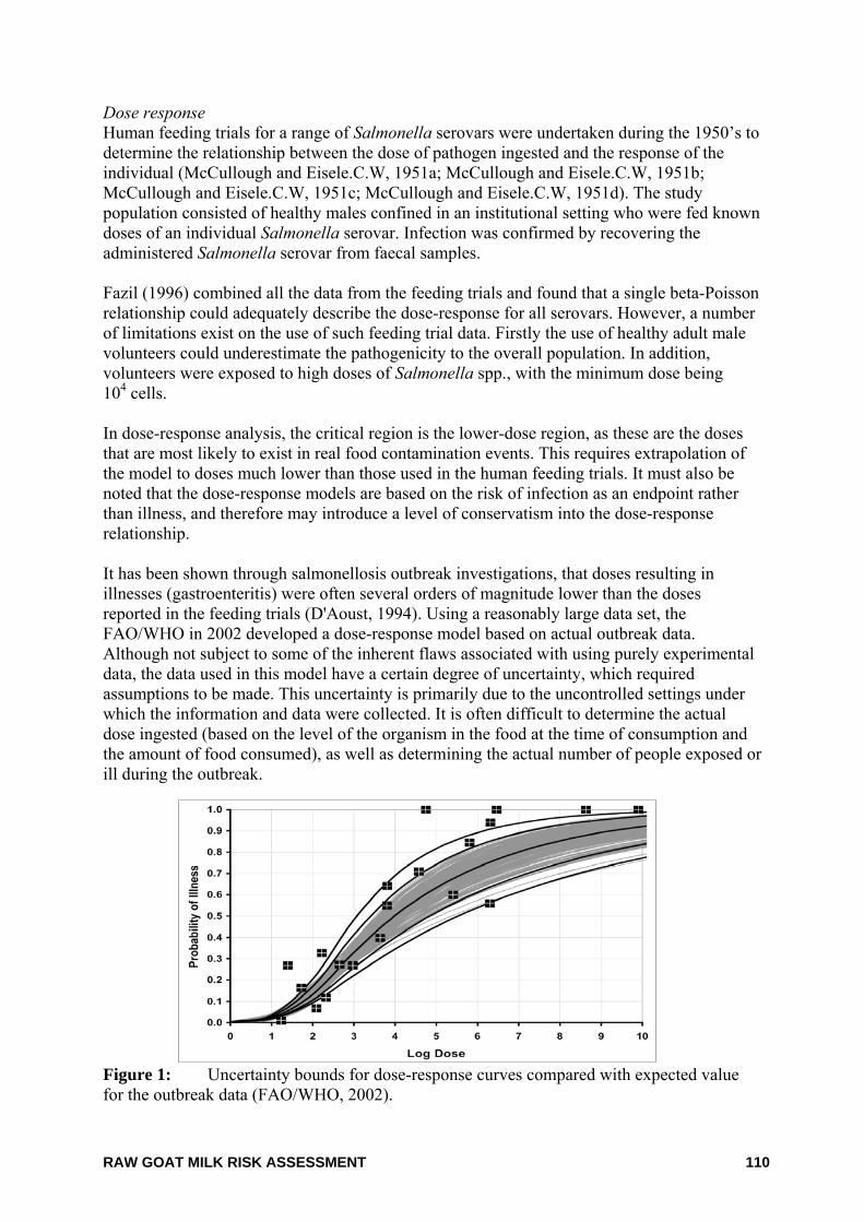
RAW GOAT MILK RISK ASSESSMENT 110
Dose response Human feeding trials for a range of Salmonella serovars were undertaken during the 1950’s to determine the relationship between the dose of pathogen ingested and the response of the individual (McCullough and Eisele.C.W, 1951a; McCullough and Eisele.C.W, 1951b; McCullough and Eisele.C.W, 1951c; McCullough and Eisele.C.W, 1951d). The study population consisted of healthy males confined in an institutional setting who were fed known doses of an individual Salmonella serovar. Infection was confirmed by recovering the administered Salmonella serovar from faecal samples. Fazil (1996) combined all the data from the feeding trials and found that a single beta-Poisson relationship could adequately describe the dose-response for all serovars. However, a number of limitations exist on the use of such feeding trial data. Firstly the use of healthy adult male volunteers could underestimate the pathogenicity to the overall population. In addition, volunteers were exposed to high doses of Salmonella spp., with the minimum dose being 104 cells. In dose-response analysis, the critical region is the lower-dose region, as these are the doses that are most likely to exist in real food contamination events. This requires extrapolation of the model to doses much lower than those used in the human feeding trials. It must also be noted that the dose-response models are based on the risk of infection as an endpoint rather than illness, and therefore may introduce a level of conservatism into the dose-response relationship. It has been shown through salmonellosis outbreak investigations, that doses resulting in illnesses (gastroenteritis) were often several orders of magnitude lower than the doses reported in the feeding trials (D'Aoust, 1994). Using a reasonably large data set, the FAO/WHO in 2002 developed a dose-response model based on actual outbreak data. Although not subject to some of the inherent flaws associated with using purely experimental data, the data used in this model have a certain degree of uncertainty, which required assumptions to be made. This uncertainty is primarily due to the uncontrolled settings under which the information and data were collected. It is often difficult to determine the actual dose ingested (based on the level of the organism in the food at the time of consumption and the amount of food consumed), as well as determining the actual number of people exposed or ill during the outbreak. Figure 1: Uncertainty bounds for dose-response curves compared with expected value for the outbreak data (FAO/WHO, 2002).
RAW GOAT MILK RISK ASSESSMENT 111
Host factors Individual susceptibility to Salmonella spp. infection and/or disease can vary significantly, depending on host factors such as pre-existing immunity, nutrition, age, ability to elicit an immune response, structural and functional anomalies of the intestinal tract, or pre-existing disease (Gerba et al., 1996; Jay et al., 2003). Individuals who are generally at greater risk of infection and/or risk of developing more severe outcomes from exposure to Salmonella spp. include the very young, the elderly, pregnant women and the immunucompromised (organ transplant patients, cancer patients and AIDS patients) (Gerba et al., 1996). References Adak, G.K., Long, S.M. and O'Brien, S.J. (2002) Trends in indigenous foodborne disease and deaths, England and Wales: 1992 to 2000. Gut 51(6):832-841.
Anon. (2003) Foodborne disease in Australia: incidence, notifications and outbreaks. Annual report of the OzFoodNet network, 2002. Commun Dis Intell. 27(2):209-243.
Ashbolt, R., Givney, R., Gregory, J.E., Hall, G., Hundy, R., Kirk, M., McKay, I., Meuleners, L., Millard, G., Raupach, J., Roche, P., Prasopa-Plaizier, N., Sama, M.K., Stafford, R., Tomaska, N., Unicomb, L. and Williams, C. (2002) Enhancing foodborne disease surveillance across Australia in 2001: the OzFoodNet Working Group. Commun Dis Intell. 26(3):375-406.
Bell, C. and Kyriakides, A. (2002) Salmonella: a practical approach to the organism and its control in foods. Blackwell Science, Oxford, UK.
Blumer, C., Roche, P., Spencer, J., Lin, M., Milton, A., Bunn, C., Gidding, H., Kaldor, J., Kirk, M., Hall, R., Della-Porta, T., Leader, R. and Wright, P. (2003) Australia's notifiable diseases status, 2001: annual report of the National Notifiable Diseases Surveillance System. Commun Dis Intell. 27(1):1-78.
Brenner, F.W., Villar, R.G., Angulo, F.J., Tauxe, R. and Swaminathan, B. (2000) Salmonella nomenclature. J Clin Microbiol 38(7):2465-2467.
Bryan, F.L. and Doyle, M.P. (1995) Health risks and consequences of Salmonella and Campylobacter jejuni in raw poultry. J Food Prot. 58(3):326-344.
Crerar, S.K., Nicholls, T.J. and Barton, M.D. (1999) Multi-resistant Salmonella Typhimurium DT104--implications for animal industries and the veterinary profession. Aust Vet J 77(3):170-171.
D'Aoust, J.Y. (1994) Salmonella and the international food trade. Int J Food Microbiol 24(1-2):11-31.
D'Aoust, J.Y. (1997) Salmonella species. In: Doyle, M.P., Beuchat, L.R., and Montville, T.J. eds. Food Microbiology: fundamentals and frontiers. ASM Press, Washington DC, USA, pp129-158.
Dalton, C.B., Gregory, J., Kirk, M.D., Stafford, R.J., Givney, R., Kraa, E. and Gould, D. (2004) Foodborne disease outbreaks in Australia, 1995 to 2000. Commun Dis Intell. 28(2):211-224.
FAO/WHO (2002) Risk Assessments of Salmonella in eggs and broiler chickens. Food and Agriculture Organization/World Health Organization, Geneva, Switzerland. ftp://ftp.fao.org/docrep/fao/005/y4392e/y4392e00.pdf.
Fazil, A.M. (1996) A quantitative risk assessment model for salmonella. Drexel University, Philadelphia P.A, Accessed on 1996.
Gerba, C.P., Rose, J.B. and Haas, C.N. (1996) Sensitive populations: who is at the greatest risk? Int J Food Microbiol 30(1-2):113-123.
RAW GOAT MILK RISK ASSESSMENT 112
Hall, G.V. (2003) Gastroenteritis survey and foodborne disease in Australia 2002. Working paper series. National Centre for Epidemiology and Population Health, Australian National University, Canberra, ACT.
Helms, M., Vastrup, P., Gerner-Smidt, P. and Molbak, K. (2003) Short and long term mortality associated with foodborne bacterial gastrointestinal infections: registry based study. BMJ 326(7385):357.
Hohmann, E.L. (2001) Nontyphoidal salmonellosis. Clin Infect Dis 32(2):263-269.
ICMSF. (1996) Salmonellae. In: Roberts, T.A., Baird-Parker, A.C., and Tompkin, R.B. eds. Microorganisms in foods 5: Microbiological specifications of food pathogens. First ed, Blackie Academic & Professional, London, UK, pp217-264.
Jay, S., Davos, D., Dundas, M., Frankish, E. and Lightfoot, D. (2003) Salmonella. In: Hocking, A.D. eds. Foodborne microorganisms of public health significance. 6 ed, Australian Institute of Food Science and Technology, NSW Branch, Food Microbiology Group, Waterloo, NSW, pp207-266.
McCullough, N. and Eisele.C.W. (1951a) Experimental human salmonellosis. I. Pathogenicity of strains of Salmonella meleagridis and Salmonella anatum obtained from spray-dried whole egg. J Infect Dis 88(3):278-289.
McCullough, N. and Eisele.C.W. (1951b) Experimental human salmonellosis. II. Immunity studies following experimental illness with Salmonella meleagridis and Salmonella anatum. J Immunol. 66(5):595-608.
McCullough, N. and Eisele.C.W. (1951c) Experimental human salmonellosis. III. Pathogenicity of strains of Salmonella newport, Salmonella derby, and Salmonella bareilly obtained from spray-dried whole egg. J Infect Dis 89(3):209-213.
McCullough, N. and Eisele.C.W. (1951d) Experimental human salmonellosis. IV. Pathogenicity of strains of Salmonella pullorum obtained from spray-dried whole egg. J Infect Dis 89(3):259-265.
Mead, P.S., Slutsker, L., Dietz, V., McCaig, L.F., Bresee, J.S., Shapiro, C., Griffin, P.M. and Tauxe, R.V. (1999) Food-related illness and death in the United States. Emerg.Infect Dis 5(5):607-625.
Roche, P., Spencer, J., Lin, M., Gidding, H., Kirk, M., Eyeson-Annan, M., Milton, A., Witteveen, D. and Merianos, A. (2001) Australia's notifiable diseases status, 1999: annual report of the National Notifiable Diseases Surveillance System. Commun Dis Intell. 25(4):190-245.
Yohannes, K., Roche, P., Blumer, C., Spencer, J., Milton, A., Bunn, C., Gidding, H., Kirk, M. and Della-Porta, T. (2004) Australia's notifiable diseases status, 2002: Annual report of the National Notifiable Diseases Surveillance System. Commun Dis Intell. 28(1):6-68.
RAW GOAT MILK RISK ASSESSMENT 113
13. Staphylococcus aureus The genus Staphylococcus is subdivided into 28 species and 8 subspecies. Staphylococcus aureus is a non-motile, Gram-positive, non-spore forming spherical bacterium. On microscopic examination, S. aureus appears in pairs, short chains, or bunched, grape-like clusters (Stewart, 2003). S. aureus is ubiquitous and inhabits the mucous membranes and skin of most warm-blooded animals, including all food animals and humans. Up to 50% of humans may carry this organism in their nasal passages and throats and on their hair and skin (FDA, 2003). S. aureus counts are often estimated by detecting coagulase-positive staphylococci, with further confirmatory tests required to specifically identify S. aureus. Nevertheless, the identification of coagulase-positive staphylococci or S. aureus is essentially an indicator test for the likelihood of enterotoxin production, as not all of these organisms have the ability to produce toxin, in addition, some strains of enterotoxin-producing staphylococci do not possess the coagulase enzyme (Stewart, 2003). Growth characteristics The temperature range for growth of S. aureus is 7 - 48°C with optimum growth occurring at 35 - 40°C. The temperature range for toxin production is 10 - 48°C with the optimum temperature being from 40 - 45°C. S. aureus grows over a wide water activity range (0.83 - 0.99) with an optimum water activity of >0.99. The pH range for growth is 4.0 - 10 and the pH range for toxin production is 4.5 - 9.6 (ICMSF, 1996). S. aureus is tolerable to salt up to 25% NaCl (water activity of 0.85). S. aureus grows under both aerobic and anaerobic conditions; however growth is better in the presence of oxygen. Toxins are also produced under both aerobic and anaerobic conditions with greatest toxin production in the presence of oxygen (Bergdoll, 1989). S. aureus is generally considered a poor competitor with other bacteria. S. aureus is readily killed at cooking and pasteurisation temperatures, however heat resistance is increased in dry, high-fat and high-salt foods. In contrast, S. aureus enterotoxins are extremely resistant to heat. Heat resistance for enterotoxin B has been reported at D149 = 100 minutes (water activity of 0.99) (ESR, 2001). Heat resistances for S. aureus vegetative cells have been reported at D60 = 0.43 - 8.0 minutes whereas a time/temperature equivalent for enterotoxin is 121°C for 3 - 8 minutes (Baird-Parker, 1990; ICMSF, 1996). The enterotoxin is not affected by frozen storage. Preservatives such as sorbate and benzoate are inhibitory to S. aureus, with their effectiveness increasing with a reduction in pH. Methyl and propyl parabens also have an effect on S. aureus, and high concentrations of carbon dioxide cause a substantial reduction in growth rates of S. aureus (Molin, 1985). Most chemical sanitisers used routinely in the food industry such as chlorine, other halogens and quaternary ammonium compounds destroy S. aureus on surfaces. However some strains, for example those that become established on poultry processing equipment, have increased resistance (Bolton et al., 1988).
RAW GOAT MILK RISK ASSESSMENT 114
Pathology of illness Staphylococcal foodborne illness is caused by the ingestion of food that contains preformed toxins produced by S. aureus. Usually this occurs when S. aureus is introduced into a food that will support growth of the organism, and that food is stored under conditions allowing the organism to grow and produce sufficient quantities of enterotoxin (Ash, 1997). Symptoms generally appear around 3 hours after ingestion but can occur in as little as 1 hour (range 1 - 6 hours) and are self-limiting (Ash, 1997; Stewart, 2003). Symptoms include nausea, vomiting, abdominal cramps of varying severity and diarrhoea. Some individuals may not demonstrate all of the symptoms associated with the illness. In severe cases, blood and mucus may be observed in stools and vomitus. Marked prostration, headaches and sweating accompany severe attacks and there may be fever or shock with subnormal temperatures and lowered blood pressure. Recovery is usually between 1 - 3 days requiring no medical treatment. Fatalities are rare, but are occasionally reported in young children and the elderly (Ash, 1997). All people are susceptible to staphylococcal food poisoning; however the intensity/severity may vary, depending on individual sensitivities. S. aureus is also an opportunistic pathogen that causes infections via open wounds. S. aureus causes several types of infection including skin eruptions and inflammations (boils, acne, sties, etc) and wounds (Ash, 1997). S. aureus can also cause respiratory infections or may become established in the gut causing enteritis. S. aureus is an important bacterial cause of mastitis (an inflammatory disease of the mammary gland) in cows (Akineden et al., 2001). Mastitis in dairy cattle is characterised by changes in the udder tissue, clots and changes in milk quality, and is sometimes accompanied by heat and pain in the udder. Mode of transmission Staphylococcal food poisoning is caused by the consumption of food containing enterotoxins produced by certain strains of S. aureus. Despite wide-spread association of S. aureus with animals, humans tend to be the main reservoir for S. aureus infections in humans. Hand contact by food handlers with ready-to-eat foods is an important means by which S. aureus may enter the food supply. Foods that present the greatest risk of causing illness are those in which the normal flora has been destroyed (e.g. cooked meats) or inhibited (e.g. cured meats containing high salt content) (Stewart, 2003). Incidence of illness Food poisoning caused by S. aureus is one of the most common types of foodborne diseases world-wide (ICMSF, 1996). The incidence of staphylococcal food poisoning is often under-reported due largely to the self-limiting nature of illness, with most people recovering within 1 - 2 days without requiring medical attention. Foods commonly associated with staphylococcal food poisoning are meat and poultry, dairy products (particularly cheese and cream due to inappropriate handling, as well as contaminated raw milk), salads, cream filled bakery products and processed meat. Improper storage/temperature abuse of food is the greatest factor attributing to outbreaks (Homberg and Blake, 1984). In July 2000, an extremely large outbreak of staphylococcal food poisoning occurred in Japan, with an estimate 13,420 people being affected (Asao et al., 2003). The source of the outbreak was traced to powdered low-fat milk produced at a single factory in Osaka and was used as an ingredient in a number of dairy products. Staphylococcal enterotoxin was detected
RAW GOAT MILK RISK ASSESSMENT 115
in the implicated milk powder; however, viable S. aureus was not isolated. This suggests that staphylococci were able to produce enterotoxin in the milk prior to pasteurisation, and remained immunologically and biologically active despite being pasteurised three times at 130°C for 2 – 4 seconds. Despite S. aureus not being a notifiable illness in Australia, in 2002, three outbreaks of food poisoning attributed to S. aureus were reported. In one outbreak, a meal of lamb, rice and potatoes was implicated, in which Bacillus cereus was also identified. Other outbreaks implicated rice served in a childcare centre and pizza as the causative agent (Ashbolt et al., 2002; Anon, 2003). An outbreak was also reported in 2001 from consumption of barbequed chicken strongly suggesting an enterotoxin-producing bacterium as the causative agent, possibly S. aureus (Armstong et al., 2002). In 2003, S. aureus was also implicated in foodborne illness after the consumption of a rice, beef and black bean sauce meal (Anon 2003). Mead et al. (1999) stated that sporadic illness from S. aureus is not reportable in the US through either passive or active systems. The authors estimated 185,060 illnesses, 1753 hospitalisations and 2 deaths per year are attributed to S. aureus illness via contaminated food (Mead et al., 1999). Between 1975 and 1982, 36% of all reported S. aureus illness in the US were attributed to red meat, 12.3% to salads, 11.3% to poultry, 5.1% to pastries and 1.4% attributed to milk products and seafoods. In 17.1% of cases the food involved was unknown (Genigeorgis, 1989). In Canada, the average number of cases of illness from S. aureus for the years 1975 - 1984 was 232 cases per year (Todd, 1992). Foods implicated included pork (ham), turkey, chicken, cheese, pasta, salads and sandwiches. In France, S. aureus was attributed to 16 of 530 foodborne disease outbreaks recorded between 1999 - 2000 (Le Loir et al., 2003). Of these outbreaks, 32% were attributed to milk products (especially cheeses),22% were attributed to meats, 15% were attributed to sausages and pies, 11% were attributed to fish and seafood, 11% were attributed to eggs and egg products and 9.5% were attributed to poultry 95% (Haeghebaert et al., 2002). In the UK for the years 1969 - 1981, 1 - 6% of all cases of bacterial food poisoning were attributed to S. aureus. From 1982 - 1990, 0.5 - 1% of all cases of bacterial food poisoning was attributed to staphylococcal food poisoning. For the years 1969 - 1990 a study of 359 incidents of staphylococcal food poisoning was investigated. Poultry and poultry products accounted for 22% of incidents, most attributed to cold cooked chicken and in nine incidents turkey was the food vehicle (Wieneke et al., 1993; Bertolatti et al., 1996). Occurrence in foods Animals carry S. aureus on various parts of their bodies. Cow’s udders and teats, and the tonsils and skin of pigs, chickens and turkeys are also known sources. Occurrence of staphylococci is common in raw milk. S. aureus in milk is related to the health status of the herd in respect to mastitis, and organisms numbers can range from <10 to several thousands per ml of milk with occasional counts of 105 cfu/ml (Asperger and Zangerl, 2002). The prevalence of coagulase-positive staphylococci (which can include S. aureus, S. intermedius and some S. hyicus) in Australian beef and sheep carcasses and boneless beef and sheep surveyed in 1998 were 24.3% (beef carcasses), 24.1% (sheep carcasses), 17.5%
RAW GOAT MILK RISK ASSESSMENT 116
(boneless beef) and 38.6% (boneless sheep), respectively (Phillips et al., 2001a; Phillips et al., 2001b; Phillips et al., 2005). Virulence and infectivity S. aureus forms a wide range of substances associated with infectivity and illness, including the heat stable enterotoxins that cause food poisoning (Ash, 1997). Eleven antigenic types of staphylococcal enterotoxins are currently recognised, with types A and D being most commonly involved in food poisoning outbreaks. To date, staphylococcal enterotoxins A, B, C1, C2, C3, D, E, G, H, I and J toxins have been identified (Balaban and Rasooly, 2000). These enterotoxins are single-chain proteins comprising a polypeptide chain containing relatively large amounts of lysine, tyrosine and aspartic and glutamic acids and characterised by containing only two residues of half cystine and one or two residues of tryptophan. Most of them possess a cystine loop required for proper conformation and which is probably involved in the emetic activity. They are highly stable, resist most proteolytic enzymes, such as pepsin or trypsin, and thus keep their activity in the digestive tract after ingestion. They also resist chymotrypsine, rennin and papain (Bergdoll, 1989). The production of enterotoxins is dependent on de novo synthesis within the cell. The quantity of toxin produced is variable and can be categorised by the type of toxin produced. Although weakly antigenic, enterotoxin antibodies have been produced in a variety of animal hosts. The mode of action of the toxin causing illness is not fully understood. However, it is thought that vomiting in response to ingestion of preformed toxin occurs due to the stimulation of local neuroreceptors in the intestinal tract which transmit the stimuli to the vomiting centre of the brain via the vagus nerve and other parts of the sympathetic nervous system (ICMSF, 1996). A number of studies have identified toxin genes present in S. aureus isolates from the milk of cows with mastitis (Akineden et al., 2001; Cenci-Goga et al., 2003; Lim et al., 2004; Zschock et al., 2004; Loncarevic et al., 2005). The rate of enterotoxigenic S. aureus isolates from dairy cattle is highly variable and demonstrates the diversity of S. aureus strains (Cenci-Goga et al., 2003). Dose response The amount of enterotoxin that must be ingested to cause illness is not known exactly, but it is generally believed to be in the range 0.1 - 1.0 µg/kg (ICMSF, 1996). Toxin levels within this range are typically reached when S. aureus populations exceed 100,000/g (Ash, 1997). Immune status All people are believed to be susceptible to staphylococcal intoxication, but the severity of symptoms may vary depending on the amount of food ingested and the susceptibility of the individual to the toxin. Food matrix The range of conditions that allow growth of staphylococci and the production of toxin vary with food type. The amount of starch and protein present in the food may enhance toxin production (Frazier and Westhoff, 1988).
RAW GOAT MILK RISK ASSESSMENT 117
References Akineden, O., Annemuller, C., Hassan, A.A., Lammler, C., Wolter, W. and Zschock, M. (2001) Toxin genes and other characteristics of Staphylococcus aureus isolates from milk of cows with mastitis. Clin.Diagn.Lab Immunol. 8(5):959-964.
Anon. (2003) Foodborne disease in Australia: incidence, notifications and outbreaks. Annual report of the OzFoodNet network, 2002. Commun Dis Intell. 27(2):209-243.
Armstong, P., Peacock, D. and Cameron, S. (2002) Gastroenteritis outbreak in a sporting team linked to a barbequed chicken. Communicable Disease Intelligence 26(3): http://www.cda.gov.au/pubs/cdi/2002/cdi2603/htm/cdi2603.htm,.
Asao, T., Kumeda, Y., Kawai, T., Shibata, T., Oda, H., Haruki, K., Nakazawa, H. and Kozaki, S. (2003) An extensive outbreak of staphylococcal food poisoning due to low-fat milk in Japan: estimation of enterotoxin A in the incriminated milk and powdered skim milk. Epidemiol Infect 130(1):33-40.
Ash, M. (1997) Staphylococcus aureus and staphylococcal enterotoxins In. A. D. Hocking, G. Arnold, I. Jenson, K. Newton and P. Sutherland (eds), Foodborne Microorganisms of Public Health Significance, 5th Edition. 313-332. AIFST (NSW Branch) Food Microbiology Group, Sydney.
Ashbolt, R., Bell, R., Crerar, S., Dalton, C., Givney, R., Gregory, J., Hall, G., Hardy, B., Hogg, G., Hundy, R., Kirk, M., Lalor, K., McKay, I., Madden, V., Markey, P., Meuleners, L., Merrett, T., Millard, G., Raupach, J., Roche, P., Sarna, M., Shadbolt, C., Stafford, R., Tomaska, N., Unicomb, L. and Williams, C. (2002) OzFoodNet: enhancing foodborne disease surveillance across Australia: quarterly report, January to March 2002. Commun Dis Intell. 26(3):430-435.
Asperger, H. and Zangerl, P. (2002) Staphlococcus aureus. In Encyclopedia of Dairy Science (eds. Roginski, Hl, Fuquay, J. W., Fox, P.). 2563-2569. Elsevier Science, London.
Baird-Parker, A.C. (1990) The stapylococci: an introduction. Journal Applied Bacteriology Symposium Supplement:1S-8S.
Balaban, N. and Rasooly, A. (2000) Staphylococcal enterotoxins. Int.J.Food Microbiol. 61:1-10.
Bergdoll, M.S. (1989) Staphylococcus aureus. In M.P Doyle (ed.), Foodborne Bacterial Pathogens. 463-513. Marcel Dekker Inc., New York.
Bertolatti, D., Hanelly, T., Bishop, M., French, E. and Marendoli, M. (1996) Staphylococcus aureus in Western Australian poultry. International Journal of Environmental Health Research 6(4):277-287.
Bolton, K.J., Dodd, E.E.R., Mead, G.C. and Waites, W.M. (1988) Chlorine resistance of strains of Staphylococcus aureus isolated from poultry processing plants. Letters in Applied Microbiology 6:31-34.
Cenci-Goga, B.T., Karama, M., Rossitto, P.V., Morgante, R.A. and Cullor, J.S. (2003) Enterotoxin production by Staphylococcus aureus isolated from mastitic cows. J.Food Prot. 66(9):1693-1696.
ESR. (2001) Staphylococcus aureus. Microbial Pathogen Datasheet. http://www.nzfsa.govt.nz/science-technology/data-sheets/staphylococcus-aureus.pdf.
FDA (2003) The bad bug book (Foodborne Pathogenic Microorganisms and Natural Toxins Handbook ). US Food and Drug Administration. http://www.cfsan.fda.gov/~mow/intro.html. Accessed on 8 August 2004.
Frazier, W.C. and Westhoff, D.C. (1988) Food Microbiology, 4th edition. McGraw Hill, Singapore.
Genigeorgis, C.A. (1989) Present state of knowledge on staphlococcal intoxication. Int.J.Food Microbiol. 9: 327-360.
RAW GOAT MILK RISK ASSESSMENT 118
Haeghebaert, S., Le Querrec, F., Gallay, A., Bouvet, P. and Gomez, M. (2002) les toxi-infections alimentaries collectives en France, en 1999 et 2000. Bull.Epidemiol.Hebdo. 23: 105-109.
Homberg, S.D. and Blake, P.A. (1984) Staphyloccocal food poisoning in the United States. J.Am.Med.Assoc. 251:487-489.
ICMSF (1996) Microorganisms in Food 5: Microbiological Specifications of Food Pathgens. Blackie Academic and Professional, London.
Le Loir, Y., Baron, F. and Gautier, M. (2003) Staphlococcus aureus and food posioning. Genetics and Molecular Reseach 2(1): 63-76.
Lim, S.K., Joo, Y.S., Moon, J.S., Lee, A.R., Nam, H.M., Wee, S.H. and Koh, H.B. (2004) Molecular typing of enterotoxigenic Staphylococcus aureus isolated from bovine mastitis in Korea. J.Vet.Med.Sci. 66(5):581-584.
Loncarevic, S., Jorgensen, H.J., Lovseth, A., Mathisen, T. and Rorvik, L.M. (2005) Diversity of Staphylococcus aureus enterotoxin types within single samples of raw milk and raw milk products. J Appl Microbiol 98(2):344-350.
Mead, P.S., Slutsker, L., Dietz, V., McCaig, L.F., Bresee, J.S., Shapiro, C., Griffin, P.M. and Tauxe, R.V. (1999) Food-related illness and death in the United States. Emerg.Infect Dis 5(5):607-625.
Molin, G. (1985) The resistance to carbon dioxide of some food related bacteria. Eur.J.Appl.Microbiol.Biotechnol. 18:214-217.
Phillips, D., Jordan, D., Morris, S. and Sumner, J.J.I. (2005) Microbiological quality fo Australian beef and sheepmeat - results of the industry's thrid national abattoir study. Meat & Livestock Australia.
Phillips, D., Sumner, J., Alexander, J.F. and Dutton, K.M. (2001a) Microbiological quality of Australian beef. J Food Prot. 64(5):692-696.
Phillips, D., Sumner, J., Alexander, J.F. and Dutton, K.M. (2001b) Microbiological quality of Australian sheep meat. J Food Prot. 64(5):697-700.
Stewart, C.M. (2003) Staphylococus aureus and staphylococcal enterotoxins. In Foodborne Microorganisms of Public Health Significance, 6th Edition, AIFST (NSW) Branch. 359-380. Southwood Press Pty Ltd, Marrickville.
Todd, E.C.D. (1992) Foodborne disease in Canada – a 10 year summary from 1975 to 1984. Journal Applied Microbiology 55(2):123-132.
Wieneke, A.A., Roberts, D. and Gilbert, R.J. (1993) Staphylococcal food poisoning in the United Kingdom, 1969-90. Epidemiology and Infection 110(3):519-531.
Zschock, M., Risse, K. and Sommerhauser, J. (2004) Occurrence and clonal relatedness of sec/tst-gene positive Staphylococcus aureus isolates of quartermilk samples of cows suffering from mastitis. Lett.Appl.Microbiol. 38(6):493-498.
RAW GOAT MILK RISK ASSESSMENT 119
14. Streptococcus spp. The term, ‘Streptococcus’, was first used to describe the chain-forming, coccoid bacteria that had been observed in wounds and discharges of animals (ICMSF, 1996). Streptococci are Gram-positive, spherical or ovoid, non-motile bacteria. The classification of the genus Streptococcus has long been in a state of flux (Jones, 1978), however, current information groups them into pyogenic streptococci and enterococci. Pyogenic streptococci include Strptococcus pyogenes and Streptococcus agalactiae. Enterococci include Enterococcus faecalis and Enterococcus faecium. The genus is sorted into Groups A, B, C, D, F and G on the basis of antigenic, haemolytic and physiological characteristics. Streptococci from Groups A and D can be transmitted to humans via food (FDA, 2003). Streptococcus zooepidemicus (Group C) has been implicated in several episodes of human illness, including death (Barrett, 1986). S. agalactiae is a major cause of bovine mastitis (ICMSF, 1996) and is a highly contagious obligate parasite of the mammary gland (Martinez et al., 2000). Growth characteristics The temperature range for growth of S. pyogenes is 10 - 45°C with optimum growth occurring at 37oC. The pH range for growth is 4.8 - 9.3 (ICMSF, 1996). S. pyogenes is tolerable to salt between 4 - 6.5%. S. pyogenes have the ability to grow aerobically or microaerophilically. Experiments conducted by Obiger (1976) found that S. pyogenes would not survive exposure to 66ºC for 20 – 40 seconds resulting in a calculated D-value at 66ºC of 0.1 -0.2 minutes. Heat resistance figures reported by (Stumbo, 1973) included a D-value at 65.6ºC of 0.2 - 2.0 and a z value of 4.4 - 6.7ºC. Based on these figures, ICMSF (1996) conclude that pasteurisation at 62ºC for 30 minutes and 70ºC for 30 seconds would ensure only a 1.6 - 2.3 decimal reduction of S. pyogenes. However, using the D-value at 66ºC of 0.2 as per Obiger (1976), pasteurisation would result in a 20 decimal reduction of S. pyogenes in milk. Group A streptococci grow poorly in raw milk, but there is some evidence that pyogenic streptococci may multiply in raw meat held at ambient temperature (Fraser et al., 1977). Pathology of illness The symptoms of group A streptococcal infection include sore and red throat, pain on swallowing, tonsillitis, high fever, headache, nausea, vomiting, malaise and rhinorrhea. A rash may occur within the first few days. Group A streptococci may also cause acute rheumatoid fever following infection of the upper respiratory tract, and acute glomerulonephritis after skin infection (ICMSF, 1996). Although rare, complications may occur when the bacteria enter the blood, muscles or lungs. These infections are termed “invasive group A streptococcal disease”. Two of the least common but most severe forms of thisdisease are necrotising fasciitis and Streptococcal Toxic Shock Syndrome. Necrotising fasciitis destroys the muscles, fat and skin tissue. Streptococcal Toxic Shock Syndrome causes blood pressure to drop rapidly and organs, such as the kidneys, liver and lungs, to fail. About 20% of patients with necrotising fasciitis and more than 50% with STSS die.
RAW GOAT MILK RISK ASSESSMENT 120
Group D streptococci infections may result in a clinical syndrome similar to staphylococcal intoxication. The symptoms commence within 2 - 36 hours of infection and include diarrhoea, abdominal cramps, nausea, vomiting, fever, chills and dizziness (FDA 2003). Mode of transmission Humans are usually the source of contamination of pyogenic streptococcal infections. Transmission occurs from infected hosts to foods. The bacteria are generally spread via direct contact with mucus from the nose or throat of infected persons, or through contact with infected wounds or sores on the skin (FDA 2003). Group A streptococci may be carried in the throat or on the skin of people with no symptoms of illness (CDC, 2005). Group C streptococci (e.g. S. zooepidemicus) is found in the nasopharynx, tonsils, respiratory tract and genital mucous of cattle and horses and has been associated with mastitis in goats, sheep and cows along with other mammals. Human infection with these organisms is most often due to direct contact with animals or the ingestion of unpasteurised milk and milk products (Kuusi et al., 2006). Incidence of illness Outbreaks of septic sort throat and scarlet fever were numerous prior to the introduction of milk pasteurisation. Most current outbreaks have involved foods such as salads, with the source of infection being an infected food handler. Outbreaks of Group D streptococcal infections are not common and have usually been the result of unsanitary preparation, storing or handling of food (FDA 2003). In 2006 an outbreak occurred involving7 cases of S. zooepidemicus caused by the consumption of unpasteurised goats milk cheese in Finland (Kuusi et al., 2006). A further outbreak of foodborne illness due to S. zooepidemicus (Group C) involving at least 11 cases occurred in the UK in 1984, with 7 deaths occurring during the outbreak. Unpasteurised milk from a dairy herd that had experienced intermittent mastitis was implicated as the source of infection (Edwards et al., 1988). Sixteen cases of invasive group C streptococcal infection were identified in northern Mexico between July 25 - September 9 1983. The organism was isolated from the blood of 15 patients and from the pericardial fluid of one patient. A homemade white cheese produced from raw cow’s milk at a small family dairy in northern Mexico was indicated as the food source of the infection, with samples testing positive for streptococci. The cows at the dairy were found to have mammary infections due to S. zooepidemicus (MMWR, 1983). In 1984, there was one outbreak of S. zooepidemicus associated with the consumption of raw milk in England. Twelve people were admitted to hospital with meningitis or endocarditis. Eight of the twelve died, although the infection was not necessarily the primary cause of death. Ten of the patients were aged over 70 years, and one was a one-day-old infant. Cows at a local dairy that had supplied the milk were subsequently found to be excreting S. zooepidemicus in their milk (Barrett 1986).
RAW GOAT MILK RISK ASSESSMENT 121
Occurrence in foods Food associated with streptococcus Group A foodborne illness include milk, ice cream, eggs, steamed lobster, ground ham, potato salad, egg salad, custard, rice pudding and shrimp salad. Foodstuffs were allowed to stand at room temperature for several hours between preparation and consumption in almost all cases. Poor hygiene, ill food handlers or the use of unpasteurised milk were the main routes for streptococcus Group A into food (FDA 2003). Food sources for streptococcus Group D foodborne illness include sausage, evaporated milk, cheese, meat croquettes, meat pie, pudding, raw milk and pasteurised milk. Under processing and/or poor food preparation is the usual mechanisms for entrance into the food chain (FDA 2003). 200 samples of raw milk from Zulia State, Venezuela were examined, with 19 samples testing positive for the presence of Streptococcus spp. Seventeen samples were positive for Enterococcus (Faria-Reyes et al., 2002). Results from the microbiological testing of 77,172 milk samples submitted to the Wisconsin Veterinary Diagnostic laboratory from January 1994 - June 2001 were analysed. Milk samples obtained included cases of clinical and subclinical mastitis as well as samples obtained from mastitis surveillance programmes. The proportion of samples from which Streptococcus spp. were isolated decreased from 8.1% in 1994 to 3.0% in 2001 (Makovec and Ruegg, 2003). Raw bulk tank milk samples from 48 dairy farms in New York State were tested over a five month testing period. Streptococci accounted for 69% of the total bacterial counts. The most commonly identified streptococcal species were Streptococcus uberis (found in 81% of the bulk milk samples), Aerococcus viridans (found in 50% of the bulk milk samples) and S. agalactiae (found in 31% of the bulk milk samples) (Zadoks et al., 2004). Virulence and infectivity Pyogenic streptococci possess specific virulence proteins which enable the organism to adhere to epithelial cells and protect the streptococci from phagocytosis (ICMSF, 1996). Dose response The infectious dose for streptococcus Group A is likely to be quite low, with less than 1,000 organisms required for infection (FDA 2003). In contrast, it is estimated that foodborne streptococcus Group D has a high infectious dose of greater than 107 organisms. Host factors All individuals in a population are equally susceptible to streptococcal illness (FDA 2003). People with chronic illnesses such as cancer, diabetes and kidney dialysis and those using medications such as steroids have a higher risk of getting invasive group A streptococcal disease (CDC, 2005).
RAW GOAT MILK RISK ASSESSMENT 122
References Barrett, N.J. (1986) Communicable disease associated with milk and dairy products in England and Wales: 1983-1984. J Infect 12(3):265-272.
CDC (2005) Group A streptococcal (GAS) disease. Centers for Disease Control and Prevention. Department of Heath and Human Services. http://www.cdc.gov/ncidod/dbmd/diseaseinfo/groupastreptococcal_g.htm. Accessed on 1 February 2005.
Edwards, A.T., Roulson, M. and Ironside, M.J. (1988) A milk-borne outbreak of serious infection due to Streptococcus zooepidemicus (Lancefield Group C). Epidemiol.Infect. 101(1):43-51.
Faria-Reyes, J., Garcia-Urdaneta, A., Izquierdo-Corser, P., Allara-Cagnasso, M. and Valero-Leal, K. (2002) Isolation of Gram-positive bacteria from raw milk with antimicrobial residues. Archivos Latinoamericanos de Nutricion.2002 52(1):68-73.
FDA (2003) The bad bug book (Foodborne Pathogenic Microorganisms and Natural Toxins Handbook ). US Food and Drug Administration. http://www.cfsan.fda.gov/~mow/intro.html. Accessed on 8 August 2004.
Fraser, C.A., Ball, L.C., Morris, C.A. and Noah, N.D. (1977) Serological characterization of group-A streptococci associated with skin sepsis in meat handlers. J.Hyg.(Lond) 78(2):283-296.
ICMSF (1996) Microorganisms in Food 5: Microbiological Specifications of Food Pathgens. Blackie Academic and Professional, London.
Jones, D. (1978) Composition and differentiation of the genus Streptococcus. Soc.Appl.Bacteriol.Symp.Ser. 7:1-49.
Kuusi, M., Lahti, E., Virolainen, A., Hatakka, M., Vuento, R., Rantala, L., Vuopio-Varkila, J., Seuna, E., Karppelin, M., Hakkinen, M., Takkinen, J., Gindonis, V., Siponen, K. and Huotari, K. (2006) An outbreak of Streptococcus equi subspecies zooepidemicus associated with consumption of fresh goat cheese. BMC.Infect Dis 6(1):36.
Makovec, J.A. and Ruegg, P.L. (2003) Results of milk samples submitted for microbiological examination in Wisconsin from 1994 to 2001. Journal of Dairy Science.2003 86(11):3466-3472.
Martinez, G., Harel, J., Higgins, R., Lacouture, S., Daignault, D. and Gottschalk, M. (2000) Characterization of Streptococcus agalactiae isolates of bovine and human origin by randomly amplified polymorphic DNA analysis. J.Clin.Microbiol. 38(1):71-78.
MMWR (1983) Group C streptococcal infections associated with eating homemade cheese - New Mexico. 1983/32 (39), pp510-516. http://www.cdc.gov/epo/mmwr/preview/mmwrhtml/00000150.htm. Accessed on 9 May 2005.
Obiger, G. (1976) [Studies of heat resistance of important pathogens during milk pasteurization.]. Archiv fuer Lebensmittelhygiene 27(4):137-144.
Stumbo, C.R. (1973) Thermal resistance in bacteria. In: Stumbo, C.R. eds. Thermbacteriology in Food Processing. Academic Press, New York and London, pp99-120.
Zadoks, R.N., Gonzalez, R.N., Boor, K.J. and Schukken, Y.H. (2004) Mastitis-causing streptococci are important contributors to bacterial counts in raw bulk tank milk. Journal of Food Protection 67(12):2644-2650.
RAW GOAT MILK RISK ASSESSMENT 123
15. Toxoplasma gondii The protozoan parasite Toxoplasma gondii is the cause of the potentially severe disease toxoplasmosis. T. gondii is a ubiquitous parasite that occurs in all areas of the world. It can infect a wide range of animals with the primary host belonging to the cat family (Felidae) and secondary hosts including all warm blooded animals (such as mammals and birds) (Tenter et al., 2000). T. gondii causes great losses in sheep and goats; however the disease is more severe in goats (Hill and Dubey, 2003). T. gondii also has the ability to survive in faeces, soil and in other environments. Growth characteristics T. gondii is an obligate intracellular coccidian protozoan which has a complex lifecycle involving sexual reproduction and oocyst production in a primary carnivorous host (cats) and asexual reproductions phase in the secondary host (generally mammals and birds, including livestock and humans). The life cycle is completed when the primary host consumes the secondary host and is infected with cysts, which have been formed previously in its tissues and the cycle continues (Jones et al., 2000). There are three identified infectious stages of T. gondii during its lifecycle; these are tachyzoites and bradyzoites, which are found within the tissue cysts of infected animals, and oocysts (containing sporozoites) (Tenter et al., 2000). Pathology of illness The majority of infections in humans tend to be asymptomatic. Additionally, initial infection usually results in lifelong immunity against further infection. It is generally only in immunocompromised individuals and in pregnancies where clinical manifestations of T. gondii arise (Tenter et al., 2000). However, immunocompetent individuals may exhibit self- limiting mild flu-like symptoms such as swollen lymph glands, malaise, fatigue, joint and muscle pain (Dawson, 2005). In immunocompromised individuals T. gondii may cause severe encephalitis (especially in patients with AIDS), eye infections, damage to the central nervous system and pulmonary infections. T. gondii infection is very serious in cases where the secondary host is pregnant as this organism has the ability to cause spontaneous abortion or severe congenital defects in the off-spring of the host (Tenter et al., 2000). If a pregnant woman, who has not been previously infected with T. gondii, becomes infected during her pregnancy the parasite can pass through the placenta to the foetus and cause congenital defects in the unborn child. These may include loss of the child’s vision, damage to the child’s central nervous system, mental retardation, abnormalities and possible neonatal death/abortion (Goldsmid et al., 2003). Mode of transmission Transmission of T. gondii occurs via the faecal-oral route, transplacental transfer between mother and foetus, and through the consumption of infected meat and/or milk containing tachyzoites or other forms of the infective parasite from the secondary host (Chiari and Neves, 1984; Skinner et al., 1990; Tenter et al., 2000; Smith, 2006).
RAW GOAT MILK RISK ASSESSMENT 124
Incidence of illness Toxoplasmosis is widespread in humans, being one of the most common parasitic zoonoses worldwide (Tenter et al., 2000). It is estimated that up to one third of the human population may have been infected with T. gondii, the majority of cases being asymptomatic (Montoya and Liesenfeld, 2004). Clinical manifestations of toxoplasmosis, caused by T. gondii, are generally confined to immunocompromised individuals and during pregnancies. Occurrence in foods Infectious oocysts tend to be very resistant particles and hence have the ability to survive well in many different environments. Contaminated drinking water and other foods exposed to water containing the organism, such as horticultural produce, may be a method of transmission of the organism to humans (Dawson 2005). One study suggests that T. gondii has the ability to survive in refrigerated unpasteurised goat’s milk (Walsh et al., 1999). Food sources that have been linked as the probable route of infection in outbreaks of T. gondii include raw goat’s milk, raw or rare lamb, beef and venison (Smith 2006). Viable infectious tissue cysts may be present in raw or undercooked meats and consumption can lead to infection. Additionally, the organism may be transmitted through the raw milk of an infected mammal such as goat or sheep’s milk (Sacks et al., 1982; Skinner et al., 1990; Tenter et al., 2000; Goldsmid et al., 2003). Consumer hygiene may be an important factor in toxoplasmosis, especially after contact with domestic cats, their faeces or through gardening and before handling and consuming foods. Cross contamination of raw meat and ready-to-eat products may also be a possible mechanism of transmission (Jones et al., 2000). Virulence and infectivity T. gondii has a high level of virulence with the ability to survive well in a wide range of environments and to infect a large variety of animals either as primary or secondary hosts (Tenter et al., 2000). Dose response There is a lack of information on the dose-response relationship for T. gondii. However, it is assumed that consumption of a single oocysts or tissue cysts is able to initiate infection in humans (Tenter et al., 2000; Hill and Dubey, 2003; Montoya and Liesenfeld 2004). Host factors Epidemiologic studies have identified risk factors such as immunocompromised individuals and those who are pregnant. Additionally, factors such as owning a cat and having poor food hygiene skills, may also place a consumer at additional risk of infection (Jones et al., 2000).
RAW GOAT MILK RISK ASSESSMENT 125
References Chiari, C.A. and Neves, D.P. (1984) Human toxoplasmosis acquired by ingestion of goat's milk. Mem.Inst.Oswaldo Cruz 79(3):337-340.
Dawson, D. (2005) Foodborne protozoan parasites. International Journal of Food Microbiology 103(2):207-227.
Goldsmid, J.M., Speare, R. and Bettiol, S. (2003) The Parasitology of Foods. In: Hocking, A.D. eds. Foodborne Microorganisms of Public Health Significance. 6th Edition ed, AIFST, Sydney, pp703-721.
Hill, D.H. and Dubey, J.P. (2003) Toxoplasma gondii. In: Torrence, M.E. and Isaacson, R.E. eds. Microbial food safety in animal agriculture. Chapter 36. Iowa State Press, Iowa, pp359-367.
Jones, J.L., Kruszon-Moran, D., Wilson, M., McQuillan, G., Navin, T. and McAuley, J.B. (2000) Toxoplasma gondii Infection in the United States: Seroprevalence and Risk Factors. Americal Journal of Epidemiology 154(4):357-365.
Montoya, J.G. and Liesenfeld, O. (2004) Toxoplasmosis. Lancet 363(9425):1965-1976.
Sacks, J.J., Roberto, R.R. and Brooks, N.F. (1982) Toxoplasmosis infection associated with raw goat's milk. JAMA 248(14):1728-1732.
Skinner, L.J., Timperley, A.C., Wightman, D., Chatterton, J.M.W. and Ho-Yen, D.O. (1990) Simultaneous diagnosis of toxoplasmosis in goats and goatowner's family. Scandinavian Journal of Infectious Diseases 22(3):359-361.
Smith, J.L. (2006) Documented outbreaks of toxoplasmosis: Transmission of Toxoplasma gondii to humans. Journal of Food Protection 56:630-639.
Tenter, A.M., Heckeroth, A.R. and Weiss, L.M. (2000) Toxoplasma gondii: from animals to humans. International Journal for Parasitology 30(12-13):1217-1258.
Walsh, C.P., Hammond, S.E., Zajac, A.M. and Lindsay, D.S. (1999) Survival of Toxoplasma gondii tachyzoites in goat milk: potential source of human toxoplasmosis. J Eukaryot.Microbiol 46(5):73S-74S.
RAW GOAT MILK RISK ASSESSMENT 126
16. Yersinia enterocolitica Yersinia is a facultative anaerobic organism, a member of the Enterobacteriaceae family (Farmer, 1995). Among 11 named species in the genus Yersinia, 3 are considered important human pathogens. Yersinia pestis is the cause of the plague. Yersinia pseudotuberculosis and Yersinia enterocolitica are enteropathogenic strains. Y. pestis and Y. pseudotuberculosis do not frequently infect humans. Y. enterocolitica is more commonly found in human clinical specimens. Y. enterocolitica are Gram-negative, small rods with dimension in the range of 0.5 - 0.81µm by 1 - 3 µm. Young cells of Y. enterocolitica are oval or coccoid shape. The organism produces peritrichous flagella and is actively motile when it is grown at 25oC but not at 35oC (Forsythe, 2000). Y. enterocolitica is often isolated from faeces but also from wounds, sputum and mesenteric lymph nodes of patients and sick animals. Y. enterocolitica is found in cows, pigs, cats, dogs and birds; and in water, soil and a variety of food. However, they are not part of the normal human flora (FDA, 2003). Growth characteristics Optimal growth temperature for Y. enterocolitica is at approximately 22 - 28oC, however the organism can grow at refrigerated temperatures and up to 44 oC. The ability to grow at –5oC has also been reported (Barton and Ribins-Browne, 2003). Its optimum pH for growth is between 7 - 8, with growth occurring between 4.6 - 10. Minimum water activity for growth is 0.945. Y. enterocolitica is able to grow in the presence or absence of oxygen, but growth in the absence of oxygen is retarded at refrigerated temperatures. It has been reported that Y. enterocolitica can survive in spring water stored at 4oC for up to 64 weeks. Survival of Y. enterocolitica is enhanced at low temperatures when the environment pH is below the minimum allowing for its growth. The D values for Y. enterocolitica are approximately 2 minutes at 55oC, 0.5 minutes at 60oC and 2 seconds at 65oC (Forsythe, 2000). The D value for Y. enterocolitica in milk at 62.8oC is 0.24 – 0.96 minutes (Lovett et al., 1982). As such, cells of Y. enterocolitica in milk are readily inactivated by pasteurisation. Pathology of illness Yersiniosis refers to the illness caused by Y. enterocolitica. Yersiniosis is characterised by gastroenteritis with diarrhoea and/or vomiting, fever and abdominal pain. Many patients seek medical attention for persistent fever, night sweats, or secondary features of the disease. Self-limiting enterocolitis is the most usual syndrome and often seen in young children (Barton and Ribins-Browne, 2003). Mesenteric lymphadenitis caused by Y. enterocolitica shows symptoms similar to appendicitis, and can be seen in older children or adolescents. Long-term sequelae as a result of infection by Y. enterocolitica include reactive arthritis, erythema nodosum, uveitis and others. Incubation period for enterocolitis is 24 - 36 hours or longer and the illness lasts usually 1 - 3 days. Duration of excretion of the organisms in the stool of infected patients ranges from 14 - 97 days (Cover and Aber, 1989).
RAW GOAT MILK RISK ASSESSMENT 127
Mode of transmission Cells of pathogenic Y. enterocolitica that are ingested and travelled through the gastrointestinal tract can bind to the epithelial cells of the ileum, penetrate the intestinal mucosa and colonise the Peyer’s Patches. Cells that multiply may spread to the mesenteric lymph nodes via the lymphatics and in rare situations may spread to the bloodstream, liver and spleen (Barton and Ribins-Browne, 2003). Pigs are the primary source of human infections of yersiniosis. Y. enterocolitica is carried in healthy pigs worldwide. Tonsils and oral cavities of pigs are generally heavily contaminated. Consumption and handling of raw pork meat are a primary source of human infection by Y. enterocolitica (Barton and Ribins-Browne, 2003). Incidence of illness Since its peak in the early 1990s, there has been a continuing decline in the number of yersiniosis in Australia, as reported by the National Notifiable Disease Surveillance Systems. As such, yersiniosis is no longer a notifiable disease since 2001 (Lin et al., 2002). The OzFoodNet recorded 117 cases of yersiniosis in 2002, representing 1.7 cases per 100,000 of the population. Most cases of foodborne yersiniosis are sporadic, but some outbreaks have been reported. In September - October of 1976, an outbreak of illness due to consumption of Y. enterocolitica contaminated chocolate milk in the US affected 218 people including 36 hospitalisation and 16 appendectomies. Investigations found that pasteurised milk was contaminated during the mixing by hand of chocolate syrup (Black et al., 1978). In October 1995, another outbreak in the US reported 10 cases of yersiniosis associated with consumption of pasteurised milk with 3 hospitalisations and 1 appendectomy. The research found that the pasteurised milk was possibly contaminated post-pasteurisation by unchlorinated rinsing water and dairy pigs were identified as the most likely source of Y. enterocolitica (Ackers et al., 2000). An investigation of an Australian outbreak of yersiniosis associated with consumption of pasteurised milk in 1987 - 1988 reported 11 cases of Y. enterocolitica enteritis among which three were presented as appendicitis (Butt et al., 1991b). Other than milk, tofu (Tacket et al., 1985), pig meat products and bean sprouts have been implicated as vehicles of outbreaks of yersiniosis. Yersiniosis caused by Y. enterocolitica appears to be a particular health problem in northern Europe, Scandinavia, parts of North America, Japan and New Zealand (Barton and Ribins-Browne, 2003). The number of reported yersiniosis is high in New Zealand where the incidence of yersiniosis is 15.1 cases per 100,000 population in 1998 and 13.9 cases in 1999 (ESR, 2001). In Finland, the reported varied from 11.7 - 17.5 cases per 100,000 population. Occurrence in foods Y. enterocolitica is ubiquitous; frequently found in soil, water, animals and can grow in a variety of foods even at refrigeration temperatures. It has been found in many food sources such as raw milk and cream, meat and meat products, oysters, vegetables, fish, and poultry (Barton and Ribins-Browne, 2003). It has also been isolated from well water, streams, lakes, and soil.
RAW GOAT MILK RISK ASSESSMENT 128
Virulence and infectivity There are 5 biotypes (described as biotype 1A, 1B, 2, 3, 4 and 5) and at least 60 O-antigen14 serological groups. Human infections are mainly caused by a small number of pathogenic bioserotypes that carry a plasmid encoding a number of virulence factors (Barton and Ribins-Browne, 2003). Bioserotype 4,O:3 is the most common pathogenic Y. enterocolitica found in humans worldwide. In addition, bioserotype 2,O:9, 2,O:5,27 and 3,O:5,27 are important human pathogens reported in Northern Europe, and Bioserotype 1B,O:8; 1B,O:13a,13b, 1B,O:20, 1B,O:21 are important pathogens in North America. The North American biotypes are more virulent than those of the Northern Europe (Barton and Ribins-Browne, 2003). The genes encoding for invasion of mammalian cells are located on the chromosome, and other virulence factors are associated with a 70-kb virulence plasmid in pathogenic bioserotypes (Forsythe, 2000). The North American biotype 1B strains carry a high pathogenicity island on their chromosome, which enhances their virulence (Barton and Ribins-Browne, 2003). In Australia, biotype O:3 and O:6,30 have been reported in outbreak investigations (Butt et al., 1991a). Dose response Although the minimum infectious dose of Y. enterocolitica is not known (Forsythe, 2000), there is estimation that the infective dose is around 106 (Health Canada, 2001) to 107 cells (Granum et al., 1995). Immune status People most susceptible to yersiniosis and the subsequent complications are the very young, the debilitated, the very old and persons undergoing immunosuppressive therapy (FDA 2003). In 2000, notification rate of yersiniosis in Australia was 3.6 cases per 100,000 for the 0 - 4 years old sub-population (male) and 1.5 cases per 100,000 for 0-4 years old (female) sub-population, and the remaining populations was at 0 - 1 cases per 100,000 (Lin et al., 2002). Food matrix Survival and growth of Y. enterocolitica in food is influenced by pH, water activity, salt content, temperature of storage, oxygen availability and carbon dioxide levels, competing microflora, and food additives in the food matrix. Y. enterocolitica has been found to multiply in cottage cheese that contained no sorbic acid. Conversely, Y. enterocolitica could not be isolated from ripening hard goats’ milk cheeses (Tornadijo et al., 1993) or Swiss-hard or semi-hard cheeses made with raw milk (Bachmann and Spahr, 1995). In the absence of competing microflora, Y. enterocolitica can multiply to high numbers in foods, such as pasteurised milk (Black et al., 1978). However, the presence of starter culture on the other hand, had an inhibitory effect on the growth of Y. enterocolitica in Turkish Feta cheese (Bozkurt and Erkmen, 2001). It has been demonstrated that the growth of Y. enterocolitica in milk could be inhibited by the presence of a bacteriocin producing Yersinia kristensenii (Toora et al., 1994) or propionicin producing Propionibacterium thoenii (Lyon et al., 1993).
14 Refers to lipopolysaccharide-protein somatic antigens of the microorganism.
RAW GOAT MILK RISK ASSESSMENT 129
References Ackers, M.L., Schoenfeld, S., Markman, J., Smith, M.G., Nicholson, M.A., DeWitt, W., Cameron, D.N., Griffin, P.M. and Slutsker, L. (2000) An outbreak of Yersinia enterocolitica O:8 infections associated with pasteurized milk 20. J Infect Dis 181(5):1834-1837.
Bachmann, H.P. and Spahr, U. (1995) The fate of potentially pathogenic bacteria in Swiss hard and semihard cheeses made from raw milk. J Dairy Sci 78(3):476-483.
Barton, M.D. and Ribins-Browne, R.M. (2003) Yersinia enterocolitica. In: Hocking, A.D. eds. Foodborne Microorganisms of Public Health Significance. Sixth ed, Chapter 19. Australian Institute of Food Science and Technology Incorporated, Waterloo DC NSW, pp577-596.
Black, R.E., Jackson, R.J., Tsai, T., Medvesky, M., Shayegani, M., Feeley, J.C., MacLeod, K.I. and Wakelee, A.M. (1978) Epidemic Yersinia enterocolitica infection due to contaminated chocolate milk 140. N.Engl .J.Med. 298(2):76-79.
Bozkurt, H. and Erkmen, O. (2001) Predictive modelling of Yersinia enterocolotica inactivation in Turkish Feta cheese during storage. J Food Eng 47:81-87.
Butt, H.L., Gordon, D.L., Lee-Archer, T., Moritz, A. and Merrell, W.H. (1991a) Relationship between clinical and milk isolates of Yersinia enterocolitica 64. Pathology 23(2):153-157.
Butt, H.L., Gordon, D.L., Lee-Archer, T., Moritz, A. and Merrell, W.H. (1991b) Relationship between clinical and milk isolates of Yersinia enterocolitica 64. Pathology 23(2):153-157.
Cover, T.L. and Aber, R.C. (1989) Yersinia enterocolitica. N.Engl.J.Med. 321:16-24.
ESR. (2001) Fact sheet of Yersinia enterocolitica.
Farmer, J.J. (1995) Enterobacteriaceae: introduction and identification. In: Murray, P.R., Baron, E.J., Pfaller, M.A., Tenover, F.C., and Yolken, R.H. eds. Manual of Clinical Microbiology. 6th editon. Washington DC: ASM Press 1995:438-48. 6th edition ed, ASM, Washington DC, pp438-448. Manual of Clinical Micobiology.
FDA (2003) The bad bug book (Foodborne Pathogenic Microorganisms and Natural Toxins Handbook ). US Food and Drug Administration. http://www.cfsan.fda.gov/~mow/intro.html. Accessed on 8 August 2004.
Forsythe, S.T. (2000) The microbiology of safe food. Blackwell Science, Oxford.
Granum, P.E., Thomas, J.M. and Alouf, J.E. (1995) A survey of bacterial toxins involved in food poisoning: a suggestion for bacterial food poisoning toxin nomenclature. Int J Food Microbiol 28(2):129-144.
Lin, M., Roche, P., Spencer, J., Milton, A., Wright, P., Witteveen, D., Leader, R., Morianos, A., Bunn, C., Gidding, H., Kaldor, J., Kirk, M., Hall, R. and Delia-Porta, A. (2002) Australia's notifiable disease status 2000. Communicable Diseases Intelligence 26(2):118-203.
Lovett, J., Bradshaw, J.G. and Peeler, J.T. (1982) Thermal inactivation of Yersinia enterocolitica in milk 117. Appl Environ Microbiol 44(2):517-519.
Lyon, W.J., Sethi, J.K. and Glatz, B.A. (1993) Inhibition of psychrotrophic organisms by propionicin PLG-1 a bacteriocin rpoduced by Propiobacterium thoenii. J Dairy Sci 76:1506-1531.
Tacket, C.O., Ballard, J., Harris, N., Allard, J., Nolan, C., Quan, T. and Cohen, M.L. (1985) An outbreak of Yersinia enterocolitica infections caused by contaminated tofu (soybean curd). Am J Epidemiol 121(5):705-711.
Toora, S., Buda-Amoaka, E., Ablett, R.F. and Smith, J. (1994) Inhibition and inactivation of pathogenic serotypes of Yersinia enterocolitica by a bacteriocin produced by Yersinia kristensenii. Lett Appl Microbiol 19:40-43.
Tornadijo, E., Fresno, J.M., Carballo, J. and Martin-Sarmiento, R. (1993) Study of Enterobacteriaceae throughout the manufacturing and ripening of hard goats' cheese 51. J Appl Bacteriol. 75(3):240-246.
RAW GOAT MILK RISK ASSESSMENT 130
17. Yersinia pseudotuberculosis Yersinia pseudotuberculosis is a Gram-negative rod shaped bacterium that has the ability to cause human gastroenteritis. It is predominately a zoonotic disease of wild and domesticated mammals and birds, with humans being involved as incidental hosts. This organism has been implicated as a waterborne and foodborne pathogen and has been recognised as a significant pathogen in many mammalian and avian animals, including humans; specifically in New Zealand, Europe, Japan, northern North America and Scandinavia (Fukushima et al., 2001; Barton and Ribins-Browne, 2003). Three species of the Yersinia genus are considered pathogenic to humans and these include Yersinia pestis (cause of the bubonic plague), Yersinia enterocolitica and Yersinia pseudotuberculosis (Revell and Miller, 2001). Growth characteristics Y. pseudotuberculosis has the ability to survive easily in water and the environment (Jalava et al., 2004), and is often particularly found in environments with cool climates (Bakholdina et al., 2004). Y. pseudotuberculosis is psychrotropic and can survive in a wide range of ecological niches, including both abiotic (water and soil) and biotic (infection of animals and humans) (Bakholdina et al., 2004). Pathology of illness Symptoms of infection with Y. pseudotuberculosis include fever, abdominal pain, diarrhoea and vomiting. These symptoms are often self-limiting and have a very low case fatality rate. More serious complications, however, may include reactive arthritis and bacteraemia. The incubation period tends to be around 5 - 10 days. This organism is not recognised as part of the normal human micro-flora (FDA, 2003). Mode of transmission Y. pseudotuberculosis is transmitted to humans and animals through the ingestion of contaminated food-stuffs, such as meats and unpasteurised milk and drinking water (Prober et al., 1979; Fukushima et al., 1997; Jalava et al., 2004). Incidence of illness There is a lack of data is available on the incidence of illness caused by Y. pseudotuberculosis. Infection, however, does seems to occur more frequently in the northern hemisphere and in cooler climate areas (Fukushima et al., 2001). Occurrence in foods An outbreak in Finland was caused by contaminated iceberg lettuce (Jalava et al., 2004). Other foods which have been implicated in previous outbreaks include drinking water and foods contaminated with infected water, It has also been postulated that this organism may be contained in the meat products of infected animals (Fukushima et al., 1997). There is a lack of information on the occurrence of Y. pseudotuberculosis in other foods. There has also been no identified links made between Y. pseudotuberculosis and raw milk contamination in the literature extensively searched (AgriQ, 2002).
RAW GOAT MILK RISK ASSESSMENT 131
Virulence and infectivity The major mechanism of virulence of Y. pseudotuberculosis is its invasiveness of cells (Martins et al., 1998) and ability to survive in macrophages and avoid destruction by the body’s immune response (Revell and Miller 2001). Dose response A large dose of around 109 organisms is generally required for infection to occur in humans (Martins et al., 1998). References AgriQ. (2002) Risk Assessment of Sheep and Goat Milk for Safefood (Production) NSW 4.0. (Unpublished Work) .
Bakholdina, S.I., Sanina, N.M., Krasikova, I.N., Popova, O.B. and Solov'eva, T.F. (2004) The impact of abiotic factors (temperature and glucose) on physicochemical properties of lipids from Yersinia pseudotuberculosis. Biochimie 86(12):875-881.
Barton, M.D. and Ribins-Browne, R.M. (2003) Yersinia enterocolitica. In: Hocking, A.D. eds. Foodborne Microorganisms of Public Health Significance. Sixth ed, Chapter 19. Australian Institute of Food Science and Technology Incorporated, Waterloo DC NSW, pp577-596.
FDA (2003) The bad bug book (Foodborne Pathogenic Microorganisms and Natural Toxins Handbook ). US Food and Drug Administration. http://www.cfsan.fda.gov/~mow/intro.html. Accessed on 8 August 2004.
Fukushima, H., Hoshina, K., Itogawa, H. and Gomyoda, M. (1997) Introduction into Japan of pathogenic Yersinia through imported pork, beef and fowl. Int.J.Food Microbiol. 35(3):205-212.
Fukushima, H., Matsuda, Y., Seki, R., Tsubokura, M., Takeda, N., Shubin, F.N., Paik, I.K. and Zheng, X.B. (2001) Geographical heterogeneity between Far Eastern and Western countries in prevalence of the virulence plasmid, the superantigen Yersinia pseudotuberculosis-derived mitogen, and the high-pathogenicity island among Yersinia pseudotuberculosis strains. Journal of Clinical Microbiology 39(10):3541-3547.
Jalava, K., Hallanvuo, S., Nakari, U.M., Ruutu, P., Kela, E., Heinasmaki, T., Siitonen, A. and Nuorti, J.P. (2004) Multiple outbreaks of Yersinia pseudotuberculosis infections in Finland. J.Clin.Microbiol. 42(6):2789-2791.
Martins, C.H., Bauab, T.M. and Falcao, D.P. (1998) Characteristics of Yersinia pseudotuberculosis isolated from animals in Brazil. J.Appl.Microbiol. 85(4):703-707.
Prober, C.G., Tune, B. and Hoder, L. (1979) Yersinia pseudotuberculosis septicemia 133. Am J Dis Child 133(6):623-624.
Revell, P.A. and Miller, V.L. (2001) Yersinia virulence: more than a plasmid. FEMS Microbiol.Lett. 205(2):159-164.
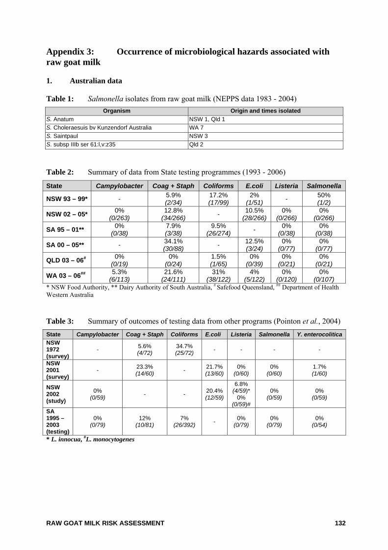
RAW GOAT MILK RISK ASSESSMENT 132
Appendix 3: Occurrence of microbiological hazards associated with raw goat milk 1. Australian data Table 1: Salmonella isolates from raw goat milk (NEPPS data 1983 - 2004)
Organism Origin and times isolated S. Anatum NSW 1, Qld 1 S. Choleraesuis bv Kunzendorf Australia WA 7 S. Saintpaul NSW 3 S. subsp IIIb ser 61:l,v:z35 Qld 2
Table 2: Summary of data from State testing programmes (1993 - 2006)
State Campylobacter Coag + Staph Coliforms E.coli Listeria Salmonella
NSW 93 – 99* - 5.9% (2/34)
17.2% (17/99)
2% (1/51) - 50%
(1/2)
NSW 02 – 05* 0% (0/263)
12.8% (34/266) - 10.5%
(28/266) 0%
(0/266) 0%
(0/266)
SA 95 – 01** 0% (0/38)
7.9% (3/38)
9.5% (26/274) - 0%
(0/38) 0%
(0/38)
SA 00 – 05** - 34.1% (30/88) - 12.5%
(3/24) 0%
(0/77) 0%
(0/77)
QLD 03 – 06# 0% (0/19)
0% (0/24)
1.5% (1/65)
0% (0/39)
0% (0/21)
0% (0/21)
WA 03 – 06## 5.3% (6/113)
21.6% (24/111)
31% (38/122)
4% (5/122)
0% (0/120)
0% (0/107)
* NSW Food Authority, ** Dairy Authority of South Australia, # Safefood Queensland, ## Department of Health Western Australia Table 3: Summary of outcomes of testing data from other programs (Pointon et al., 2004) State Campylobacter Coag + Staph Coliforms E.coli Listeria Salmonella Y. enterocolitica NSW 1972 (survey)
- 5.6% (4/72)
34.7% (25/72) - - - -
NSW 2001 (survey)
- 23.3% (14/60) - 21.7%
(13/60) 0%
(0/60) 0%
(0/60) 1.7% (1/60)
NSW 2002 (study)
0% (0/59) - - 20.4%
(12/59)
6.8% (4/59)*
0% (0/59)#
0% (0/59)
0% (0/59)
SA 1995 – 2003 (testing)
0% (0/79)
12% (10/81)
7% (26/392) - 0%
(0/79) 0%
(0/79) 0%
(0/54)
* L. innocua, #L. monocytogenes
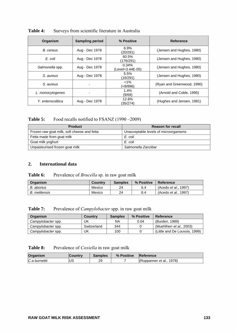
RAW GOAT MILK RISK ASSESSMENT 133
Table 4: Surveys from scientific literature in Australia
Organism Sampling period % Positive Reference
B. cereus Aug - Dec 1978 6.9% (20/291) (Jensen and Hughes, 1980)
E. coli Aug - Dec 1978 60.5% (176/291) (Jensen and Hughes, 1980)
Salmonella spp. Aug - Dec 1978 0.34% (Level=3.44E-05) (Jensen and Hughes, 1980)
S. aureus Aug - Dec 1978 5.5% (16/291) (Jensen and Hughes, 1980)
S. aureus - <1% (<8/896) (Ryan and Greenwood, 1990)
L. monocytogenes - 1.4% (9/69) (Arnold and Coble, 1995)
Y. enterocolitica Aug - Dec 1978 12.8% (35/274) (Hughes and Jensen, 1981)
Table 5: Food recalls notified to FSANZ (1990 –2009)
Product Reason for recall Frozen raw goat milk, soft cheese and fetta Unacceptable levels of microorganisms Fetta made from goat milk E. coli Goat milk yoghurt E. coli Unpasteurised frozen goat milk Salmonella Zanzibar
2. International data Table 6: Prevalence of Brucella sp. in raw goat milk Organism Country Samples % Positive Reference B. abortus Mexico 24 6.4 (Acedo et al., 1997) B. melitensis Mexico 24 8.4 (Acedo et al., 1997)
Table 7: Prevalence of Campylobacter spp. in raw goat milk Organism Country Samples % Positive Reference Campylobacter spp. UK NA 0.04 (Burden, 1989) Campylobacter spp. Switzerland 344 0 (Muehlherr et al., 2003) Campylobacter spp. UK 100 0 (Little and De Louvois, 1999)
Table 8: Prevalence of Coxiella in raw goat milk Organism Country Samples % Positive Reference C.a burnettii US 29 7 (Ruppanner et al., 1978)
RAW GOAT MILK RISK ASSESSMENT 134
Table 9: Prevalence of E. coli in raw goat milk Organism Country Samples % Positive Reference STEC Switzerland 344 16.3 (Muehlherr et al., 2003) EHEC UK 94 0.7 (Anon, 1999) E. coli UK 2462 10 (Roberts, 1985) E. coli US 2911 1.6 (White and Hinckley, 1999) E coli Austria 204 1.5 (Pernthaner et al., 1993) E. coli O157:H7 Italy 60 1.7 (Foschino et al., 2002) E. coli O157:H7 UK 100 0 (Little and De Louvois, 1999)
Table 10: Prevalence of Listeria spp. in raw goat milk Organism Country Samples % Positive Reference L. monocytogenes UK 100 0 (Little and De Louvois, 1999) L. monocytogenes India 64 1.56 (Barbuddhe et al., 2000) L. monocytogenes UK 94 2.09 (Anon, 1999) L. monocytogenes Spain 1445 2.56 (Gaya et al., 1996) L. monocytogenes US 450 3.8 (Abou-Eleinin et al., 2000) L. monocytogenes Portugal 39 0 (Guerra et al., 2001) L. innocua Spain 1445 1.73 (Gaya et al., 1996) L. innocua US 450 5.8 (Abou-Eleinin et al., 2000) Listeria spp. Portugal 39 5 (Guerra et al., 2001) Table 11: Prevalence of Mycobacterium avium subsp. paratuberculosis in raw goat milk Organism Country Samples % Positive Reference MAP (Mycobacterium) Norway 340
7.1 (PCR) (Djonne et al., 2003)
0 (culture) MAP Switzerland 344 23 (Muehlherr et al., 2003) MAP (Goat identified was infected with Johne's disease) India 20 1 (Singh and Vihan, 2004)
MAP UK 90 <1 (PCR) (Grant et al., 2001) Table 12: Prevalence of Salmonella spp. in raw goat milk Organism Country Samples % Positive Reference Salmonella spp. Spain 1445 0 (Gaya et al., 1996) Salmonella spp. Europe 50 0 (Abo-Elnaga et al., 1985) Salmonella spp. UK 2463 0 (Roberts, 1985) Salmonella spp. Switzerland 344 0 (Muehlherr et al., 2003) Salmonella spp. Italy 60 0 (Foschino et al., 2002) Salmonella spp. UK 100 0 (Little and De Louvois, 1999) Salmonella spp. Bulgaria 60 0 (Vashin et al., 1999)
RAW GOAT MILK RISK ASSESSMENT 135
Table 13: Prevalence of Staphylococcus spp. in raw goat milk Organism Country Samples % Positive Reference S. aureus Europe 50 0 (Abo-Elnaga et al., 1985) S. aureus US 2911 11 (White and Hinckley, 1999) S. aureus France 238 2 (De Buyser et al., 1987) S. aureus Norway 213 96.2 (Jorgensen et al., 2005) S. aureus Switzerland 344 31.7 (Muehlherr et al., 2003) S. aureus Italy 60 43 (Foschino et al., 2002) S. aureus Austria 359 17.6 (Deinhofer and Pernthaner, 1995) S. aureus UK 2,493 4 (Roberts, 1985) S. aureus UK 100 15 (Little and De Louvois, 1999) S. aureus Greece 1350 10 (Kalogridou-Vassiliadou, 1991) S. aureus Iraq 297 3 (Al-Graibawi et al., 1986) Coag -ve staph Austria 204 55 (Pernthaner et al., 1993) Coag +ve staph Austria 204 37.3 (Pernthaner et al., 1993) Staphylococcus spp. Austria 204 1.5 (Pernthaner et al., 1993) Table 14: Prevalence of Streptococcus spp.in raw goat milk Organism Country Samples % Positive Reference Streptococcus spp. US 2911 4.1 (White and Hinckley, 1999) Streptococcus spp. Austria 204 6.2 (Pernthaner et al., 1993) Streptococcus spp. Austria 2243 1.6 (Deinhofer and Pernthaner, 1995)
Table 15: Prevalence of Toxoplasma spp. in raw goat milk Organism Country Samples % Positive Reference T. gondii US 232 9 (Smith, 1993a) T. gondii US - 7 (Walsh et al., 1999) Table 16: Prevalence of Yesinia spp. in raw goat milk Organism Country Samples % Positive Reference Y. enterocolitica UK 2,493 0.08 (Roberts, 1985) Y. enterocolitica UK - 0.08 (Burden, 1989)
RAW GOAT MILK RISK ASSESSMENT 136
Appendix 4: Foodborne illness associated with consumption of raw goat milk 1. Australian data Table 1: Outbreaks of illness associated with raw goat milk
Year Country Cases Causative Agent Comment Reference
1990 Australia 9 Salmonella choleraesuis var. Kunzendorf
Consumption of unpasteurised goats milk (Iveson et al., 1990)
1984 Australia 2 Cryptosporidium parvum Consumption of unpasteurised goats milk five days prior to onset of illness - mother and 1yr old child
(Smith, 1993a) (WHO, 1984)
2. International data Table 2: Outbreaks of illness associated with raw goat milk
Year Country Cases (deaths)
Causative Agent Comment Reference
2001 Austria 2 E. coli O157 Isolated from dairy cow and goat, raw milk (Allerberger et al., 2001)
2001 Canada 5 E. coli O157:H7 Source of implicated goat's milk was a co-operative farm
(McIntyre, 2001)
2001 Sweden 1 E. coli O157 Consumption of unpasteurised goats milk
(Steen et al., 2001) SA risk assessment
1998 Scotland 1 E. coli O157 Unpasteurised goat milk from farm gate
(Handysides and Cowden, 1998)
1995 Czech Republic 5 E. coli O157 Unpasteurised goat milk
from farm (Bielaszewska et al., 1997)
1992 France 40 Coxiella burnettii Persons who worked on farm and consumed unpasteurised milk products
(Fishbein and Raoult, 1992)
1991 USA 3 Campylobacter jejuni Consumed on farm (CDC, 2002)
1990 UK 1 Toxoplasma gondii Consumption of unpasteurised goats milk (Skinner et al., 1990)
1989 UK 3 Campylobacter jejuni Consumption of unpasteurised goats milk (Burden, 1989)
1988 Israel 3 Staphylococcus aureus Milk from a goat with overt mastitis (Gross et al., 1988)
1985 Scotland 2 Staphylococcus aureus Goats miik from farm - unpasteurised (Sharp, 1989)
1985 UK 1 Campylobacter jejuni Consumption of unpasteurised goats milk (Hutchinson et al., 1985a)
1985 UK 3 Campylobacter jejuni Consumption of unpasteurised goats milk (Hutchinson et al., 1985b)
1984 Brazil 6 Brucella melitensis Consumption of unpasteurised goats milk and / or cheese
(Chiari and Neves, 1984)
1984 USA 2 Toxoplasma gondii Consumption of raw goats milk (Smith, 1993a)
1984 USA 6 Toxoplasma gondii Consumption of raw goats milk (Smith, 1993a)
1983 USA 6 Campylobacter jejuni Associated with dairy that produced raw goat milk (Harris et al., 1987)
1978 USA 10 Toxoplasma gondii Consumption of unpasteurised goats milk (Sacks et al., 1982)
1975 USA 1 Toxoplasma gondii Consumption of unpasteurised goats milk (Riemann et al., 1975)
RAW GOAT MILK RISK ASSESSMENT 137
Table 3: Outbreaks of illness associated with raw goat milk cheese
Year Country Cases (deaths)
Product Causative Agent Comment Reference
2006 Finland 7
Soft unpasteurised goats milk cheese
Streptococcus equi subspecies zooepidemicus
Small farm production (Kuusi et al., 2006)
2006 France 3 Raw goats milk cheese E. coli 0157
Unpasteurised raw goat cheese produced by a local provider
(Espie et al., 2006)
2005 France 18 Raw goats milk cheese
Salmonella Stourbridge
Cheese was made from the unpasteurised milk of a single herd of 260 goats
(Vaillant and Espie, 2005)
2004 Italy 4 Unpasteurised goats milk cheese
Brucella melitensis
Symptoms included fever and lumbar pain (Taliani et al., 2004)
2002 Spain 11 Raw goats cheese
Brucella melitensis serovar 3
Unpasteurised raw goat cheese produced in a farmhouse
(Mendez et al., 2003)
1999 Canada ? Cheese from goats milk Coxiella burnettii
Associated with contact with goat placenta, smoking tobacco
(Hatchette et al., 2001)
1995 Malta 135 (1) Soft cheese made with raw goats milk
Brucella mellitensis
Consumption of raw milk cheese (Anon, 1995)
1994 France 4 Raw goats milk cheese E. coli 0103 Goats milk suspected (Ammon, 1997)
1993 France 273 (1) Unpasteurised goats milk cheese
S. enterica Paratyphi B phage type 1 var 3
Brand A unpasteurised goat milk cheese, Out break was possibly related to contaminated goats milk cheese
(Desenclos et al., 1996)
1992 France 4 (1)
Unpasteurised fromage frais goats/cows cheese
E. coli Acute haemolytic uraemic syndrome (HUS)
(Deschenes et al., 1996)
1990 France 277 Contaminated goats milk cheese
S. enterica Paratyphi B
Out break was possibly related to contaminated goats milk cheese
(Desenclos et al., 1996)
1988 England 1 Goats milk soft cheese Listeria spp. Immuno-compromised case (Azadian et al.,
1989)
1983 USA 31 Raw goats cheese
Brucella melitensis
Mexican raw goats milk cheese
(Thapar and Young, 1986)
1973 USA 3 Mexican fresh raw cheese
Brucella melitensis
Mexican raw goats milk cheese (Eckman, 1975)
1973 Mexico 6 Fresh raw goats cheese
Brucella melitensis
Mexican raw goats milk cheese
(Young and Suvannoparrat, 1975)
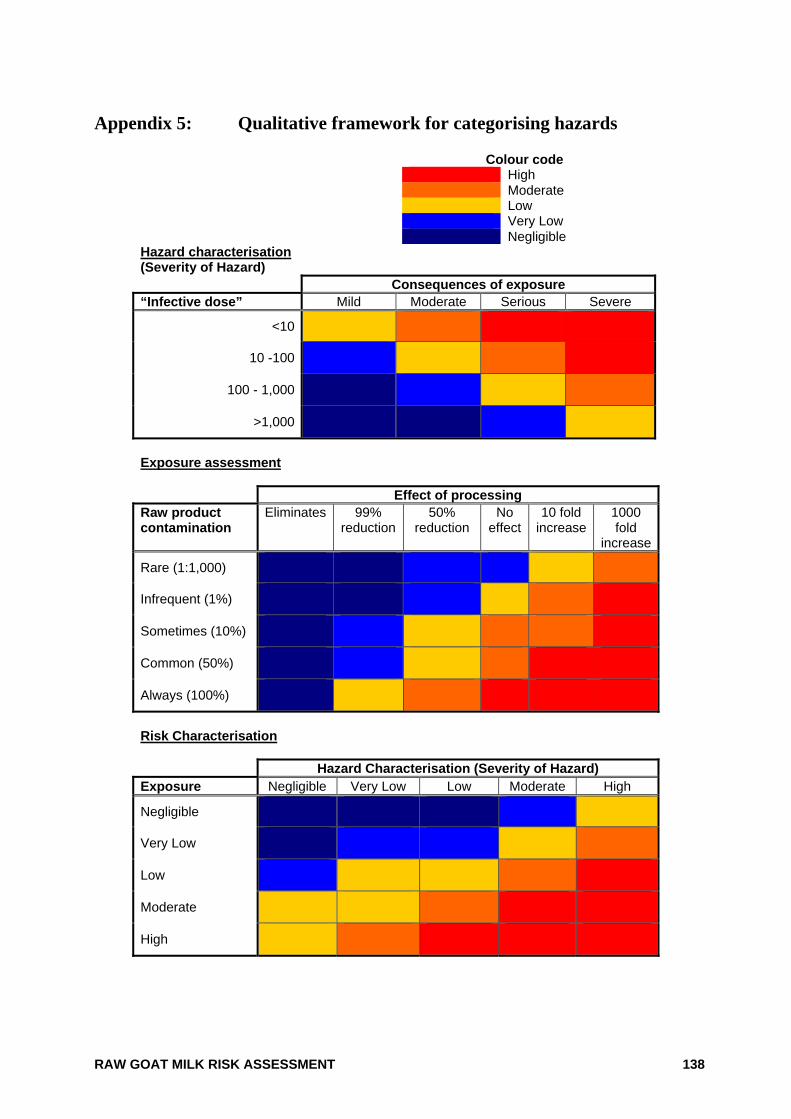
RAW GOAT MILK RISK ASSESSMENT 138
Appendix 5: Qualitative framework for categorising hazards
Colour code High Moderate Low Very Low Negligible
Hazard characterisation (Severity of Hazard) Consequences of exposure “Infective dose” Mild Moderate Serious Severe
<10
10 -100
100 - 1,000
>1,000
Exposure assessment Effect of processing Raw product contamination
Eliminates 99% reduction
50% reduction
No effect
10 fold increase
1000 fold
increase
Rare (1:1,000)
Infrequent (1%)
Sometimes (10%)
Common (50%)
Always (100%)
Risk Characterisation Hazard Characterisation (Severity of Hazard) Exposure Negligible Very Low Low Moderate High
Negligible
Very Low
Low
Moderate
High
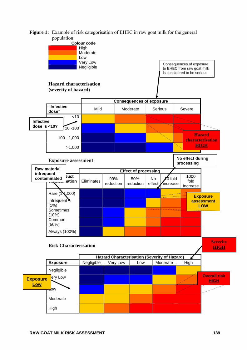
RAW GOAT MILK RISK ASSESSMENT 139
Figure 1: Example of risk categorisation of EHEC in raw goat milk for the general population
Colour code High Moderate Low Very Low Negligible
Hazard characterisation (severity of hazard) Consequences of exposure “Infective dose” Mild Moderate Serious Severe
<10
10 -100
100 - 1,000
>1,000
Exposure assessment Effect of processing Raw product contamination Eliminates 99%
reduction50%
reductionNo
effect10 fold
increase
1000 fold
increase
Rare (1:1,000)
Infrequent (1%)
Sometimes (10%)
Common (50%)
Always (100%)
Risk Characterisation Hazard Characterisation (Severity of Hazard) Exposure Negligible Very Low Low Moderate High
Negligible
Very Low
Low
Moderate
High
Consequences of exposure to EHEC from raw goat milk is considered to be serious
Infective dose is <10?
No effect during processing
Raw material infrequent contaminated
Exposure assessment
LOW
Exposure Low
Overall risk HIGH
Hazard characterisation
HIGH
Severity HIGH
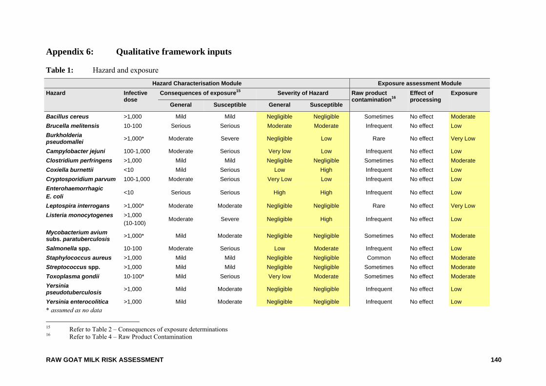
RAW GOAT MILK RISK ASSESSMENT 140
Appendix 6: Qualitative framework inputs Table 1: Hazard and exposure
Hazard Characterisation Module Exposure assessment Module Hazard Infective
dose Consequences of exposure15 Severity of Hazard Raw product
contamination16 Effect of processing
Exposure
General Susceptible General Susceptible
Bacillus cereus >1,000 Mild Mild Negligible Negligible Sometimes No effect Moderate Brucella melitensis 10-100 Serious Serious Moderate Moderate Infrequent No effect Low Burkholderia pseudomallei >1,000* Moderate Severe Negligible Low Rare No effect Very Low
Campylobacter jejuni 100-1,000 Moderate Serious Very low Low Infrequent No effect Low Clostridium perfringens >1,000 Mild Mild Negligible Negligible Sometimes No effect Moderate Coxiella burnettii <10 Mild Serious Low High Infrequent No effect Low Cryptosporidium parvum 100-1,000 Moderate Serious Very Low Low Infrequent No effect Low Enterohaemorrhagic E. coli <10 Serious Serious High High Infrequent No effect Low
Leptospira interrogans >1,000* Moderate Moderate Negligible Negligible Rare No effect Very Low Listeria monocytogenes >1,000
(10-100) Moderate Severe Negligible High Infrequent No effect Low
Mycobacterium avium subs. paratuberculosis >1,000* Mild Moderate Negligible Negligible Sometimes No effect Moderate
Salmonella spp. 10-100 Moderate Serious Low Moderate Infrequent No effect Low Staphylococcus aureus >1,000 Mild Mild Negligible Negligible Common No effect Moderate Streptococcus spp. >1,000 Mild Mild Negligible Negligible Sometimes No effect Moderate Toxoplasma gondii 10-100* Mild Serious Very low Moderate Sometimes No effect Moderate Yersinia pseudotuberculosis >1,000 Mild Moderate Negligible Negligible Infrequent No effect Low
Yersinia enterocolitica >1,000 Mild Moderate Negligible Negligible Infrequent No effect Low * assumed as no data
15 Refer to Table 2 – Consequences of exposure determinations 16 Refer to Table 4 – Raw Product Contamination
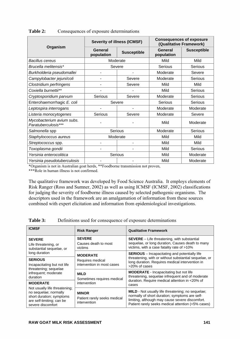
RAW GOAT MILK RISK ASSESSMENT 141
Table 2: Consequences of exposure determinations
Organism Severity of illness (ICMSF) Consequences of exposure
(Qualitative Framework) General
population Susceptible General population
Susceptible
Bacillus cereus Moderate Mild Mild Brucella melitensis* Severe Serious Serious Burkholderia pseudomallei - - Moderate Severe Campylobacter jejuni/coli - Severe Moderate Serious Clostridium perfringens - Severe Mild Mild Coxiella burnettii** - - Mild Serious Cryptosporidium parvum Serious Severe Moderate Serious Enterohaemorrhagic E. coli Severe Serious Serious Leptospira interrogans - - Moderate Moderate Listeria monocytogenes Serious Severe Moderate Severe Mycobacterium avium subs. Paratuberculosis*** - - Mild Moderate
Salmonella spp Serious Moderate Serious Staphylococcus aureus Moderate Mild Mild Streptococcus spp. - - Mild Mild Toxoplasma gondii - - Mild Serious Yersinia enterocolitica Serious Mild Moderate Yersinia pseudotuberculosis - - Mild Moderate *Organism is not in Australian goat herds, **Foodborne transmission not proven, ***Role in human illness is not confirmed. The qualitative framework was developed by Food Science Australia. It employs elements of Risk Ranger (Ross and Sumner, 2002) as well as using ICMSF (ICMSF, 2002) classifications for judging the severity of foodborne illness caused by selected pathogenic organisms. The descriptors used in the framework are an amalgamation of information from these sources combined with expert elicitation and information from epidemiological investigations. Table 3: Definitions used for consequence of exposure determinations ICMSF Risk Ranger Qualitative Framework
SEVERE Life threatening, or substantial sequelae, or long duration
SEVERE Causes death to most victims
SEVERE – Life threatening, with substantial sequelae, or long duration, Causes death to many victims, with a case fatality rate of >10%
MODERATE Requires medical intervention in most cases
SERIOUS – Incapacitating and potentially life threatening, with or without substantial sequelae, or long duration. Requires medical intervention in >20% of cases
SERIOUS Incapacitating but not life threatening; sequelae infrequent; moderate duration
MILD Sometimes requires medical intervention
MODERATE - Incapacitating but not life threatening, sequelae infrequent and of moderate duration. Require medical attention in <20% of cases MODERATE
Not usually life threatening; no sequelae; normally short duration; symptoms are self-limiting; can be severe discomfort
MINOR Patient rarely seeks medical intervention
MILD - Not usually life threatening; no sequelae; normally of short duration; symptoms are self-limiting, although may cause severe discomfort. Patient rarely seeks medical attention (<5% cases)
RAW GOAT MILK RISK ASSESSMENT 142
Table 4: Raw product contamination
Organism Raw product contamination Justification
Bacillus cereus Sometimes (10%) No data. Expert panel consultation.
Brucella melitensis Infrequent (1%) International data 6.4 – 8.4%, Disease not found in Australia. Expert panel consultation.
Burkholderia pseudomallei Rare (1:1000) No data. Expert panel consultation.
Campylobacter jejuni Infrequent (1%) Australian data 1.39%, International data 0 – 0.04%. Expert panel consultation.
Clostridium perfringens Sometimes (10%) No data. Expert panel consultation.
Coxiella burnettii Infrequent (1%) International data 7%. Expert panel consultation.
Cryptosporidium parvum Infrequent (1%) No data. Expert panel consultation.
Enterohaemorrhagic E. coli
Infrequent (1%) (Assumed from E.coli
data)
Australian data 7.37% (E. coli), International data 0 – 16.3%. Expert panel consultation.
Leptospira interrogans Rare (1:1000) No data. Expert panel consultation.
Listeria monocytogenes Infrequent (1%) Australian data 0-6.8 %, International data 0 – 5.8%. Expert panel consultation.
Mycobacterium avium subs. paratuberculosis Sometimes (10%) International data 0 – 23%. Expert panel
consultation.
Salmonella spp. Infrequent (1%) Australian data 0.2 %, International data 0 %. Expert panel consultation.
Staphylococcus aureus Common (50%) Australian data up to 23.3%, International data 0 – 96.2%. Expert panel consultation.
Streptococcus spp. Sometimes (10%) International data 1.6 – 6.2%. Expert panel consultation.
Toxoplasma gondii Sometimes (10%) International data 7 – 9%. Expert panel consultation.
Yersinia pseudotuberculosis Infrequent (1%) No data. Expert panel consultation.
Yersinia enterocolitica Infrequent (1%) International data 0.08%. Expert panel consultation.
RAW GOAT MILK RISK ASSESSMENT 143
Appendix 7: Outcomes of State risk assessments 1. Overview South Australia This study was undertaken following a risk profile of the primary industry sector commissioned by the Department of Primary Industries and Resources (Sumner, 2002). The study aimed to identify appropriate food safety risk management options, both policy and regulation for the dairy goat milk industry. The study adopted a qualitative risk ranking approach, based on ICMSF principles and considered hazard severity; occurrence of the hazard in foods; potential for growth; effects of production, processing and handling (including a consumer terminal step); and epidemiological data. Queensland The risk assessment utilised methodology developed to rank food safety hazards based on internationally accepted principles of risk assessment including Codex Alimentarius Commission CAC/GL-30 1999. The semi-quantitative approach assigned risk scores (maximum of 100) to hazards and determined total assessed risk scores for each of four population segments based on exposure and severity of consequence. New South Wales A risk assessment was conducted to analyse the risks associated with the production, sale and consumption of products made from goat milk. Conclusions are based on the assessment of risk relating to the 11 licensed operators working in the goat industry in NSW. The risk assessment was conducted as two separate parts; a qualitative analysis of risk for each hazard identified in the Hazard Analysis, and; a stochastic semi-quantitative model (Excel-@Risk) to scope the public health significance of Salmonella, Staphylococcus aureus and Listeria monocytogenes. 2. Uncertainty and assumptions South Australia The study reported several areas of uncertainty in the assessment: • susceptibility of the exposed population (an assumption was made that goat milk is fed
disproportionately to infants) • frequency of consumption and distribution of consumption within the population • probability of raw product contamination • the effect of existing controls • dose response • frequency of use of a terminal kill step (e.g. boiling) • underreporting of illness • emergence of new microbiological hazards
RAW GOAT MILK RISK ASSESSMENT 144
Queensland The Queensland study reported the following assumptions: • it was estimated that consumption of raw goat milk in the ‘general population’ was
between 1 and 20% • it was estimated that consumption of raw goat milk in the ‘niche population’ was
between 81 and 99% • dose response data used was in the form of published point estimates of infective dose
where available New South Wales Several areas of constraint were identified in the study including: • knowledge of current industry hygiene and processing practices for dairy goat farmers
outside of the industry quality assurance scheme • significant lack of information in the literature on the incidence, prevalence and growth
of organisms in goat’s milk • information pertaining to the consumer population and consumption patterns
An estimate of 8% of the adult population in New South Wales was used to calculate consumption figures however it was noted that very little data was available. The study further stated that evidence suggested that products were consumed on a daily basis in some households and more frequently where goat’s milk was being used to feed infants. Assumptions for growth predictions modelled using the USDA Pathogen Modelling Program included; growth conditions, competitive microflora, temperature ranges, initial microbial load, composition of goat’s milk and the pathogen level of concern (LOC). The LOC for pathogens was based on 1 x 10⁶ cfu/g, except for pathogens with a reported low infective dose, such as E. coli and Salmonella spp. Assumptions for the stochastic semi-quantitative model included: • conditions for the milk harvesting, processing and storage/transport of product • hazard ranking utilising decision analysis-like methodology • consumption and exposure information
3. Conclusions South Australia The study reported that a broad range of microbiological hazards have been recorded internationally and domestically as causing illness due to consumption of raw goat milk. Many of these hazards are capable of causing severe illness and death. A number of hazards were rated as high risk for susceptible populations (C. parvum, EHEC, L. monocytogenes, Salmonella and T. gondii). C. jejuni/coli, Salmonella and EHEC were rated as medium risk for the general population. C. parvum, L. monocytogenes, S. aureus and T. gondii were all rated as low risk for the general population.
RAW GOAT MILK RISK ASSESSMENT 145
The reported stated the low general exposure must be weighed against the exposure of lactose intolerant, susceptible infants. Queensland study Findings were reported for four population segments within each of two groups. The two groups consisted of the general population group where consumption of raw goat milk was likely to be consumed by less than 20% of the population, and the “niche” market where raw goat milk is the milk of choice. Each of these groups was then categorised into four segments: general population, babies/infants, immunocompromised/very old and hypersensitive/intolerant. For the normal population where goat milk is generally not consumed there was an overall low risk, with E. coli O157:H7 and other pathogenic E. coli a medium risk to babies and infants, and L. monocytogenes a medium risk to babies/infants and the immunocompromised. For the niche market where raw goat milk is consumed as the milk of choice, there was an overall increase in risk compared to the normal market population. S. aureus enterotoxin and Burkholderia pseudomallei were considered a medium risk to all populations segments, while E. coli O157:H7 was a medium risk to the general population and immunocompromised and a high risk to babies/infants. L. monocytogenes, while considered a low risk to the general population, was a high risk to both babies/infants and the immunocompromised. NSW study The study concluded that insufficient data was available to make any statement, qualitative or quantitative on the risks and public health significance associated with the hazards identified in the hazard analysis. Particular mention was made of the paucity of information relating to the incidence of specific hazards in goat’s milk, incidence of food poisoning outbreaks, consumption data, consumer handling of product prior to consumption and the lack of information pertaining to unlicensed operators. Modelling of the public health significance for Salmonella, S. aureus and L. monocytogenes indicates that a single contamination event resulting from contamination and subsequent abuse of the product could lead to severe public health consequences. A major contamination of product (all product from one farm) over 2 to 3 days resulted in some deaths expected for Salmonella and L. monocytogenes, while contamination with S. aureus could result in 30 to 40 people requiring intensive medical treatment. Item contamination (a single package of cheese or litre of milk) would be less serious with Salmonella and S. aureus causing 2 to 3 people to experience mild illness, although one high risk event with L. monocytogenes and a pregnant woman could not be dismissed.
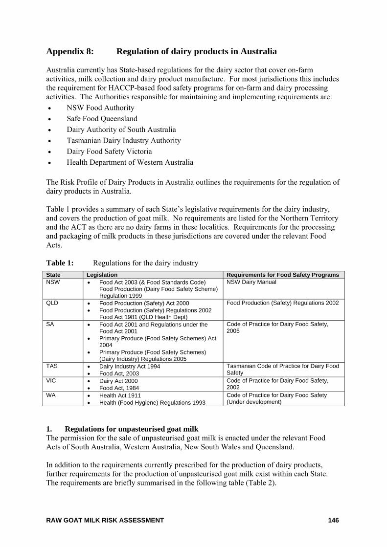
RAW GOAT MILK RISK ASSESSMENT 146
Appendix 8: Regulation of dairy products in Australia Australia currently has State-based regulations for the dairy sector that cover on-farm activities, milk collection and dairy product manufacture. For most jurisdictions this includes the requirement for HACCP-based food safety programs for on-farm and dairy processing activities. The Authorities responsible for maintaining and implementing requirements are: • NSW Food Authority • Safe Food Queensland • Dairy Authority of South Australia • Tasmanian Dairy Industry Authority • Dairy Food Safety Victoria • Health Department of Western Australia
The Risk Profile of Dairy Products in Australia outlines the requirements for the regulation of dairy products in Australia. Table 1 provides a summary of each State’s legislative requirements for the dairy industry, and covers the production of goat milk. No requirements are listed for the Northern Territory and the ACT as there are no dairy farms in these localities. Requirements for the processing and packaging of milk products in these jurisdictions are covered under the relevant Food Acts. Table 1: Regulations for the dairy industry State Legislation Requirements for Food Safety ProgramsNSW • Food Act 2003 (& Food Standards Code)
Food Production (Dairy Food Safety Scheme) Regulation 1999
NSW Dairy Manual
QLD • Food Production (Safety) Act 2000 • Food Production (Safety) Regulations 2002
Food Act 1981 (QLD Health Dept)
Food Production (Safety) Regulations 2002
SA • Food Act 2001 and Regulations under the Food Act 2001
• Primary Produce (Food Safety Schemes) Act 2004
• Primary Produce (Food Safety Schemes) (Dairy Industry) Regulations 2005
Code of Practice for Dairy Food Safety, 2005
TAS • Dairy Industry Act 1994 • Food Act, 2003
Tasmanian Code of Practice for Dairy Food Safety
VIC • Dairy Act 2000 • Food Act, 1984
Code of Practice for Dairy Food Safety, 2002
WA • Health Act 1911 • Health (Food Hygiene) Regulations 1993
Code of Practice for Dairy Food Safety (Under development)
1. Regulations for unpasteurised goat milk The permission for the sale of unpasteurised goat milk is enacted under the relevant Food Acts of South Australia, Western Australia, New South Wales and Queensland. In addition to the requirements currently prescribed for the production of dairy products, further requirements for the production of unpasteurised goat milk exist within each State. The requirements are briefly summarised in the following table (Table 2).
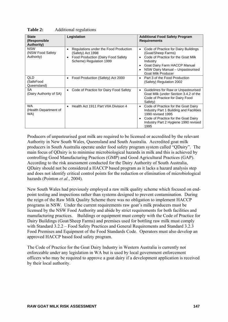
RAW GOAT MILK RISK ASSESSMENT 147
Table 2: Additional regulations State (Responsible Authority)
Legislation Additional Food Safety Program Requirements
NSW (NSW Food Safety Authority)
• Regulations under the Food Production (Safety) Act 1998
• Food Production (Dairy Food Safety Scheme) Regulation 1999
• Code of Practice for Dairy Buildings (Goat/Sheep Farms)
• Code of Practice for the Goat Milk Industry
• Goat Dairy Farm HACCP Manual • NSW Dairy Manual – Unpasteurised
Goat Milk Producer QLD (SafeFood Queensland)
• Food Production (Safety) Act 2000 • Part 3 of the Food Production (Safety) Regulation 2002
SA (Dairy Authority of SA)
• Code of Practice for Dairy Food Safety
• Guidelines for Raw or Unpasteurised Goat Milk (under Section 3.4.2 of the Code of Practice for Dairy Food Safety)
WA (Health Department of WA)
• Health Act 1911 Part VIIA Division 4 • Code of Practice for the Goat Dairy Industry Part 1 Building and Facilities 1990 revised 1995
• Code of Practice for the Goat Dairy Industry Part 2 Hygiene 1990 revised 1995
Producers of unpasteurised goat milk are required to be licensed or accredited by the relevant Authority in New South Wales, Queensland and South Australia. Accredited goat milk producers in South Australia operate under food safety program system called “QDairy”. The main focus of QDairy is to minimise microbiological hazards in milk and this is achieved by controlling Good Manufacturing Practices (GMP) and Good Agricultural Practices (GAP). According to the risk assessment conducted for the Dairy Authority of South Australia, QDairy should not be considered a HACCP based program as it lacks a hazard analysis step and does not identify critical control points for the reduction or elimination of microbiological hazards (Pointon et al., 2004). New South Wales had previously employed a raw milk quality scheme which focused on end-point testing and inspections rather than systems designed to prevent contamination. During the reign of the Raw Milk Quality Scheme there was no obligation to implement HACCP programs in NSW. Under the current requirements raw goat’s milk producers must be licensed by the NSW Food Authority and abide by strict requirements for both facilities and manufacturing practices. Buildings or equipment must comply with the Code of Practice for Dairy Buildings (Goat/Sheep Farms) and premises used for bottling raw milk must comply with Standard 3.2.2 – Food Safety Practices and General Requirements and Standard 3.2.3 Food Premises and Equipment of the Food Standards Code. Operators must also develop an approved HACCP based food safety program. The Code of Practice for the Goat Dairy Industry in Western Australia is currently not enforceable under any legislation in WA but is used by local government enforcement officers who may be required to approve a goat dairy if a development application is received by their local authority.
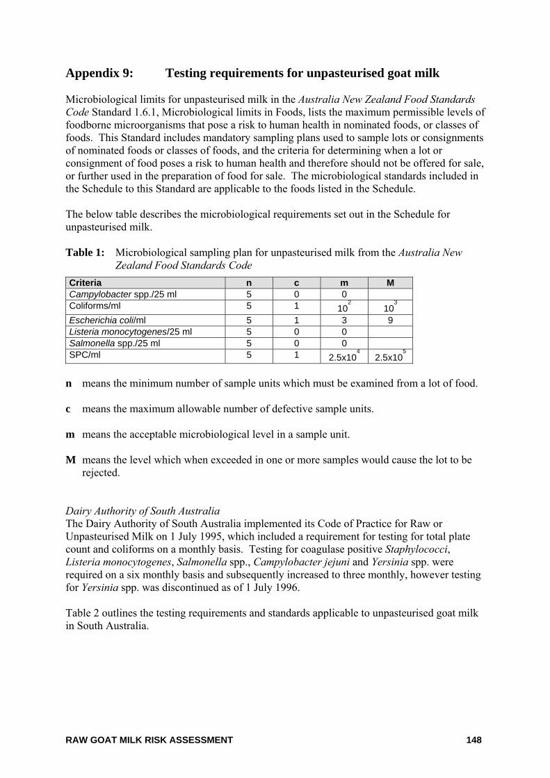
RAW GOAT MILK RISK ASSESSMENT 148
Appendix 9: Testing requirements for unpasteurised goat milk Microbiological limits for unpasteurised milk in the Australia New Zealand Food Standards Code Standard 1.6.1, Microbiological limits in Foods, lists the maximum permissible levels of foodborne microorganisms that pose a risk to human health in nominated foods, or classes of foods. This Standard includes mandatory sampling plans used to sample lots or consignments of nominated foods or classes of foods, and the criteria for determining when a lot or consignment of food poses a risk to human health and therefore should not be offered for sale, or further used in the preparation of food for sale. The microbiological standards included in the Schedule to this Standard are applicable to the foods listed in the Schedule. The below table describes the microbiological requirements set out in the Schedule for unpasteurised milk. Table 1: Microbiological sampling plan for unpasteurised milk from the Australia New
Zealand Food Standards Code Criteria n c m M Campylobacter spp./25 ml 5 0 0 Coliforms/ml 5 1 10
2 10
3
Escherichia coli/ml 5 1 3 9 Listeria monocytogenes/25 ml 5 0 0 Salmonella spp./25 ml 5 0 0 SPC/ml 5 1 2.5x10
4 2.5x10
5
n means the minimum number of sample units which must be examined from a lot of food. c means the maximum allowable number of defective sample units. m means the acceptable microbiological level in a sample unit. M means the level which when exceeded in one or more samples would cause the lot to be
rejected. Dairy Authority of South Australia The Dairy Authority of South Australia implemented its Code of Practice for Raw or Unpasteurised Milk on 1 July 1995, which included a requirement for testing for total plate count and coliforms on a monthly basis. Testing for coagulase positive Staphylococci, Listeria monocytogenes, Salmonella spp., Campylobacter jejuni and Yersinia spp. were required on a six monthly basis and subsequently increased to three monthly, however testing for Yersinia spp. was discontinued as of 1 July 1996. Table 2 outlines the testing requirements and standards applicable to unpasteurised goat milk in South Australia.
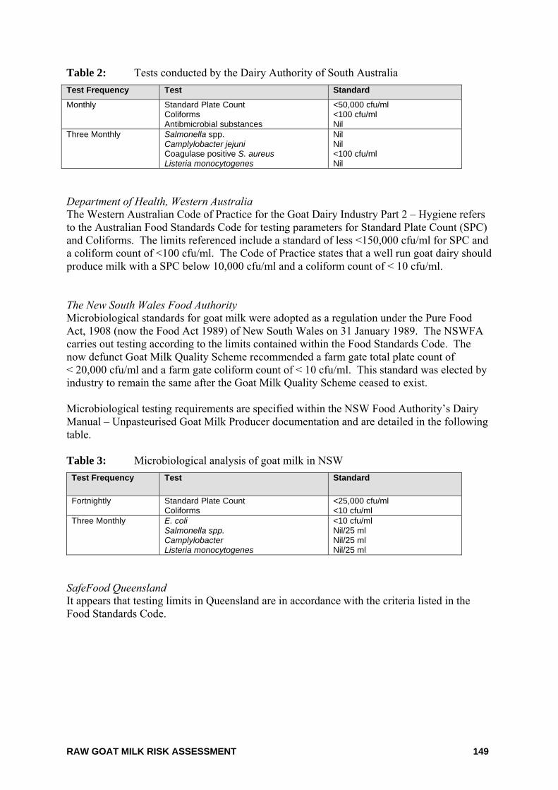
RAW GOAT MILK RISK ASSESSMENT 149
Table 2: Tests conducted by the Dairy Authority of South Australia Test Frequency Test Standard
Monthly Standard Plate Count Coliforms Antibmicrobial substances
<50,000 cfu/ml <100 cfu/ml Nil
Three Monthly Salmonella spp. Camplylobacter jejuni Coagulase positive S. aureus Listeria monocytogenes
Nil Nil <100 cfu/ml Nil
Department of Health, Western Australia The Western Australian Code of Practice for the Goat Dairy Industry Part 2 – Hygiene refers to the Australian Food Standards Code for testing parameters for Standard Plate Count (SPC) and Coliforms. The limits referenced include a standard of less <150,000 cfu/ml for SPC and a coliform count of <100 cfu/ml. The Code of Practice states that a well run goat dairy should produce milk with a SPC below 10,000 cfu/ml and a coliform count of < 10 cfu/ml. The New South Wales Food Authority Microbiological standards for goat milk were adopted as a regulation under the Pure Food Act, 1908 (now the Food Act 1989) of New South Wales on 31 January 1989. The NSWFA carries out testing according to the limits contained within the Food Standards Code. The now defunct Goat Milk Quality Scheme recommended a farm gate total plate count of < 20,000 cfu/ml and a farm gate coliform count of < 10 cfu/ml. This standard was elected by industry to remain the same after the Goat Milk Quality Scheme ceased to exist. Microbiological testing requirements are specified within the NSW Food Authority’s Dairy Manual – Unpasteurised Goat Milk Producer documentation and are detailed in the following table. Table 3: Microbiological analysis of goat milk in NSW
Test Frequency Test Standard
Fortnightly Standard Plate Count Coliforms
<25,000 cfu/ml <10 cfu/ml
Three Monthly E. coli Salmonella spp. Camplylobacter Listeria monocytogenes
<10 cfu/ml Nil/25 ml Nil/25 ml Nil/25 ml
SafeFood Queensland It appears that testing limits in Queensland are in accordance with the criteria listed in the Food Standards Code.
RAW GOAT MILK RISK ASSESSMENT 150
Appendix 10: References
Abo-Elnaga, I.G., Hessain, A. and Sarhan, H.R. (1985) Bacteria and food poisoning organisms in milk. Nahrung 29(4):375-380.
Abou-Eleinin, A.A., Ryser, E.T. and Donnelly, C.W. (2000) Incidence and seasonal variation of Listeria species in bulk tank goat's milk. J Food Prot 63(9):1208-1213.
Abud, G. (2005) Goat milk volumes produced in 2003-4. 12 April 2006.
Abud, G. and Stubbs, A. (2005) Farming and Marketing Goat and Sheep Milk Products - Progress Report, December 2005. Rural Industries Research and Development Corporation.Accessed on December 2005.
Acedo, E., Diaz, M.E. and Leon, A.B. (1997) Incidence of Brucella spp. in raw milk and fresh regional cheese. Alimentaria 281:57-60.
AgriQ. (2000) Hazard Analysis of Sheep and Goat Milk Products in New South Wales for SafeFood (Production) NSW. (Unpublished Work).
AgriQ. (2002) Risk Assessment of Sheep and Goat Milk for Safefood (Production) NSW 4.0. (Unpublished Work).
Al-Graibawi, M.A., Sharma, V.K. and Al-Shammari, A.J. (1986) Microbial pathogens from goat mastitis and phage-typing of Staphylococcus aureus isolates. Comp Immunol.Microbiol.Infect.Dis. 9(1):23-28.
Allerberger, F., Wagner, M., Schweiger, P., Rammer, H.P., Resch, A., Dierich, M.P., Friedrich, A.W. and Karch, H. (2001) Escherichia coli O157 infections and unpasteurised milk. Euro.Surveill 6(10):147-151.
Ammon, A. (1997) Surveillance of enterohaemorrhagic E. coli (EHEC) infections and haemolytic uraemic syndrome (HUS) in Europe. Euro.Surveill 2(12):91-96.
Anon. (1995) Brucellosis associated with unpasteurised milk products abroad. Commun Dis Rep Weekly 5(32): 151.
Anon. (1998) IFST position statement - Mycobacterium paratuberculosis and milk. Food Science & Technology Today 12(4):223-228.
Anon. (1999) Microbiological survey of unpasteurised sheep, goats and buffaloes milk. http://archive.food.gov.uk/maff/pdf/microsurv/sheep_goats_buff.pdf.
Anon. (2002) Annual report of the National Notifiable Diseases Surveillance System. CDI 26(2):178.
Anon (2004a) Association between Johne's Disease and Crohn's Disease: A Microbiological Review. Food Standards Australia New Zealand - Technical Report Series No. 35. http://www.foodstandards.gov.au/_srcfiles/edit_Report_JD and CD- Final Dec 2004.pdf. Accessed on 10 May 2005.
RAW GOAT MILK RISK ASSESSMENT 151
Anon (2004b) Leptospirosis - Bacteriological Information. The Leptospirosis Information Centre. http://www.leptospirosis.org/bacteria. Accessed on 21 June 2006.
Arnold, G.J. and Coble, J. (1995) Incidence of Listeria species in foods in NSW. Food Australia 47(2):71-75.
Australian Quarantine and Inspection Service (1999) Importation of dairy products into Australia for human consumption. pp1-105. http://www.daff.gov.au/content/publications.cfm?ObjectID=D3144F08-E8DF-4446-AB164EEE55209326.
Azadian, B.S., Finnerty, G.T. and Pearson, A.D. (1989) Cheese-borne listeria meningitis in immunocompetent patient. Lancet 1(8633):322-323.
Baranton, G. and Postic, D. (2006) Trends in leptospirosis epidemiology in France. Sixty-six years of passive serological surveillance from 1920 to 2003. International Journal of Infectious Diseases 10(2):162-170.
Barbuddhe, S.B., Malik, S.V., Bhilegaonkar, K.N., Kumar, P. and Gupta, L.K. (2000) Isolation of Listeria monocytogenes and anti-listeriolysin O detection in sheep and goats. Small Ruminant Research 38:151-155.
Bergoinier, D., De Cremoux, R., Rupp, R., Lagriffoul, G. and Berthelot, X. (2003) Mastitis of dairy small ruminants. Vererinary Research 34(5):689-716.
Bielaszewska, M., Janda, J., Blahova, K., Minarikova, H., Jikova, E., Karmali, M.A., Laubova, J., Sikulova, J., Preston, M.A., Khakhria, R., Karch, H., Klazarova, H. and Nyc, O. (1997) Human Escherichia coli O157:H7 infection associated with the consumption of unpasteurized goat's milk. Epidemiol Infect 119(3):299-305.
Billon, P. (2002) Goat Husbandry - Milking Management. In: Roginski, H., Fuquay, J.W., and Fox, P.F. eds. Encyclopedia of Dairy Sciences. Chapter Goat Husbandry. Elsevier Science, Academic Press, London, pp. 1243-1253.
Bolin, C.A. and Koellner, P. (1988) Human to human transmission of leptospira interrogans by milk. The Journal of Infectical Diseases 158(1):246-247.
Burden, P. (1989) Public health aspects of milk and milk products. Goat Veterinary Society Journal 10(2):73-78.
Bureau of Animal Welfare. (2001) Agriculture Notes - Code of accepted farming practice for the welfare of goats (Victoria). AG0591: Victorian Department of Natural Resources and Environment, .
Cappucci, D.T.J., Daniels, R.B., Perelli-Minetti, J.E., Furlong, H.J. and Wilcox, M.A. (1978) Caprine mastitis associated with Yersinia pseudotuberculosis. J.Am.Vet.Med.Assoc. 173(12):1589-1590.
CDC (2005) Centres for Disease Control and Prevension – Leptospirosis. http://www.cdc.gov/ncidod/dbmd/diseaseinfo/leptospirosis_t.htm.
RAW GOAT MILK RISK ASSESSMENT 152
CDC (2002) Foodborne Outbreak Response Surveillance Unit. http://www2a.cdc.gov/ncidod/foodborne/fbsearch.asp. Accessed on 10 May 2005.
Chacon, O., Bermudez, L.E. and Barletta, R.G. (2004) Johne's disease, inflammatory bowel disease, and Mycobacterium paratuberculosis. Annual Review of Microbiology 58:329-363.
Chen,Y., Ross, W.H., Gray, M.J., Wiedmann, M., Whiting, R.C., Scott, V.N. (2006) Attributing risk to Listeria monocytogenes subgroups: Dose response in relation to genetic lineages. Journal of Food Protection 69(2):335-344.
Chiari, C.A. and Neves, D.P. (1984) Human toxoplasmosis acquired by ingestion of goat's milk. Mem.Inst.Oswaldo Cruz 79(3):337-340.
Chiodini, R.J. (1989) Crohn's disease and the mycobacterioses: a review and comparison of two disease entities Clin Microbiol Rev 2(1):90-117.
Choy, J.L., Mayo, M., Janmaat, A. and Currie, B.J. (2000) Animal melioidosis in Australia. Acta Trop 74(2-3):153-158.
Christiansson, A., Bertilsson, J. and Svensson, B. (1999) Bacillus cereus spores in raw milk: factors affecting the contamination of milk during the grazing period. Journal of Dairy Science.1999 82(2):305-314.
Cook, G.M. and Sandeman, R.M. (2000) Sources and characterisation of spore-forming bacteria in raw milk. Australian Journal of Dairy Technology 55(3):119-126.
Dairy Authority of South Australia (2005) Dairy Authority of South Australia Annual Report 2004-2005. Report No. 12th Annual Report, Dairy Authority of South Australia.
Dance, D.A.B. (2000) Ecology of Burkholderia pseudomallei and the interactions between environmental Burkholderia spp. and human-animal hosts. Acta Tropica 74(2-3):159-168.
De Buyser, M.L., Dilasser, F., Hummel, R. and Bergdoll, M.S. (1987) Enterotoxin and toxic shock syndrome toxin-l production by staphylococci isolated from goat's milk. Int J Food Microbiol 5:301-309.
de Graaf, D.C., Vanopdenbosch, E., Ortega-Mora, L.M., Abbassi, H. and Peeters, J.E. (1999) A review of the importance of cryptosporidiosis in farm animals. International Journal for Parasitology 29:1269-1287.
Deinhofer, M. and Pernthaner, A. (1995) Staphyloccous spp. as mastitis-related pathogens in goats milk. Veterinary Microbiology 43:161-166.
Deschenes, G., Casenave, C., Grimont, F., Desenclos, J.C., Benoit, S., Collin, M., Baron, S., Mariani, P., Grimont, P.A. and Nivet, H. (1996) Cluster of cases of haemolytic uraemic syndrome due to unpasteurised cheese. Pediatr.Nephrol. 10(2):203-205.
Desenclos, J.C., Bouvet, P., Benz-Lemoine, E., Grimont, F., Desqueyroux, H., Rebiere, I. and Grimont, P.A. (1996) Large outbreak of Salmonella enterica serotype paratyphi B infection caused by a goats' milk cheese, France, 1993: a case finding and epidemiological study. BMJ 312(7023):91-94.
RAW GOAT MILK RISK ASSESSMENT 153
Desmarchelier, P.M. (2001) Pathogenic microbiological contaminants of milk. Australian Journal of Dairy Technology 56(2):123-125.
Djonne, B., Jensen, M.R., Grant, I.R. and Holstad, G. (2003) Detection by immunomagnetic PCR of Mycobacterium avium subsp paratuberculosis in milk from dairy goats in Norway. Veterinary Microbiology 92(1-2):135-143.
Dontorou, A., Papadopolou, C., Filioussis, G., Apostolou, I., Economou, V., Kansouzidou, A. and Levidiotou, S. (2004) Isolation of a rare Escherichia coli O157:H7 strain from farm animals in Greece. Comp Immunol.Microbiol Infect Dis 27(3):201-207.
Eckman, M.R. (1975) Brucellosis linked to Mexican cheese. JAMA 232(6):636-637.
Espie, E., Vaillant, V., Mariani-Kurkdjian, P., Grimont, F., Martin-Schaller, R., De Valk, H. and Vernozy-Rozand, C. (2006) Escherichia coli O157 outbreak associated with fresh unpasteurized goats' cheese. Epidemiology and Infection 134(1):143-146.
Feller, M., Huwiler, K., Stephan, R., Altpeter, E., Shang, A., Furrer, H., Pfyffer, G.E., Jemmi, T., Baumgartner, A., Egger, M. (2007) Mycobacterium avium subspecies paratuberculosis and Crohn's disease; a systematic review and meta-analysis. Lancet Infectious Diseases 7:607-613.
Fenlon, D.R., Wilson, J. and Donachie, W. (1996) The incidence and level of Listeria monocytogenes contamination of food sources at primary production and initial processing. J Appl Bacteriol. 81(6):641-650.
Fishbein, D.B. and Raoult, D. (1992) A cluster of Coxiella burnetii infections associated with exposure to vaccinated goats and their unpasteurized dairy products. Am J Trop.Med Hyg. 47(1):35-40.
Foschino, R., Invernizzi, A., Barucco, R. and Stradiotto, K. (2002) Microbial composition, including the incidence of pathogens, of goat milk from the Bergamo region of Italy during a lactation year. Journal of Dairy Research 69(2):213-225.
FSANZ (2006) A Risk Profile of Dairy Products in Australia. Food Standards Australia New Zealand. http://www.foodstandards.gov.au/standardsdevelopment/proposals/proposalp296primaryp2806.cfm.
Gaya, P., Saralegui, C., Medina, M. and Nunez, M. (1996) Occurrence of Listeria monocytogenes and other Listeria spp. in raw caprine milk. J.Dairy Sci. 79(11):1936-1941.
Grant, I.R., O'Riordan, L.M., Ball, H.J. and Rowe, M.T. (2001) Incidence of Mycobacterium paratuberculosis in raw sheep and goats' milk in England, Wales and Northern Ireland. Veterinary Microbiology 79(2):123-131.
Grau, F.H., Brownlie, L.E., Roberts, E.A. (1968) Effect of some preslaughter treatments on the Salmonella population in the bovine reumen and faeces. Journal Applied Bacteriology 31(1)157-163.
RAW GOAT MILK RISK ASSESSMENT 154
Grau, F.H., Brownlie, L.E., Smith, M.G. (1969) Effects of food intake on numbers of Salmonellae and Escherichia coli in rumen and faeces of sheep. Journal Applied Bacteriology 32(1)112-117.
Gross, E.M., Wolff, A., Sheinman, R., Weizman, Z., Picard, E. and Platzner, N. (1988) Milkborne gastroentertis due to Staphylococcus aureus enterotoxin B from a goat with mastitis. Am J Trop Med Hyg 39(1):103-104.
Guerra, M.M., McLauchlin, J. and Berndtson, E. (2001) Listeria in ready-to-eat and unprocessed foods produced in Portugal. Food Microbiology 18(4):423-429.
Haenlein, G.F.W. (2002) Relationship of somatic cell counts in goat milk to mastitis and productivity. Small Ruminant Research 45(2):163-178.
Handysides, S., Cowden, J. (1998) Unpasteurised milk. Euro.Surveill 2(11):1246.
Harris, N.V., Kimball, T.J., Bennett, P., Johnson, Y., Wakely, D. and Nolan, C.M. (1987) Campylobacter jejuni enteritis associated with raw goat's milk. Am J Epidemiol 126(2):179-186.
Harris, J.E. and Lammerding, A.M. (2001) Crohn's disease and Mycobacterium avium subsp paratuberculosis: Current issues. Journal of Food Protection 64(12):2103-2110.
Hatchette, T.F., Hudson, R.C., Schlech, W.F., Campbell, N.A., Hatchette, J.E., Ratnam, S., Raoult, D., Donovan, C. and Marrie, T.J. (2001) Goat-associated Q fever: a new disease in Newfoundland. Emerg.Infect Dis 7(3):413-419.
Health Canada (2001) Yersinia enterocolitica and Yersinia pseudotuberculosis, Material Safety Data Sheet, http://www.phac-aspc.gc.ca/msds-ftss/msds168e.html Accessed 26 April 2005
Herd Improvement and Producers Association. (2006) Goat Milk and Cheese Supermarket Sales. (Unpublished Work).
Hill, D.H. and Dubey, J.P. (2003) Toxoplasma gondii. In: Torrence, M.E. and Isaacson, R.E. eds. Microbial food safety in animal agriculture. Chapter 36. Iowa State Press, Iowa, pp. 359-367.
Hughes, D. and Jensen, N. (1981) Yersinia enterocolitica in raw goat's milk 128. Appl Environ Microbiol 41(1):309-310.
Hutchinson, D.N., Bolton, F.J., Hinchliffe, P.M., Dawkins, H.C., Horsley, S.D., Jessop, E.G., Robertshaw, P.A. and Counter, D.E. (1985a) Evidence of udder excretion of Campylobacter jejuni as the cause of milk-borne campylobacter outbreak. J Hyg.(Lond) 94(2):205-215.
Hutchinson, D.N., Bolton, F.J., Jelley, W.C., Mathews, W.G., Telford, D.R., Counter, D.E., Jessop, E.G. and Horsley, S.D. (1985b) Campylobacter enteritis associated with consumption of raw goat's milk. Lancet 1(8436):1037-1038.
ICMSF. (1996) Microorganisms in Food 5: Microbiological Specifications of Food Pathgens. In: International Commission on Microbiological Specifications for Foods. eds. 1st ed, Blackie Academic and Professional, London.
RAW GOAT MILK RISK ASSESSMENT 155
ICMSF. (2002) Microorganisms in Foods 7: Microbiological Testing in Food Safety Management. In: International Commission on Microbiological Specifications for Foods. eds. Kluwer Academic/Plenum Publishers, New York.
Iveson J B, Wymer V, Kittson E M, Curtis R B and Mogyorosy R. (1990) Salmonella cholersesuis var. Kunzendorf Australia a new bioserovar isolated from humans and goats milk Western Australia. Commun Dis Intell Bull 90(24):19-21.
Jensen, N. and Hughes, D. (1980) Public health aspects of raw goats milk produced throughout New South Wales. Food Technology in Australia 32(7):336-341.
Jorgensen, H.J., Mork, T., Hogasen, H.R. and Rorvik, L.M. (2005) Enterotoxigenic Staphylococcus aureus in bulk milk in Norway. Journal of Applied Microbiology.2005 99(1):158-166.
Kalogridou-Vassiliadou, D. (1991) Mastitis-related pathogens in goat milk. Small Ruminant Research 4(2):203-212.
Kaneene, J.B. and Potter, R.C. (2003) Campylobacter spp. In: Torrence, M.E. and Isaacson, R.E. eds. Microbial Food Safety in Animal Agriculture - Current Topics. First ed, Chapter 19. Iowa State Press, Iowa, pp. 175-182.
Kasimoglu, A. (2002) Determination of Brucella spp. in raw milk and Turkish white cheese in Kinkkale. Dtsch Tierarztl Wochenschr. 109(7):324-326.
Kuusi, M., Lahti, E., Virolainen, A., Hatakka, M., Vuento, R., Rantala, L., Vuopio-Varkila, J., Seuna, E., Karppelin, M., Hakkinen, M., Takkinen, J., Gindonis, V., Siponen, K. and Huotari, K. (2006) An outbreak of Streptococcus equi subspecies zooepidemicus associated with consumption of fresh goat cheese. BMC.Infect Dis 6(1):36.
Kyozaire, J.K., Veary, C.M., Petzer, I.M., Donkin, E.F. (2005) Microbiological quality of goat’s milk obtained under different production systems. J S Afr Vet Assoc 76(2):69-73.
Lejeune, J.T., Besser, T.E., Merrill, N.L., Rice, D.H. and Hancock, D.D. (2001) Livestock drinking water microbiology and the factors influencing the quality of drinking water offered to cattle. J Dairy Sci 84(8):1856-1862.
Lipiec, M. (2003) Paratuberculosis as a zoonosis. Medycyna Weterynaryjna 59(3):191-194.
Little, C.L. and De Louvois, J. (1999) Health risks associated with unpasteurized goats' and ewes' milk on retail sale in England and Wales. A PHLS Dairy Products Working Group Study. Epidemiol Infect 122(3):403-408.
Maurin, M. and Raoult, D. (1999) Q fever. Clin.Microbiol.Rev. 12(4):518-553.
McGregor, B. (1997) Dairy Goats and goat milk products. In: Hyde, K. eds. The New Rural Industries - A handbook for Farmers and Investors. Rural Industries Research and Development Corporation.
McIntyre, L. (2001) Escherichia Coli O157 Outbreak Associated With The Ingestion Of Unpasteurized Goat's Milk In British Columbia, 2001. Canada Communicable Diseases Report 28-01.
RAW GOAT MILK RISK ASSESSMENT 156
Mendez, M.C., Paez, J.A., Cortes-Blanco, M., Salmoral, C.E., Mohedano, M.E., Plata, C., Varo, B.A. and Martiinez, N.F. (2003) Brucellosis outbreak due to unpasteurized raw goat cheese in Andalucia (Spain), January - March 2002. Euro.Surveill 8(7):164-168.
Miles, D. and Van Den Hout, S. (2000) Business Profile - Sheep and goat Dairy Industries in NSW. Report No. Final report, Safe Food Production NSW - Dairy Division.
Muehlherr, J.E., Zweifel, C., Corti, S., Blanco, J.E. and Stephan, R. (2003) Microbiological quality of raw goat's and ewe's bulk-tank milk in Switzerland. Journal of Dairy Science 86(12):3849-3856.
Olechnowicz, J. and Jaskowski, J.M. (2004) Somatic cells in goat milk. Medycyna Weterynaryjna 60(12):1263-1266.
OzFoodNet (2005) Foodborne outbreaks associated with dairy products — Analysis of OzFoodNet data, 1995 – June 2004. (Unpublished Work).
Pernthaner, A., Piroutz-Pittmann, M.E., Schoder, G., Deutz, A. and Baumgartner, W. (1993) The effect of stage of lactation on cell counts in goats milk. Tieraerztliche Umschau 48(4):222-225.
Peterson,R.E., Klopfenstein, T.J., Moxley, R.A., Erickson, G.E., Hinkley, S., Bretschneider, G., Berberov, E.M., Rogan, D., Smith, D.R. (2007) Effect of a vaccine product containing type III secreted proteins on the probability of Escherichia coli O157:H7 fecal shedding and mucosal colonization in feed lot cattle. Journal of Food Protection 70:2568-2577.
Pointon, A., Slade, J., Dowsett, P., Lennon, P., Little, F., Holds, G., Kiermeier, A. and Rice, S. (2004) Risk-Based Assessment of Unpasteurised Goat Milk. South Australian Research and Development Institute.
QDPI. (2004) Queensland Raw Goat Milk Risk Assessment. Safe Food Queensland. (Unpublished Work).
QHSS (2004) National Leptospirosis Surveillance Report Number 13. In: Queensland Health Scientific Services. eds. Report No. 13, WHO/FAO/OIE Collaborating Centre for Reference and Research on Leptospirosis. Accessed on 2004.
Ralph, A., McBride, J. and Currie, B.J. (2004) Transmission of Burkholderia pseudomallei via breast milk in northern Australia. Pediatr Infect Dis J 23(12):1169-1171.
Rampling, A. (1998) The microbiology of milk and milk products. In: Topley and Wilson's microbiology and microbial infections, 9th edn, Vol. 2, Chapter 16. London: Arnold. In: Topley and Wilson's microbiology and microbial infections. 9th Vol 2 ed, Chapter 16. Arnold, London.
Rice, E.W. and Johnson, C.H. (2000) Short communication: survival of Escherichia coli O157:H7 in dairy cattle drinking water. J Dairy Sci 83(9):2021-2023.
Riemann, H.P., Meyer, M.E., Theis, J.H., Kelso, G. and Behymer, D.E. (1975) Toxoplasmosis in an infant fed unpasteurized goat milk. J Pediatr 87(4):573-576.
RAW GOAT MILK RISK ASSESSMENT 157
Roberts, D. (1985) Microbiological aspects of goat's milk. A Public Health Laboratory Service survey. J Hyg.(Lond) 94(1):31-44.
Robertson, L.J., Campbell, A.T. and Smith, H.V. (1992) Survival of Cryptosporidium parvum oocysts under various environmental pressures. Appl Environ Microbiol 58(11):3494-3500.
Ross, T. and Sumner, J. (2002) A simple spreadsheet-based food safety risk assessment tool. International-Journal-of-Food-Microbiology 77:39-53.
Ruppanner, R., Riemann, H.P., Farver, T.B., West, G., Behymer, D.E. and Wijayasinghe, C. (1978) Prevalence of Coxiella burnetii (Q fever) and Toxoplasma gondii among dairy goats in California. Am J Vet Res 39(5):867-870.
Ryan, D. and Greenwood, P.L. (1990) Prevalence of udder bacteria in milk samples from four dariy goat herds. Aust Vet J 67(10):362-363.
Sacks, J.J., Roberto, R.R. and Brooks, N.F. (1982) Toxoplasmosis infection associated with raw goat's milk. JAMA 248(14):1728-1732.
Schukken, Y.H., Grohn, Y.T. and Wiedmann, M. (2003) Epidemiology of Listeria. In: Torrence, M.E. and Isaacson, R.E. eds. Microbial Food Safety in Animal Agriculture. First ed, Chapter 23. IOWA State Press, IOWA, pp. 221-232.
Scientific Committee on Animal Health and Animal Welfare (2000) Possible links between Crohn's disease and Paratuberculosis. European Commission, Directorate-General Health & Consumer Protection, Brussels, Belgium.
Sharp, J.C. (1989) Milk-borne infection. J.Med.Microbiol. 29(4):239-242.
Sheldrake, R.F. and Hoare, R.J.T. (1980) Unknown. Journal Dairy Research 43(27).
Singh, S.V. and Vihan, V.S. (2004) Detection of Mycobacterium avium subspecies paratuberculosis in goat milk. Small Ruminant Research 54(3):231-235.
Skinner, L.J., Timperley, A.C., Wightman, D., Chatterton, J.M.W. and Ho-Yen, D.O. (1990) Simultaneous diagnosis of toxoplasmosis in goats and goatowner's family. Scandinavian Journal of Infectious Diseases 22(3):359-361.
Smith, J.L. (1993a) Cryptosporidium and Giardia as agents of foodborne disease. Journal of Food Protection 56(5):451-461.
Smith, J.L. (1993b) Documented outbreaks of toxoplasmosis: Transmission of Toxoplasma gondii to humans. Journal of Food Protection 56(7):630-639.
Stanley, K., Cunningham, R. and Jones, K. (1998) Isolation of Campylobacter jejuni from groundwater. J Appl Microbiol 85(1):187-191.
Steen, M., Hedin, G., Hokeberg, I., Eriksson, E., Wallden, B. and Nagelhus, O. (2001) Enterohaemorrhagic Escherichia coli from goat milk was the cause of gastrointestinal disease in a young woman. Svensk Veterinartidning.2001 53(16):861-865.
RAW GOAT MILK RISK ASSESSMENT 158
Streeter, R.N., Hoffsis, G.F., BechNielsen, S., Shulaw, W.P., Rings, M. (1995) Isolation of Mycobacterium-Paratuberculosis from colostrum and milk of subclinically infected cows American Journal of Veterinary Research 56(10):1322-1324.
Stubbs, A. and Abud, G. (2002) Dairy Goat Manual - A report for the Rural Industries Research and Development Corporation. Report No. 02/025, Rural Industries Research and Development Corporation.
Stubbs, A. and Abud, G. (2003) Dairy Goat Farming Practices for Specialty Cheese and Other Products. Report No. 03/057, Rural Industries Research and Development Corporation.
Sumner, J. (2002) Food safety risk profile for primary industries in South Australia. Primary Industries and Resources South Australia. http://www.foodsafetysa.com.au/files/pages/SA_PI_Risk_profile.pdf.
Sweeney, R.W., Whitlock, R.H. and Rosenberger, A.E. (1992) Mycobacterium paratuberculosis cultured from milk and supramammary lymph nodes of infected asymptomatic cows. J Clin Microbiol 30(1):166-171.
Taliani, G., Bartoloni, A., Tozzi, A., Bartalesi, F., Corti, G. and Paradisi, F. (2004) Lumbar pain in a married couple who likes cheese: brucella strikes again! Clin.Exp.Rheumatol. 22(4):477-480.
Tenter, A.M., Heckeroth, A.R. and Weiss, L.M. (2000) Toxoplasma gondii: from animals to humans. International Journal for Parasitology 30(12-13):1217-1258.
Thapar, M.K. and Young, E.J. (1986) Urban outbreak of goat cheese brucellosis. Pediatr.Infect.Dis. 5(6):640-643.
Thomas, A.D., Spinks, G.A., D'Arcy, T.L., Norton, J.H. and Trueman, K.F. (1988) Evaluation of four serological tests for the diagnosis of caprine melioidosis. Aust Vet J 65(9):261-264.
Thompson, D.E., (1994) The role of mycobacteria in Crohn's disease. J Med Microbiol 41(2):74-94.
Tomita, G.M. and Hart, S.P. (2000) The Mastitis Problem. E (Kika) de la Garza American Institute for Goat Research. www.luresext.edu/goats/library/field/tomita01.pdf.
Uzal, F.A. (2004) Diagnosis of Clostridium perfringens intestinal infections in sheep and goats. Anaerobe 10:135-143.
Vaillant V, Espie, E. (2005) Cases of Salmonella Stourbridge infection in France, April-June 2005. Eurosurveillance Weekly 10(29):2752.
Vanderlinde, P. (2004) Qualitative Risk Assessment of Raw Milk Roquefort Cheese. Final Report, Food Science Australia, Prepared for Food Standards Australia New Zealand, October 2004.
Van der Lugt, J.J. and Henton, M.M. (1995) Melioidosis in a goat. J S Afr.Vet Assoc 66(2):71-73.
RAW GOAT MILK RISK ASSESSMENT 159
Vashin, I., Mihailova, G., Tiankov, Sv. and Kafedjiev, V. (1999) Microbiological analysis of bulk raw goat milk . Bulgarian Journal of Agricultural Science 5(3):487-491.
Wallace, J.S. (1999) The ecological cycle of Escherichia coli 0157:H7. In: Stewart, C.S. and Hint, H.I. eds. Escherichia coli 0157 in farm animals. CAB International, London, pp. 195-223.
Walsh, C.P., Hammond, S.E., Zajac, A.M. and Lindsay, D.S. (1999) Survival of Toxoplasma gondii tachyzoites in goat milk: potential source of human toxoplasmosis. J Eukaryot.Microbiol 46(5):73S-74S.
White, E.C. and Hinckley, L.S. (1999) Prevalence of mastitis pathogens in goat milk. Small Ruminant Research 33(2):117-121.
Wilson, D.J., Stewart, K.N., Sears, P.M. (1995) Effects of stage of lactation, production, parity and season on somatic cell counts in infected and uninfected dariy goats. Small Ruminant Research 16:165-169.
WHO. (1984) Cryptosporidiosis surveillance. Wkly Epidem Rec 10:72-73.
Wilson, D.J., Stewart, K.N., Sears,P.M. (1995) Effects of stage of lactation, production, parity and season on somatic cell counts in intected and uninfected dairy goats. Small Ruminant Research 16:165-169.
Young, E.J. and Suvannoparrat, U. (1975) Brucellosis outbreak attributed to ingestion of umpasteurized goat cheese. Arch.Intern.Med. 135(2):240-243.
Zeng, S., Escobar, E.N., Popham, T. (1997) Daily variations in somatic cell count, composition, and production of Alpin goat milk. Small Ruminant Research 26:253-260.