Embed Size (px)
Citation preview

X-RAY SPECTROMETRYX-Ray Spectrom. 2008; 37: 424–434Published online 9 May 2008 in Wiley InterScience(www.interscience.wiley.com) DOI: 10.1002/xrs.1072
Microanalysis and synthesis of calcite. Growthmechanisms on prehistoric paintings in the Large Cave,Arcy-sur-Cure (Yonne, France)
E. Chalmin,1 E. Sansot,2 G. Orial,3 F. Bousta3 and I. Reiche2∗
1 European Synchrotron Radiation Facility, Grenoble, France2 Laboratoire du Centre de recherche et de restauration des musees de France, UMR 171 CNRS, GDR 2114 ChimArt 2, Paris, France3 Laboratoire de Recherche des Monuments Historiques, Champs sur Marne, France
Received 21 February 2008; Accepted 21 March 2008
The formation of subsequent layers of calcium carbonate (CaCO3) in karst caves is a well-knownphenomenon observed especially on the prehistoric paintings in the Large Cave (Grande Grotte) ofArcy-sur-Cure, in France. About 40 natural samples coming from the Large Cave were already investigatedin a previous work and classified as a function of their crystal morphology, texture, composition andappearance. The presence of at least two calcite types was revealed on the walls: translucent and opaquewhite or grey calcites. The latter opaque calcites completely obstruct the paintings. The formation ofthese different calcites depends on various geochemical and geophysical parameters and could also beinfluenced by biotic processes. Questions remain about formation mechanisms leading to different calcitetypes and, in particular, about the role of micro-organisms as well as interactions between prehistoric paintlayers and calcite layers.
Additional natural calcites were studied in order to confirm the first classification. Various synthesesof abiotic calcite were performed to study the influence of environmental parameters on calcite growth.Moreover, several series of biotic calcite were synthesised in a specific medium with two bacteria isolatedfrom the cave in order to test their calcifying properties. All natural and synthetic samples were analysedusing complementary microanalytical laboratory techniques. Micro-x-ray fluorescence analysis (micro-XRF) and x-ray absorption near-edge structure spectroscopy (XANES) based on synchrotron radiationalso enabled a more detailed distinguishing of different minerals within the complex biotic and abioticmixtures and layered synthetic samples at microscopic scale.
Comparison of natural and synthetic samples gives more detailed insights into the formationmechanisms of the different calcite types found in the Grande Grotte and allows proposing an adaptedconservation strategy of its prehistoric paintings. Copyright 2008 John Wiley & Sons, Ltd.
ARCHAEOLOGICAL CONTEXT AND AIMS
Prehistoric paintings were discovered in 1990–1991 in theLarge Cave called the Grande Grotte in the karst massif ofArcy-sur-Cure (28 000–24 500 BP, Yonne, France). This rockart was, and is, the object of many interactions with calciumcarbonate concretions. Basically, two types of calcite havebeen observed on the walls: translucent layers and opaquelayers. This last opaque layer had, in the past, a protectiveeffect although totally hiding the paintings. In order to bringto light the figures, a restoration of the paintings has beenstarted some years ago by removing the opaque white calcitelayer mechanically while retaining a translucent calcite layeron the paintings.1 In addition, other calcite types are presentin the lakes of the cave (floating calcite rafts at the surface ofthe lakes and soft calcite at the bottom of the lakes).
A multi-disciplinary project was started to understandthe formation of different calcium carbonate types in this cave
ŁCorrespondence to: I. Reiche, Laboratoire du Centre de rechercheet de restauration des musees de France, UMR 171 CNRS, GDR2114 ChimArt 2, Paris, France. E-mail: [email protected]
and to evaluate its role on prehistoric paintings. In previousworks, about 40 natural samples at different locations in thecave were studied using mainly laboratory-based analyti-cal methods and classical microbial as well as biomolecularanalyses to characterise the chemical composition, struc-tural features such as texture, and to show evidence ofan eventually biological origin of these samples.2 Differentmicrostructures could be evidenced in the calcites sampled inthe cave. Crystals with sizes in the range of millimetres werefound in translucent layers with a columnar fabric, whereasmicro-crystalline calcite was evidenced in opaque zones. For-mer studies showed that such differences in microstructureand texture are induced by varying growth mechanisms thatcan not only be completely abiotic but also biotic.3 – 5 Bacte-ria could be isolated from the natural calcite samples of theLarge Cave, indicating an important microbiological activity.The diversity of the bacteria was already evidenced thanks toeffective microbiological methods. However, their possiblerole in the calcite growth mechanisms needs to be elucidatedunambiguously.2,6 The presence of micro-organisms is anacute problem in many prehistoric decorated caverns whose
Copyright 2008 John Wiley & Sons, Ltd.

Microanalysis of calcite on prehistoric cave paintings coupled with synthesis 425
conservation is seriously endangered. In addition, calciteinteraction with compounds of iron oxide-bearing paint lay-ers of the prehistoric rock art was evaluated. However, onthe basis of the natural calcites available in the cave it couldnot be investigated if other pigments used for rock art showthe same behaviour.6
In this work, synthetic calcites prepared using varyingabiotic and biotic conditions were studied. They weresynthesised in solution with various cations, with thepresence of bacteria extracted from the cave and on limestonesupport covered with different pigments and calcite growthinhibitors. It is expected that the comparison of thecharacteristics of natural calcites with those of synthetic ones(especially those prepared in biotic media and on limestonesupport) gives additional insights into the calcite growthmechanisms, the interactions between calcites and the paintlayers, and especially into the role of micro-organisms inthis phenomenon. However, it has to be noticed that thetimescale of the laboratory experiments cannot be comparedto that of the natural calcite formations.
Synthetic calcites were studied using the same ana-lytical methodology as applied to the natural ones stud-ied previously.2 Furthermore, other microanalytical meth-ods such as synchrotron micro-x-ray fluorescence analysis(micro-XRF) and micro-x-ray absorption near-edge structurespectroscopy (micro-XANES) were used to study natural andsynthetic samples at a smaller scale. Thanks to this refinedapproach, the parameters influencing the calcite growth andthe interaction of calcite with the prehistoric pigments couldbe better evaluated. This is very important information inorder to propose a smooth conservation method for theprehistoric paintings in the Grande Grotte of Arcy-sur-Cure.
SAMPLE DESCRIPTION AND METHODS
Natural samplesAbout 40 natural calcite (NC) samples were taken fromdifferent locations, namely ceiling, wall, the surface of thelake or the bottom of the lake in the Grande Grotte of Arcy-sur-Cure. These calcites present various colours (grey, white,yellowish and brownish) and translucency (translucent,opaque) as well as sampling forms (powder samples,stalactite cross-sections). A first description of a sample series(NC#1–7) is given in previous publications.2,6 Therefore, asynopsis of these first results is given to illustrate the diversityof natural calcites present in the cave in addition to some newresults obtained from further natural samples (NC#8–12)showing successive natural calcite layers (Table 1). Thesesamples were analysed by means of different elementaland structural investigations combined with synchrotron-induced microanalysis (micro-XANES and micro-XRF).
Biotic and abiotic synthesis protocolsBiotic synthesisIn a first approach, cultures were performed in a liquidmedium to verify if the extracted bacteria are able to inducecalcite growth. If such is the case, culture conditions will berefined in a later step to obtain conditions similar to thatof the cave environment. About 20 biotic calcites (BC#1–20)
were synthesised using the bacteria Bacillus sp. (B50) andPseudomonas fluorescens (B51). These bacteria were isolatedfrom natural calcites and water taken from the cave andcultivated in appropriate media prior to synthesis. A liquidmedium of Nutrical (N) (6 g yeast extract, 6 g peptone, 2.2 gCaCl2, 2 g KNO3, 7 g sea salt and 1.2 g amine as activator ofcalcite growth for one litre of solution adjusted at pH 7) wasused to stimulate the bacteria to form calcite. A second calcitegrowth experiment was conducted in a modified Nutricalmedium by means of another calcium source: Ca�CH3COO�2
(2.2 g/l) instead of CaCl2 (N0). For each medium, solutionswere sampled at time zero (t0) without bacteria, t0 withbacteria, after 2, 10, 22 and 30 days of incubation at 22 °Cwith constant slow stirring (120 min�1) (BC#01–20). Allsamples (biomass and organic and mineral residual) werecentrifuged, rinsed with distilled water and then dried. Apart of the samples was heated at 500 °C for 3 h to eliminatethe organic residuals. Biomass and formed minerals werealso weighted after incubating. Heated samples were alsoweighted after the heat treatment to estimate the quantity ofthe newly formed mineral.
Synthesis on limestone supportAbout 30 syntheses of abiotic calcite (AC#01–09 1–3) wereperformed on limestone support (NC#12) taken from thecave. Limestone supports were covered by three kinds ofpigments: hematite, an iron oxide with the chemical formulaFe3O4 (samples AC#01, 04, 07), pyrolusite, a manganeseoxide with the chemical formula MnO2 (samples AC#02,05, 08) and charcoal (samples AC#03, 06, 09). The sup-ports were partially immersed in a calcifying solution (0.1 M
CaCl2 C 0.75 M NH4Cl) in CO2 atmosphere at room temper-ature (degassing of NH4HCO3/NH4CO2 powder, pressurenot controlled). Various parameters such as addition of clay(samples AC#04–06) and humic acid (Aldrich) (samplesAC#07–09) were tested. Blocs were sampled after 10 (sam-ples AC#01–09 1), 30 (samples AC#01–09 2) and 45 days(samples AC#01–09 3).
MethodsBacterial counting, isolation and identificationIdentification of bacteria and counting was performed onsamples of floating calcite, calcite from the bottom of the lake,water and micro-crystalline wall calcite. All samples showedthe presence of different bacteria. Cultivable bacteria wereisolated from two different sample types, floating calciumcarbonate and water flowing from a pool inside the cave.They were the most enriched in bacteria. Each sample wasduplicated. Solid samples were crushed in a mortar with apestle and suspended in Tween 80 surfactant (0.5%). Aliquots(1 ml) of the suspension were plated on plate count agar(PCA) (peptone 5.20 g, yeast extract 2.5 g, D-glucose 1 g,agar 14 g, per litre of distilled water) and incubated at 30 °Cfor 3 days. Colony-forming units (CFU) were scored andcalculated per gram of sample. Individual colonies were thenselected by morphology and isolated by repeated streakingon PCA (three times). The API Tests (Biomerieux, Marcyl’Etoile, France) were used to identify each strain.
Copyright 2008 John Wiley & Sons, Ltd. X-Ray Spectrom. 2008; 37: 424–434DOI: 10.1002/xrs

426 E. Chalmin et al.
Table 1. Description of selected samples of calcite from the Grande Grotte of Arcy-sur-Cure
No. ofnaturalsample
Type ofcalcite Colour
Morphology,structure andcrystal sizes
Chemicalcomposition Location
NC#01 Opaque calcite White Micritic calcite (<10 µm) Relatively pure calcite ‘Chaos’ roomNC#02 Opaque calcite Grey Micritic calcite (<10 µm) Relatively pure calcite ‘Chaos’ roomNC#03 Two layers of
translucent andopaque calcite
Yellowish and white Micritic, but presence ofsome microspariticcrystals with sizes up to20 µm and orientedsparitic (<1 mm) calcite,micro-crystalline fabric(mcf) and columnarfabric (cf), respectively
Calcite C inclusions ofaluminosilicates
‘Salle des vagues’ceiling
NC#04 Soft calcite Brownish Rhombohedral calcite(10–20 µm)
Calcite C aluminosilicates,sulphate
‘Lake’ sediment
NC#05 Stalactite withpaint layer
White, yellowish, reddepending on thelayer
Micritic and spariticcalcite, hematite in thepaint layer
Very pure calcite,aluminosilicates and ironoxide (hematite)
‘Salle des vagues’ceiling
NC#06 Translucent calcitecorresponding tosample NC#5
Yellowish Sparitic calcite (>500 µm) Very pure calcite ‘Salle des vagues’ceiling
NC#07 Floating calcite White Rhombohedral calcite(10–20 µm)
Calcite C aluminosilicates,sulphate
Pool in the ‘passagedu defile’
NC#08 Translucent andopaque calcite
Yellow and white Oriented sparitic(100–800 µm) andmicritic (10 µm) calcite
Calcite C aluminosilicates ‘Salle des noyaux decerises’ ceiling
NC#09 Translucent andopaque calcite
Yellow and white Idem NC#08 Idem NC#08 ‘Salle des noyaux decerises’ ceiling
NC#10 Layered calcite(translucent andopaque) with blackinclusions
White, light brown,yellow-orange andblack
Oriented sparitic calcite,small blacknon-identified interface,micritic andmicrosparitic calcite(10–100 µm)
Calcite C aluminosilicates ‘Salle des cascades’ceiling
NC#11 Layered calcite(translucent andopaque) with blackinclusions
White, light brown,yellow-orange andblack
Idem NC#10 ‘Salle des cascades’ceiling
NC#12 Limestone blockand deposit
Brownish and blackdeposit
Heterodisperse calcite(10 µm- few mm) andquartz
CaCO3 C SiO2 Caluminosilicates
‘Salle des cascades’ground
Optical and electron microscopic methodsPowdered samples and cross-sections were observed byoptical microscope (OM) and scanning electron microscope(SEM) to analyse the colour, morphology, crystallinityand texture of CaCO3 as well as the paint layers. Theobservations were carried out with an optical microscopeand a Philips XL 30 CP SEM instrument (operating voltage20 kV), respectively.
Chemical analysisMicro-PIXE. Chemical analyses were performed using micro-particle-induced x-ray emission (micro-PIXE) at the externalmicro-beam of the 2 MV tandem accelerator AGLAE facilityinstalled at the C2RMF.7 The experimental micro-PIXE
conditions are a proton beam of 3 MeV extracted in a Heatmosphere, a beam diameter of about 40 µm on the sampleat a distance of 2 mm and 2 Si(Li) detectors for low Zand high Z elements. Quantitative spectrum evaluation wasperformed using the GUPIX programme.8
Synchrotron micro-XRF. In addition, synthetic and naturalsamples were analysed by means of micro-XRF at 6.55 keV(Mn K-edge), or 4.15 keV (Ca K-edge) at the ID21 beamline at the European Synchrotron Radiation Facility (ESRF)using a Si(111) double-crystals monochromator.9 The micro-beam was focussed, thanks to a zone plate, to 0.5 ð 1.5 µm2
at the Mn K-edge and 0.34 ð 0.75 µm2 at the Ca K-edge.This method was used only for mapping major elements
Copyright 2008 John Wiley & Sons, Ltd. X-Ray Spectrom. 2008; 37: 424–434DOI: 10.1002/xrs

Microanalysis of calcite on prehistoric cave paintings coupled with synthesis 427
on a microscopic scale in transverse sections in order tostudy the interaction of Mn with the synthetic calcite layerformed on the top of the paint layer as well as that withthe limestone support. The second objective was to map Caand P distributions in the powdered biotic synthetic sampleswith a good spatial resolution.
Structural characterisation methodsLaboratory XRD. The XRD measurements were carried outon a Siemens Krystalloflex 810 diffractometer (experimentalconditions: �–2�, 5–80°/0.02°/12 s/step, Co K˛ radiation at6.93 keV). The sample powders were separated by means ofa 50-µm sieve.
In addition, x-ray diffraction experiments with a grazingincidence angle in a locked coupled mode were realised onthe limestone support covered by newly formed calcite. Thisanalysis mode enables reduction of the analytical depth andenhancement of the contribution of the surface. The incidentangle � was gradually chosen from � D 9.5°, 6.5°, 3.5° to0.5°, to reduce the analytical depth, while the 2� angle ofthe diffractograms ranges between 20 and 60°, with a stepof 0.04°. The angular area (20–60°) is restricted, comparedto powder analyses, in order to limit the acquisition time.Soller slits of 230 mm are used to increase the parallelism ofthe incident beam.
Micro-XANES at Ca K- and Mn K-edge. Calcium and MnK-edge micro-XANES spectra were collected under high-resolution conditions in energy (¾0.5 eV) with a micro-beamwhen possible in transmission and otherwise in fluorescencemode. This method enables us to get information on thevalence state and on the chemical environment of the targetelement. The comparison of the x-ray absorption near-edgestructure spectroscopy (XANES) spectrum of the sample withthat of a reference data base allows qualitative identificationand enables distinguishing crystallographic structures.10 Pre-edge features and main edge crests were collected with 0.4 eVsteps between 6.50 and 6.70 keV for Mn, and between 4.00and 4.15 keV for Ca. The XANES spectra were acquired influorescence mode, thanks to a SDD, and in transmissionmode with a photodiode. At the Mn K-edge, a Kapton filter(128 µm) was added ahead of the detector nozzle in order tocut the lower energy intensity and to avoid saturation dueto the high concentration of calcium without reducing theweak Mn signal.
RESULTS
Natural samplesGeneral and morphological studyA general description of the crystal habit, size, textureand composition of the natural calcites studied is givenin Table 1.2 The opaque calcite is micritic or microspariticof micro-crystalline fabric (mcf) whatever its colour. Crystalsizes are heterodisperse ranging from the nanometre to thetens of micrometre scale. The observation of the translucentcalcite confirmed its constitution of large rhombohedralcrystals with a length-to-width ratio from 2 : 1 to 5 : 1reaching up to millimetre sizes. Such crystal arrangement
is called columnar fabric.5 The soft and floating calcites hadrhombohedral crystal habits and sizes ranging from 10 to20 µm.
The observation of the stalactite cross-section NC#05with the red paint remain, and the other layered calcites(NC#08–11) revealed different colours and fabrics for eachlayer (Fig. 1). For instance, the sample NC#05 presentinga trapped paint layer was characterised by white micriticcalcite of micro-crystalline fabric and a yellowish translucentlayer with large sparitic crystals of columnar fabric (cf)whose growth did not seem to be disturbed by the presenceof reddish brown paint and clay-containing layers. It was notvery clear if there were not actually two different layers, oneiron oxide and one clay layer, which can be distinguished onthe calcite thin section (Fig. 1).
Structural informationIn addition to a morphological study and XRD experimentsthat evidenced mainly calcite and some quartz, micro-XANES at the Ca K-edge was performed on each calcitetype to confirm the presence of pure calcite and to get newinsights into its microstructure.
Powdered samples from the walls (NC#01, 02, 06),floating (NC#07) and soft calcites (NC#04) presented slightdifferences in the XANES feature compared to syntheticcalcium carbonate (Fig. 2(a)).
Micro-XANES on different layers of the stalactite sectionNC#05 showed that similar spectrum features were observedfor white opaque layers at the inside and on the outside ofthe section with respect to the paint layer. Their shape couldbe clearly distinguished from the XANES feature of thetranslucent calcite layers inside and outside compared to thepaint layer (Fig. 2(b)). A variation in intensity was observedin the fine structure of the white line (4.051–4.055 keV) ofthe XANES feature. The contribution of calcite could beobserved, even in the clay- and pigment-bearing layers
Figure 1. Cross-section of the stalactite (NC#05) containing aprehistoric painting layer observed by optical microscopy.
Copyright 2008 John Wiley & Sons, Ltd. X-Ray Spectrom. 2008; 37: 424–434DOI: 10.1002/xrs

428 E. Chalmin et al.
Figure 2. Ca K-edge XANES spectra acquired (a) on naturalsamples of powdered calcite from Arcy-sur-Cure (NC#04, 07,06, 01) compared to a synthetic calcite as reference (intransmission mode), and (b) on cross-section of stalactite withvarious calcite layers (NC#05) in fluorescence mode(ID21, ESRF).
visible on the micrograph (Fig. 1). It was possible todistinguish the calcite associated with the clay showingspectrum features similar to the translucent one, andthe calcite associated with the pigment having analogouspatterns as the opaque one. This finding confirmed thepresence of two layers trapped in the calcite layers ofNC#05: one containing mainly iron oxide and anotherthat principally bears clay. However, XANES intensityvariations (particularly for the white line) observed onopaque and translucent layers of NC#05 were not foundon the corresponding powdered samples. Information oncrystal orientation was lost in powdered samples, and crystalsize might be changed by powder sampling and sample
preparation. The origin of the slight intensity changes in theXANES feature is not well understood yet.
Chemical compositionThe chemical analysis of all powder samples revealed verypure calcite containing not only few amounts of traceelements such as Mn, Fe and Zn but also Mg and Sr.Micro-PIXE analysis showed Zn contents ranging from 10 to100 µg/g, Sr, an element generally associated with calcite, atless than 30 µg/g and Mn concentrations below 100 µg/g foreach sample. A larger range was observed for the Fe content.Especially for brownish calcites, the Fe concentration reachedup to 12 wt% (concentration was calculated as oxide). Thishigh Fe content could be explained by the presence of ironoxides mixed with some aluminosilicates. Aluminosilicategrains were confirmed by the presence of Si, Al and Mg,generally at less than 1 wt%.
Biological studyMicrobial activity was observed in calcites in contact with thelake water (NC#04, NC#07) and in those coming from the wall(NC#03). It was also evidenced in water samples as observedfrom the presence of significant amounts of bacteria (morethan 3 ð 103 CFU ml�1). The most frequently encounteredspecies are Bacillus sp. (B50) and P. fluorescens (B51). It isimportant to note that only a small percentage of the totalbacteria present in the environment could be cultivated. TheDNA analysis of the isolated bacteria (from opaque, floating,soft calcite and water) presented elsewhere,2 partly confirmsthe presence of the bacteria. The presence of a great quantityof bacteria in these natural calcites enabled us to consider therole of the microbial activity in the formation of these calcitetypes.
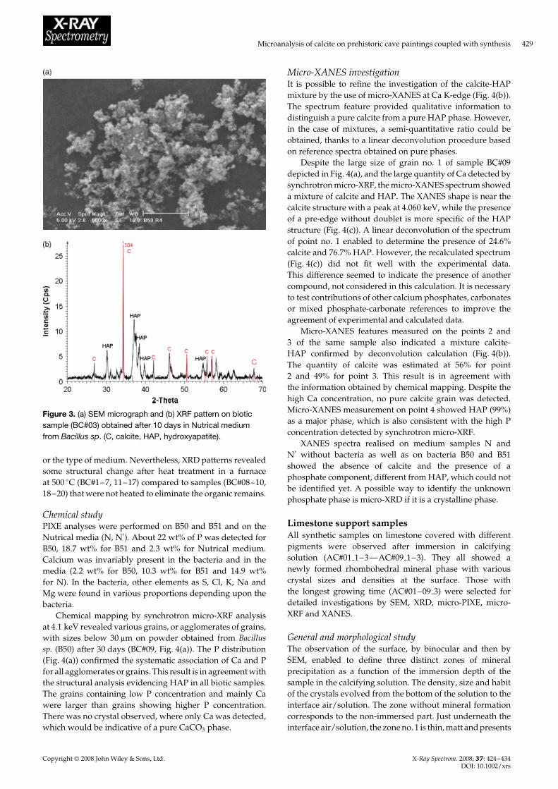
Biotic synthetic samplesGeneral and morphological studyThe mineral phase represented 5.8 wt% of the total mass after2 days of synthesis, and 7 wt% after 10 days. The mineralgrains observed were very fine and without any specifichabit, even after 10, 22 or 30 days of growth (Fig. 3(a)).Moreover, no evolution in size was observed as a functionof incubating time under the chosen conditions. No effectof the type of bacteria or of the medium was visible on themorphology and the grain size.
Structural informationXRD patterns were measured for identification of the newlyformed minerals after powder recovery from the incubatingmedium. The presence of CaCO3 component was hardlydetected beyond 2 days of incubation. After 10 days ofgrowth (BC#03 and BC#13, Fig. 3(b)) the crystallised calciteappeared clearly. No calcite was observed in the sample ofthe bacteria alone (B50, B51) and in the medium alone (N,N0), confirming calcite growth induced by the presence ofbacteria in an adapted medium.
Hydroxyapatite (HAP, Ca10�PO4�6�OH�2) was alwaysdetected from the first 2 days until 30 days of growth byXRD (Fig. 3(b)). The crystallinity and the quantity of HAPdid not change with the type of bacteria, the incubating time
Copyright 2008 John Wiley & Sons, Ltd. X-Ray Spectrom. 2008; 37: 424–434DOI: 10.1002/xrs
Microanalysis of calcite on prehistoric cave paintings coupled with synthesis 429
Figure 3. (a) SEM micrograph and (b) XRF pattern on bioticsample (BC#03) obtained after 10 days in Nutrical mediumfrom Bacillus sp. (C, calcite, HAP, hydroxyapatite).
or the type of medium. Nevertheless, XRD patterns revealedsome structural change after heat treatment in a furnaceat 500 °C (BC#1–7, 11–17) compared to samples (BC#08–10,18–20) that were not heated to eliminate the organic remains.
Chemical studyPIXE analyses were performed on B50 and B51 and on theNutrical media (N, N0). About 22 wt% of P was detected forB50, 18.7 wt% for B51 and 2.3 wt% for Nutrical medium.Calcium was invariably present in the bacteria and in themedia (2.2 wt% for B50, 10.3 wt% for B51 and 14.9 wt%for N). In the bacteria, other elements as S, Cl, K, Na andMg were found in various proportions depending upon thebacteria.
Chemical mapping by synchrotron micro-XRF analysisat 4.1 keV revealed various grains, or agglomerates of grains,with sizes below 30 µm on powder obtained from Bacillussp. (B50) after 30 days (BC#09, Fig. 4(a)). The P distribution(Fig. 4(a)) confirmed the systematic association of Ca and Pfor all agglomerates or grains. This result is in agreement withthe structural analysis evidencing HAP in all biotic samples.The grains containing low P concentration and mainly Cawere larger than grains showing higher P concentration.There was no crystal observed, where only Ca was detected,which would be indicative of a pure CaCO3 phase.
Micro-XANES investigationIt is possible to refine the investigation of the calcite-HAPmixture by the use of micro-XANES at Ca K-edge (Fig. 4(b)).The spectrum feature provided qualitative information todistinguish a pure calcite from a pure HAP phase. However,in the case of mixtures, a semi-quantitative ratio could beobtained, thanks to a linear deconvolution procedure basedon reference spectra obtained on pure phases.
Despite the large size of grain no. 1 of sample BC#09depicted in Fig. 4(a), and the large quantity of Ca detected bysynchrotron micro-XRF, the micro-XANES spectrum showeda mixture of calcite and HAP. The XANES shape is near thecalcite structure with a peak at 4.060 keV, while the presenceof a pre-edge without doublet is more specific of the HAPstructure (Fig. 4(c)). A linear deconvolution of the spectrumof point no. 1 enabled to determine the presence of 24.6%calcite and 76.7% HAP. However, the recalculated spectrum(Fig. 4(c)) did not fit well with the experimental data.This difference seemed to indicate the presence of anothercompound, not considered in this calculation. It is necessaryto test contributions of other calcium phosphates, carbonatesor mixed phosphate-carbonate references to improve theagreement of experimental and calculated data.
Micro-XANES features measured on the points 2 and3 of the same sample also indicated a mixture calcite-HAP confirmed by deconvolution calculation (Fig. 4(b)).The quantity of calcite was estimated at 56% for point2 and 49% for point 3. This result is in agreement withthe information obtained by chemical mapping. Despite thehigh Ca concentration, no pure calcite grain was detected.Micro-XANES measurement on point 4 showed HAP (99%)as a major phase, which is also consistent with the high Pconcentration detected by synchrotron micro-XRF.
XANES spectra realised on medium samples N andN0 without bacteria as well as on bacteria B50 and B51showed the absence of calcite and the presence of aphosphate component, different from HAP, which could notbe identified yet. A possible way to identify the unknownphosphate phase is micro-XRD if it is a crystalline phase.
Limestone support samplesAll synthetic samples on limestone covered with differentpigments were observed after immersion in calcifyingsolution (AC#01 1–3—AC#09 1–3). They all showed anewly formed rhombohedral mineral phase with variouscrystal sizes and densities at the surface. Those withthe longest growing time (AC#01–09 3) were selected fordetailed investigations by SEM, XRD, micro-PIXE, micro-XRF and XANES.
General and morphological studyThe observation of the surface, by binocular and then bySEM, enabled to define three distinct zones of mineralprecipitation as a function of the immersion depth of thesample in the calcifying solution. The density, size and habitof the crystals evolved from the bottom of the solution to theinterface air/solution. The zone without mineral formationcorresponds to the non-immersed part. Just underneath theinterface air/solution, the zone no. 1 is thin, matt and presents
Copyright 2008 John Wiley & Sons, Ltd. X-Ray Spectrom. 2008; 37: 424–434DOI: 10.1002/xrs

430 E. Chalmin et al.
(b) (c)
(a)
Figure 4. Biotic calcite BC#09 (prepared in Nutrical medium from Bacillus sp. during 30 days, without heat treatment): (a) Ca and Pmapping at 4.1 keV (pixel size: 0.5 ð 0.5 µm2) and location of micro-XANES measurement points. (b) Ca K-edge micro-XANESspectra on different points of the mapping zone (in transmission mode, beam size: 0.34 ð 0.75 µm2). The mixture corresponds to amixture of hydroxyapatite (HAP) and calcite. (c) Comparison between Ca K-edge XANES of calcite and HAP references,micro-XANES on point 1 (in transmission mode) and recalculated spectrum from linear deconvolution calculation.
small crystals as for the zone no. 3. Zone 3 is larger atthe bottom of the solution (Fig. 5(a)). Large rhombohedralcrystals (10–100 µm) were observed in the brilliant zone no.2 in intermediate position (Fig. 5(b)). The thickness of thislayer could be estimated at 50–70 µm (Fig. 5(c)).
No effect of the nature of the pigment layer was observedon the mineral morphology. Only the size of crystals evolvedas a function of the immersion time. However, the additionof clay to the pigment (AC#04–06 1–3) seemed to reducethe size and the number of crystals formed. The samebehaviour was observed for the addition of humic acid(AC#07–09 1–3), known as inhibitor of calcite growth. Inthese cases, the limestone support was less densely coveredby the newly formed calcite layer.
Structural informationThanks to grazing incidence XRD, it was possible to restrictthe structural analysis (with an estimated analytical depthof around 100 µm) to the mineral layer at the surface of thelimestone covered with pigments without any contributionof the limestone signal. The analysed surface area increasedcompared to traditional XRD due to the decreased incidentangle of the x-ray beam. However, the three zones at thesurface of the sample were large enough to be analysedseparately. The XRD patterns on AC#01 3 enabled us tovisualise only the contribution of well-crystallised calcite foran angle � of 0.5° on zone no. 2 (Fig. 5(d)). Thus, newlyformed calcite could be shown on the synthetic limestonesamples covered with pigments.
Copyright 2008 John Wiley & Sons, Ltd. X-Ray Spectrom. 2008; 37: 424–434DOI: 10.1002/xrs

Microanalysis of calcite on prehistoric cave paintings coupled with synthesis 431
(a) (b)
(c)(d)
Figure 5. (a) Scheme of zones of calcite formation on synthetic samples immersed in a calcifying solution, as for instance, AC#01 3(Limestone support covered with hematite after 45 days of growth), (b) SEM micrograph of largest rhombohedral crystals observedon the synthetic sample in the zone no. 2, (c) SEM micrograph of limestone cross-section showing the thickness of newly formedcalcite at the surface (50–70 µm), (d) grazing incidence x-ray diffraction at various angles on the surface of sample AC#01 3.
A cross-section of the synthetic sample covered withblack Mn oxide and newly formed calcite (AC#02 3) wasstudied by means of synchrotron micro-XRF and XANESat Mn K-edge (Fig. 6). The micro-x-ray beam (0.5 ð 1.5 µm2)allowed analysis with a spatial resolution in the micrometerrange and differentiating each layer: the limestone support,the homogeneous pigment layer composed by pyrolusite,and the newly formed calcite (Fig. 6(a)). Micro-XANESspectra were acquired at different points across eachlayer (Fig. 6(b)). They showed that the Mn K-edge featurecorresponds basically to that of a pyrolusite structure. Nostructural modification of the pigment was observed in anylayer. The noise in the Mn K-edge XANES spectra on calciteand limestone was due to the very low Mn concentration.
DISCUSSIONCalcite characteristics in the Large Cave ofArcy-sur-Cure, and main parameters influencingtheir growthMicritic, or micro-crystalline calcites and sparitic calcites ofcolumnar fabric are found in the opaque and transparentlayers, respectively, on the cave walls. Difference in crystalfabric is clearly at the origin of varying macroscopic aspectsof calcite. Micro-crystalline calcites are opaque, whereas
calcite layers with columnar fabrics are translucent due to abetter diffusion of light. Different growth kinetics, discharge,fluid supersaturation, outgassing, the presence of growthinhibitors, ion transport mechanisms from the solution to thecrystal surface, as well as temperature changes lead to theobserved variations in crystal size, morphology and fabric.According to Frisia et al.,5 columnar fabrics with the presenceof flat faces are characterised by few dislocations and formedthrough the screw dislocation growth mechanism. Suchmechanism is generally observed under low supersaturationconditions linked with a constant flow. That means a slowand continuous growth process. No biological process seemsto interfere in this completely abiotic crystal growth. Thecrystallite surfaces and sizes can, however, vary for columnarfabrics. This phenomenon can be explained by different evo-lutions as a function of summer and winter times. By contrast,micro-crystalline calcites are characterised by a high numberof defects as dislocations, microtwins, lamellae and subgrainboundaries.5 This indicates that they were formed under con-ditions that deviate strongly from the equilibrium inducedfor instance by a strong supersaturation, changing flow anddrip rate, varying temperature or changing CO2 pressure.Many nucleation sites are present for crystal growth. Micro-organisms can also interfere in their formation process.
Copyright 2008 John Wiley & Sons, Ltd. X-Ray Spectrom. 2008; 37: 424–434DOI: 10.1002/xrs

432 E. Chalmin et al.
The detailed observation of the synthetic calcite crystalsformed at the surface of the limestone samples coveredwith different pigments enables additional insights into theparameters influencing the calcite growth under naturalconditions. It was observed that the crystal size andappearance was not the same as a function of the immersiondepth in the calcifying solution. This variation in crystal sizecan be explained by a CO2 concentration gradient insidethe solution. As a function of the depth, the ratio CO2/Cadecreases and influences the saturation degree. Optimalcrystallisation conditions are reached two or three mmunder the surface level of the solution. Therefore, the largestand most brilliant calcite crystals are formed in this zone.Above and below this zone, either the CO2 concentrationis not optimal to allow regular calcite growth leadingto micro-crystalline calcites having a matt macroscopicappearance. Thus, CO2 pressure seems to be, in Ca-richenvironments such as karst caves, the controlling factor forcalcite formation.
The observation of the stalactite cross-section (NC#05)allows evidencing of the variations in habit, fabric and sizeof calcite crystals. The clear transition between the micro-crystalline and the columnar calcite layers can thereforebe explained by a variation in growth mechanisms andkinetics, induced by changes in the environmental conditionsas variations of CO2 pressure hydrological conditions ortemperature.11 Change in the vegetative cover, as forestclearance, inducing modifications in CO2 pressure in thecave atmosphere and water could also be at the origin of thevariation of calcite fabric.12 It is also possible that there wasa long period when calcite growth was completely absentbecause of low temperature and low CO2 concentrationsin the cave atmosphere. Genty et al.13 showed by datingstalagmite samples from the Chauvet Cave (Ardeche, France)that there was a period of about 9000 years (between 25 000and 15 000 years ago) marked by the absence of any calciteformation in the cave. This phenomenon could be related toknown climatic events in the past. The colder climate induceda deceleration of calcite growth. Thus, it would be interestingto date precisely the calcite layers from the Large Cave ofArcy-sur-Cure to know if such change in climate is at theorigin of the different calcite formations and if there is alsoa long period characterised by the absence of calcite growth.The age of the paintings, 28 000–24 500 B.P., just coincideswith the beginning of the cooling period of the continentalclimate in northern Europe, and as a consequence, theobserved slowing down of the calcite growth in caves leadingin this case to the complete stop of the growth of the spariticcalcite of columnar fabric. Micro-crystalline calcite wouldthen be formed much later.13 Moreover, a detailed study ofthe variations of the chemical and isotopic composition ofthe speleothem laminae at the Large Cave of Arcy-sur-Curecoupled with precise dating would allow reconstruction ofseasonal changes in temperature or at the scale of somecenturies.14
Prehistoric paint layer and interactions with calciteMicro-XANES analyses could show in the stalactite cross-section (NC#05) containing a prehistoric paint layer, that
(a)
(b)
Figure 6. (a) Mapping at 6.7 keV on limestone support coveredwith pyrolusite after 45 days of growth (AC#02 3) (pixel size:5 ð 5 µm2) and (b) associated Mn K-edge XANES spectrum foreach layer (in fluorescence mode, beam size: 0.5 ð 1.5 µm2).
it can actually be divided into two zones, a clay-richzone and a pigment-rich zone. The colouring elementscontained in the paint and clay layers were not transportedto neighbouring calcite layers as evidenced by measuringmicro-PIXE concentration profile.2 Additionally, it wasshown that the growth of large crystals of columnar fabric intranslucent calcite was not disturbed by the application of thepaint layer. The calcite crystals completely trap the small ironoxide (hematite, Fe2O3) and clay (aluminosilicates) particlescontained in the paint layer, which have a protective effect,as in frescos.
At this state of the study, it is not possible to determine anatural or intended anthropic origin of this small clay layer.It can be postulated that its presence is linked to a specialpreparation mode of the painting, added intentionally, ormore simply to the use of natural ochre. The hypothesis ofnatural ochre seems more consistent with other observationson pigments sampled and analysed from the ‘Grande Grotte’
Copyright 2008 John Wiley & Sons, Ltd. X-Ray Spectrom. 2008; 37: 424–434DOI: 10.1002/xrs

Microanalysis of calcite on prehistoric cave paintings coupled with synthesis 433
of Arcy-sur-Cure. Baffier et al.15 showed that both iron oxidesand coloured clays were used for the wall decorations in thecave.
The analysis of the cross-section of the synthetic limestonesamples covered with pigments confirmed the absenceof alteration of the paint layer, for both Mn and Feoxides. Moreover, no structural modification of the pigmentoccurred after calcite growth as evidenced by micro-XANES.All these observations indicated that calcite has a protectingeffect by entrapping not only the Fe-containing pigments butalso the Mn-containing pigments. The same effect should bevalid for charcoal-based pigments.
Role of bacteria on calcite growth in theArcy-sur-Cure CaveBacteria-induced calcite growth and comparison withcrystal features observed on natural calcitesThe culture of bacteria isolated from cave samples in aspecific medium proves the calcifying properties of thesebacteria. Indeed, the presence of tiny crystals of calcite wasdetected after 10 days of growth, and the amount of mineralphase increases with the incubation time. Micritic calciteseemed to be most similar to this synthetic biotic calcite, evenif the latter was mixed with other mineral phases. Takinginto account the extreme conditions and the short timescaleof the culture used for evidencing the calcifying propertiesof the bacteria at this first stage, it seems evident that theconditions used in the laboratory for bacteria-induced calcitegrowth did not reproduce the conditions that existed in thecave. Therefore, conclusions on the role of bacteria in calcitegrowth of Arcy-sur-Cure have to be carefully drawn from thedirect comparison of these biotics with the natural calcites.
According to literature sources, bacteria can have distincteffects on the calcite formation. On one hand, they can favourthe nucleation of calcite acting analogously as a catalyser,but on the other hand, they are supposed to inhibit calcitecrystal growth.3 This second effect is likely to explain thesmall grain sizes of biotic calcites and the fact that theobserved calcite size does not increase with the incubationtime. Other investigations showed that bacteria can havecalcifying properties even if the mechanisms evidenced inlaboratory remain difficult to apply to in situ phenomena.15
A detailed study on prehistoric paintings of several Spanishcaves, extensively visited or not, also showed an importantbacterial diversity. However, the biocorrosive potential anda possible damage of the paintings could not be evidenced.4
Origin of hydroxyapatite in biotic calcitesWhatever the conditions of biotic synthesis, the systematicformation of HAP next to calcite was observed. As a majorphase it was formed individually or in mixture with calciteor other compounds in the same agglomerates. The HAPmight originate from P and Ca in the bacteria themselvesand in the medium, respectively. Phosphorus could comefrom phospholipids in the bacteria, the main constituents ofthe bacteria membrane. A Ca source is added to the mediumfor culture. Because of the very low HAP saturation index,it is very likely that this phase precipitates first when P isliberated in the solution from the bacteria, or its formation isfavoured by a biomineralogical process.16 Benzerara et al.17
showed by Ca L2,3, as well as C K-edge Near Edge X-rayAbsorption Fine Structure (NEXAFS) studies on calcifiedbacteria, that carbonates can be incorporated in the HAPstructure, very likely by substitution for phosphate groups,and/or to a lesser extent, for hydroxyl ions, thus forminga carbonate-hydroxyapatite phase (dahllite, JCPDS 9–348).This phase exactly corresponds to that of the mineral phaseobserved in other biological mineral tissues as bone, dentineor ivory. Therefore, it can be postulated that a carbonate-HAP forms first, and in a later step calcite is precipitated.The heat treatment of the biotic samples used to recoverthe new minerals formed induced change in crystallinityof the apatite phase. According to studies on heat-inducedmodifications of mineral tissues as bone it can be assumedthat the carbonate-HAP phase is modified in the same wayas these mineral tissues. In such bio-mineral tissues, calcite isformed after heating to 500 °C.18 So the heating step duringthe biotic synthesis has to be changed or cancelled whilestudying the HAP origin.
CONCLUSIONS
The study of new natural calcites from the Large Caveof Arcy-sur-Cure confirmed first observations of differentcalcite types on the walls and their differentiation. Indeed,translucent calcite characterised by large sparitic crystals ofcolumnar fabric were most likely formed by a completelyabiotic process under very stable continuous conditionsnext to equilibrium, whereas opaque calcites are micro-crystalline and are probably formed under conditionsdeviating strongly from the equilibrium, induced for instanceby a strong supersaturation, changing flow and drip rate,varying temperature or changing CO2 pressure. Duringcalcite synthesis on limestone blocs covered with differentpaint layers prepared under abiotic conditions immersed ina calcifying solution, it was observed that the size of thecalcite crystals is basically dependent on the immersiondepth of the sample in the solution, and hence, of theCO2 concentration which decreases in the solution as afunction of the depth. This result indicates that one ofthe most critical environmental factors in karst caves fordetermining calcite growth mechanism is CO2 pressure thatshould be controlled and maintained stable for favouringtranslucent calcite growth on the walls. On this basis, themost appropriate conservation method of the prehistoricpaintings in this cave and in prehistoric caves in generalwould be the stabilisation and the control of CO2 pressurefluctuations and streams. The CO2 pressure should be held ata value allowing formation of translucent calcite on the cavewalls that does not hamper the visibility of the paintings,but on the contrary would act as an additional protectivevarnish.
Secondly, the study confirmed that the calcite is notaltering the paint layers. Actually Fe or Mn oxides arebasically maintained in their pigment layer and are notchemically changed because of the interactions with thecalcite. The translucent calcite traps the pigments and reactsas in frescos with the pigments on the wall. Thus, calcite hasa protecting effect. In addition, no transport or diffusion of
Copyright 2008 John Wiley & Sons, Ltd. X-Ray Spectrom. 2008; 37: 424–434DOI: 10.1002/xrs

434 E. Chalmin et al.
the colouring species as Fe or Mn could be observed eveninto micritic calcites. This justifies the conservation measurestaken at the Large Cave by eliminating the micritic opaquecalcites mechanically on the surface of the paintings. Thesemeasures do not endanger the integrity of the paintings andtheir conservation.
Third, investigations of biotic calcites evidenced the cal-cifying properties of the bacteria Bacillus sp. and P. fluorescensisolated from natural calcite samples originating from theGrande Grotte of Arcy-sur-Cure. However, conclusions onthe role of bacteria in the growth of natural calcites aredifficult to draw. In addition, it was observed that anothermineral phase, HAP, a calcium phosphate, is systematicallyformed in all biotic samples next to calcite. This phenomenonwas already observed in other researches on calcifying bacte-ria. The origin of the HAP phase and its role in the formationof calcite has still to be clarified. Refined structural analysesby micro-XRD and soft x-ray spectromicroscopy are pro-posed to solve the problem of the exact carbonate origin inthese biotic calcites.
However, the possibility of determining a clear markerof biologically induced calcite in prehistoric caves wouldallow a better differentiation of the origins of calcites foundin the paintings. The understanding of calcite origin couldhelp to define precise markers of similar artistic techniquesand contemporaneous prehistoric creations of paintings onthe basis of pigment groups formed by the association ofdifferent minerals. For instance, it could be evidenced bystudying Lascaux paint samples that some pigments weremixed with HAP, very likely traces of antler.19 Therefore, itis of great archaeological significance to clarify the origin ofpresent phosphates and calcites.
For the moment, no phosphate-containing mineral hasbeen observed in the paints or calcite samples of the GrandeGrotte of Arcy-sur-Cure. Even if opaque calcites constitutedfrom small crystals are observed, it is very likely that thecontribution of micro-organisms to opaque calcite growthin the cave is very limited, and can be completely excludedfor translucent calcites. It seems at this stage of the researchthat the microbial activity and the calcite growth cannotbe considered as an important alteration factor of theprehistoric paints in the Grande Grotte of Arcy-sur-Cure.On the contrary, controlled calcite growth can contribute toa better conservation of the paintings on the walls.
AcknowledgementsThe owner of the Arcy-sur-Cure cave (Le Comte de La Varende)is thanked for allowing us to study the cave. We are grateful toDominique Baffier, the conservator, and Michel Girard, archaeolo-gist, for their collaboration and their advice in choosing the samples.
The CORA association (Jean-Claude Liger, Daniele Molez, GillesSouchet) is acknowledged for help and support on-site, as well asE. Guillamet. Thin sections of stalactites were kindly prepared byA. Leclaire (LC2RMF). Vincent De Andrade (ID21, ESRF) is thankedfor the development of the calculation procedure by linear deconvo-lution. Laurent Charlet (LGIT, UMR 9995, Grenoble), Michel Menu,Colette Vignaud, Fanny d’Orlye (LC2RMF, UMR 171 CNRS) andRoberto Geremia (LECA, UMR 5553, Grenoble) are acknowledgedfor their contribution in the PNRC project. Alexandre Francois(LRMH) is thanked for the microbiological experimentations. Thisproject was supported by the PNRC programme 2004 financed bythe French Ministry of Culture and Communication as well as bythe GDR 2114 ChimArt 2.
REFERENCES1. Girard M, Baffier D, Brunet J, Guillamet E. L’art Avant l’histoire,
la Conservation de l’art prehistorique. 10emes journees d’etudesde la SFIIC, Paris, 2001.
2. Chalmin E, D’Orlye F, Zinger L, Charlet L, Geremia R, Orial G,Menu M, Baffier D, Reiche I. J. Geol. Soc. London 2007; 279: 185.
3. Schultze-Lam S, Harauz G, Beveridge TJ. J. Bacteriol. 1992; 174:7971.
4. Schabereiter-Gurtner C, Saiz-Jimenez C, Pinar G, Lubitz W,Rolleke S. FEMS Microbiol. Ecol. 2004; 47: 235.
5. Frisia S, Borsato A, Fairchild IJ, McDermott F. J. Sediment. Res.2000; 70: 1183.
6. Reiche I, Chalmin E, d’Orlye F, Sansot E, Menu M, Charlet L,Orial G, Geremia R, Baffier D, Girard M. 36th InternationalSymposium on Archaeometry, ISA2006. Laval’s series ofPublications in Archeometry: Quebec City, 2006.
7. Calligaro T, Dran J-C, Salomon J, Walter P. Nucl. Instrum.Methods Phys. Res. B 2004; 226: 29.
8. Maxwell JA, Campbell JL, Teesdale WJ. Nucl. Instrum. MethodsPhys. Res. B 1988; 43: 218.
9. Susini J, Salome M, Fayard B, Ortega R, Kaulich B. Surf. Rev. Lett.2002; 9: 203.
10. Chalmin E, Farges F, Brown GE Jr. Contrib. Mineral. Petrol. (Inpress).
11. White WB. Paleoclimate records from speleothems in limestonecaves. In Studies of Cave Sediment. Physical and Chemical Recordsof Paleoclimate, Sasowsky ID, Mylroie J (eds), Kluwer AcademicPublishers: Dordrecht / Boston / London 2003; 135.
12. Perrette Y, Delannoy JJ, Bolvin H, Cordonnier M, Destombes JL,Zhilinskaya EA, Aboukais A. Chem. Geol. 2000; 162: 221.
13. Genty D, Ghaleb B, Plagnes V, Causse C, Valladas H, Blamart D,Massault M, Geneste J-M, Clottes J. CR Palevol. 2004; 3: 629.
14. Kuczumow A, Genty D, Chevallier P, Nowak J, Florek M,Buczynska A. X-Ray Spectrom. 2005; 34: 502.
15. Baffier D, Girard M, Menu M, Vignaud C. L’anthropologie 1999;Tome 103: 1.
16. Benzerara K, Miller VM, Barell G, Kumar V, Miot J, Brown GE Jr,Lieske JC. J. Investig. Med. 2006; 54: 1.
17. Benzerara K, Yoon TH, Tyliszczak T, Constantz B, SpormannAM, Brown GE Jr. Geobiology 2004; 2: 249.
18. Reiche I, Vignaud C, Menu M. Archaeometry 2002; 44: 447.19. Chadefaux C, Vignaud C, Menu M, Reiche I. Archaeometry 2008;
doi:10.1111/j.1475-4754.2008.00373.x.
Copyright 2008 John Wiley & Sons, Ltd. X-Ray Spectrom. 2008; 37: 424–434DOI: 10.1002/xrs