Embed Size (px)
Citation preview

Methods in Molecular Biology
Molecular Biology Bio4751 Spring 2003Gary A. Bulla, PhD
a. Northern
1. Lyse with detergent, 2. Phenol extract (removes proteins)3. Precipitate RNA4. Load onto agarose gel5. Transfer RNA to nylon membrane6. Add radioactive DNA or RNA to detect individual species
Nylon membrane
probed with labeled a1-antitrypsin RNA, then
tubulin DNA
RNA quantitation
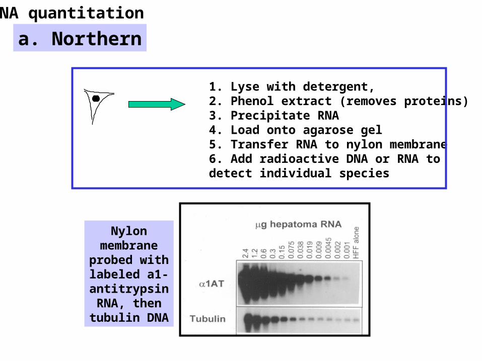
b. RNase protection1. Lyse with detergent 2. Phenol extract (removes proteins)3. Precipitate RNA4. Incubate with radioactive antisense RNA 5. Degrade single strand RNA with RNases6. Load on 8% PAGE
AAAAAmRNA
Radioactive antisense RNA
HybridizeRNase treat
1. Heat , PAGE2. Expose to film
RNA quantitation

Primer extension analysis
Prevent reinitiation
TATAA
RNA pol II holoenzymeHistone/DNA ratios
Primer
Also used to identify locations where transcription starts
c. Primer extensionRNA quantitation
mRNA
+dNTPs
Heat, PAGE, probe

Enhancer
Western Transfect C ells
35S amino acid, immunoprecipitate
electrophoresis
Molecular Biology Bio4751 Spring 2003Gary A. Bulla, PhD
Cell extract PAGE (+ SDS)
Rx---
Transfer to nylon membrane
Incubate with anti-Rx
Anti-myc
Alkaline peroxidase
Anti-Anti-
Alkaline peroxidase
Western (protein detection)
Anti-Rx
A. Immunoblot
B. Radiolabeling

Genetically Modified FoodsIncludes frost-resistant tomatoes
Disease-resistant sweet potatoes
Muscle-rich cattle
…..and many others
• Zambia’s government rejected 1000s of tons of corn from US because it may contain some GM kernels
•Approx 2.9 people at risk of starvation from drought-induced famine since 2001
•35,000 will die by 2003 if food not provided (WHO) •GM corn produces a bacterial toxin that is toxic to insects•GM corn used world-wide for 6 years without adverse effects (FDA)
Last month-

How can we detect measure gene activation?
DNA
RNA
Protein
Northern
Western
RNase Protection
How do we examine DNA-protein interactions?
Mad
Electrophoretic Mobility Shift Assay (EMSA) (aka gel shift)
DNaseI protection
How do we examine protein-protein interactions?
GST pull-down
EMSA Supershift
Co-immunoprecipitation
Primer extension
Photo-crosslinking
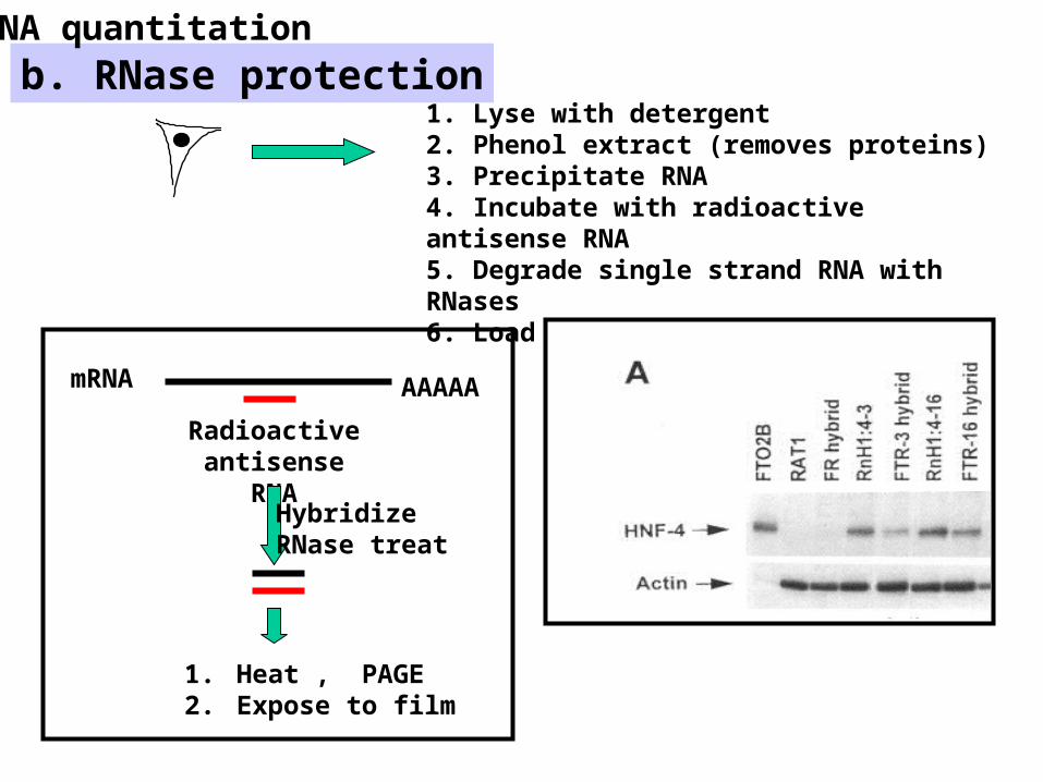
How can we measure promoter activity?
CAT-261 +44
1AT
Answer- Link it to a gene that is easy to monitor
Luciferase
B-galactosidase
(Chloramphenicol acetyl transferase)
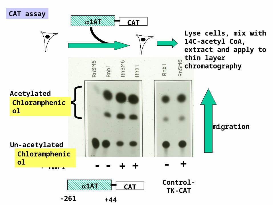
CAT1AT
+ HNF1
CAT
-261 +44
Control- TK-CAT
+ + +-- -
1AT
Acetylated
Un-acetylated
CAT assay
Lyse cells, mix with 14C-acetyl CoA, extract and apply to thin layer chromatography
migration
Chloramphenicol
Chloramphenicol

Protein-DNA interactions
EMSA (Gel Shift)
Photo-crosslinking
DNAseI footprinting
fos jun
TATATRE
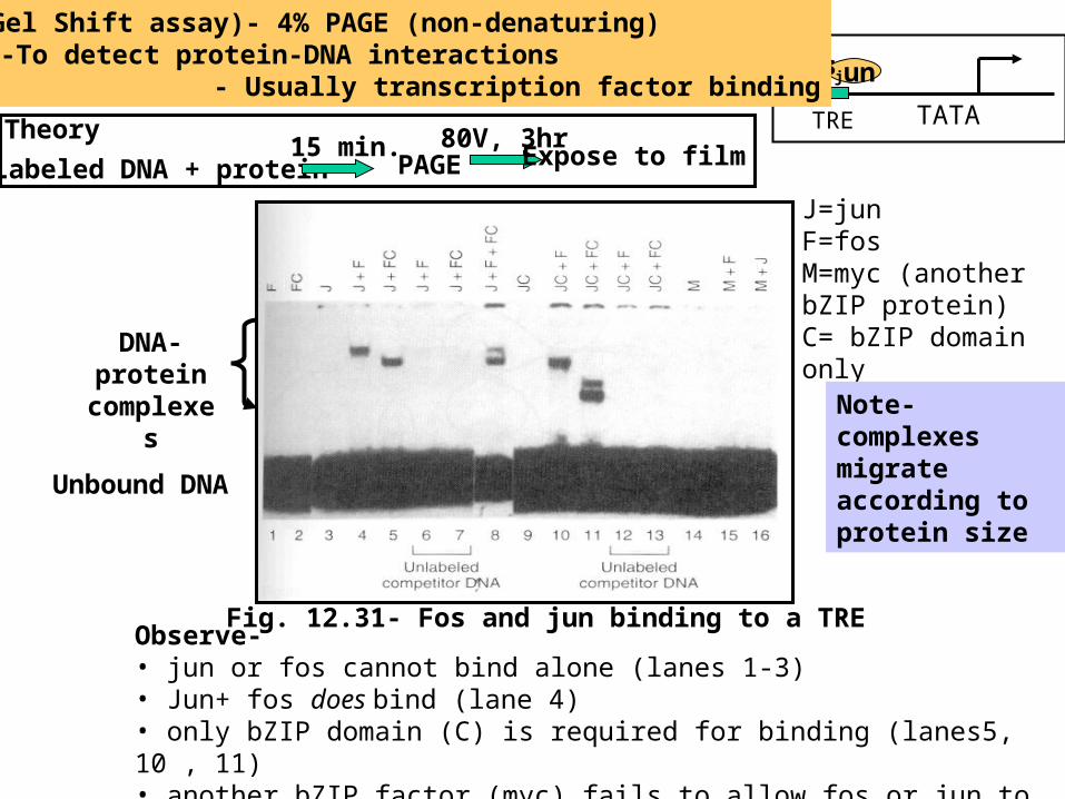
J=junF=fosM=myc (another bZIP protein)C= bZIP domain only
Fig. 12.31- Fos and jun binding to a TREObserve- • jun or fos cannot bind alone (lanes 1-3)• Jun+ fos does bind (lane 4)• only bZIP domain (C) is required for binding (lanes5, 10 , 11)• another bZIP factor (myc) fails to allow fos or jun to bind (lanes 14-15)
EMSA (Gel Shift assay)- 4% PAGE (non-denaturing)-To detect protein-DNA interactions
- Usually transcription factor binding
Labeled DNA + protein15 min.
PAGE
Unbound DNA
DNA-protein
complexes
TheoryExpose to film
80V, 3hr
Note- complexes migrate according to protein size

Footprinting by DNaseI and Cu++
Observe- •TFIID binds poorly•A + D binds strongly•B doesn’t enhance binding of D+A
Experiment
TFIID, A and/or B added to DNA
treat with DNaseI or Cu++
polyacrylamide gel electrophoresis(PAGE)
DNase I digestion products
32P
DNaseI footprintingFig. 11.4
DNAseI footprinting

Fig. 6.25
Fig. 6.24
• To identify proteins which bind DNAPhotocrosslinking)
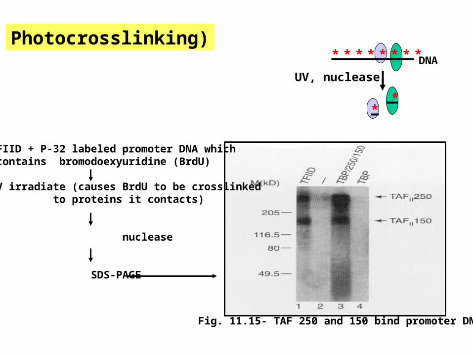
Fig. 11.15- TAF 250 and 150 bind promoter DNA
TFIID + P-32 labeled promoter DNA which contains bromodoexyuridine (BrdU)
UV irradiate (causes BrdU to be crosslinked to proteins it contacts)
nuclease
SDS-PAGE
DNA* * * * * * * *
UV, nuclease
**
Photocrosslinking)

Health stupidity reigns supreme
• Magnet therapy
• electronic ab exercisers- called “pump fiction” by Federal Trade Commission -ftc/gov/opa/2002/05/projectabsurd.htm
• Acupuncture-No proven benefit in controlled studies
• Chiropractic medicine- only useful for lower back pain. Period.
To make a lie into a truth-
“Say it loud, say it often” G. Gordon Liddy
All are largely accepted based upon “wart phenomenon”

How can we detect measure gene activation?
DNA
RNA
Protein
Northern
Western
RNase Protection
How do we examine DNA-protein interactions?
Mad
Electrophoretic Mobility Shift Assay (EMSA) (aka gel shift)
DNaseI protection
How do we examine protein-protein interactions?
GST pull-down
EMSA Supershift
Co-immunoprecipitation
Primer extension
Photo-crosslinking

Protein-protein interactions

Method- Epitope Tagging Ligate a small peptide onto a protein, introduce that protein into cells, then lyse the cells, and use antibodies raised against the small peptide to bind the protein plus any proteins interacting with that protein.
Gene of interest
ATG TAA
Poly-Adenylation sequence
PromoterFLAG epitope
(7-9 amino acids)
TAA
ATG
Epitope-tagged protein3A, slide 5
How do we determine identify protein-protein interactions?

Figure 10.13
Method- Epitope Tagging
RNA polymerase II structure- yeast has 12 subunits

MadSin3A
HDACFlag
Example- Epitope tagging experiment
Ac
Epitope-tagged histone deacetylase (HDAC2) to generate FLAG-HDAC2
Introduce FLAG-HDAC2 + Mad1 plasmids into cells
Prepare cell extracts
Immunoprecipitate with anti-FLAG Ab
PAGE
Probe with anti- SinA or anti-Mad1
Transfer to membrane
FLAG
HDAc
How do we determine identify protein-protein interactions?
Co-immunoprecipitation
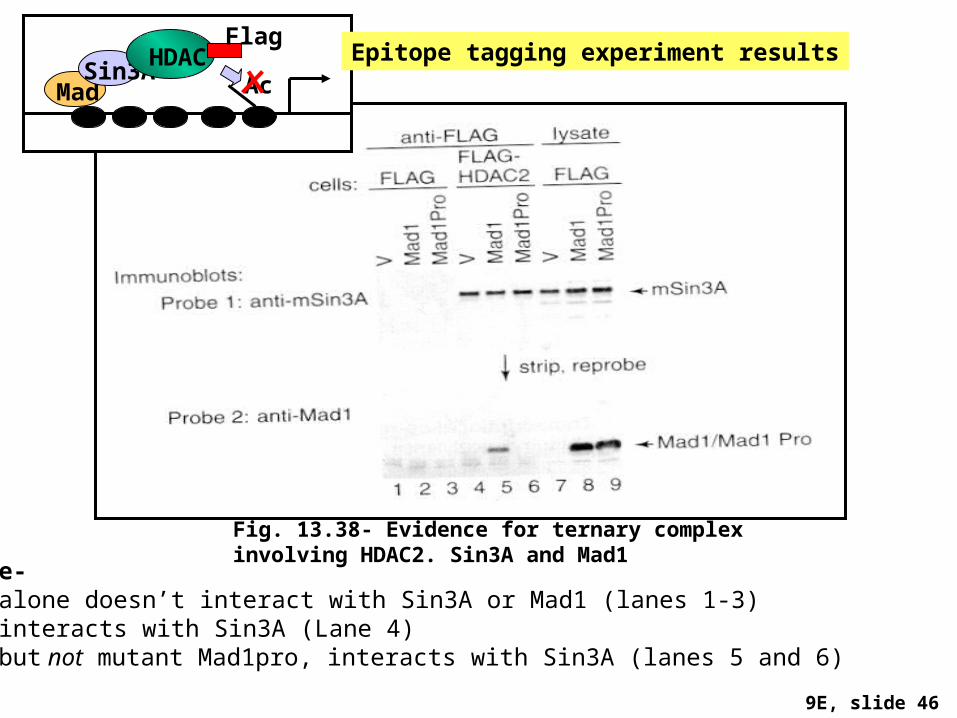
Observe- FLAG alone doesn’t interact with Sin3A or Mad1 (lanes 1-3)HDAC2 interacts with Sin3A (Lane 4)Mad1, but not mutant Mad1pro, interacts with Sin3A (lanes 5 and 6)
Fig. 13.38- Evidence for ternary complex involving HDAC2. Sin3A and Mad1
9E, slide 46
MadSin3A
HDACFlag
AcEpitope tagging experiment results

Anti-FLAG
FLAG
CBP
HNF1 myc
Anti-myc
Alkaline peroxidase
M2 High Sensitivity Capture Assay
96-well format
CMV PolyAFLAG CBP
CMV PolyAC-myc HNF1Co-transfect Cos7 cells
Another clever assay for protein-protein interaction

Assay- 1. separate nuclear protein on SDS-PAGEimpregnated with histones2. incubate gel with tritium -labeled AcCoA, wash away
Fig. 13.33 Activity gel assay for HAT activity
H4
Ac
AcAc
AcH3
H2BH2A
**
**
How do we determine whether a protein is a histone acetyltransferase (HAT)?
All nuclear proteins

How do we identify methylated DNA?
Methylation analysis: The results of MspI and HpaII cleavage are compared by Southern analysis
CCGG CCmGG CCGG
DNA probe
Msp
I
Hpa
II
•Digest genomic DNA with enzyme pair •Load onto agarose gel•Southern transfer•probe with 32-P DNA

Inactive Active
DNAseI DNAseI
Remove proteins Remove proteins
Cut with restriction enzyme
6kb 4kb 5kb 3kb
6kb4kb
5kb3kb
How does one find “open” vs “Closed” DNADNase sensitivity assay
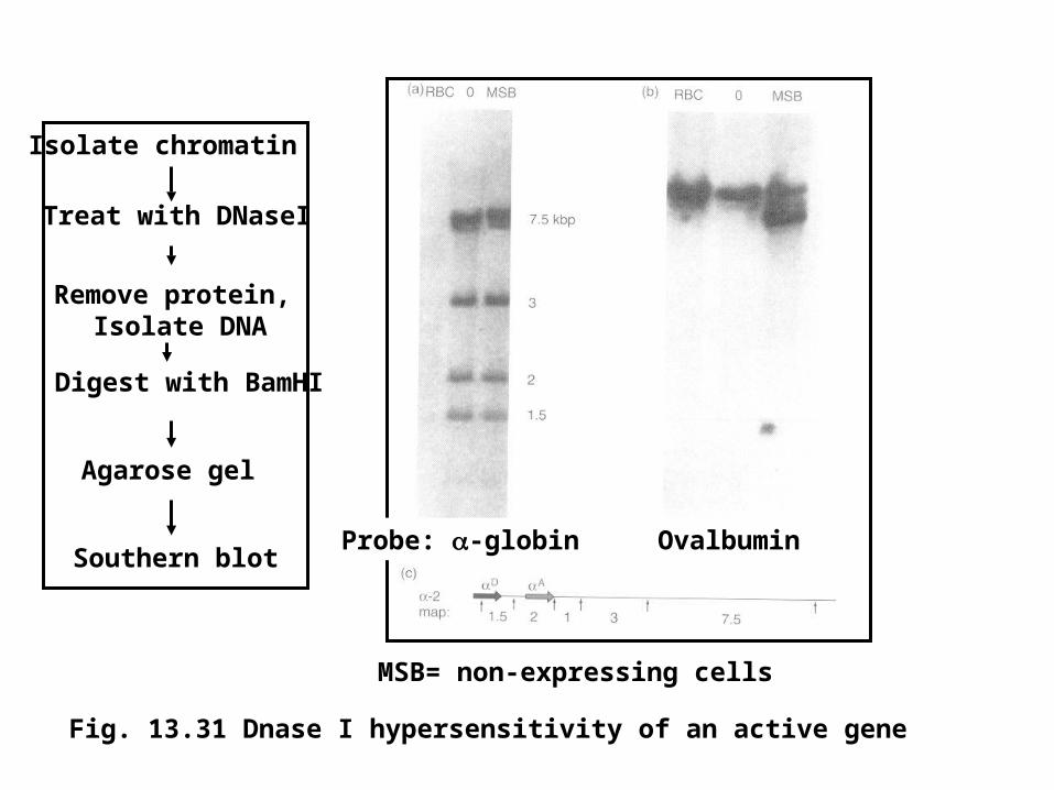
Fig. 13.31 Dnase I hypersensitivity of an active gene
Remove protein, Isolate DNA
Treat with DNaseI
Isolate chromatin
Digest with BamHI
Southern blot
Agarose gel
MSB= non-expressing cells
Ovalbumin Probe: -globin

Transcription run-off assay• To monitor transcriptional activity of a gene
Measure transcription directly. Thus post-transcriptional processing in not a concern

Figure 5.27
8B, slide 18

375 nt transcript
Note- Each lane contains RNA pol II + TFIIA,B,E and F
Fig. 11.18- TBP alone can’t respond to Sp1
SP1
TATAA
bh= bacterially derived human TBPvh=virus derived human TBP
TBP
Transcription run-off assay