Embed Size (px)
Citation preview

266JOP. Journal of the Pancreas - http://www.serena.unina.it/index.php/jop - Vol. 15 No. 3 – May 2014. [ISSN 1590-8577]
CASE REPORT
JOP. J Pancreas (Online) 2014 May 27; 15(3):266-268.
Metastatic Ampullary Adenocarcinoma Presenting as a Hydrocele: A Case Report
Whitney O Lane, Rex C Bentley, Herbert I Hurwitz, Leigh A Howard, Thomas J Polascik, Mark R Anderson, Dan G Blazer
Duke University School of Medicine, Duke University Medical Center, Durham, USA
ABSTRACTContext Metastases from ampullary malignancies are common, but spread to the testicle and paratesticular tissue is exceedingly rare with only 2 reported cases in the literature. Case Report We report a case of a 70 year-old male with a history of ampullary adenocarcinoma status post pancreaticoduodenectomy who presented with a symptomatic right-sided hydrocele. Subsequent pathology revealed metastatic ampullary adenocarcinoma. Conclusions Metastasis to the testicle and paratesticular tissue from ampullary malignancies is rare, but must be considered in the evaluation of scrotal masses in patients with a history of ampullary malignancy.
Received April 14, 2014 – Accepted May 02, 2014Key words Pancreatic Neoplasms; Pancreaticojejunostomy; Testicular Hydrocele; Testicular NeoplasmsCorrespondence Whitney O LaneDuke University School of Medicine, Duke University Medical Center, Durham NC 27710, USAPhone: +81-52.853.8211, Fax: +81-52.852.0952E-mail: [email protected]
INTRODUCTION
Ampullary malignancies have a higher rate of resectability and a more favorable prognostic outcome when compared to other periampullary tumors [1]. Despite this, 30% of patients with ampullary malignancies will have positive lymph nodes status at the time of resection and 50% of these patients will recur, most commonly locoregionally or with liver metastasis [2-4]. Metastasis to the testis and paratesticular tissue is rare with only 2 cases reported [5, 6]. Of those reports, only one case presented with a testicular mass as the initial site of treatment failure following resection [6]. We report a case of metastatic ampullary adenocarcinoma following pancreaticoduodenectomy with a hydrocele as the initial presentation of metastatic spread.
CASE REPORTAn A 70-year-old male originally presented with abdominal pain, chills and was found to have a total bilirubin of 7 and alkaline phosphatase of 770. He subsequently underwent endoscopic retrograde cholangiopancreatography (ERCP) with biliary stent placement and brushings. Pathology revealed atypical ductal cells suspicious for adenocarcinoma. At that time, Ca 19-9 was found to be elevated to 561 U/ml (reference range: <40U/ml). Subsequently, the patient underwent Endoscopic Ultrasound (EUS) and Computed Tomography (CT) scan,
both revealing dilatation of the biliary tree, but no discrete pancreatic, ampullary, or biliary mass was identified.
Due to concern for periampullary malignancy, the patient underwent a pancreaticoduodenectomy. Pathology revealed an ampullary adenocarcinoma with negative surgical margins and 1/9 positive lymph nodes (Figure 1). He subsequently underwent 5 weeks of adjuvant chemoradiation with capecitabine and a total of 4500cGy of radiation therapy, followed by 4 months of adjuvant gemcitabine therapy alone. Following completion of adjuvant therapy, he was clinically well, with no evidence of recurrent disease.
At a routine follow-up eighteen months after resection, his CA 19-9 was elevated to 249 U/ml (reference range: <40 U/ml). A CT scan at that time showed no radiographic evidence of metastatic disease and given that the patient was asymptomatic and clinically stable, it was determined that he should continue with routine surveillance.
Figure 1. High power view of the patient’s primary ampullary adenocarcinoma 21 months prior. The primary tumor is extremely similar histologically to the metastasis in the hydrocele sac. Compare to figure 4. (hematoxylin and eosin stained sections, 200X original magnification).
267JOP. Journal of the Pancreas - http://www.serena.unina.it/index.php/jop - Vol. 15 No. 3 – May 2014. [ISSN 1590-8577]
JOP. J Pancreas (Online) 2014 May 27; 15(3):266-268.
presentation of underlying malignancy or concurrently with the pancreatic primary. This case is unique as it is only the second reported case of metastatic spread presenting as a testicular mass following surgical resection.
Multiple theories of metastasis to the testicle and paratesticular tissues have been proposed, including retrograde venous extension, arterial embolization, retrograde lymphatic extension, transperitoneal seeding, and direct invasion [7, 8]. As there was evidence of lymphatic involvement at the time of the initial resection, the suspected route of tumor spread in this case is either lymphatic or direct transperitoneal seeding from peritoneal carcinomatosis.
Although metastasis to the testicle and paratesticular tissue from an ampullary primary is uncommon, it should certainly be considered in the evaluation of a scrotal mass in patients with a history of periampullary malignancy. Unfortunately, as in this case, patients with metastatic periampullary malignancies have a poor prognosis. The
Twenty-one months following pancreaticoduodenectomy, he was evaluated in clinic for a symptomatic right-sided groin mass and a CT scan showed free fluid in the right inguinal canal (Figure 2). He was subsequently taken to the OR and underwent outpatient right-sided hydrocele repair and excision of a non-communicating loculated cyst of the spermatic cord. Postoperatively, he recovered uneventfully. Surprisingly, despite no gross evidence of metastatic disease in the sac, pathology revealed metastatic moderately differentiated adenocarcinoma consistent with a pancreaticobiliary primary (Figure 3, 4, 5).
One month following the hydrocele repair, a restaging CT scan revealed no definite peritoneal nodularity, but repeat CA 19-9 was 1387 U/ml(reference range: <40 U/ml). Given the concern for occult peritoneal carcinomatosis, the patient was started on gemcitabine single agent therapy.
DISCUSSIONMetastatic ampullary malignancies to the testicle and paratesticular tissue are extremely rare, with only several additional cases of periampullary carcinoma with spread to the testicle or paratesticular tissues. Previous case reports have suggested that most cases are discovered as the first
Figure 2. CT-scan of the abdomen and pelvis showing right-sided inguinal fluid collection.
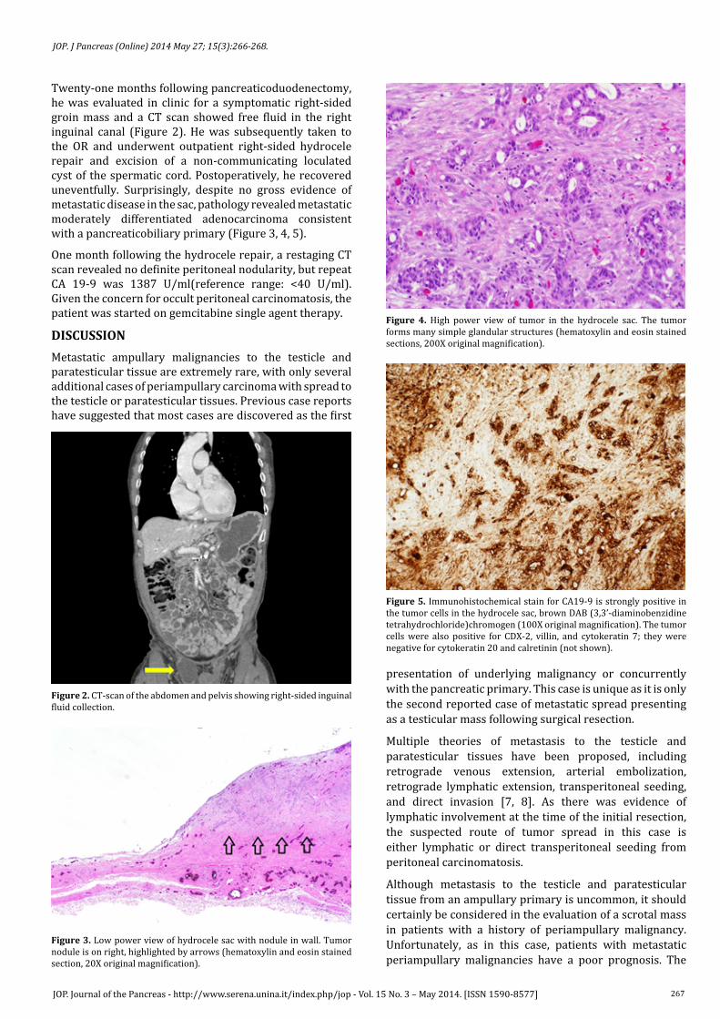
Figure 3. Low power view of hydrocele sac with nodule in wall. Tumor nodule is on right, highlighted by arrows (hematoxylin and eosin stained section, 20X original magnification).
Figure 4. High power view of tumor in the hydrocele sac. The tumor forms many simple glandular structures (hematoxylin and eosin stained sections, 200X original magnification).
Figure 5. Immunohistochemical stain for CA19-9 is strongly positive in the tumor cells in the hydrocele sac, brown DAB (3,3’-diaminobenzidine tetrahydrochloride)chromogen (100X original magnification). The tumor cells were also positive for CDX-2, villin, and cytokeratin 7; they were negative for cytokeratin 20 and calretinin (not shown).
268JOP. Journal of the Pancreas - http://www.serena.unina.it/index.php/jop - Vol. 15 No. 3 – May 2014. [ISSN 1590-8577]
JOP. J Pancreas (Online) 2014 May 27; 15(3):266-268.
median survival in patients receiving chemotherapy is 6 months, while those with untreated disease have a median survival of 2-3 months [9].
Conflict of InterestAuthors declare that they have no conflict of interest to disclose.
References
1. Howe JR, Klimstra DS, Moccia RD, Conlon KC, Brennan MF. Factors predictive of survival in ampullary carcinoma. Ann Surg, 1998. 228(1): 87-94. [PMID:9671071]
2. Talamini MA, Moesinger RC, Pitt HA, Sohn TA, Hruban RH, Lillemoe KD, Yeo CJ, et al. Adenocarcinoma of the ampulla of Vater. A 28-year experience. Ann Surg. 1997; 225(5):590-599; discussion 599-600. [PMID:9193186]
3. Winter JM, Cameron JL, Olino K, Herman JM, de Jong MC, Hruban RH, Wolfgang CL, et al. Clinicopathologic analysis of ampullary neoplasms in
450 patients: implications for surgical strategy and long-term prognosis. J Gastrointest Surg. 2010; 14(2):379-387. [PMID:19911239]
4. Park JS, Yoon DS, Kim KS, Choi JS, Lee WJ, Chi HS, Kim BR. Factors influencing recurrence after curative resection for ampulla of Vater carcinoma. J Surg Oncol. 2007; 95(4):286-290. [PMID:17326125]
5. Sawa TE, Duun S, Andersen JT. Paratesticular Tumor: A metastasis from primary pancreas cancer. Scand J Urol Nephrol. 2000; 34(1):70-71. [PMID:10757275]
6. Dookeran KA, Lotze MT, Sikora SS, Rao UN. Pancretic and ampullary carcinoma with intrascrotal metastases. Br J Surg. 1997; 84(2):198-199. [PMID:9052433]
7. Rosser CJ, Gerrard E. Metastatic adenocarcinoma of the pancreas to the testicle: A case report. Am J Clin Oncol. 1999; 22(6):619-620. [PMID:10597749]
8. Seo IY, Kim SG, Han WC, Rim JS. Paratesticular mucinous cystadenocarcinoma: Metastasis from pancreatic cancer. Int J Urol. 2004; 11(12):1147-1149. [PMID:15663694]
9. Stathis A, Moore MJ. Advanced pancreatic carcinoma: current treatment and future challenges. Nat Rev Clin Oncol. 2010; 7(3): 163-172. [PMID:20101258]