Embed Size (px)
Citation preview
Hindawi Publishing CorporationCase Reports in Obstetrics and GynecologyVolume 2013, Article ID 108582, 3 pageshttp://dx.doi.org/10.1155/2013/108582
Case ReportMature Ovarian Teratoma with Carcinoid Tumor in a28-Year-Old Patient
Stamatios Petousis,1 Ioannis Kalogiannidis,1 Chrysoula Margioula-Siarkou,1
Alexandros Traianos,1 Dimosthenis Miliaras,2 Apostolos Kamparoudis,3
Apostolos Mamopoulos,1 and David Rousso1
1 3rd Department of Obstetrics and Gynaecology, Aristotle University of Thessaloniki, Konstantinoupoleos 49, 54642, Greece2 Laboratory of Histology, Aristotle University of Thessaloniki, 54124 Panemistimioupolis, Greece3 5th Surgical Department, Aristotle University of Thessaloniki, 54124 Panemistimioupolis, Greece
Correspondence should be addressed to Stamatios Petousis; [email protected]
Received 31 May 2013; Accepted 8 July 2013
Academic Editors: D. Hochner-Celnikier, E. F. C. Murta, and M. A. Osmanagaoglu
Copyright © 2013 Stamatios Petousis et al. This is an open access article distributed under the Creative Commons AttributionLicense, which permits unrestricted use, distribution, and reproduction in any medium, provided the original work is properlycited.
Introduction. Coexistence of carcinoid tumor inside a mature cystic teratoma is an extremely rare phenomenon, especially inyoung women. We present the case of a 28-year-old woman diagnosed with a right ovarian carcinoid and treated uneventfullywith conservative surgical approach. Case Report. A 28-year-old woman, gravid 0, parity 0, presented to our department for herannual gynecological examination and Pap smear test. During her examination, a mobile cystic mass was detected in the rightlower abdomen. Ultrasound indicated a right ovarian mass 10.5 × 6.3 cm, confirmed by CT scan. Further investigation revealedAFP levels (1539 ng/mL). The ovarian mass was excised by laparoscopy, leaving intact the remaining right ovary. Frozen sectionsshowed amature cystic teratoma.However, paraffin sections revealed the presence of a small carcinoid within the teratoma’s gastric-type mucosa. The patient was set to a close followup. Nine months postoperatively, ultrasound pelvis imaging and CT scan of theabdomen as well as serum tumor markers have shown no evidence of recurrence disease. Conclusion. Despite the weak evidence,fertility spare surgical approach for women wanting to preserve their genital tract might be a reasonable option.
1. Introduction
Mature cystic teratomas (MCT) represent 10–20% of allovarian neoplasms [1].Theymainly present in young women,and by definition they are characterized by benign histologicfeatures. Malignant transformation of teratoma (TMT), pre-dominantly to squamous cell carcinomas, may be observedin 1–3% of mature teratomas [2]. However, despite the rela-tively low incidence of malignant transformation, a numberof other malignant tumors arising from MCT have beenreported, including adenocarcinoma, thyroid carcinoma,sebaceous carcinoma, malignant melanoma, and sarcoma,while cases with metastatic behavior have also been reported[3–6].
The occasion of a carcinoid tumor arising from MCTis very rare with a very small number of published cases[7, 8]. Furthermore, patients with TMT are at least 15 years
older than the average patient with mature cystic teratomaand the majority of reports concern postmenopausal women[9]. Therefore, the coexistence of a carcinoid tumor insidea mature ovarian cystic teratoma in young patients is anextremely rare phenomenon of high clinical interest thatposes a challenging dilemma about the optimal therapeuticstrategy, especially for women desiring to preserve theirfertility.
We present a case of a 28-year-old nulliparous womandiagnosed with carcinoid tumor arising from a matureovarian cystic teratoma.
2. Case Report
A 28-year-old woman, gravid 0, parity 0, presented to ourdepartment for her annual gynecological examination and
2 Case Reports in Obstetrics and Gynecology
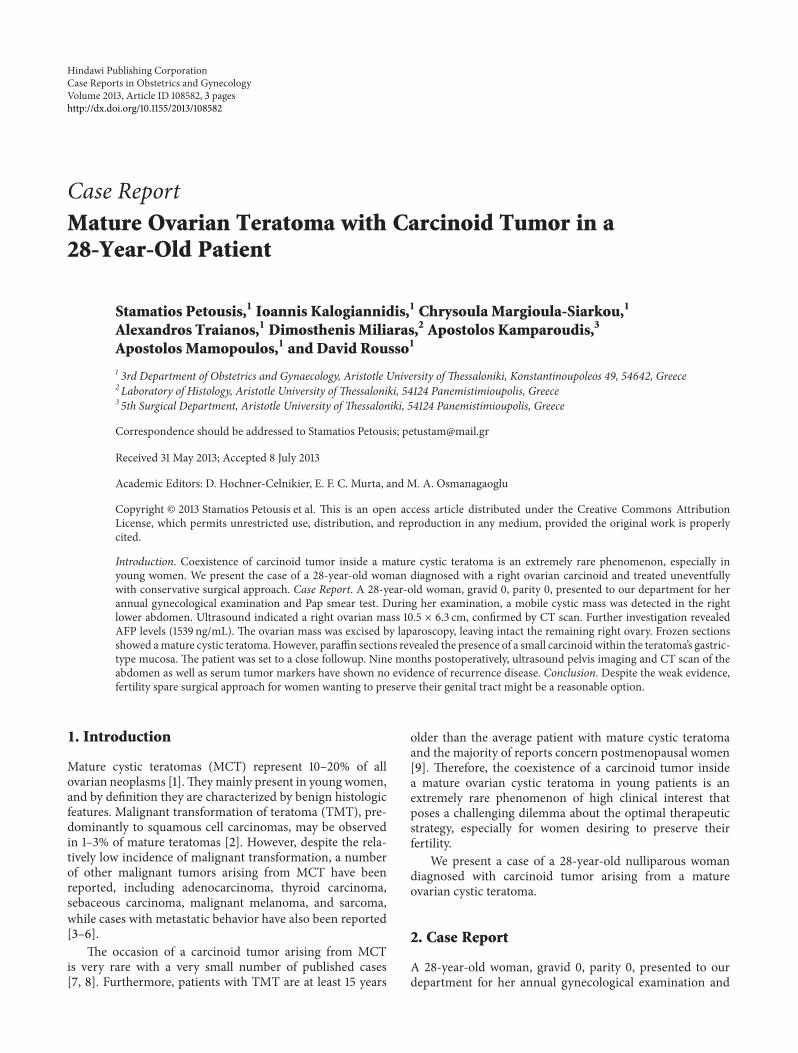
Figure 1: A carcinoid tumor is seen in the upper half of the picture.An island of mature hyaline cartilage (lower left), and lobules ofmucous and serous glands (lower part towards the center) are alsoseen (H&E, ×100).
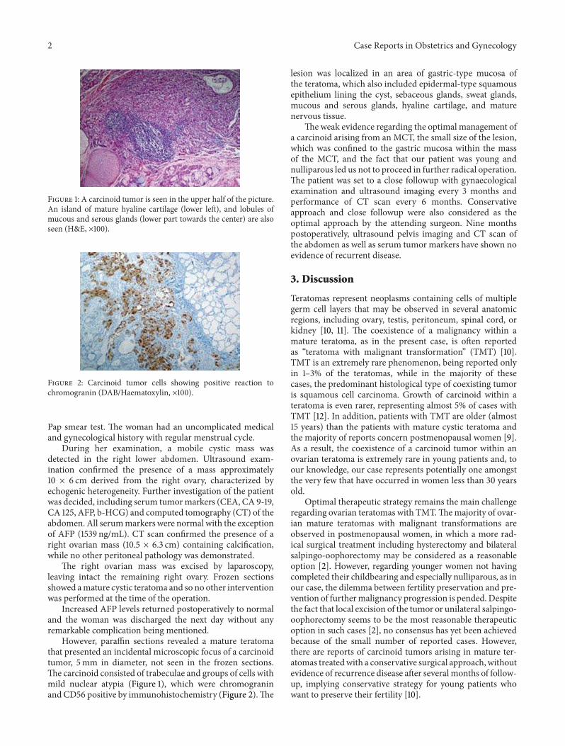
Figure 2: Carcinoid tumor cells showing positive reaction tochromogranin (DAB/Haematoxylin, ×100).
Pap smear test. The woman had an uncomplicated medicaland gynecological history with regular menstrual cycle.
During her examination, a mobile cystic mass wasdetected in the right lower abdomen. Ultrasound exam-ination confirmed the presence of a mass approximately10 × 6 cm derived from the right ovary, characterized byechogenic heterogeneity. Further investigation of the patientwas decided, including serum tumormarkers (CEA, CA 9-19,CA 125, AFP, b-HCG) and computed tomography (CT) of theabdomen. All serummarkers were normal with the exceptionof AFP (1539 ng/mL). CT scan confirmed the presence of aright ovarian mass (10.5 × 6.3 cm) containing calcification,while no other peritoneal pathology was demonstrated.
The right ovarian mass was excised by laparoscopy,leaving intact the remaining right ovary. Frozen sectionsshowed amature cystic teratoma and so no other interventionwas performed at the time of the operation.
Increased AFP levels returned postoperatively to normaland the woman was discharged the next day without anyremarkable complication being mentioned.
However, paraffin sections revealed a mature teratomathat presented an incidental microscopic focus of a carcinoidtumor, 5mm in diameter, not seen in the frozen sections.The carcinoid consisted of trabeculae and groups of cells withmild nuclear atypia (Figure 1), which were chromograninandCD56 positive by immunohistochemistry (Figure 2).The
lesion was localized in an area of gastric-type mucosa ofthe teratoma, which also included epidermal-type squamousepithelium lining the cyst, sebaceous glands, sweat glands,mucous and serous glands, hyaline cartilage, and maturenervous tissue.
The weak evidence regarding the optimal management ofa carcinoid arising from an MCT, the small size of the lesion,which was confined to the gastric mucosa within the massof the MCT, and the fact that our patient was young andnulliparous led us not to proceed in further radical operation.The patient was set to a close followup with gynaecologicalexamination and ultrasound imaging every 3 months andperformance of CT scan every 6 months. Conservativeapproach and close followup were also considered as theoptimal approach by the attending surgeon. Nine monthspostoperatively, ultrasound pelvis imaging and CT scan ofthe abdomen as well as serum tumor markers have shown noevidence of recurrent disease.
3. Discussion
Teratomas represent neoplasms containing cells of multiplegerm cell layers that may be observed in several anatomicregions, including ovary, testis, peritoneum, spinal cord, orkidney [10, 11]. The coexistence of a malignancy within amature teratoma, as in the present case, is often reportedas “teratoma with malignant transformation” (TMT) [10].TMT is an extremely rare phenomenon, being reported onlyin 1–3% of the teratomas, while in the majority of thesecases, the predominant histological type of coexisting tumoris squamous cell carcinoma. Growth of carcinoid within ateratoma is even rarer, representing almost 5% of cases withTMT [12]. In addition, patients with TMT are older (almost15 years) than the patients with mature cystic teratoma andthe majority of reports concern postmenopausal women [9].As a result, the coexistence of a carcinoid tumor within anovarian teratoma is extremely rare in young patients and, toour knowledge, our case represents potentially one amongstthe very few that have occurred in women less than 30 yearsold.
Optimal therapeutic strategy remains the main challengeregarding ovarian teratomas with TMT.Themajority of ovar-ian mature teratomas with malignant transformations areobserved in postmenopausal women, in which a more rad-ical surgical treatment including hysterectomy and bilateralsalpingo-oophorectomy may be considered as a reasonableoption [2]. However, regarding younger women not havingcompleted their childbearing and especially nulliparous, as inour case, the dilemma between fertility preservation and pre-vention of furthermalignancy progression is pended. Despitethe fact that local excision of the tumor or unilateral salpingo-oophorectomy seems to be the most reasonable therapeuticoption in such cases [2], no consensus has yet been achievedbecause of the small number of reported cases. However,there are reports of carcinoid tumors arising in mature ter-atomas treatedwith a conservative surgical approach, withoutevidence of recurrence disease after several months of follow-up, implying conservative strategy for young patients whowant to preserve their fertility [10].
Case Reports in Obstetrics and Gynecology 3
Clinical and histopathologic prognostic factors of theneoplasm represent the basic determinants of the therapeuticstrategy in such rare cases without evidence-based guidelines.Cyst wall invasion, intraoperative rupture of the ovarianmass, tumor dissemination, and adhesions aremainly consid-ered as unfavorable prognostic factors [12]. Furthermore, theobservation of clinical symptoms and signs such as flushing,edema, diarrhea, abdominal cramps, respiratory distress,and cardiac dysfunction that are caused by the secretionof vasoactive factors from the neuroendocrine cells, whichis usually referred to as the “carcinoid syndrome,” may beindicative of a rather aggressive biological behavior [10].However, in our case, no unfavorable pathologic or clinicalsigns or symptoms were observed. Therefore, consideringthe low metastatic potential of the present tumor, the youngage, and the nulliparous status of our patient, no furtherradical surgical approach was decided, while close followupaccording to the oncologic standards was organized.
In conclusion, despite the weak evidence related to thecoexistence of a carcinoid tumor within a mature teratomain younger patients, it seems that fertility spare surgicalapproach for women wanting to preserve their genital tractmay be a reasonable option. However, further evidenceis needed in order to support definitively the option ofconservative treatment in such cases without compromisingpatient’s survival.
References
[1] S. Khanna, V. Srivastava, S. Saroj, S. P. Mishra, and S. P. Gupta,“An unusual presentation of ovarian teratoma: a case teratoma,”Case Reports in Emergency Medicine, vol. 2012, Article ID845198, 2 pages, 2012.
[2] S. M. Kim, H. S. Choi, J. S. Byun et al., “Mucinous adeno-carcinoma and strumal carcinoid tumor arising in one maturecystic teratoma of the ovary with synchronous cervical cancer,”Journal of Obstetrics and Gynaecology Research, vol. 29, no. 1, pp.28–32, 2003.
[3] M. S. Krumerman andA. Chung, “Squamous carcinoma arisingin benign cystic teratoma of the ovary. A report of four cases andreview of the literature,” Cancer, vol. 39, no. 3, pp. 1237–1242,1977.
[4] O. M. Curling, P. N. Potsides, and C. N. Hudson, “Malignantchange in benign cystic teratoma of the ovary,” British Journalof Obstetrics and Gynaecology, vol. 86, no. 5, pp. 399–402, 1979.
[5] S. Chadha and A. Schaberg, “Malignant transformation inbenign cystic teratomas: dermoids of the ovary,” EuropeanJournal of Obstetrics Gynecology and Reproductive Biology, vol.29, no. 4, pp. 329–338, 1988.
[6] S. Kanayama, Y. Yamada, Y. Tanase et al., “A case of early-stageovarian carcinoid tumormetastasized to the liver,” Case Reportsin Obstetrics and Gynecology, vol. 2012, Article ID 961087, 5pages, 2012.
[7] F. Flam and C. Silfversward, “Combination of granulosa celltumour and carcinoid in an imitation of appendix vermiculariswithin a mature teratoma—a unique case,” European Journal ofObstetrics Gynecology and Reproductive Biology, vol. 56, no. 2,pp. 139–142, 1994.
[8] P. Chatzipantelis, A. Mavrogiorgis, E. Kairi-Vassilatou, and A.Pafiti, “Ovarian neoplasm composed of an insular carcinoid
tumor and a borderline mucinous cystadenoma arising in amature cystic teratoma: a case report,” European Journal ofGynaecological Oncology, vol. 27, no. 6, pp. 636–637, 2006.
[9] B.Djordjevic, E.D. Euscher, andA.Malpica, “Growing teratomasyndrome of the ovary: review of literature and first report ofa carcinoid tumor arising in a growing teratoma of the ovary,”American Journal of Surgical Pathology, vol. 31, no. 12, pp. 1913–1918, 2007.
[10] T. Yamasaki, Y. Yagihashi, T. Shirahase, T. Hashimura, and C.Watanabe, “Primary carcinoid tumor arising in a retroperi-toneal mature teratoma in an adult,” International Journal ofUrology, vol. 11, no. 10, pp. 912–915, 2004.
[11] E. Kurzer, R. J. Leveillee, and G.Morillo, “Rare case of carcinoidtumor arising within teratoma in kidney,” Urology, vol. 66, no.3, pp. 658.e5–658.e6, 2005.
[12] D. S. Arora and S. Haldane, “Carcinosarcoma arising in adermoid cyst of the ovary,” Journal of Clinical Pathology, vol. 49,no. 6, pp. 519–521, 1996.


![PARIPEX - INDIAN JOURNAL OF RESEARCH | Volume-8 | Issue-10 ... · teratoma is known as a monodemal teratoma.[1] Immature teratoma (IT) is a preferred term for the malignant ovarian](https://img.dokumen.tips/doc/110x75/603e5f8d2bf3bd27e47c8252/paripex-indian-journal-of-research-volume-8-issue-10-teratoma-is-known.jpg)

![A case of ovarian adenosquamous carcinoma arising from … · 2017. 8. 29. · oma include malignant transformation of a preexisting ovarian teratoma [2, 3] and endometriosis [4]](https://img.dokumen.tips/doc/110x75/60e9eb065f970107452b4437/a-case-of-ovarian-adenosquamous-carcinoma-arising-from-2017-8-29-oma-include.jpg)


