Embed Size (px)
Citation preview
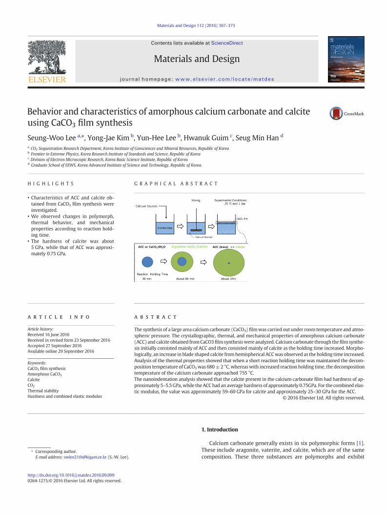
Materials and Design 112 (2016) 367–373
Contents lists available at ScienceDirect
Materials and Design
j ourna l homepage: www.e lsev ie r .com/ locate /matdes
Behavior and characteristics of amorphous calcium carbonate and calciteusing CaCO3 film synthesis
Seung-Woo Lee a,⁎, Yong-Jae Kim b, Yun-Hee Lee b, Hwanuk Guim c, Seug Min Han d
a CO2 Sequestration Research Department, Korea Institute of Geosciences and Mineral Resources, Republic of Koreab Frontier in Extreme Physics, Korea Research Institute of Standards and Science, Republic of Koreac Division of Electron Microscopic Research, Korea Basic Science Institute, Republic of Koread Graduate School of EEWS, Korea Advanced Institute of Science and Technology, Republic of Korea
H I G H L I G H T S G R A P H I C A L A B S T R A C T
• Characteristics of ACC and calcite ob-tained from CaCO3 film synthesis wereinvestigated.
• We observed changes in polymorph,thermal behavior, and mechanicalproperties according to reaction hold-ing time.
• The hardness of calcite was about5 GPa, while that of ACC was approxi-mately 0.75 GPa.
⁎ Corresponding author.E-mail address: [email protected] (S.-W. Lee).
http://dx.doi.org/10.1016/j.matdes.2016.09.0990264-1275/© 2016 Elsevier Ltd. All rights reserved.
a b s t r a c t
a r t i c l e i n f oArticle history:Received 16 June 2016Received in revised form 23 September 2016Accepted 27 September 2016Available online 29 September 2016
The synthesis of a large area calcium carbonate (CaCO3) filmwas carried out under room temperature and atmo-spheric pressure. The crystallographic, thermal, and mechanical properties of amorphous calcium carbonate(ACC) and calcite obtained fromCaCO3film synthesiswere analyzed. Calcium carbonate through thefilm synthe-sis initially consisted mainly of ACC and then consisted mainly of calcite as the holding time increased. Morpho-logically, an increase in blade shaped calcite fromhemispherical ACCwas observed as the holding time increased.Analysis of the thermal properties showed that when a short reaction holding time was maintained the decom-position temperature of CaCO3was 680±2 °C, whereaswith increased reaction holding time, the decompositiontemperature of the calcium carbonate approached 755 °C.The nanoindentation analysis showed that the calcite present in the calcium carbonate film had hardness of ap-proximately 5–5.5 GPa,while the ACC had an average hardness of approximately 0.75GPa. For the combined elas-tic modulus, the value was approximately 59–60 GPa for calcite and approximately 25–30 GPa for the ACC.
© 2016 Elsevier Ltd. All rights reserved.
Keywords:CaCO3 film synthesisAmorphous CaCO3
CalciteCO2
Thermal stabilityHardness and combined elastic modulus
1. Introduction
Calcium carbonate generally exists in six polymorphic forms [1].These include aragonite, vaterite, and calcite, which are of the samecomposition. These three substances are polymorphs and exhibit
368 S.-W. Lee et al. / Materials and Design 112 (2016) 367–373
different crystal structures for the same composition. They have alsobeen classified as CaCO3·H2O, with monomolecular bonding of watermolecules, CaCO3·6H2O (Ikaite), in which there is bonding by 6 watermolecules, and amorphous calcium carbonate (ACC), which does nothave an established crystal structure [2].
Calcium carbonate is a known biological material often found innature as well as an inorganic substance synthesized from living or-ganisms. Biological materials are defined as substances that are syn-thesized (e.g., bone, shell, skin, etc.) by humans and other livingorganisms for their own survival; these materials have received con-siderable attention from researchers in the chemistry and elementalfields due to their excellent elemental characteristics. Calcium car-bonate, which can be easily identified in nature, is a substanceforming the clamshell (shell) or exoskeleton of a spongy body organ-ism and has been a subject of continued interest due to the ease in se-curing sample materials and due to its excellent mechanicalproperties [3,4].
Amorphous calcium carbonate can be thought of as the initialstarting material for the formation of calcium carbonate. It exhibitsvery weak thermal stability and thus has been known to easily tran-sit into the crystal phase at room temperature and atmospheric pres-sure [1,5–9]. However, more information is needed to understandthe calcium carbonate phase transition. It is difficult to identify thepresence of amorphous calcium carbonate in nature and obtain suf-ficient sample material for analysis. Additionally, a small sampleamount leads to severe limitations to possible experimental analy-ses. Considering these factors, it may be easier to obtain amorphouscalcium carbonate through an in vitro CaCO3 experiment rather thanin vivo.
However, investigation of the calcium carbonate phase transitionthrough in vitroCaCO3 experiments is also challenging. In vitro synthesisof calcium carbonate is generally performed by either the CO2 diffusionmethod using the diffusion of ammonium carbonate [10,11] or precipi-tated calcium carbonate (PCC) synthesis [12,13]. However, the CO2 dif-fusion method only synthesizes a very small amount of calciumcarbonate that is not sufficient for quantitative analysis of the crystallinestructure using X-ray diffraction (XRD). In the case of precipitated calci-um carbonate synthesis, it is difficult to determinewhen the behavior ofthe amorphous phase should be analyzed due to the rapid synthesis ofcalcium carbonate.
In contrast, with CaCO3 film synthesis [14,15], a number of grams ofcalcium carbonate per unit mass can be synthesized and the effect ofsoluble material on the phase transition can be detected.
The study of the phase transition of calcium carbonate is an aca-demic field related to the carbonization process that is activelyresearched for carbon capture & utilization (CCU) as well as carboncapture & storage (CCS) and biomineralization [16,17], which is a re-search field related to tissue engineering and the reaction path ofbiogenic calcium carbonate [18,19]. Practically, studies of calciumcarbonate are important for large-scale industrial use because thecrystal phase and shape are adjusted through phase transition.Therefore, it is a research field encompassing academic and practicalresearch, with high value information produced through theresearch.
In this study, calcium carbonate film synthesis, which can be used toobtain large-scale calcium carbonate films at room temperature and at-mospheric pressure was used to observe the morphological propertiesand the resulting thermal properties of amorphous calcium carbonateand crystalline calcium carbonate synthesized at different reactionholding times. Additionally, elemental differences between the amor-phous and crystal phases were observed in the area where the twophases coexist. The results were compared to the materials obtainedby precipitated calcium carbonate synthesis to highlight the character-istics of calcium carbonate film synthesis as well as to show the advan-tages of calcium carbonate film synthesis for studying the phasetransition of calcium carbonate.
2. Experimental section
2.1. Synthesis of CaCO3 film
Ca(OH)2 (50 g, Sigma) was introduced into a glass beaker (2 L),and then, distilled water (1 L) was added. After stirring (400 RPM)for 1 min, the admixture of Ca(OH)2 and distilled water was placedon a table. The experiment was carried out under atmospheric condi-tions (25 ± 2 °C and 1 atm). The supply of CO2 was used by the spon-taneous dissociation of atmospheric carbon dioxide through theslurry solution. Gaseous CO2 dissolves in the slurry solution andthen reacts with water to form carbonate ions. A CaCO3 film was syn-thesized at the interface between air and the admixture solution in-cluding Ca(OH)2 and distilled water. The CaCO3 film was separatedfrom the solution by a glass substrate. The film on the glass substratewas then put in a desiccator for drying in atmospheric conditions.After drying, characterization via XRD, FE-SEM, and nanoindentationwas performed.
The time of calcium hydroxide and distilled water mixing was re-corded as the experimental starting reaction time. The time perioduntil the separation of CaCO3 particles or film was defined as the reac-tion holding time. As the reaction holding time elapsed, particle forma-tion was observed at the interface between the Ca(OH)2 solution andthe atmosphere.
2.2. Characterization (XRD, FE-SEM, & TG-DTA)
To analyze the crystal structure and crystallinity, wide angle X-raydiffraction (XRD, DMAX 2200 PV, RIGAKU) was used with Cu Kα radia-tion via a rotating anode at 30 kV and 20 mA.
To analyze the surface morphology and elemental composition, afield emission–scanning electron microscope (FE-SEM, S-4800,Hitachi) and an ultra-high analytical field emission-scanning elec-tron microscope (UHA FE-SEM, MERLIN) equipped with an energydispersive X-ray spectrometer (EDS, Bruker XFlash 6) were used. Athermogravimetric differential thermal analysis (TG-DTA, DTG-60H, Shimadzu) was used to identify the decomposition tempera-tures of CaCO3 and the CaCO3 film. The measurements were carriedout in nitrogen at temperature in the 25–900 °C range at a heatingrate of 10 °C/min.
2.3. Nanoindentation
Nanoindentation tests were performed using a TI-750Ubinanoindenter (Hysitron Inc., Minneapolis, MN) equipped withNano-DMA. Prior to indentation tests, the air-facing surface of thesynthesized CaCO3 film was attached to the cleaned slide glass withcrystal bond wax with its opposite solution-facing surface exposed.Scanning probe microscope (SPM) imaging with a three-sided dia-mond Berkovich indenter was then obtained to perform indentationon the crystalline calcium carbonate (calcite) and amorphous calci-um carbonate (ACC) with minimum surface roughness. The speci-men was loaded up to the maximum load (Pmax) of 10 mN with aconstant loading rate (dP/dt) of 1 mN/s, held at Pmax for 2 s, andthen unloaded. The validity of each test was confirmed by post-testSPM imaging. More than 6 nanoindentation tests were carried outin each phase and each sample to ensure measurement reliability.From the measured load-displacement (P-h) curves, the hardness(H) and combined elastic modulus (E) were calculated based onthe Oliver-Pharr method [20], in which the contact area functionwas calibrated using a fused quartz reference material. The com-bined elastic modulus implies not only elastic deformation of the in-dented CaCO3 film but also elastic deformation of the diamondindenter during mechanical contacts.
369S.-W. Lee et al. / Materials and Design 112 (2016) 367–373
3. Results and discussion
3.1. Crystallographic and thermal properties of the amorphous calciumcarbonate
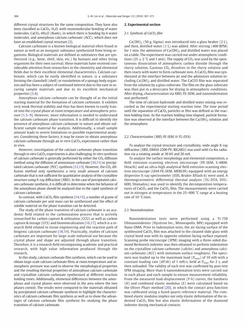
Calcium carbonate synthesized over the reaction holding time usingcalcium carbonate film synthesis was analyzed by XRD. As reactionholding time is increased, the component of the film is changed to cal-cite from ACC. Interestingly, characteristics of amorphous calcium car-bonate (dotted line in Fig. 1a) and CaCO3·6H2O (I in Fig. 1a and b)were observed during the initial reaction holding time (5 min) of thecalcium carbonate film synthesis. However, after 30 min of reactionholding time, therewas a rapid decrease in the intensity of CaCO3·6H2Ocharacteristics and a rapid appearance of the (104) crystal face, which isthe cleavage fracture crystal face of calcite, and this gives rise to themain characteristic calcite peak. Additionally,when the reaction holdingtime reached 95 min, the characteristic peak of CaCO3·6H2O could nolonger be observed, and it was difficult to clearly observe the character-istic peak of amorphous calcium carbonate due to the development ofcalcite (Fig. 1c). In contrast, the characteristic peak of the (104) crystalface of calcite became well developed with increased reaction holdingtime. Although a difference was observed near 2θ = 20–40° at 95 minof reaction holding time, this resulted from the influence of coexistingphases of amorphous calciumcarbonate and crystalline calcium carbon-ate (Fig. 1d). For greater reaction holding times, the identification ofamorphous calcium carbonate by wide angle XRD analysis waschallenging.
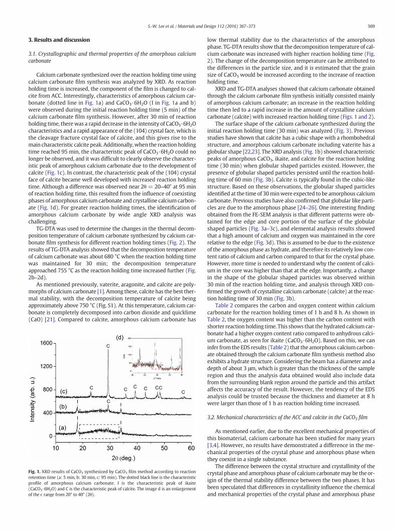
TG-DTA was used to determine the changes in the thermal decom-position temperature of calcium carbonate synthesized by calcium car-bonate film synthesis for different reaction holding times (Fig. 2). Theresults of TG-DTA analysis showed that the decomposition temperatureof calcium carbonate was about 680 °C when the reaction holding timewas maintained for 30 min; the decomposition temperatureapproached 755 °C as the reaction holding time increased further (Fig.2b–2d).
As mentioned previously, vaterite, aragonite, and calcite are poly-morphs of calcium carbonate [1]. Among these, calcite has the best ther-mal stability, with the decomposition temperature of calcite beingapproximately above 750 °C (Fig. S1). At this temperature, calcium car-bonate is completely decomposed into carbon dioxide and quicklime(CaO) [21]. Compared to calcite, amorphous calcium carbonate has
Fig. 1. XRD results of CaCO3 synthesized by CaCO3 film method according to reactionretention time (a: 5 min, b: 30 min, c: 95 min). The dotted black line is the characteristicprofile of amorphous calcium carbonate. I is the characteristic peak of ikaite(CaCO3·6H2O) and C is the characteristic peak of calcite. The image d is an enlargementof the c range from 20° to 40° (2θ).
low thermal stability due to the characteristics of the amorphousphase. TG-DTA results show that the decomposition temperature of cal-cium carbonate was increased with higher reaction holding time (Fig.2). The change of the decomposition temperature can be attributed tothe differences in the particle size, and it is estimated that the grainsize of CaCO3 would be increased according to the increase of reactionholding time.
XRD and TG-DTA analyses showed that calcium carbonate obtainedthrough the calcium carbonate film synthesis initially consisted mainlyof amorphous calcium carbonate; an increase in the reaction holdingtime then led to a rapid increase in the amount of crystalline calciumcarbonate (calcite) with increased reaction holding time (Figs. 1 and 2).
The surface shape of the calcium carbonate synthesized during theinitial reaction holding time (30 min) was analyzed (Fig. 3). Previousstudies have shown that calcite has a cubic shape with a rhombohedralstructure, and amorphous calcium carbonate including vaterite has aglobular shape [22,23]. The XRD analysis (Fig. 1b) showed characteristicpeaks of amorphous CaCO3, ikaite, and calcite for the reaction holdingtime (30 min) when globular shaped particles existed. However, thepresence of globular shaped particles persisted until the reaction hold-ing time of 60 min (Fig. 3b). Calcite is typically found in the cubic-likestructure. Based on these observations, the globular shaped particlesidentified at the time of 30minwere expected to be amorphous calciumcarbonate. Previous studies have also confirmed that globular like parti-cles are due to the amorphous phase [24–26]. One interesting findingobtained from the FE-SEM analysis is that different patterns were ob-tained for the edge and core portion of the surface of the globularshaped particles (Fig. 3a–3c), and elemental analysis results showedthat a high amount of calcium and oxygen was maintained in the corerelative to the edge (Fig. 3d). This is assumed to be due to the existenceof the amorphous phase as hydrate, and therefore its relatively low con-tent ratio of calcium and carbon compared to that for the crystal phase.However, more time is needed to understand why the content of calci-um in the core was higher than that at the edge. Importantly, a changein the shape of the globular shaped particles was observed within30 min of the reaction holding time, and analysis through XRD con-firmed the growth of crystalline calcium carbonate (calcite) at the reac-tion holding time of 30 min (Fig. 3b).
Table 2 compares the carbon and oxygen content within calciumcarbonate for the reaction holding times of 1 h and 8 h. As shown inTable 2, the oxygen content was higher than the carbon content withshorter reaction holding time. This shows that thehydrated calcium car-bonate had a higher oxygen content ratio compared to anhydrous calci-um carbonate, as seen for ikaite (CaCO3·6H2O). Based on this, we caninfer from the EDS results (Table 2) that the amorphous calciumcarbon-ate obtained through the calcium carbonate film synthesis method alsoexhibits a hydrate structure. Considering the beamhas a diameter and adepth of about 3 μm, which is greater than the thickness of the sampleregion and thus the analysis data obtained would also include datafrom the surrounding blank region around the particle and this artifactaffects the accuracy of the result. However, the tendency of the EDSanalysis could be trusted because the thickness and diameter at 8 hwere larger than those of 1 h as reaction holding time increased.
3.2. Mechanical characteristics of the ACC and calcite in the CaCO3 film
As mentioned earlier, due to the excellent mechanical properties ofthis biomaterial, calcium carbonate has been studied for many years[3,4]. However, no results have demonstrated a difference in the me-chanical properties of the crystal phase and amorphous phase whenthey coexist in a single substance.
The difference between the crystal structure and crystallinity of thecrystal phase and amorphous phase of calcium carbonatemay be the or-igin of the thermal stability difference between the two phases. It hasbeen speculated that differences in crystallinity influence the chemicaland mechanical properties of the crystal phase and amorphous phase
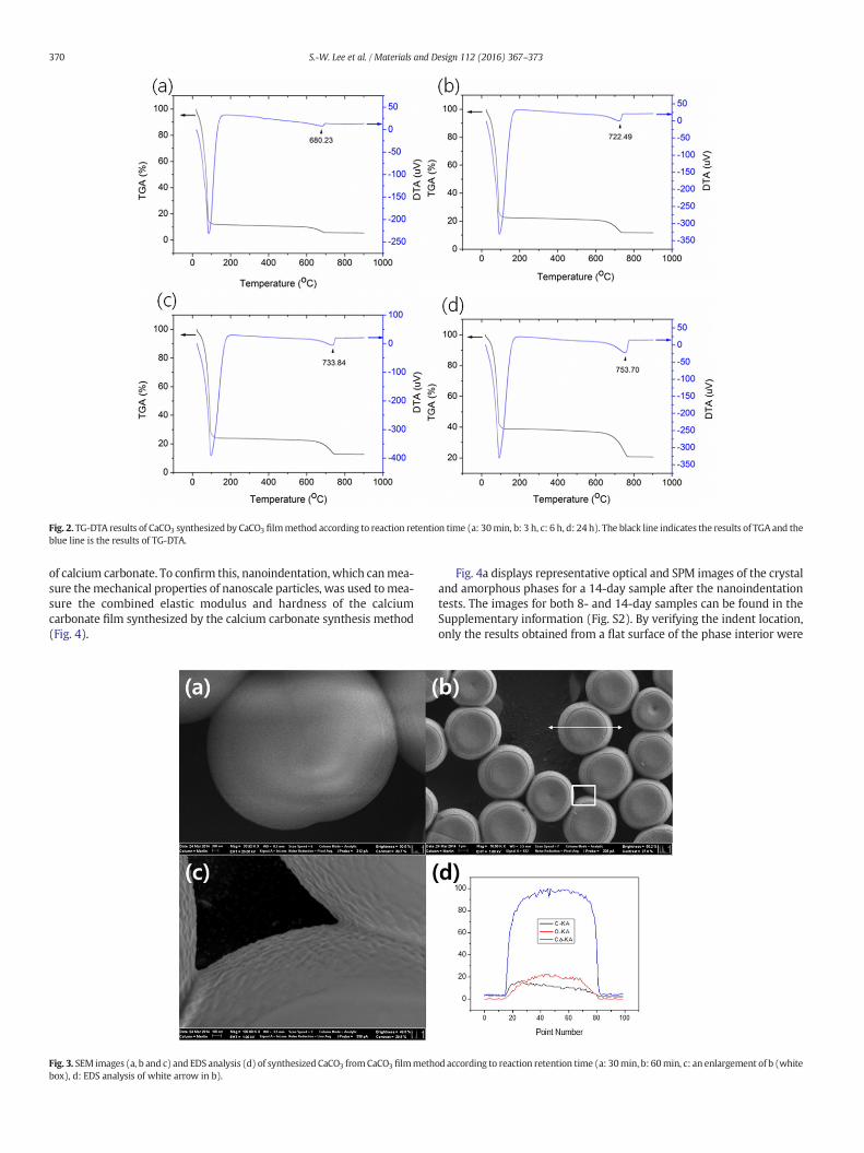
Fig. 2. TG-DTA results of CaCO3 synthesized by CaCO3 filmmethod according to reaction retention time (a: 30min, b: 3 h, c: 6 h, d: 24 h). The black line indicates the results of TGA and theblue line is the results of TG-DTA.
370 S.-W. Lee et al. / Materials and Design 112 (2016) 367–373
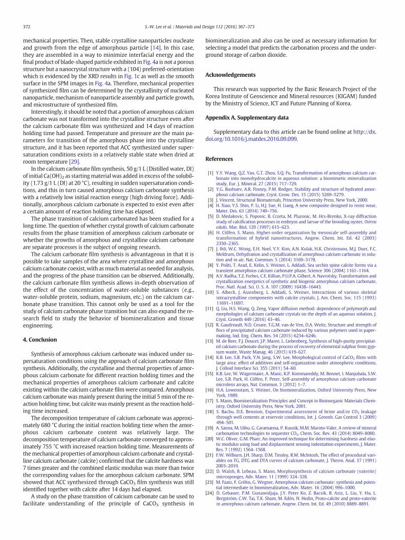
of calcium carbonate. To confirm this, nanoindentation, which canmea-sure themechanical properties of nanoscale particles, was used tomea-sure the combined elastic modulus and hardness of the calciumcarbonate film synthesized by the calcium carbonate synthesis method(Fig. 4).
Fig. 3. SEM images (a, b and c) and EDS analysis (d) of synthesized CaCO3 fromCaCO3 filmmethbox), d: EDS analysis of white arrow in b).
Fig. 4a displays representative optical and SPM images of the crystaland amorphous phases for a 14-day sample after the nanoindentationtests. The images for both 8- and 14-day samples can be found in theSupplementary information (Fig. S2). By verifying the indent location,only the results obtained from a flat surface of the phase interior were
od according to reaction retention time (a: 30min, b: 60min, c: an enlargement of b (white
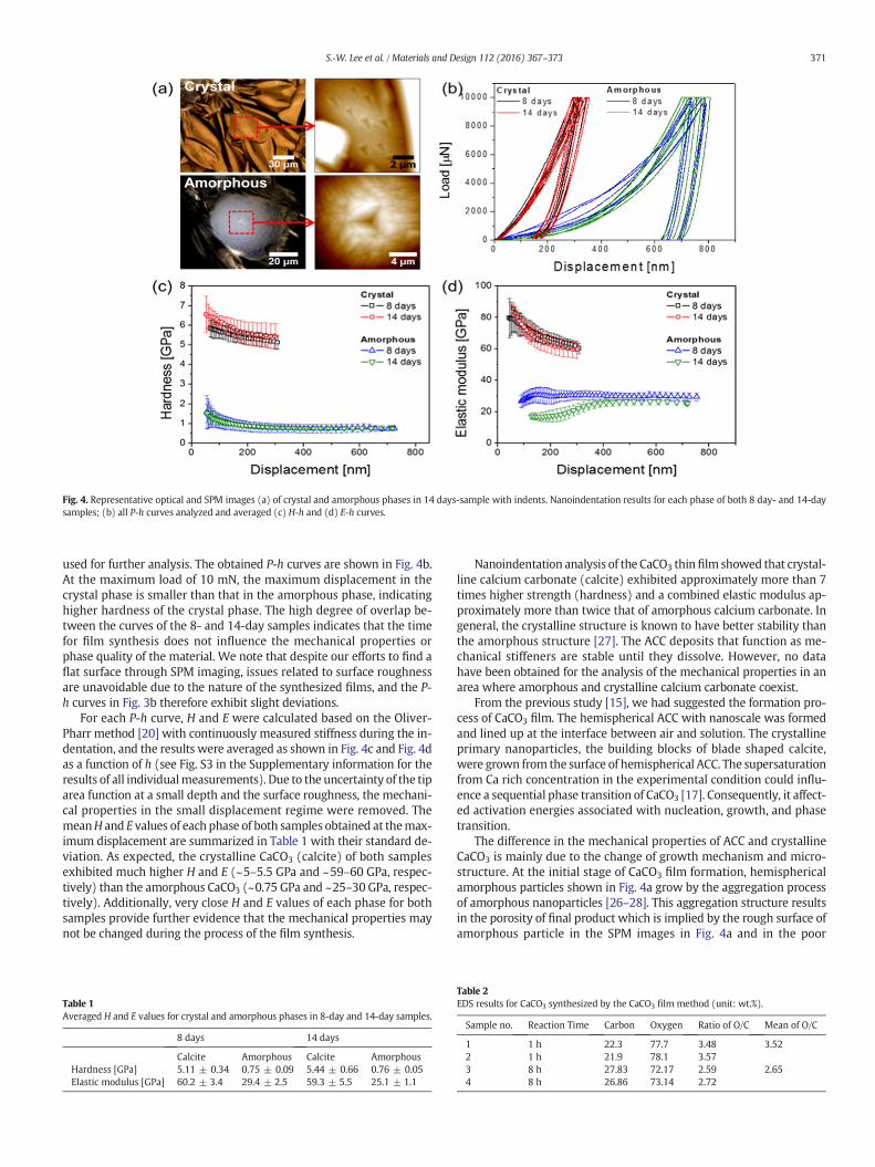
Fig. 4. Representative optical and SPM images (a) of crystal and amorphous phases in 14 days-sample with indents. Nanoindentation results for each phase of both 8 day- and 14-daysamples; (b) all P-h curves analyzed and averaged (c) H-h and (d) E-h curves.
371S.-W. Lee et al. / Materials and Design 112 (2016) 367–373
used for further analysis. The obtained P-h curves are shown in Fig. 4b.At the maximum load of 10 mN, the maximum displacement in thecrystal phase is smaller than that in the amorphous phase, indicatinghigher hardness of the crystal phase. The high degree of overlap be-tween the curves of the 8- and 14-day samples indicates that the timefor film synthesis does not influence the mechanical properties orphase quality of the material. We note that despite our efforts to find aflat surface through SPM imaging, issues related to surface roughnessare unavoidable due to the nature of the synthesized films, and the P-h curves in Fig. 3b therefore exhibit slight deviations.
For each P-h curve, H and E were calculated based on the Oliver-Pharr method [20] with continuously measured stiffness during the in-dentation, and the results were averaged as shown in Fig. 4c and Fig. 4das a function of h (see Fig. S3 in the Supplementary information for theresults of all individualmeasurements). Due to the uncertainty of the tiparea function at a small depth and the surface roughness, the mechani-cal properties in the small displacement regime were removed. ThemeanH and E values of each phase of both samples obtained at themax-imum displacement are summarized in Table 1 with their standard de-viation. As expected, the crystalline CaCO3 (calcite) of both samplesexhibited much higher H and E (~5–5.5 GPa and ~59–60 GPa, respec-tively) than the amorphous CaCO3 (~0.75 GPa and ~25–30 GPa, respec-tively). Additionally, very close H and E values of each phase for bothsamples provide further evidence that the mechanical properties maynot be changed during the process of the film synthesis.
Table 1Averaged H and E values for crystal and amorphous phases in 8-day and 14-day samples.
8 days 14 days
Calcite Amorphous Calcite AmorphousHardness [GPa] 5.11 ± 0.34 0.75 ± 0.09 5.44 ± 0.66 0.76 ± 0.05Elastic modulus [GPa] 60.2 ± 3.4 29.4 ± 2.5 59.3 ± 5.5 25.1 ± 1.1
Nanoindentation analysis of the CaCO3 thinfilm showed that crystal-line calcium carbonate (calcite) exhibited approximately more than 7times higher strength (hardness) and a combined elastic modulus ap-proximately more than twice that of amorphous calcium carbonate. Ingeneral, the crystalline structure is known to have better stability thanthe amorphous structure [27]. The ACC deposits that function as me-chanical stiffeners are stable until they dissolve. However, no datahave been obtained for the analysis of the mechanical properties in anarea where amorphous and crystalline calcium carbonate coexist.
From the previous study [15], we had suggested the formation pro-cess of CaCO3 film. The hemispherical ACC with nanoscale was formedand lined up at the interface between air and solution. The crystallineprimary nanoparticles, the building blocks of blade shaped calcite,were grown from the surface of hemispherical ACC. The supersaturationfrom Ca rich concentration in the experimental condition could influ-ence a sequential phase transition of CaCO3 [17]. Consequently, it affect-ed activation energies associated with nucleation, growth, and phasetransition.
The difference in the mechanical properties of ACC and crystallineCaCO3 is mainly due to the change of growth mechanism and micro-structure. At the initial stage of CaCO3 film formation, hemisphericalamorphous particles shown in Fig. 4a grow by the aggregation processof amorphous nanoparticles [26–28]. This aggregation structure resultsin the porosity of final product which is implied by the rough surface ofamorphous particle in the SPM images in Fig. 4a and in the poor
Table 2EDS results for CaCO3 synthesized by the CaCO3 film method (unit: wt.%).
Sample no. Reaction Time Carbon Oxygen Ratio of O/C Mean of O/C
1 1 h 22.3 77.7 3.48 3.522 1 h 21.9 78.1 3.573 8 h 27.83 72.17 2.59 2.654 8 h 26.86 73.14 2.72
372 S.-W. Lee et al. / Materials and Design 112 (2016) 367–373
mechanical properties. Then, stable crystalline nanoparticles nucleateand growth from the edge of amorphous particle [14]. In this case,they are assembled in a way to minimize interfacial energy and thefinal product of blade-shaped particle exhibited in Fig. 4a is not a porousstructure but a nanocrystal structure with a (104) preferred-orientationwhich is evidenced by the XRD results in Fig. 1c as well as the smoothsurface in the SPM images in Fig. 4a. Therefore, mechanical propertiesof synthesized film can be determined by the crystallinity of nucleatednanoparticle, mechanism of nanoparticle assembly and particle growth,and microstructure of synthesized film.
Interestingly, it should be noted that a portion of amorphous calciumcarbonate was not transformed into the crystalline structure even afterthe calcium carbonate film was synthesized and 14 days of reactionholding time had passed. Temperature and pressure are the main pa-rameters for transition of the amorphous phase into the crystallinestructure, and it has been reported that ACC synthesized under super-saturation conditions exists in a relatively stable state when dried atroom temperature [29].
In the calcium carbonate film synthesis, 50 g/1 L (Distilled water, DI)of initial Ca(OH)2 as startingmaterialwas added in excess of the solubil-ity (1.73 g/1 L (DI) at 20 °C), resulting in sudden supersaturation condi-tions, and this in turn caused amorphous calcium carbonate synthesiswith a relatively low initial reaction energy (high driving force). Addi-tionally, amorphous calcium carbonate is expected to exist even aftera certain amount of reaction holding time has elapsed.
The phase transition of calcium carbonated has been studied for along time. The question of whether crystal growth of calcium carbonateresults from the phase transition of amorphous calcium carbonate orwhether the growths of amorphous and crystalline calcium carbonateare separate processes is the subject of ongoing research.
The calcium carbonate film synthesis is advantageous in that it ispossible to take samples of the area where crystalline and amorphouscalcium carbonate coexist, with asmuchmaterial as needed for analysis,and the progress of the phase transition can be observed. Additionally,the calcium carbonate film synthesis allows in-depth observation ofthe effect of the concentration of water-soluble substances (e.g.,water-soluble protein, sodium, magnesium, etc.) on the calcium car-bonate phase transition. This cannot only be used as a tool for thestudy of calcium carbonate phase transition but can also expand the re-search field to study the behavior of biomineralization and tissueengineering.
4. Conclusion
Synthesis of amorphous calcium carbonate was induced under su-persaturation conditions using the approach of calcium carbonate filmsynthesis. Additionally, the crystalline and thermal properties of amor-phous calcium carbonate for different reaction holding times and themechanical properties of amorphous calcium carbonate and calciteexisting within the calcium carbonate film were compared. Amorphouscalcium carbonate wasmainly present during the initial 5min of the re-action holding time, but calcitewasmainly present as the reaction hold-ing time increased.
The decomposition temperature of calcium carbonate was approxi-mately 680 °C during the initial reaction holding time when the amor-phous calcium carbonate content was relatively large. Thedecomposition temperature of calcium carbonate converged to approx-imately 755 °C with increased reaction holding time. Measurements ofthemechanical properties of amorphous calcium carbonate and crystal-line calcium carbonate (calcite) confirmed that the calcite hardness was7 times greater and the combined elastic modulus wasmore than twicethe corresponding values for the amorphous calcium carbonate. SPMshowed that ACC synthesized through CaCO3 film synthesis was stillidentified together with calcite after 14 days had elapsed.
A study on the phase transition of calcium carbonate can be used tofacilitate understanding of the principle of CaCO3 synthesis in
biomineralization and also can be used as necessary information forselecting a model that predicts the carbonation process and the under-ground storage of carbon dioxide.
Acknowledgements
This research was supported by the Basic Research Project of theKorea Institute of Geoscience and Mineral resources (KIGAM) fundedby the Ministry of Science, ICT and Future Planning of Korea.
Appendix A. Supplementary data
Supplementary data to this article can be found online at http://dx.doi.org/10.1016/j.matdes.2016.09.099.
References
[1] Y.Y. Wang, Q.Z. Yao, G.T. Zhou, S.Q. Fu, Transformation of amorphous calcium car-bonate into monohydrocalcite in aqueous solution: a biomimetic mineralizationstudy, Eur. J. Mineral. 27 (2015) 717–729.
[2] Y.G. Bushuev, A.R. Finney, P.M. Rodger, Stability and structure of hydrated amor-phous calcium carbonate, Cryst. Grow. Des. 15 (2015) 5269–5279.
[3] J. Vincent, Structural Biomaterials, Princeton University Press, New York, 2000.[4] H. Xiao, Y.S. Shin, P. Li, H.J. Sue, H. Liang, A new composite designed to resist wear,
Mater. Des. 63 (2014) 749–756.[5] D. Medakovic, S. Popovic, B. Crzeta, M. Plazonic, M. Hrs-Brenko, X-ray diffraction
study of calcification processes in embryos and larvae of the brooding oyster, Ostreaedulis. Mar. Biol. 129 (1997) 615–623.
[6] H. Cölfen, S. Mann, Higher-order organization by mesoscale self-assembly andtransformation of hybrid nanostructures, Angew. Chem. Int. Ed. 42 (2003)2350–2365.
[7] J. Ihli, W.C. Wong, E.H. Noel, Y.Y. Kim, A.N. Kulak, H.K. Christenson, M.J. Duer, F.C.Meldrum, Dehydration and crystallization of amorphous calcium carbonate in solu-tion and in air, Nat. Commun. 5 (2014) 3169–3178.
[8] Y. Politi, T. Arad, E. Kelin, S. Weiner, L. Addadi, Sea urchin spine calcite forms via atransient amorphous calcium carbonate phase, Science 306 (2004) 1161–1164.
[9] A.V. Radha, T.Z. Forbes, C.E. Killian, P.U.P.A. Gilbert, A. Navrotsky, Transformation andcrystallization energetics of synthetic and biogenic amorphous calcium carbonate,Proc. Natl. Acad. Sci. U. S. A. 107 (2009) 16438–16443.
[10] S. Albeck, J. Aizenberg, L. Addadi, S. Weiner, Interactions of various skeletalintracrystalline components with calcite crystals, J. Am. Chem. Soc. 115 (1993)11691–11697.
[11] Q. Liu, H.S. Wang, Q. Zeng, Vapor diffusion method: dependence of polymorph andmorphologies of calcium carbonate crystals on the depth of an aqueous solution, J.Cryst. Growth 449 (2016) 43–46.
[12] R. Gaudreault, N.D. Cesare, T.G.M. van de Ven, D.A. Weitz, Structure and strength offlocs of precipitated calcium carbonate induced by various polymers used in paper-making, Ind. Eng. Chem. Res. 54 (2015) 6234–6246.
[13] M. de Beer, F.J. Doucet, J.P. Maree, L. Liebenberg, Synthesis of high-purity precipitat-ed calcium carbonate during the process of recovery of elemental sulphur from gyp-sum waste, Waste Manag. 46 (2015) 619–627.
[14] K.B. Lee, S.B. Park, Y.N. Jang, S.W. Lee, Morphological control of CaCO3 films withlarge area: effect of additives and self-organization under atmospheric conditions,J. Colloid Interface Sci. 355 (2011) 54–60.
[15] K.B. Lee, W. Wagermaier, A. Masic, K.P. Kommareddy, M. Bennet, I. Manjubala, S.W.Lee, S.B. Park, H. Cölfen, F. Peter, Self-assembly of amorphous calcium carbonatemicrolens arrays, Nat. Commun. 3 (2012) 1–7.
[16] H.A. Lowenstam, S. Weiner, On biomineralization, Oxford University Press, NewYork, 1989.
[17] S. Mann, Biomineralization Principles and Concept in Bioinorganic Materials Chem-istry, Oxford University Press, New York, 2001.
[18] S. Bachu, D.E. Bennion, Experimental assessment of brine and/or CO2 leakagethrough well cements at reservoir conditions, Int. J. Greenh. Gas Control 3 (2009)494–501.
[19] A. Sanna, M. Uibu, G. Caramanna, P. Kuusik, M.M. Maroto-Valer, A review of mineralcarbonation technologies to sequester CO2, Chem. Soc. Rev. 43 (2014) 8049–8080.
[20] W.C. Oliver, G.M. Pharr, An improved technique for determining hardness and elas-tic modulus using load and displacement sensing indentation experiments, J. Mater.Res. 7 (1992) 1564–1568.
[21] F.W. Wilburn, J.H. Sharp, D.M. Tinsley, R.M. McIntosh, The effect of procedural vari-ables on TG, DTG and DTA curves of calcium carbonate, J. Therm. Anal. 37 (1991)2003–2019.
[22] D. Walsh, B. Lebeau, S. Mann, Morphosynthesis of calcium carbonate (vaterite)microsponges, Adv. Mater. 11 (1999) 324–328.
[23] M. Faatz, F. Gröhn, G. Wegner, Amorphous calcium carbonate: synthesis and poten-tial intermediate in biomineralization, Adv. Mater. 16 (2004) 996–1000.
[24] D. Gebauer, P.M. Gunawidjaja, J.Y. Peter Ko, Z. Bacsik, B. Aziz, L. Liu, Y. Hu, L.Bergström, C.W. Tai, T.K. Sham, M. Edén, N. Hedin, Proto-calcite and proto-vateritein amorphous calcium carbonate, Angew. Chem. Int. Ed. 49 (2010) 8889–8891.

373S.-W. Lee et al. / Materials and Design 112 (2016) 367–373
[25] C. Zhong, C.C. Chu, On the origin of amorphous cores in biomimetic CaCO3 spheru-lites: new insights into spherulitic crystallization, Cryst. Grow. Des. 10 (2010)5043–5049.
[26] S.W. Lee, Y.I. Kim, K.B. Lee, J.H. Bang, C.W. Jun, Y.N. Jang, Effect of serine and arginineon the phase transition from amorphous CaCO3 and CaCO3·6H2O to calcite film,Mater. Trans. 53 (2012) 1732–1738.
[27] L. Addadi, S. Raz, S. Weiner, Taking advantage of disorder: amorphous calcium car-bonate and its roles in biomineralization, Adv. Mater. 15 (2003) 959–970.
[28] D. Gebauer, A. Vӧlkel, H. Cӧlfen, Stable prenucleation calcium carbonate clusters, Sci-ence 322 (2008) 1819–1822.
[29] K. Gorna, M. Hund, M. Vucak, F. Gröhn, G.Wegner, Amorphous calcium carbonate inform of spherical nanosized particles and its application as fillers for polymers,Mater. Sci. Eng. A 477 (2008) 217–225.


![Product Information Compression die for testing flexible foams › - › media › files › share... · Item No. Standard Type Indenter geometry Indenter shape Test load Fmax [kN]](https://img.dokumen.tips/doc/110x75/5f29564adbcad74f962732d8/product-information-compression-die-for-testing-flexible-foams-a-a-media-a.jpg)