Embed Size (px)
Citation preview
Management of
Diabetic Ketoacidosis
and Hyperosmolar
Hyperglycemic State
in Adults
}{This continuing education activity can also be completed online at www.MedEdToday.com.
Release date: November 9, 2005 Expiration date: November 30, 2007
Estimated time to complete activity: 1.6–2.0 hours
Sponsored by Postgraduate Institute for Medicine
This activity is supported by an educational grantfrom Novo Nordisk Inc.
A Continuing Education Monograph for Pharmacists and Nurses
Program GoalThis monograph is a review of the pathophysiology, diagnosis, treatment, and complications of diabeticketoacidosis (DKA) and hyperosmolar hyperglycemic state (HHS) in adults.
Target AudienceThis educational activity has been designed to meet theeducational needs of pharmacists and registered nursesinvolved in the management of patients with diabetes.
PurposeProvide clinicians treating patients with diabetes the latest information about the pathophysiology, diagnosis,and management of DKA and HHS to help reduce theincidence, morbidity, and mortality of the disorder.
Statement of Need/Program OverviewDKA and HHS are life-threatening emergencies and arethe most serious complications of diabetes. DKA is causedby relative or absolute insulin deficiency, which results inhyperglycemia, ketonemia, dehydration, electrolyte imbalances, and acidosis. Treatment protocols for adultsgenerally advocate a more rapid and aggressive reversalof DKA than is advised for children. DKA consumes a significant proportion of all direct medical costs for adultsand children with type 1 diabetes. The management ofDKA requires a full complement of hospital, emergency,and intensive care services. In the United States, morethan 100,000 individuals are hospitalized each year forDKA and the mortality rate is 2% to 5%.
Most cases of DKA can be prevented by using an effectivediabetes management plan that includes patient self-care.Early identification of precipitating signs and symptomsand prompt, appropriate intervention can reduce the frequency of DKA episodes that result in a medical emergency.
The healthcare team must stay informed about the recentdevelopments in diabetes care and have the necessaryclinical skills to prevent and manage DKA episodes.Through good self-management and intensive glycemiccontrol, the incidence, morbidity, and mortality of DKAcan be reduced.
Educational Objectives After completing this educational activity, participantsshould be able to:
• Differentiate between DKA and HHS • Describe the epidemiology and pathophysiology of DKA• Identify the signs, symptoms, and complications of DKA• Discuss methods of treatment and recovery care for DKA• Describe potential complications during treatment of
DKA and special considerations for pregnant and elderly individuals
• Provide strategies for the prevention of DKA
Editorial Review Board John B. Buse, MD, PhD, FACEChief, Division of General Internal MedicineDirector, Diabetes Care CenterUniversity of North Carolina School of MedicineChapel Hill, North Carolina
Marsha Menke, RN, BSN, MS, CDE, CPTDirector, Diabetes Care CenterGenesis Medical CenterDavenport, Iowa
Thomas S. Sisca, PharmD, FCCP, BCPSClinical Pharmacist, Antithrombosis SpecialistAntithrombosis Services/Anticoagulation ClinicShore Health SystemMemorial HospitalEaston, Maryland
Pharmacist Continuing EducationAccreditation StatementPostgraduate Institute for Medicine is accredited by the Accreditation Council for Pharmacy Education as a provider of continuing pharmacy education.
Credit DesignationPostgraduate Institute for Medicine designates this continuingeducation activity for 1.6 contact hours (0.16 CEU) of theAccreditation Council for Pharmacy Education.
Universal Program Number – 809-999-05-064-H01.
{ }i
Nursing Continuing EducationCNA/ANCCThis educational activity for 2.0 contact hours is providedby the Postgraduate Institute for Medicine (PIM). ThePostgraduate Institute for Medicine is approved as aprovider of continuing education in nursing by theColorado Nurses Association, which is accredited as an approver of continuing education in nursing by theAmerican Nurses Credentialing Center’s Commission on Accreditation.
California Board of Registered NursingPostgraduate Institute for Medicine is approved by theCalifornia Board of Registered Nursing, Provider Number13485 for 2.0 contact hours.
Disclosure of Conflicts of InterestPostgraduate Institute for Medicine (PIM) assesses conflictof interest with its instructors, planners, managers, andother individuals who are in a position to control the content of CME activities. All relevant conflicts of interestthat are identified are thoroughly vetted by PIM for fairbalance, scientific objectivity of studies utilized in thisactivity, and patient care recommendations. PIM is committed to providing its learners with high-quality CME activities and related materials that promoteimprovements or quality in healthcare and not a specificproprietary business interest of a commercial interest.
Method of ParticipationThere are no fees for participating and receiving CE creditfor this activity. During the period November 9, 2005through November 30, 2007, participants must: 1) read thelearning objectives and faculty disclosures; 2) study theeducational activity; 3) complete the post-test by recordingthe best answer to each question in the answer key on the evaluation form; 4) complete the evaluation form; and5) mail or fax the evaluation form with answer key to thePostgraduate Institute for Medicine.
A statement of credit will be issued only upon receipt of a completed activity evaluation form and a completedpost-test with a score of 70% or better. Your statement ofcredit will be mailed to you within 3 weeks.
MediaMonograph
Disclosure of Unlabeled UseThis educational activity may contain discussion of published and/or investigational uses of agents that arenot indicated by the US Food and Drug Administration.Postgraduate Institute for Medicine (PIM), Scherer ClinicalCommunications, and Novo Nordisk Inc. do not recommendthe use of any agent outside of the labeled indications.
The opinions expressed in the educational activity arethose of the faculty and do not necessarily represent theviews of PIM, Scherer Clinical Communications, and Novo Nordisk Inc. Please refer to the official prescribing
{ }ii
The following faculty reported a real or apparent conflict of interest:
NAME OF FACULTY OR PRESENTER REPORTED AREAS OF CONFLICT
John B. Buse, MD, PhD, FACE Consulting Fees: Amylin, BD, Eli Lilly and Company, Insulet
Research: Amylin, BD, Dexcom, Eli Lilly and Company, Sanofi-Aventis
Ownership/Stocks: Insulet
Marsha Menke, RN, BSN, MS, CDE, CPT No significant financial relationships related to this activity
Thomas S. Sisca, PharmD, FCCP, BCPS Consulting Fees: Sanofi-Aventis, Bristol-Myers Squibb
The following planners and managers reported a real or apparent conflict of interest:
NAME OF PLANNER OR MANAGER REPORTED AREAS OF CONFLICT
Peter Macholdt No financial relationships related to this activity
Jan Hixon, RN, MSN No financial relationships related to this activity
information for each product for discussion of approvedindications, contraindications, and warnings.
DisclaimerParticipants have an implied responsibility to use thenewly acquired information to enhance patient outcomesand their own professional development. The informationpresented in this activity is not meant to serve as a guideline for patient management. Any procedures, medications, or other courses of diagnosis or treatmentdiscussed or suggested in this activity should not be used by clinicians without evaluation of their patient’s conditions and possible contraindications on dangers inuse, review of any applicable manufacturer’s product information, and comparison with recommendations ofother authorities.
Copyright © 2005, Scherer Clinical Communications. All rights reserved.
Published by Scherer Clinical Communications117 West Prospect Street, Hopewell, NJ 08525
Printed in USA.
{ }iii

{ }iv
Table of ContentsINTRODUCTION 1
DEFINITION 1–2
EPIDEMIOLOGY 2
PATHOPHYSIOLOGY 2–3
Events 3
Hyperglycemia 3
Dehydration 3
Ketone production 3
Ionic changes 3–4
Compensatory Attempts 4
TRIGGERS OF DKA AND HHS 4–5
WARNING SIGNS AND SYMPTOMS 5–6
DIAGNOSIS 6
Anion Gap 6–7
TREATMENT GUIDELINES 7–10
RECOVERY CARE 10
ELECTROLYTE MANAGEMENT DURING RECOVERY 10
COMPLICATIONS 10–11
PREVENTION 11–12
Outpatient prevention measures 12
Preventing recurrent diabetic ketoacidosis 12
SPECIAL CIRCUMSTANCES 12
Pregnancy 12
Elderly 12
ECONOMIC ISSUES 13
SUMMARY 13
APPENDIX A 14
APPENDIX B 15
REFERENCES 16
POST-TEST 17–18
EVALUATION FORM 19–20
IntroductionEach year more than 100,000 people are hospitalized fordiabetic ketoacidosis (DKA) in the United States, resultingin cumulative annual hospital costs that may be more than $1 billion.1 DKA is a life-threatening illness, and the mortality rate for DKA, estimated between 2% to 5%, hasremained relatively unchanged since the 1970s.2,3 Theaverage length of hospital stay for patients diagnosedwith DKA, however, has declined from 7.9 days in 1980 to3.9 days in 2002.4 More than 25% of the costs for directmedical care for adults with type 1 diabetes covers DKA;this increases to 50% in those experiencing recurrentepisodes.5
Among the benefits of effective glycemic control isreduced incidence of DKA or hyperosmolar hyperglycemicstate (HHS) and associated morbidity and mortality. BothDKA and HHS are life-threatening emergencies.6,7 TheAmerican Diabetes Association (ADA) recommends thatadults with diabetes maintain preprandial plasma glucoselevels between 90 and 130 mg/dL, postprandial levels<180 mg/dL, and an A1C <7% while pointing out thatmore stringent glycemic goals (ie, a normal A1C <6%) may further reduce complications at the cost of increasedrisk of hypoglycemia.8 The American College ofEndocrinology has somewhat different targets, recom-mending preprandial plasma glucose levels ≤110 mg/dL,postprandial levels ≤140 mg/dL, and an A1C ≤6.5%.9
Comprehensive education in self-management is a criticalelement for achieving and maintaining optimal glycemiccontrol. The physician and patient must set realistic treatment goals with the diabetes management team andfamily members. An effective program requires ongoingsupport from the clinical care team.
Healthcare professionals who treat patients with diabetesmust know how to prevent and manage DKA. This mono-graph defines DKA and describes symptoms and warningsigns, diagnosis, current practices, complications, preventionmethods, special circumstances, and economic issues.
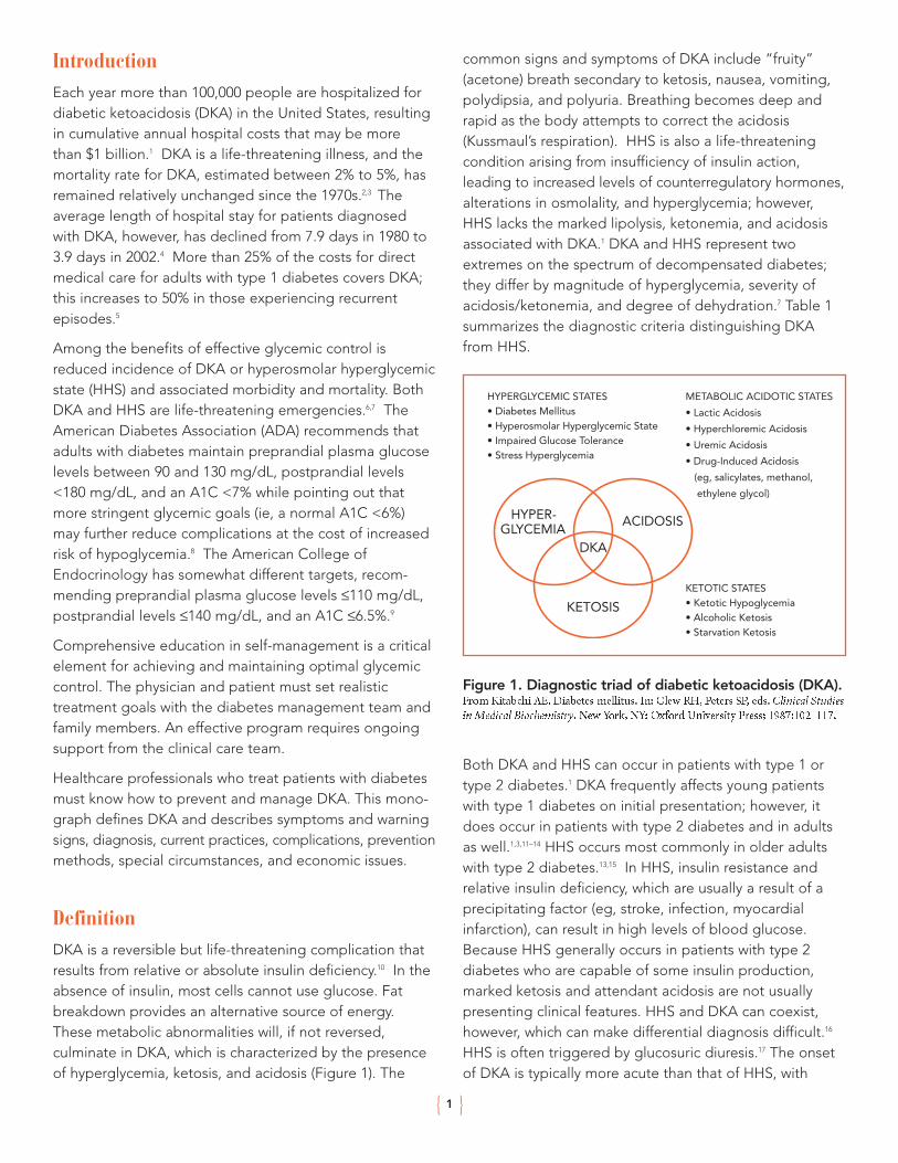
DefinitionDKA is a reversible but life-threatening complication thatresults from relative or absolute insulin deficiency.10 In theabsence of insulin, most cells cannot use glucose. Fatbreakdown provides an alternative source of energy.These metabolic abnormalities will, if not reversed, culminate in DKA, which is characterized by the presenceof hyperglycemia, ketosis, and acidosis (Figure 1). The
common signs and symptoms of DKA include “fruity” (acetone) breath secondary to ketosis, nausea, vomiting, polydipsia, and polyuria. Breathing becomes deep andrapid as the body attempts to correct the acidosis(Kussmaul’s respiration). HHS is also a life-threateningcondition arising from insufficiency of insulin action, leading to increased levels of counterregulatory hormones,alterations in osmolality, and hyperglycemia; however,HHS lacks the marked lipolysis, ketonemia, and acidosisassociated with DKA.1 DKA and HHS represent twoextremes on the spectrum of decompensated diabetes;they differ by magnitude of hyperglycemia, severity of acidosis/ketonemia, and degree of dehydration.7 Table 1summarizes the diagnostic criteria distinguishing DKAfrom HHS.
Figure 1. Diagnostic triad of diabetic ketoacidosis (DKA). From Kitabchi AE. Diabetes mellitus. In: Glew RH, Peters SP, eds. Clinical Studiesin Medical Biochemistry. New York, NY: Oxford University Press; 1987:102–117.
Both DKA and HHS can occur in patients with type 1 ortype 2 diabetes.1 DKA frequently affects young patientswith type 1 diabetes on initial presentation; however, itdoes occur in patients with type 2 diabetes and in adultsas well.1,3,11–14 HHS occurs most commonly in older adultswith type 2 diabetes.13,15 In HHS, insulin resistance and relative insulin deficiency, which are usually a result of aprecipitating factor (eg, stroke, infection, myocardialinfarction), can result in high levels of blood glucose.Because HHS generally occurs in patients with type 2 diabetes who are capable of some insulin production,marked ketosis and attendant acidosis are not usually presenting clinical features. HHS and DKA can coexist,however, which can make differential diagnosis difficult.16
HHS is often triggered by glucosuric diuresis.17 The onsetof DKA is typically more acute than that of HHS, with
{ }1
HYPER-GLYCEMIA
ACIDOSIS
KETOSIS
DKA
HYPERGLYCEMIC STATES• Diabetes Mellitus• Hyperosmolar Hyperglycemic State• Impaired Glucose Tolerance• Stress Hyperglycemia
METABOLIC ACIDOTIC STATES
• Lactic Acidosis
• Hyperchloremic Acidosis
• Uremic Acidosis
• Drug-Induced Acidosis
(eg, salicylates, methanol,
ethylene glycol)
KETOTIC STATES• Ketotic Hypoglycemia• Alcoholic Ketosis• Starvation Ketosis
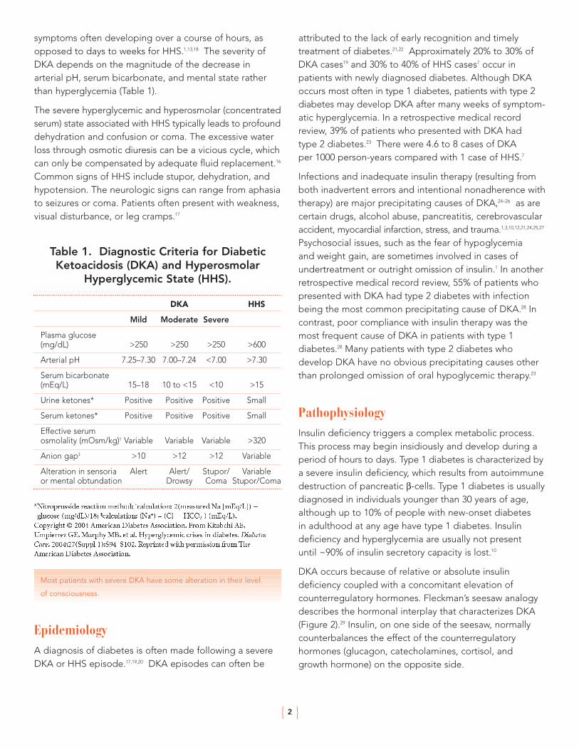
symptoms often developing over a course of hours, asopposed to days to weeks for HHS.1,13,18 The severity ofDKA depends on the magnitude of the decrease in arterial pH, serum bicarbonate, and mental state ratherthan hyperglycemia (Table 1).
The severe hyperglycemic and hyperosmolar (concentratedserum) state associated with HHS typically leads to profounddehydration and confusion or coma. The excessive waterloss through osmotic diuresis can be a vicious cycle, whichcan only be compensated by adequate fluid replacement.16
Common signs of HHS include stupor, dehydration, andhypotension. The neurologic signs can range from aphasiato seizures or coma. Patients often present with weakness,visual disturbance, or leg cramps.17
Table 1. Diagnostic Criteria for DiabeticKetoacidosis (DKA) and Hyperosmolar
Hyperglycemic State (HHS).
*Nitroprusside reaction method; †calculation: 2(measured Na [mEq/L]) + glucose (mg/dL)/18; ‡calculation: (Na+) – (Cl– + HCO3–) (mEq/L).
Copyright © 2004 American Diabetes Association. From Kitabchi AE,Umpierrez GE, Murphy MB, et al. Hyperglycemic crises in diabetes. DiabetesCare. 2004;27(Suppl 1):S94–S102. Reprinted with permission from TheAmerican Diabetes Association.
Most patients with severe DKA have some alteration in their level
of consciousness.
EpidemiologyA diagnosis of diabetes is often made following a severeDKA or HHS episode.17,19,20 DKA episodes can often be
attributed to the lack of early recognition and timely treatment of diabetes.21,22 Approximately 20% to 30% ofDKA cases19 and 30% to 40% of HHS cases7 occur inpatients with newly diagnosed diabetes. Although DKAoccurs most often in type 1 diabetes, patients with type 2diabetes may develop DKA after many weeks of symptom-atic hyperglycemia. In a retrospective medical recordreview, 39% of patients who presented with DKA had type 2 diabetes.23 There were 4.6 to 8 cases of DKA per 1000 person-years compared with 1 case of HHS.7
Infections and inadequate insulin therapy (resulting fromboth inadvertent errors and intentional nonadherence withtherapy) are major precipitating causes of DKA,24–26 as arecertain drugs, alcohol abuse, pancreatitis, cerebrovascularaccident, myocardial infarction, stress, and trauma.1,3,10,12,21,24,25,27
Psychosocial issues, such as the fear of hypoglycemia and weight gain, are sometimes involved in cases ofundertreatment or outright omission of insulin.1 In anotherretrospective medical record review, 55% of patients whopresented with DKA had type 2 diabetes with infectionbeing the most common precipitating cause of DKA.28 Incontrast, poor compliance with insulin therapy was themost frequent cause of DKA in patients with type 1 diabetes.28 Many patients with type 2 diabetes who develop DKA have no obvious precipitating causes otherthan prolonged omission of oral hypoglycemic therapy.23
PathophysiologyInsulin deficiency triggers a complex metabolic process.This process may begin insidiously and develop during aperiod of hours to days. Type 1 diabetes is characterized bya severe insulin deficiency, which results from autoimmunedestruction of pancreatic β-cells. Type 1 diabetes is usuallydiagnosed in individuals younger than 30 years of age,although up to 10% of people with new-onset diabetes in adulthood at any age have type 1 diabetes. Insulin deficiency and hyperglycemia are usually not present until ~90% of insulin secretory capacity is lost.10
DKA occurs because of relative or absolute insulin deficiency coupled with a concomitant elevation of counterregulatory hormones. Fleckman’s seesaw analogydescribes the hormonal interplay that characterizes DKA(Figure 2).29 Insulin, on one side of the seesaw, normallycounterbalances the effect of the counterregulatory hormones (glucagon, catecholamines, cortisol, and growth hormone) on the opposite side.
{ }2
DKA HHS
Mild Moderate Severe
Plasma glucose (mg/dL) >250 >250 >250 >600
Arterial pH 7.25–7.30 7.00–7.24 <7.00 >7.30
Serum bicarbonate (mEq/L) 15–18 10 to <15 <10 >15
Urine ketones* Positive Positive Positive Small
Serum ketones* Positive Positive Positive Small
Effective serum osmolality (mOsm/kg)† Variable Variable Variable >320
Anion gap‡ >10 >12 >12 Variable
Alteration in sensoria Alert Alert/ Stupor/ Variableor mental obtundation Drowsy Coma Stupor/Coma
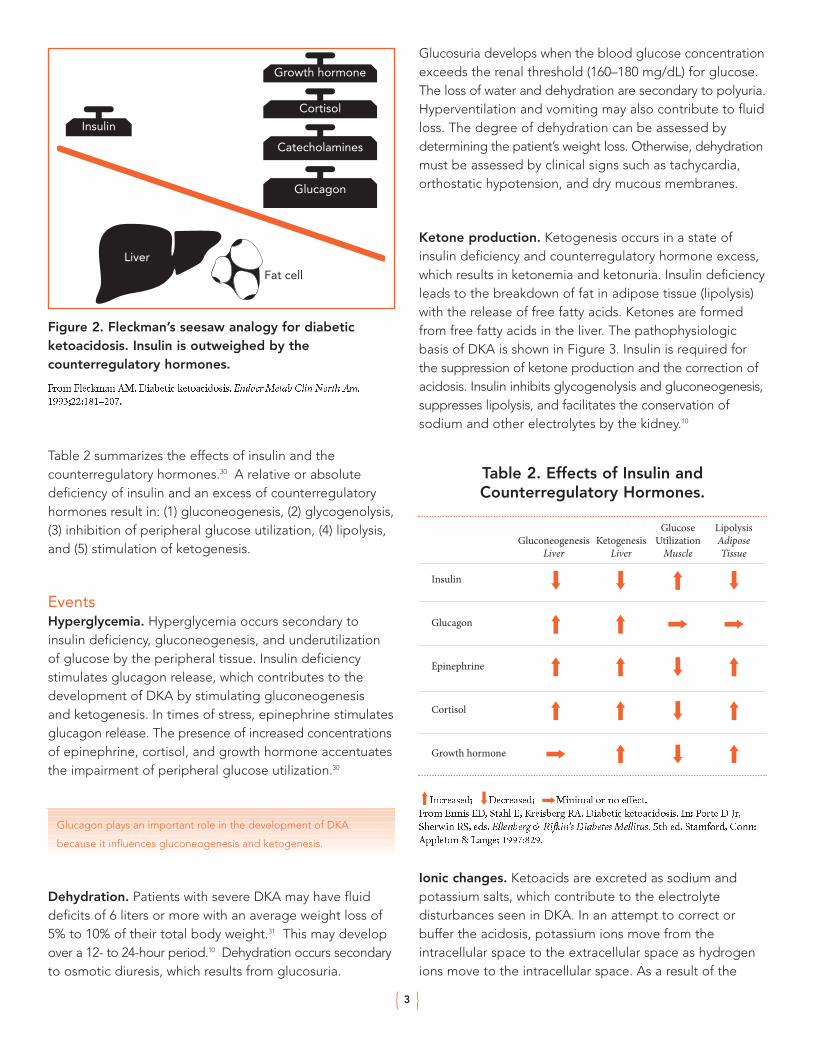
Figure 2. Fleckman’s seesaw analogy for diabeticketoacidosis. Insulin is outweighed by the counterregulatory hormones.
From Fleckman AM. Diabetic ketoacidosis. Endocr Metab Clin North Am.1993;22:181–207.
Table 2 summarizes the effects of insulin and the counterregulatory hormones.30 A relative or absolute deficiency of insulin and an excess of counterregulatoryhormones result in: (1) gluconeogenesis, (2) glycogenolysis,(3) inhibition of peripheral glucose utilization, (4) lipolysis,and (5) stimulation of ketogenesis.
EventsHyperglycemia. Hyperglycemia occurs secondary toinsulin deficiency, gluconeogenesis, and underutilizationof glucose by the peripheral tissue. Insulin deficiency stimulates glucagon release, which contributes to thedevelopment of DKA by stimulating gluconeogenesis and ketogenesis. In times of stress, epinephrine stimulatesglucagon release. The presence of increased concentrationsof epinephrine, cortisol, and growth hormone accentuatesthe impairment of peripheral glucose utilization.30
Glucagon plays an important role in the development of DKA
because it influences gluconeogenesis and ketogenesis.
Dehydration. Patients with severe DKA may have fluiddeficits of 6 liters or more with an average weight loss of5% to 10% of their total body weight.31 This may developover a 12- to 24-hour period.10 Dehydration occurs secondaryto osmotic diuresis, which results from glucosuria.
Glucosuria develops when the blood glucose concentrationexceeds the renal threshold (160–180 mg/dL) for glucose.The loss of water and dehydration are secondary to polyuria.Hyperventilation and vomiting may also contribute to fluidloss. The degree of dehydration can be assessed by determining the patient’s weight loss. Otherwise, dehydrationmust be assessed by clinical signs such as tachycardia,orthostatic hypotension, and dry mucous membranes.
Ketone production. Ketogenesis occurs in a state ofinsulin deficiency and counterregulatory hormone excess,which results in ketonemia and ketonuria. Insulin deficiencyleads to the breakdown of fat in adipose tissue (lipolysis)with the release of free fatty acids. Ketones are formedfrom free fatty acids in the liver. The pathophysiologicbasis of DKA is shown in Figure 3. Insulin is required forthe suppression of ketone production and the correction ofacidosis. Insulin inhibits glycogenolysis and gluconeogenesis,suppresses lipolysis, and facilitates the conservation of sodium and other electrolytes by the kidney.10
Table 2. Effects of Insulin andCounterregulatory Hormones.
Increased; Decreased; Minimal or no effect.From Ennis ED, Stahl E, Kreisberg RA. Diabetic ketoacidosis. In: Porte D Jr,Sherwin RS, eds. Ellenberg & Rifkin’s Diabetes Mellitus. 5th ed. Stamford, Conn:Appleton & Lange; 1997:829.
Ionic changes. Ketoacids are excreted as sodium andpotassium salts, which contribute to the electrolyte disturbances seen in DKA. In an attempt to correct orbuffer the acidosis, potassium ions move from the intracellular space to the extracellular space as hydrogenions move to the intracellular space. As a result of the
{ }3
Insulin
Liver
Growth hormone
Cortisol
Catecholamines
Glucagon
Fat cell
GluconeogenesisLiver
Insulin
Glucagon
Epinephrine
Cortisol
Growth hormone
KetogenesisLiver
GlucoseUtilizationMuscle
LipolysisAdiposeTissue
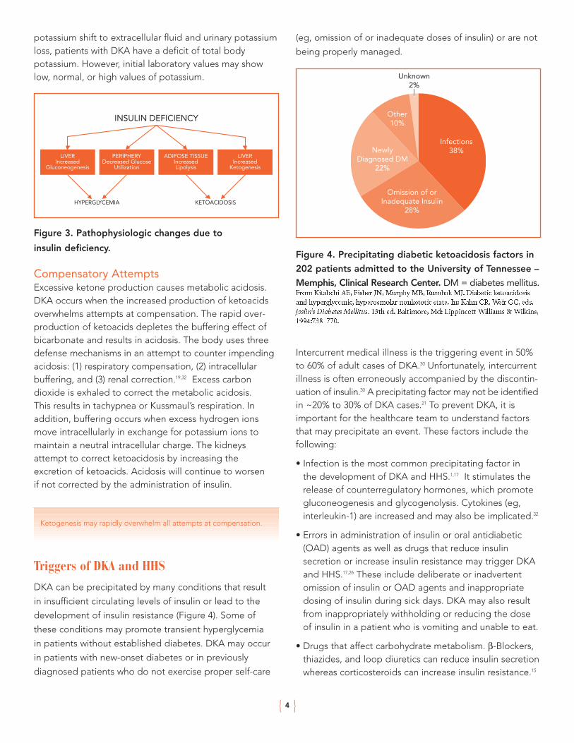
potassium shift to extracellular fluid and urinary potassiumloss, patients with DKA have a deficit of total body potassium. However, initial laboratory values may showlow, normal, or high values of potassium.
Figure 3. Pathophysiologic changes due to
insulin deficiency.
Compensatory AttemptsExcessive ketone production causes metabolic acidosis.DKA occurs when the increased production of ketoacidsoverwhelms attempts at compensation. The rapid over-production of ketoacids depletes the buffering effect ofbicarbonate and results in acidosis. The body uses threedefense mechanisms in an attempt to counter impendingacidosis: (1) respiratory compensation, (2) intracellularbuffering, and (3) renal correction.19,32 Excess carbon dioxide is exhaled to correct the metabolic acidosis. This results in tachypnea or Kussmaul’s respiration. Inaddition, buffering occurs when excess hydrogen ionsmove intracellularly in exchange for potassium ions tomaintain a neutral intracellular charge. The kidneysattempt to correct ketoacidosis by increasing the excretion of ketoacids. Acidosis will continue to worsen if not corrected by the administration of insulin.
Ketogenesis may rapidly overwhelm all attempts at compensation.
Triggers of DKA and HHS
DKA can be precipitated by many conditions that result
in insufficient circulating levels of insulin or lead to the
development of insulin resistance (Figure 4). Some of
these conditions may promote transient hyperglycemia
in patients without established diabetes. DKA may occur
in patients with new-onset diabetes or in previously
diagnosed patients who do not exercise proper self-care
(eg, omission of or inadequate doses of insulin) or are not
being properly managed.
Figure 4. Precipitating diabetic ketoacidosis factors in
202 patients admitted to the University of Tennessee –
Memphis, Clinical Research Center. DM = diabetes mellitus.From Kitabchi AE, Fisher JN, Murphy MB, Rumbak MJ. Diabetic ketoacidosisand hyperglycemic, hyperosmolar nonketotic state. In: Kahn CR, Weir GC, eds.Joslin’s Diabetes Mellitus. 13th ed. Baltimore, Md: Lippincott Williams & Wilkins,1994:738–770.
Intercurrent medical illness is the triggering event in 50%to 60% of adult cases of DKA.30 Unfortunately, intercurrentillness is often erroneously accompanied by the discontin-uation of insulin.30 A precipitating factor may not be identifiedin ~20% to 30% of DKA cases.21 To prevent DKA, it isimportant for the healthcare team to understand factorsthat may precipitate an event. These factors include thefollowing:
• Infection is the most common precipitating factor in the development of DKA and HHS.1,17 It stimulates therelease of counterregulatory hormones, which promotegluconeogenesis and glycogenolysis. Cytokines (eg,interleukin-1) are increased and may also be implicated.32
• Errors in administration of insulin or oral antidiabetic(OAD) agents as well as drugs that reduce insulin secretion or increase insulin resistance may trigger DKAand HHS.17,26 These include deliberate or inadvertentomission of insulin or OAD agents and inappropriatedosing of insulin during sick days. DKA may also resultfrom inappropriately withholding or reducing the doseof insulin in a patient who is vomiting and unable to eat.
• Drugs that affect carbohydrate metabolism. β-Blockers,thiazides, and loop diuretics can reduce insulin secretionwhereas corticosteroids can increase insulin resistance.15
{ }4
INSULIN DEFICIENCY
LIVERIncreased
Gluconeogenesis
PERIPHERYDecreased Glucose
Utilization
HYPERGLYCEMIA KETOACIDOSIS
ADIPOSE TISSUEIncreasedLipolysis
LIVERIncreased
Ketogenesis
Infections38%
Omission of orInadequate Insulin
28%
NewlyDiagnosed DM
22%
Unknown2%
Other10%
• Use of atypical antipsychotic agents (eg, clozapine, olanzapine, quetiapine, or risperidone) may significantlyimpair glucose metabolism and lead to DKA.33
• Cardiovascular events can occur as a complication ofDKA or may trigger DKA because of acutely increasedinsulin requirements. Cardiovascular events are a majorcause of DKA-associated death. Myocardial infarctionshould always be considered a possibility in an elderlypatient with DKA.34 In some patients, warning signs or symptoms of acute myocardial infarction may beabsent. This could result in a treatment delay becausethe patient may not seek medical attention at the critical time.
• Substance abuse causes about 10% of the DKA cases.Patients with diabetes who are under the influence of alcohol or illicit drugs may not be able to administerinsulin appropriately, which can result in insulin deficiency and DKA.35
• DKA secondary to an increase in counterregulatory hormones can develop during pregnancy. The secondand third trimesters of pregnancy are associated withincreased insulin requirements and insulin resistance.36
• Psychiatric problems or any dramatic emotional responsesto stress can contribute to the development of DKA secondary to increased levels of counterregulatory hormones.
• Fasting and dehydration can contribute to DKA.
• Insulin infusion pump malfunction can lead to an interruption in insulin delivery and result in DKA.37 In theDiabetes Control and Complications Trial, DKA eventswere higher in the insulin pump group (1.8 events/100patient-years) when compared with the multiple-doseinsulin group (0.8 event/100 patient-years). Completeinsulin deficiency occurs in patients with type 1 diabeteswithin a few hours after a pump malfunction. Pumppatients must understand the importance of maintainingappropriate insulin delivery to minimize the risk of DKA.Good self-care can prevent pump malfunction in themajority of cases. Routine monitoring of blood glucoseand attentiveness to symptoms of hyperglycemia shouldhelp pump users promptly identify insulin infusion problems (eg, empty insulin reservoir, displaced needle,improper placement of the pump reservoir, and infusionline blockage). Inflammation or infection at the injectionsite can be minimized by adhering to good hygiene andappropriate changes of the catheter insertion site.
• Other precipitating factors of DKA include pancreatitis,cerebrovascular accident, trauma, and drugs that affect carbohydrate metabolism (thiazide diuretics, corticosteroids).1
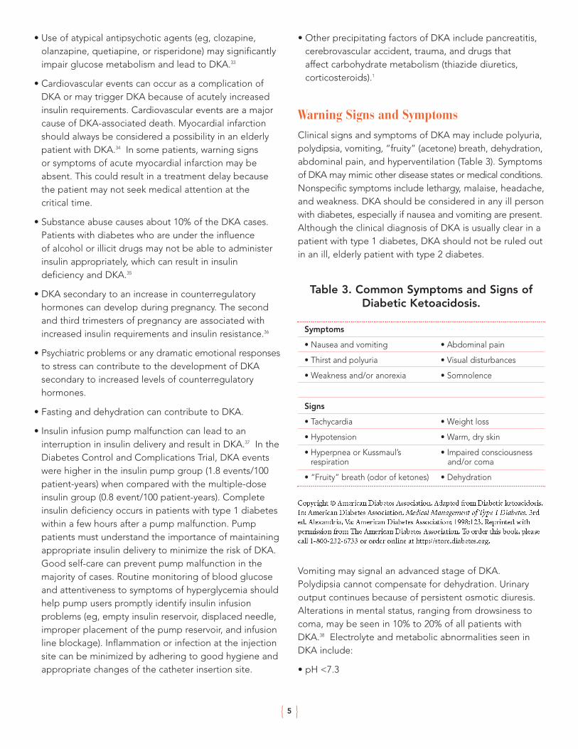
Warning Signs and SymptomsClinical signs and symptoms of DKA may include polyuria,polydipsia, vomiting, “fruity” (acetone) breath, dehydration,abdominal pain, and hyperventilation (Table 3). Symptomsof DKA may mimic other disease states or medical conditions.Nonspecific symptoms include lethargy, malaise, headache,and weakness. DKA should be considered in any ill personwith diabetes, especially if nausea and vomiting are present.Although the clinical diagnosis of DKA is usually clear in apatient with type 1 diabetes, DKA should not be ruled outin an ill, elderly patient with type 2 diabetes.
Table 3. Common Symptoms and Signs ofDiabetic Ketoacidosis.
Copyright © American Diabetes Association. Adapted from Diabetic ketoacidosis.In: American Diabetes Association. Medical Management of Type 1 Diabetes. 3rded. Alexandria, Va: American Diabetes Association; 1998:123. Reprinted withpermission from The American Diabetes Association. To order this book, pleasecall 1-800-232-6733 or order online at http://store.diabetes.org.
Vomiting may signal an advanced stage of DKA.Polydipsia cannot compensate for dehydration. Urinaryoutput continues because of persistent osmotic diuresis.Alterations in mental status, ranging from drowsiness tocoma, may be seen in 10% to 20% of all patients withDKA.38 Electrolyte and metabolic abnormalities seen inDKA include:
• pH <7.3
{ }5
Symptoms
• Nausea and vomiting • Abdominal pain
• Thirst and polyuria • Visual disturbances
• Weakness and/or anorexia • Somnolence
Signs
• Tachycardia • Weight loss
• Hypotension • Warm, dry skin
• Hyperpnea or Kussmaul’s • Impaired consciousness respiration and/or coma
• “Fruity” breath (odor of ketones) • Dehydration

• Hypokalemia (usually after therapy has been initiated)
• Hypernatremia or hyponatremia
• Hyperosmolality
• Hyperglycemia
• Hypertriglyceridemia
DiagnosisThe diagnosis of DKA must be made rapidly to preventmorbidity and mortality. It can be difficult to make a diag-nosis in the elderly. The patient may present with some orall of the symptoms previously discussed. The diagnosiscan be made after obtaining a medical history and physicalexamination. The patient’s medical history should betaken with special attention paid to recent infections,compliance with insulin or OAD therapy, concomitantmedication use, and concurrent medical illnesses. It isimportant for the physician to obtain thorough informationabout the patient’s diabetic history. If the patient is coma-tose, family members should be carefully questioned.
Hypotension, tachycardia, and tachypnea may be present.A rectal temperature measure may be required if tachypneaprecludes oral measurement. An elevated body tempera-ture warrants a careful examination and evaluation forpossible infection. However, patients may not be febrile,even in the presence of an underlying infection.38 A subnormal temperature can result from vasodilation.Hypothermia is an alarming sign and may be associatedwith increased risk of death.38 Dehydration may result inpoor skin turgor, warm dry skin, dry mucous membranes,tachycardia, and orthostatic hypotension. A change in systolic pressure of more than 10 mm Hg represents afluid volume deficit and may be indicative of systemicdehydration. Urinary output may decrease to less than 30 mL/hr and the patient may develop anuria.38
DKA must be differentiated from other causes of acidosis.The diagnosis of DKA can be confused with alcoholicketoacidosis or starvation ketosis because both conditionscause ketonemia and acidosis. Isolated alcoholic ketoacidosisis usually characterized by mild to moderate metabolicacidosis, an increased anion gap, and normoglycemia orhypoglycemia. Starvation ketosis is usually characterizedby a normal arterial pH, a mildly increased anion gap, and
the absence of significant ketonemia.16 The diagnostic criteria and typical total body deficits in DKA are listed inTable 4.
Table 4. Diagnostic Criteria and Typical TotalBody Deficits in Diabetic Ketoacidosis.
kbw = kilogram body weight.*Not all patients will meet all diagnostic criteria, depending on hydration status,previous administration of diabetes treatment, and other factors.
† Nitroprusside reaction method.From Kitabchi AE, Wall BM. Management of diabetic ketoacidosis. Am FamPhysician. 1999;60:456.
Anion GapArterial pH is a measure of the acidity or alkalinity of theblood. The arterial pH in a patient with DKA reflects thedegree of respiratory compensation and severity of acid-base disturbance. In DKA, ketoacids ionize at physiologicpH and the hydrogen ion of the ketoacids is bufferedessentially mole for mole by bicarbonate, which results inthe consumption and decrease in the serum bicarbonate.
The anion gap is the difference between serum cationsand anions. In the circulation, Na+ is the predominantcation while Cl– and HCO3
– are the main anions. Duringketogenesis, metabolic acidosis occurs because bicarbonateconcentration is reduced, resulting in an increased aniongap. In DKA, the increase in the anion gap is usuallyequivalent to the decrease in the bicarbonate concentration.
{ }6
Diagnostic criteria*
Blood glucose >250 mg/dL (13.9 mmol/L)
Arterial pH <7.3
Serum bicarbonate <15 mEq/L
Urinary ketone† Positive
Serum ketone Positive at 1:2 dilutions
Serum osmolality Variable
Typical deficits
Water 6 L or 100 mL/kbw
Sodium 7–10 mEq/kbw
Potassium 3–5 mEq/kbw
Phosphate ~1 mmol/kbw
Treatment GuidelinesThe goal of treatment is to reverse the underlying meta-bolic abnormalities, restore intravascular volume, improvetissue perfusion, and normalize the serum glucose.Emergency treatment of DKA and HHS involves correctingdehydration, hyperglycemia, and electrolyte imbalances.One of the mainstays of treatment for DKA and HHS isfluid replacement.17,18 The degree of hyperglycemia, acidosis, dehydration, and impairment of consciousnessmay vary depending on the severity of metabolicderangement, nutritional status, duration of DKA, con-comitant medications and illnesses, and the degree ofinsulin deficiency. The causes should also be identifiedand treated with steps to prevent recurrence.18
The reversal of these abnormalities should be undertakenwith meticulous care and frequent monitoring to correctand avoid serious electrolyte imbalances and fluid overload. Appendix A provides an example of a DKA flow sheet used by healthcare professionals to recordtreatment and successive changes in the patient’s status.During the first 12 hours of therapy, the frequency ofreevaluation depends on the patient’s condition. Typically,the reevaluation is hourly for the first 4 hours and continuesevery 2 to 4 hours, depending on the patient’s condition.
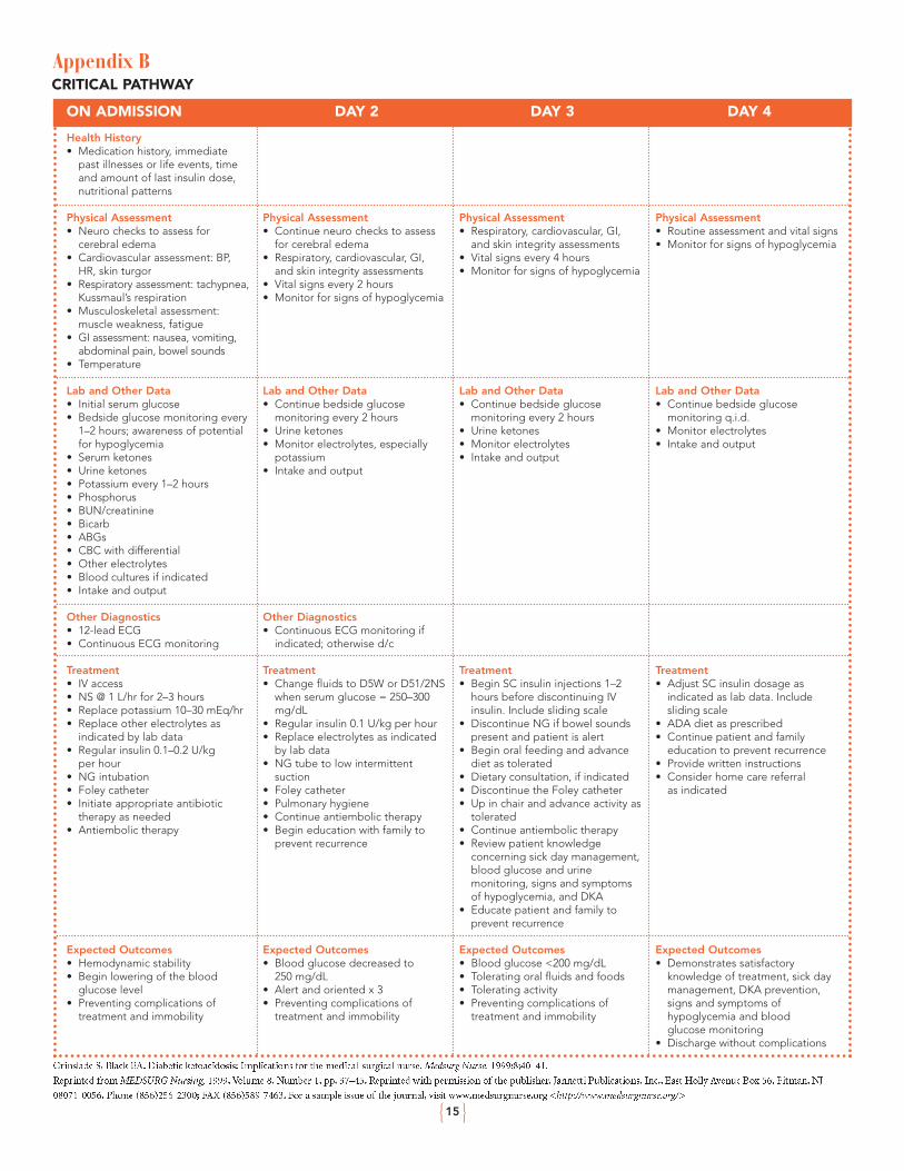
The healthcare team may follow a “critical pathway,” acomprehensive guide for treatment that includes antici-pated interventions and expected outcomes. Appendix Bis an example of a critical pathway used to guide a multi-disciplinary team through the management of DKA. Theseprotocols improve the quality of care received by patientsand are cost-effective.38 The ADA guidelines for fluidreplacement, insulin administration, and potassiumreplacement in adult patients with DKA are depicted inFigure 5. Adequate rehydration restores and maintainsintravascular volume and preserves adequate renal bloodflow. Insulin enhances the intracellular movement ofpotassium during the treatment of DKA.
{ }7
The anion gap is calculated by subtracting the sum of the chloride and bicarbonate concentrations from the “uncorrected” serum sodium concentration:
[Na+ – (Cl– + HCO3-)]
Normal Individual
Na, 140; Cl, 105; HCO3, 27
Anion gap: (140 – [105 + 27]) = 8
An anion gap of 8 is normal.
Patient with DKA
Na, 130; Cl, 98; HCO3, 10
Anion gap: (130 – [98 + 10]) = 22
This anion gap of 22 is elevated.
The range for normal anion gap can vary depending upon thelaboratory performing the test. Normal values are guidelinesand specific to each laboratory.
Parameter DKA* Normal Values†
Plasma glucose (mg/dL) 475 60–110
Serum osmolality (mOsm/kg) 309 275–293
Sodium (mEq/L) 131 135–145
Potassium (mEq/L) 4.8 3.5–5.0
HCO3– (mEq/L) 9 23–29
BUN (mg/dL) 21 8–20
Arterial pH <7.3 7.35–7.45
Ketonuria ≥3+ NA
Growth hormone (ng/mL) 7.9 0–5
Cortisol, 8 a.m. (µg/dL) 49 5–20
Free fatty acids (mmol/L) 2.26 0.19–0.9
Glucagon (pg/mL) 400–500 50–200
Lactate, plasma (mmol/L) 4.6
Venous 0.5–2.02
Arterial 0.5–1.6
β-Hydroxybutyrate (mmol/L) 13.7 NA
Catecholamines, total free (µg/mL) 1.78 ± 4 4–126 µg/24 hr
BUN = blood urea nitrogen.*All average laboratory values for DKA were taken from Ennis ED, Stahl E,Kreisberg RA. Diabetic ketoacidosis. In: Porte D Jr, Sherwin RS, eds. Ellenberg &Rifkin’s Diabetes Mellitus. 5th ed. Stamford, Conn: Appleton & Lange; 1997:831.
† All normal laboratory values were taken from Spraycar M, ed. Stedman’s MedicalDictionary. 26th ed. Philadelphia, Pa: Williams & Wilkins; 1995:1992–2010;Beers MH, Berkow R, eds. The Merck Manual of Diagnosis and Therapy. 17th ed.Centennial Edition. 1999. Available at: http://www.merck.com/pubs/mmanual/.Accessed May 6, 2002; and Tierney LM Jr, McPhee SJ, Papadakis MA, eds.Current Medical Diagnosis & Treatment. 41st ed. New York, NY: The McGraw-Hill Companies; 2002:1711–1719.
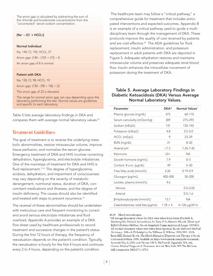
Table 5. Average Laboratory Findings inDiabetic Ketoacidosis (DKA) Versus Average
Normal Laboratory Values.
Table 5 lists average laboratory findings in DKA and compares them with average normal laboratory values.30
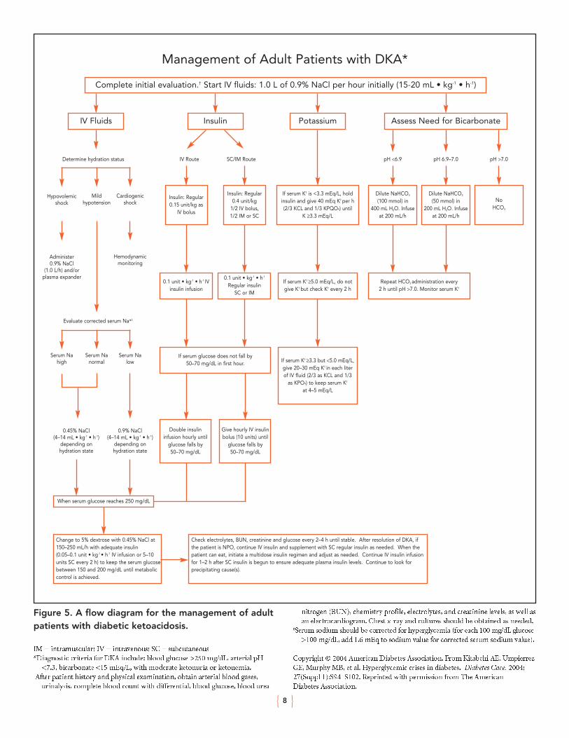
Figure 5. A flow diagram for the management of adultpatients with diabetic ketoacidosis.
IM = intramuscular; IV = intravenous; SC = subcutaneous*Diagnostic criteria for DKA include: blood glucose >250 mg/dL, arterial pH
<7.3, bicarbonate <15 mEq/L, with moderate ketonuria or ketonemia.†After patient history and physical examination, obtain arterial blood gases,
urinalysis, complete blood count with differential, blood glucose, blood urea
nitrogen (BUN), chemistry profile, electrolytes, and creatinine levels, as well asan electrocardiogram. Chest x-ray and cultures should be obtained as needed.
‡Serum sodium should be corrected for hyperglycemia (for each 100 mg/dL glucose>100 mg/dL, add 1.6 mEq to sodium value for corrected serum sodium value).
Copyright © 2004 American Diabetes Association. From Kitabchi AE, UmpierrezGE, Murphy MB, et al. Hyperglycemic crises in diabetes. Diabetes Care. 2004;27(Suppl 1):S94–S102. Reprinted with permission from The American Diabetes Association.
{ }8
Management of Adult Patients with DKA*
Complete initial evaluation.† Start IV fluids: 1.0 L of 0.9% NaCl per hour initially (15-20 mL • kg-1 • h-1)
IV Fluids Insulin
Insulin: Regular0.15 unit/kg as
IV bolus
Insulin: Regular0.4 unit/kg
1/2 IV bolus,1/2 IM or SC
0.1 unit • kg-1 • h-1 IVinsulin infusion
If serum glucose does not fall by50–70 mg/dL in first hour. If serum K† ≥3.3 but <5.0 mEq/L,
give 20–30 mEq K† in each literof IV fluid (2/3 as KCL and 1/3
as KPO4) to keep serum K†
at 4–5 mEq/L
0.1 unit • kg-1 • h-1
Regular insulinSC or IM
Double insulininfusion hourly until
glucose falls by50–70 mg/dL
When serum glucose reaches 250 mg/dL
Change to 5% dextrose with 0.45% NaCl at150–250 mL/h with adequate insulin(0.05–0.1 unit • kg-1 • h-1 IV infusion or 5–10 units SC every 2 h) to keep the serum glucose between 150 and 200 mg/dL until metabolic control is achieved.
Check electrolytes, BUN, creatinine and glucose every 2–4 h until stable. After resolution of DKA, ifthe patient is NPO, continue IV insulin and supplement with SC regular insulin as needed. When the patient can eat, initiate a multidose insulin regimen and adjust as needed. Continue IV insulin infusionfor 1–2 h after SC insulin is begun to ensure adequate plasma insulin levels. Continue to look forprecipitating cause(s).
Give hourly IV insulinbolus (10 units) until
glucose falls by50–70 mg/dL
If serum K† ≥5.0 mEq/L, do notgive K† but check K† every 2 h
Repeat HCO3 administration every2 h until pH >7.0. Monitor serum K†
If serum K† is <3.3 mEq/L, holdinsulin and give 40 mEq K† per h(2/3 KCL and 1/3 KPQO4) until
K ≥3.3 mEq/L
Dilute NaHCO3
(100 mmol) in400 mL H2O. Infuse
at 200 mL/h
Dilute NaHCO3
(50 mmol) in200 mL H2O. Infuse
at 200 mL/h
NoHCO3
Potassium Assess Need for Bicarbonate
Determine hydration status
Hypovolemicshock
Mildhypotension
Evaluate corrected serum Na♣‡
Cardiogenicshock
Administer0.9% NaCl
(1.0 L/h) and/orplasma expander
0.45% NaCl(4–14 mL • kg-1 • h-1)
depending onhydration state
0.9% NaCl(4–14 mL • kg-1 • h-1)
depending onhydration state
Serum Nahigh
Serum Nanormal
Serum Nalow
Hemodynamicmonitoring
IV Route SC/IM Route pH <6.9 pH 6.9–7.0 pH >7.0
The ADA guidelines for insulin therapy in DKA are depicted in Table 5. Because of its quick onset of action,regular insulin by continuous intravenous infusion is thepreferred treatment for DKA in adults unless the episodeis mild. Following exclusion of hypokalemia, the ADAguidelines recommend administration of an intravenousbolus of regular insulin at 0.15 unit/kg body weight, followed by continuous infusion of regular insulin at adose of 0.1 unit/kg per hour. This will usually decreaseplasma glucose concentration by 50–75 mg/dL per hour,which is similar to a higher dose insulin regimen (Figure 5).If the plasma glucose levels do not decrease by at least 50 mg/dL from the original value in the first hour, the hydration status should be checked. If the hydration statusis acceptable, then the insulin infusion may be doubledevery hour until plasma glucose shows a steady decline of50 to 75 mg/dL per hour. Once the plasma glucose reaches250 mg/dL, the insulin infusion rate can sometimes bedecreased to 0.05 to 0.1 unit/kg per hour, and 5% to 10%dextrose can be added to the intravenous fluids.1
Modern management of DKA emphasizes the use of lower doses of
insulin to avoid hypoglycemia and other complications of treatment.
Timely potassium replacement and careful monitoringduring therapy for DKA are essential to avoid potentiallylife-threatening hypokalemia. Extra caution is necessary in patients with severe hypokalemia associated with dehydration and hyperglycemia to avoid potentially fatalcardiac arrhythmias. Potassium is generally initiated afterserum levels decrease <5.0 mEq/L (see Figure 5).Potassium replacement is started earlier in patients with serum potassium levels <3.3 mEq/L, exhibiting electrocardiogram (ECG) signs of flat or inverted T waves,depressed ST segments, and emergence of U waves. Bothinsulin and bicarbonate therapy lower serum potassiumlevels, and in cases where hypokalemia is significant, orwhen serum potassium levels are <3.3 mEq/L, insulin therapy should be postponed until serum potassium hasbeen adequately replenished.1
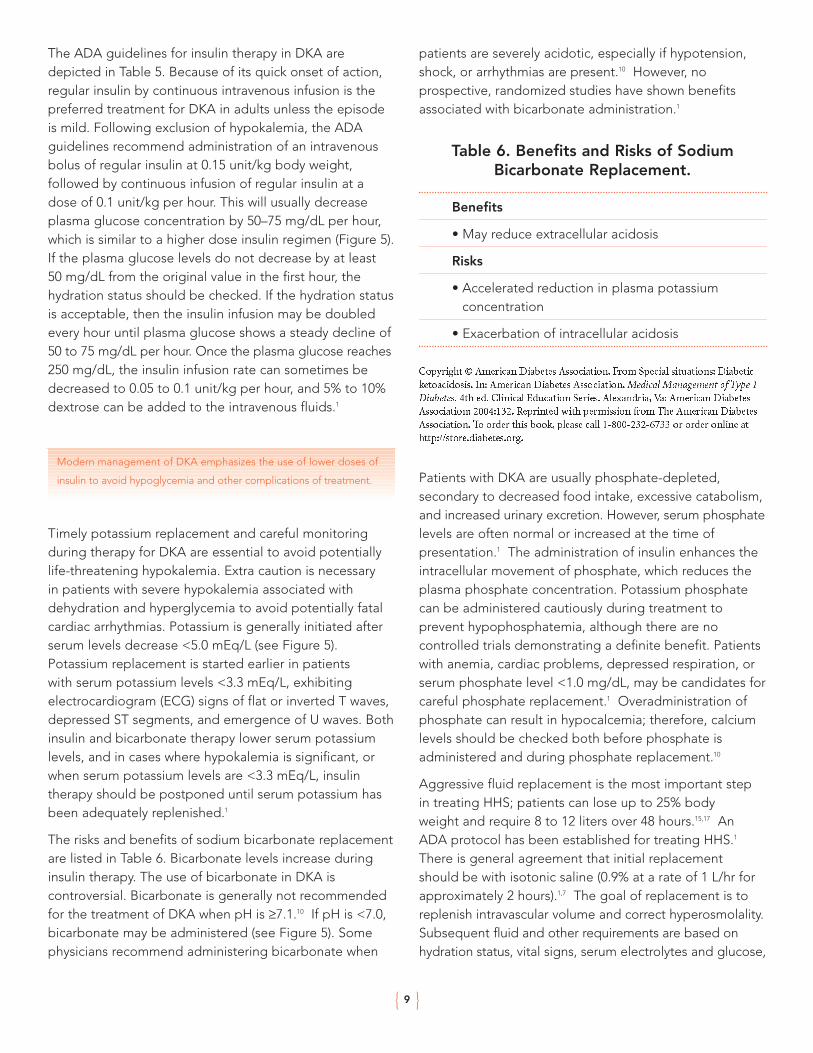
The risks and benefits of sodium bicarbonate replacementare listed in Table 6. Bicarbonate levels increase duringinsulin therapy. The use of bicarbonate in DKA is controversial. Bicarbonate is generally not recommendedfor the treatment of DKA when pH is ≥7.1.10 If pH is <7.0,bicarbonate may be administered (see Figure 5). Somephysicians recommend administering bicarbonate when
patients are severely acidotic, especially if hypotension,shock, or arrhythmias are present.10 However, no prospective, randomized studies have shown benefitsassociated with bicarbonate administration.1
Table 6. Benefits and Risks of SodiumBicarbonate Replacement.
Copyright © American Diabetes Association. From Special situations: Diabeticketoacidosis. In: American Diabetes Association. Medical Management of Type 1Diabetes. 4th ed. Clinical Education Series. Alexandria, Va: American DiabetesAssociation; 2004:132. Reprinted with permission from The American DiabetesAssociation. To order this book, please call 1-800-232-6733 or order online athttp://store.diabetes.org.
Patients with DKA are usually phosphate-depleted, secondary to decreased food intake, excessive catabolism,and increased urinary excretion. However, serum phosphatelevels are often normal or increased at the time of presentation.1 The administration of insulin enhances theintracellular movement of phosphate, which reduces theplasma phosphate concentration. Potassium phosphatecan be administered cautiously during treatment to prevent hypophosphatemia, although there are no controlled trials demonstrating a definite benefit. Patientswith anemia, cardiac problems, depressed respiration, orserum phosphate level <1.0 mg/dL, may be candidates forcareful phosphate replacement.1 Overadministration ofphosphate can result in hypocalcemia; therefore, calciumlevels should be checked both before phosphate isadministered and during phosphate replacement.10
Aggressive fluid replacement is the most important step in treating HHS; patients can lose up to 25% body weight and require 8 to 12 liters over 48 hours.15,17 AnADA protocol has been established for treating HHS.1
There is general agreement that initial replacementshould be with isotonic saline (0.9% at a rate of 1 L/hr forapproximately 2 hours).1,7 The goal of replacement is toreplenish intravascular volume and correct hyperosmolality.Subsequent fluid and other requirements are based onhydration status, vital signs, serum electrolytes and glucose,
{ }9
Benefits
• May reduce extracellular acidosis
Risks
• Accelerated reduction in plasma potassium concentration
• Exacerbation of intracellular acidosis
and urine output.1,7 Potassium, sodium, and phosphateare replaced on the basis of laboratory measures. In somecases, anticoagulants may be needed to prevent venousthromboembolism. Insulin is administered after an adequate amount of fluid replacement.15,17 Administeringinsulin before fluids can exacerbate hypotension and causevascular collapse or death through insulin-induced cellularuptake of water.17 Care in the selection of the intravenousfluid is critical as well. Whereas initial hydration in the setting of hypotension and tachycardia should be withblood, colloid, or isotonic fluids, as hemodynamic stabilityis achieved, most patients can be switched to hypotonic fluids such as one-half normal saline to avoid the hyperchloremic metabolic acidosis which often accompaniesoverhydration with normal saline. Comprehensive guidelines have been formulated by the ADA to treatboth DKA and HHS.24
Recovery CarePatients with DKA require intensive medical care, monitoring,follow-up, and education. Two important managementprinciples are followed during the recovery period: (1) continue administering insulin; and (2) allow patients toeat. Intravenous fluids and short-acting insulin are continueduntil acidosis is corrected and the patient can ingest foodwithout vomiting. Early feeding is considered because theadded carbohydrate in the presence of insulin assists inthe clearance of ketones. After the patient begins to eat,several days will still be required to correct all biochemicalabnormalities associated with an episode of DKA.10 Thepatient need not remain hospitalized beyond the timerequired to replete fluids and electrolytes, to ascertainand treat precipitating causes, and to demonstrate thatthe patient and caregivers can self-manage the diabetes.
When the decision is made to begin feeding, the patientis switched from intravenous to subcutaneous insulin.Since subcutaneous insulin acts more slowly than intravenous insulin, the transition to subcutaneous insulinis initiated with caution to avoid recurrence of acidosisduring the transition phase. The insulin drip is discontinued1 to 2 hours after the administration of subcutaneousinsulin. Glucose levels are monitored 2 hours later and atleast every 4 hours subsequently until a relatively stablesubcutaneous insulin regimen is established. Therapy isusually required for 24 to 48 hours.26 Resolution of DKA isindicated by blood glucose <200 mg/dL, a bicarbonate≥18 mEq/L, and venous pH >7.3.26
Electrolyte Management During RecoveryHyperchloremic acidosis with a normal anion gap is acommon occurrence during recovery. During an episodeof DKA, sodium is excreted as the sodium salt of ketoacids.Relative hyperchloremia can occur when treatment withintravenous solutions containing equal parts of sodiumand chloride is used.29 Because chloride losses are smallerthan sodium losses, relative hyperchloremia occurs withtherapy. The correction of DKA causes sodium bicarbonateto shift into the intracellular space leaving chloride overrepresented in the extracellular space.29 If the aniongap gradually normalizes during therapy, a subsequentperiod of hyperchloremia-related nonanion gap acidosis is of no clinical concern.
ComplicationsThe majority of DKA cases are treated successfully without complications. However, potentially life-threateningcomplications are possible and include: hypoglycemia,hypokalemia, hyperchloremia, thromboembolic events,congestive heart failure, cerebral edema, and acute respiratory distress syndrome.
Hypoglycemia can occur if an excess of insulin is administeredrelative to glucose supply. Severe hypoglycemia can be a life-threatening complication. Sweating, tremors, andpalpitations may occur with mild hypoglycemia; loss ofconsciousness and convulsions can occur with severehypoglycemia. Patients should receive hourly monitoringof plasma glucose initially and after adjustments in intravenous insulin or administered carbohydrates todetect a rapidly decreasing blood glucose level and toprevent the development of hypoglycemia.
Hypokalemia is potentially life-threatening if potassiumreplacement is delayed or inadequate. Hypokalemia usuallyoccurs after the initiation of insulin therapy, secondary tothe intracellular movement of potassium. Increased potassiumsupplementation may be needed if bicarbonate therapy is required to correct ketoacidosis. Bicarbonate promotesthe intracellular movement of potassium and increases therisk of hypokalemia.
Hyperchloremia, or hyperchloremic normal anion gapmetabolic acidosis, is present in ~10% of patients admittedwith DKA and is common in patients recovering fromDKA.24 It is usually caused by an excessive use of salinefor fluid and electrolyte replacement during treatment.However, other causes of hyperchloremia include loss of
{ }10
potential bicarbonate due to excretion of ketoanions aspotassium and sodium salts, decrease in availability ofbicarbonate in proximal tubule which leads to an increasein chloride reabsorption, and reduction of bicarbonate andother buffering capacities in other body compartments.24
Thromboembolic events may cause death in adults withDKA.19 Prolonged stasis, immobility, and hemoconcentrationare major precipitating factors of a thromboembolicevent.19 Antithrombotic therapy in the form of externalcompression devices or subcutaneous heparin is warrantedin more severe or prolonged cases.
Congestive heart failure can develop with fluid replacementtherapy, especially if an acute myocardial infarction or anunderlying diabetic cardiomyopathy is missed.
Cerebral edema is a rare, often fatal complication thatusually occurs within the first 4 to 24 hours after the initiation of therapy. It is more common in children, especially those younger than 4 to 5 years of age withDKA and new-onset diabetes. There are no establishedwarning signs or clinical predictors.38 Frequent ongoingneurologic assessments of the patient are critical duringfluid replacement and insulin therapy.38 Signs and symptoms may include headache, lethargy, abnormalpupil response, behavioral changes, seizures, bradycardia,papilledema, or unconsciousness.
Cerebral edema is an extremely serious, although rare, complication
of DKA and occurs more often in children than adults.
Acute respiratory distress syndrome is a form of acutelung injury that may occur as a result of various insults.This may lead to decreased intravascular osmotic pressureand fluid shifts resulting in pulmonary edema. Sepsis is thepredominant risk factor.39,40 Other predisposing conditionsinclude multiple transfusions, severe nonthoracic trauma,pulmonary contusion, aspiration of gastric contents, multiple fractures, drug overdose, and pneumonia.39,40
Caution in the rate of replacement of intravenous fluidsshould be exercised in patients with hypoxia and with suspected intrapulmonary or systemic infection.
PreventionPreventing DKA is one of the primary goals in diabetesmanagement. The diabetes care team, including physicians,nurses, pharmacists, and diabetes educators, should trainpatients with diabetes to recognize the early signs and
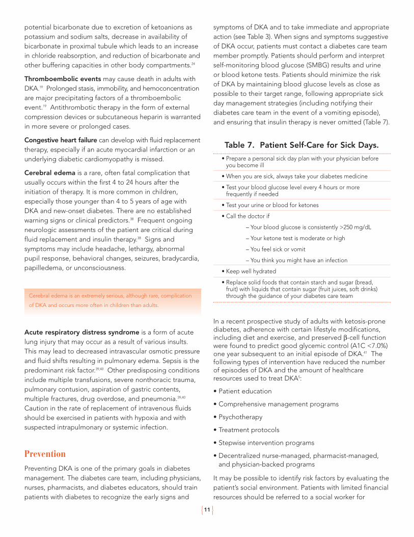
symptoms of DKA and to take immediate and appropriateaction (see Table 3). When signs and symptoms suggestiveof DKA occur, patients must contact a diabetes care teammember promptly. Patients should perform and interpretself-monitoring blood glucose (SMBG) results and urine or blood ketone tests. Patients should minimize the risk of DKA by maintaining blood glucose levels as close as possible to their target range, following appropriate sickday management strategies (including notifying their diabetes care team in the event of a vomiting episode),and ensuring that insulin therapy is never omitted (Table 7).
Table 7. Patient Self-Care for Sick Days.
In a recent prospective study of adults with ketosis-pronediabetes, adherence with certain lifestyle modifications,including diet and exercise, and preserved β-cell functionwere found to predict good glycemic control (A1C <7.0%)one year subsequent to an initial episode of DKA.41 Thefollowing types of intervention have reduced the numberof episodes of DKA and the amount of healthcareresources used to treat DKA5:
• Patient education
• Comprehensive management programs
• Psychotherapy
• Treatment protocols
• Stepwise intervention programs
• Decentralized nurse-managed, pharmacist-managed,and physician-backed programs
It may be possible to identify risk factors by evaluating thepatient’s social environment. Patients with limited financialresources should be referred to a social worker for
{ }11
• Prepare a personal sick day plan with your physician beforeyou become ill
• When you are sick, always take your diabetes medicine
• Test your blood glucose level every 4 hours or more frequently if needed
• Test your urine or blood for ketones
• Call the doctor if
– Your blood glucose is consistently >250 mg/dL
– Your ketone test is moderate or high
– You feel sick or vomit
– You think you might have an infection
• Keep well hydrated
• Replace solid foods that contain starch and sugar (bread,fruit) with liquids that contain sugar (fruit juices, soft drinks)through the guidance of your diabetes care team
assistance. A referral to appropriate community resourcesand associations for support and education may helppatients and their families.
The risk of DKA in patients who use continuous subcutaneousinsulin infusion pumps can be reduced by limiting the useof these devices to patients who are highly motivated andproperly trained in its usage. The patient must have aclear understanding of the pump function, exercise goodhygiene, plan for sick days, adjust insulin appropriately,and recognize the signs and symptoms of DKA. Patientsare instructed to perform frequent SMBG tests (at least 4 times a day), perform urine or blood ketone tests whenappropriate, and respond appropriately to signs andsymptoms of hyperglycemia and hypoglycemia.Healthcare professionals who are experienced in pumpuse should educate and guide these patients. Improvedpump technology reduces the equipment malfunctionsthat can lead to DKA.
Outpatient Prevention MeasuresIntervention with supplemental injections of short-actinginsulin or insulin analogs and oral administration of fluidsare often initiated in patients with mild ketosis. In theabsence of nausea and vomiting, patients with mild tomoderate ketosis (glucose level >250 mg/dL, with moderateto large amounts of urinary ketones detected by urineketone dipstick) may respond to supplemental doses ofshort-acting insulin or analogs, which can be repeated in 4 hours or less if the problem has not resolved. The development of nausea and vomiting or failure to clearketones necessitates further evaluation in a hospital emergency room.
Preventing Recurrent Diabetic KetoacidosisFrequent, recurrent DKA indicates the need for a detailedassessment by the diabetes care team. The team mustdetermine whether the diabetes management plan isappropriate and identify any factors that promote noncompliance. Typical factors include:
• Inadequate patient education
• Cognitive impairment
• Substance abuse
• Lack of motivation
• Financial issues
• Emotional or psychiatric disorders
• Sensory impairment
• Problem with insulin/insulin delivery system
If patients or their families cannot achieve effective self-management, the team should focus on teaching them to identify the symptoms that require prompt medicalattention. Patients who have repeated episodes of DKAmay need psychological consultation.
Special CircumstancesPregnancyDKA occurs in 1% to 3% of pregnancies complicated by diabetes.42 It is an acute medical emergency.42 Anepisode of DKA lasting 2 to 5 hours or longer can result in the death of the fetus. DKA most commonly occurs inthe second and third trimesters when increased insulinresistance is present. Prenatal and perinatal counseling is imperative in pregnant patients with diabetes. DKA frequently occurs when pregnancy appears in the presence of undiagnosed diabetes. Factors that predispose pregnant women to DKA are43:
• Accelerated starvation (eg, eating disorder)
• Dehydration secondary to vomiting
• Lowered buffering capacity
• Increased production of insulin antagonists
• Stress
Other precipitating factors of DKA were reviewed earlier(see “Triggers of DKA and HHS”). Fetal loss, the most serious consequence, occurs in about 9% of pregnantwomen presenting with DKA, even with the use of insulinand fluid therapy.42 The mechanism of fetal mortality isunclear and could be related to fetal distress and hypoxia.Maternal ketoacids cross the placenta and cause fetal acidosis. Management of this condition is the same as for a nonparous individual.
ElderlyThere are special considerations involved in caring for theelderly with DKA because of the high mortality rate in thispopulation. Elderly patients have an increased risk for fluidoverload, decreased pulmonary capacity, silent cardiacevents, and thromboembolic events.38 It is critical that cardiopulmonary function be monitored, good pulmonaryhygiene be practiced, and abdominal complaints be evaluated in the elderly. Initial therapy is usually conductedin an intensive care unit. Once patients are stabilized, theyare transferred to a general unit.
{ }12
Economic IssuesThe costs of DKA prevention with medication, monitoringSMBG, urine, and ketones, and patient education are afraction of the cost of an emergency hospitalization.Location of treatment, intensive care facility versus generalward, plays a substantial role in the overall expense ofDKA. The treatment of DKA requires a full complement ofhospital, emergency, and intensive services, which consumessignificant healthcare resources. This is especially true forpatients hospitalized for multiple episodes. Treating apatient with DKA requires a full-time team of specialists toadminister intravenous medications and fluids and performcareful hourly monitoring, frequent laboratory tests requiringrapid turnaround time and interpretation, and reliablebedside blood glucose monitoring.44
The newer rapid-acting insulin analogs, insulin aspart andinsulin lispro, have been tested in therapy for DKA.Conclusions drawn from these studies have been thatrapid-acting analogs, delivered subcutaneously every 1 to2 hours, may provide an alternative to intravenous regularinsulin for the treatment of mild and moderate DKA.45,46
Although insulin analogs cost more than formulations ofregular human insulin, the option of subcutaneous treat-ment could potentially reduce the cost of DKA therapy if it could be done in less intensive surroundings. However,the presumption that intravenous insulin therapy requiresa critical care facility, whereas subcutaneous insulin injectiontherapy can be carried out in a less intensive setting, willnot apply to all institutions.44
From population-based studies, the annual incidence ratefor DKA is estimated to range from 4.6 to 8 episodes per1000 patients with diabetes.1 With the mean medical carefor a patient with DKA estimated at $13,000 per episode,the overall hospital cost for patients with DKA may bemore than $1 billion per year.1,24 A substantial proportionof the cost for direct medical care for adults with type 1diabetes is attributable to DKA. It is estimated that aboutone half of the DKA cost derives from patients who experience multiple episodes.5
Two studies show that specialists (endocrinologists) providemore cost-effective care compared with nonspecialists.Patients under the care of specialists have a shorter hospitalstay, fewer medical procedures, lower medical costs($10,109 for nonspecialist care vs $5463 for specialist care),and a lower rate of recurrent DKA.47,48 Early conversion tooral feeding and subcutaneous insulin therapy are alsoassociated with a shorter hospital stay.
SummaryDKA is a reversible but potentially life-threatening illnessthat results from relative or absolute insulin deficiency.Without insulin the body cannot utilize glucose as a fuelsource and must obtain an alternative source of energy.This triggers a complex metabolic process that causes the breakdown of fat in adipose tissue and electrolyte disturbances that ultimately produces ketones and makes the blood acidic. The characteristics of DKA arehyperglycemia, ketosis, and acidosis. The clinical signsand symptoms include polyuria, polydipsia, progressive dehydration, and Kussmaul’s respiration.
DKA usually can be prevented in the outpatient settingthrough effective patient education and timely interventionby the diabetes care team. Prompt and appropriate treatmentof DKA most often results in satisfactory outcomes. Criticalcare personnel must know how to stabilize the patient’scondition rapidly, administer appropriate medical treatment,and initiate a thorough evaluation to identify possible precipitating factors. Insulin administration, rehydration,and electrolyte replacement are the cornerstones of treatment.Early conversion to oral feeding and subcutaneous insulintherapy after the metabolic derangement is corrected is associated with a shorter length of hospital stay.Healthcare professionals who treat patients with diabetes should heed these important observations and recommendations:
• Treatment should be initiated immediately when DKA isdiagnosed or suspected.
• An excellent clinical outcome is usually possible withmeticulous care of patients with DKA.
• Successful treatment of patients with DKA is based onunderstanding that the etiology of DKA is severe insulindeficiency.
• Management of DKA includes administering insulin, correcting metabolic abnormalities by replacing fluidsand electrolytes, identifying and treating precipitatingcauses, and monitoring for complications.
• Subcutaneous use of rapid-acting insulin analogs mayprovide an alternative to the use of intravenous insulintherapy in the treatment of mild and moderate DKA.
• Prevention and timely reversal of DKA can reduce morbidity and mortality and the cost of treatment.
{ }13
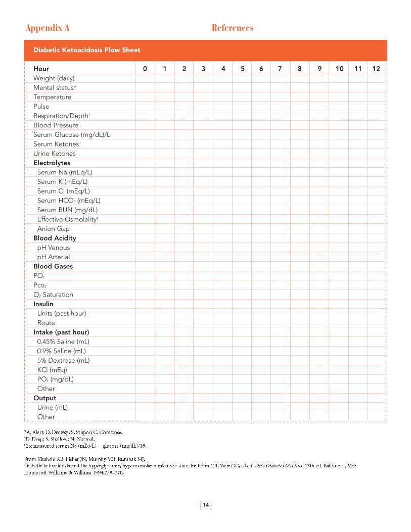
Appendix A
*A, Alert; D, Drowsy; S, Stupor; C, Comatose.†D, Deep; S, Shallow; N, Normal.‡2 x measured serum Na (mEq/L) + glucose (mg/dL)/18.
References
{ }14
Diabetic Ketoacidosis Flow Sheet
Hour 0 1 2 3 4 5 6 7 8 9 10 11 12Weight (daily)Mental status*TemperaturePulseRespiration/Depth†
Blood PressureSerum Glucose (mg/dL)/LSerum KetonesUrine KetonesElectrolytes
Serum Na (mEq/L)Serum K (mEq/L)Serum Cl (mEq/L)Serum HCO3 (mEq/L)Serum BUN (mg/dL)Effective Osmolality‡
Anion GapBlood Acidity
pH VenouspH Arterial
Blood GasesPO2
Pco2
O2 SaturationInsulin
Units (past hour)Route
Intake (past hour)0.45% Saline (mL)0.9% Saline (mL)5% Dextrose (mL)KCl (mEq)PO4 (mg/dL)Other
OutputUrine (mL)Other
From Kitabchi AE, Fisher JN, Murphy MB, Rumbak MJ.Diabetic ketoacidosis and the hyperglycemic, hyperosmolar nonketotic state. In: Kahn CR, Weir GC, eds. Joslin’s Diabetes Mellitus. 13th ed. Baltimore, Md:Lippincott Williams & Wilkins; 1994:738–770.
Grinslade S, Black EA. Diabetic ketoacidosis: Implications for the medical-surgical nurse. Medsurg Nurse. 1999:8;40–41.Reprinted from MEDSURG Nursing, 1999, Volume 8, Number 1, pp. 37–45. Reprinted with permission of the publisher, Jannetti Publications, Inc., East Holly Avenue Box 56, Pitman, NJ08071-0056. Phone (856)256-2300; FAX (856)589-7463. For a sample issue of the journal, visit www.medsurgnurse.org <http://www.medsurgnurse.org/>
CRITICAL PATHWAY
ON ADMISSION DAY 2 DAY 3 DAY 4
Health History• Medication history, immediate
past illnesses or life events, timeand amount of last insulin dose,nutritional patterns
Physical Assessment• Neuro checks to assess for
cerebral edema • Cardiovascular assessment: BP,
HR, skin turgor• Respiratory assessment: tachypnea,
Kussmaul’s respiration• Musculoskeletal assessment:
muscle weakness, fatigue• GI assessment: nausea, vomiting,
abdominal pain, bowel sounds• Temperature
Lab and Other Data• Initial serum glucose• Bedside glucose monitoring every
1–2 hours; awareness of potentialfor hypoglycemia
• Serum ketones • Urine ketones• Potassium every 1–2 hours• Phosphorus• BUN/creatinine • Bicarb• ABGs• CBC with differential• Other electrolytes• Blood cultures if indicated• Intake and output
Other Diagnostics• 12-lead ECG• Continuous ECG monitoring
Treatment• IV access• NS @ 1 L/hr for 2–3 hours• Replace potassium 10–30 mEq/hr• Replace other electrolytes as
indicated by lab data• Regular insulin 0.1–0.2 U/kg
per hour• NG intubation• Foley catheter • Initiate appropriate antibiotic
therapy as needed• Antiembolic therapy
Expected Outcomes• Hemodynamic stability• Begin lowering of the blood
glucose level • Preventing complications of
treatment and immobility
Physical Assessment• Continue neuro checks to assess
for cerebral edema • Respiratory, cardiovascular, GI,
and skin integrity assessments• Vital signs every 2 hours• Monitor for signs of hypoglycemia
Lab and Other Data• Continue bedside glucose
monitoring every 2 hours• Urine ketones• Monitor electrolytes, especially
potassium • Intake and output
Other Diagnostics• Continuous ECG monitoring if
indicated; otherwise d/c
Treatment• Change fluids to D5W or D51/2NS
when serum glucose = 250–300mg/dL
• Regular insulin 0.1 U/kg per hour• Replace electrolytes as indicated
by lab data • NG tube to low intermittent
suction• Foley catheter• Pulmonary hygiene• Continue antiembolic therapy• Begin education with family to
prevent recurrence
Expected Outcomes• Blood glucose decreased to
250 mg/dL• Alert and oriented x 3• Preventing complications of
treatment and immobility
Physical Assessment• Respiratory, cardiovascular, GI,
and skin integrity assessments• Vital signs every 4 hours • Monitor for signs of hypoglycemia
Lab and Other Data• Continue bedside glucose
monitoring every 2 hours• Urine ketones• Monitor electrolytes• Intake and output
Treatment• Begin SC insulin injections 1–2
hours before discontinuing IVinsulin. Include sliding scale
• Discontinue NG if bowel soundspresent and patient is alert
• Begin oral feeding and advancediet as tolerated
• Dietary consultation, if indicated • Discontinue the Foley catheter• Up in chair and advance activity as
tolerated • Continue antiembolic therapy• Review patient knowledge
concerning sick day management,blood glucose and urine monitoring, signs and symptomsof hypoglycemia, and DKA
• Educate patient and family to prevent recurrence
Expected Outcomes• Blood glucose <200 mg/dL• Tolerating oral fluids and foods• Tolerating activity• Preventing complications of
treatment and immobility
Physical Assessment• Routine assessment and vital signs• Monitor for signs of hypoglycemia
Lab and Other Data• Continue bedside glucose
monitoring q.i.d.• Monitor electrolytes• Intake and output
Treatment• Adjust SC insulin dosage as
indicated as lab data. Include sliding scale
• ADA diet as prescribed • Continue patient and family
education to prevent recurrence • Provide written instructions • Consider home care referral
as indicated
Expected Outcomes• Demonstrates satisfactory
knowledge of treatment, sick daymanagement, DKA prevention,signs and symptoms of hypoglycemia and blood glucose monitoring
• Discharge without complications
Appendix B
{ }15
1. Kitabchi AE, Umpierrez GE, Murphy MB, et al. Hyperglycemic crises in diabetes. Diabetes Care. 2004;27(Suppl 1):S94–S102.
2. Wagner A, Risse A, Brill HL, et al. Therapy of severe diabetic ketoacidosis.Zero-mortality under very-low-dose insulin application. Diabetes Care.1999;22:674–677.
3. Umpierrez GE, Murphy MB, Kitabchi AE. Diabetic ketoacidosis and hyperglycemic hyperosmolar syndrome. Diabetes Spectr. 2002;15:28–36.
4. National Center for Chronic Disease Prevention and Health Promotion.National Diabetes Surveillance System Data and Trends: Diabetic Keto-acidosis as First-Listed Diagnosis. http://www.cdc.gov/diabetes/statistics/dkafirst/table2link.htm. US Dept of Health and Human Services, Centers for Disease Control and Prevention; 2005.
5. Javor KA, Kotsanos JG, McDonald RC, et al. Diabetic ketoacidosis chargesrelative to medical charges of adult patients with type I diabetes. DiabetesCare. 1997;20:349–354.
6. Charfen MA, Fernandez-Frackelton M. Diabetic ketoacidosis. Emerg MedClin North Am. 2005;23:609–628.
7. Nugent BW. Hyperosmolar hyperglycemic state. Emerg Med Clin North Am.2005;23:629–648.
8. American Diabetes Association. Standards of medical care in diabetes.Diabetes Care. 2005;28(Suppl 1):S4–S36.
9. American Association of Clinical Endocrinologists and the American Collegeof Endocrinology. The American Association of Clinical EndocrinologistsMedical Guidelines for the Management of Diabetes Mellitus: The AACESystem of Intensive Diabetes Self-Management—2002 Update. EndocrinePractice. 2002;8:40–82.
10. American Diabetes Association. Special situations: Diabetic ketoacidosis. In:Medical Management of Type 1 Diabetes. 4th ed. Alexandria, Va: AmericanDiabetes Association; 2004:127–135.
11. Newton CA, Raskin P. Diabetic ketoacidosis in type 1 and type 2 diabetesmellitus: Clinical and biochemical differences. Arch Intern Med.2004;164:1925–1931.
12. Casteels K, Mathieu C. Diabetic ketoacidosis. Rev Endocr Metab Disord.2003;4:159–166.
13. Chiasson JL, Aris-Jilwan N, Belanger R, et al. Diagnosis and treatment ofdiabetic ketoacidosis and the hyperglycemic hyperosmolar state. CMAJ.2003;168:859–866.
14. White NH. Management of diabetic ketoacidosis. Rev Endocr Metab Disord.2003;4:343–353.
15. Moore T. Diabetic emergencies in adults. Nurs Stand. 2004;18:45–52.16. Buse JB, Polonsky KS. Diabetic ketoacidosis, hyperglycemic hyperosmolar
nonketotic coma, and hypoglycemia. In: Hall JB, Schmidt GA, Woods LDH,eds. Principles of Critical Care. 2nd ed. New York, NY: McGraw-Hill;1998:1183–1193.
17. Stoner GD. Hyperosmolar hyperglycemic state. Am Fam Physician.2005;71:1723–1730.
18. Gaglia JL, Wyckoff J, Abrahamson MJ. Acute hyperglycemic crisis in the elderly. Med Clin North Am. 2004;88:1063–1084.
19. Bell DS, Alele J. Diabetic ketoacidosis. Why early detection and aggressivetreatment are crucial. Postgrad Med. 1997;101:193–194.
20. Westphal SA. The occurrence of diabetic ketoacidosis in non-insulin-dependentdiabetes and newly diagnosed diabetic adults. Am J Med. 1996;101:19–24.
21. Brink SJ. Diabetic ketoacidosis. Acta Paediatr Suppl. 1999;88:14–24.22. Yu EH, Wu TJ. Clinical profiles in adult diabetic ketoacidotic patients in a
tertiary referral medical center in southern Taiwan. J Formos Med Assoc.1998;97:85–89.
23. Balasubramanyam A, Zern JW, Hyman DJ, Pavlik V. New profiles of diabeticketoacidosis: Type 1 vs type 2 diabetes and the effect of ethnicity. Arch InternMed. 1999;159:2317–2322.
24. Kitabchi AE, Umpierrez GE, Murphy MB, et al. Management of hyper-glycemic crises in patients with diabetes. Diabetes Care. 2001;24:131–153.
25. English P, Williams G. Hyperglycaemic crises and lactic acidosis in diabetesmellitus. Postgrad Med J. 2004;80:253–261.
26. Trachtenbarg DE. Diabetic ketoacidosis. Am Fam Physician.2005;71:1705–1714.
27. Wallace TM, Matthews DR. Recent advances in the monitoring and management of diabetic ketoacidosis. QJM. 2004;97:773–780.
28. Yan SH, Sheu WH, Song YM, Tseng LN. The occurrence of diabetic ketoacidosis in adults. Intern Med. 2000;39:10–14.
29. Fleckman AM. Diabetic ketoacidosis. Endocr Metab Clin North Am.1993;22:181–207.
30. Ennis ED, Stahl E, Kreisberg RA. Diabetic ketoacidosis. In: Porte D Jr,Sherwin RS, eds. Ellenberg & Rifkin’s Diabetes Mellitus. 5th ed. Stamford,Conn: Appleton and Lange; 1997:827–844.
31. Kitabchi AE, Wall BM. Management of diabetic ketoacidosis. Am FamPhysician. 1999;60:455–464.
32. Silink M. Practical management of diabetic ketoacidosis in childhood andadolescence. Acta Paediatr Suppl. 1998;425:63–66.
33. Jin H, Mayer JM, Jeste DV. Phenomenology of and risk factors for new-onsetdiabetes mellitus and diabetic ketoacidosis associated with atypical antipsychotics: An analysis of 45 published cases. Ann Clin Psychiatry.2002;14:59–64.
34. Miller J. Management of diabetic ketoacidosis. J Emerg Nurs.1999;25:514–519.
35. Warner EA, Greene GS, Buchsbaum MS, et al. Diabetic ketoacidosis associated with cocaine use. Arch Intern Med. 1998;158:1799–1802.
36. Bedalov A, Balasubramanyam A. Glucocorticoid-induced ketoacidosis in gestational diabetes: Sequela of the acute treatment of preterm labor. A casereport. Diabetes Care. 1997;20:922–924.
37. Castillo MJ, Scheen AJ, Lefebvre PJ. Treatment with insulin infusion pumpsand ketoacidotic episodes: From physiology to troubleshooting. DiabetesMetab Rev. 1995;11:161–177.
38. Grinslade S, Buck EA. Diabetic ketoacidosis: Implications for the medical-surgical nurse. Medsurg Nurs. 1999;8:37–45.
39. Eisner MD, Thompson T, Hudson LD, et al. Efficacy of low tidal volume ventilation in patients with different clinical risk factors for acute lung injuryand the acute respiratory distress syndrome. Am J Respir Crit Care Med.2001;164:231–236.
40. Montgomery AB, Stager MA, Carrico CJ, Hudson LD. Causes of mortality inpatients with the adult respiratory distress syndrome. Am Rev Respir Dis.1985;132:485–489.
41. Maldonado M, D’Amico S, Otiniano M, et al. Predictors of glycaemic controlin indigent patients presenting with diabetic ketoacidosis. Diabetes ObesMetab. 2005;7:282–289.
42. Ramin KD. Diabetic ketoacidosis in pregnancy. Obstet Gynecol Clin NorthAm. 1999;26:481–488.
43. Chauhan SP, Perry KG Jr, McLaughlin BN, et al. Diabetic ketoacidosis complicating pregnancy. J Perinatol. 1996;16:173–175.
44. Haas RM, Hoffman AR. Treatment of diabetic ketoacidosis: Should mode ofinsulin administration dictate use of intensive care facilities? Am J Med.2004;117:357–358.
45. Umpierrez GE, Latif K, Stoever J, et al. Efficacy of subcutaneous insulinlispro versus continuous intravenous regular insulin for the treatment ofpatients with diabetic ketoacidosis. Am J Med. 2004;117:291–296.
46. Umpierrez GE, Cuervo R, Karabell A, et al. Treatment of diabetic ketoacidosiswith subcutaneous insulin aspart. Diabetes Care. 2004;27:1873–1878.
47. Levetan CS, Salas JR, Wilets IF, Zumoff B. Impact of endocrine and diabetesteam consultation on hospital length of stay for patients with diabetes. Am JMed. 1995;99:22–28.
48. Levetan CS, Passaro MD, Jablonski KA, Ratner RE. Effect of physician specialty on outcomes in diabetic ketoacidosis. Diabetes Care.1999;22:1790–1795.
{ }16
1. Diabetic ketoacidosis is characterized by __________.a. Plasma glucose >250 mg/dLb. Ketosisc. Acidosisd. Lipolysise. All of the above
2. All of the following EXCEPT __________ are major precipitating causes of DKA.
a. Infectionb. Hypoglycemia c. Pancreatitisd. Traumae. Myocardial infarction
3. DKA occurs in __________ of patients with newly diagnosed diabetes.
a. 5%–10%b. 10%–20%c. 20%–30%d. 30%–40%e. 40%–50%
4. Which of the following generally differentiates HHS from DKA?
a. Onset of DKA is more rapid than HHS b. Minimal or no increase in lipolysisc. Severity of ketosisd. pH may be within normal limitse. All of the above
5. Vital signs in DKA typically reveal __________.a. Hypertension, bradycardia,
and normal respirationb. Orthostatic hypotension,
bradycardia, and tachypneac. Orthostatic hypotension,
tachycardia, and tachypnead. Hypertension, palpitation,
and tachypnea
6. Sodium bicarbonate therapy for DKA __________.a. Is not recommended when pH is ≥7.1b. Can exacerbate intracellular acidosisc. Can cause hypoglycemiad. Both a & b
7. Which of the following is a FALSE statement? a. DKA never occurs in type 2 diabetes.b. Hyperventilation is a consequence
of acidosis.c. Stress associated with intercurrent
illnesses may increase the body’s requirement for insulin.
d. Diabetic neuropathy can result in the absence ordelay of signs and symptoms of a myocardialinfarction.
8. Which action is not recommended for a patient withdiabetes during sick days?
a. Stop taking insulin when not eating.b. Report infections to the physician.c. Perform frequent tests of ketones.d. Keep well hydrated.
9. Rapid-acting insulin analogs may provide an alternative to intravenous regular human insulin for treating __________.
a. Mild to moderate DKAb. DKA of any severityc. Hypoglycemic comad. Cerebral edemae. None of the above
10. How does the body compensate for acidosis?a. Renal elimination of ketonesb. Bicarbonate buffering c. Retention of carbon dioxided. a and be. All of the above
{ }17
Post-TestThis program can also be completed online at www.MedEdToday.com. Please provide 1 answer for each question (10 correct answers are necessary for successful completion). Place all answers on the exam answer key provided on the evaluation form following this post-test.
11. All of the following are objectives of the healthcareteam for patients who have diabetes except__________.a. Teach patients to recognize the early signs of DKAb. Teach patients to perform self-monitoring of
ketonesc. Teach patients to omit their insulin dose during
sick daysd. Identify risk factors that may require
special intervention
12. Potential complications during treatment for DKAinclude __________.a. Hypoglycemiab. Hypochloremiac. Schizophreniad. All of the above
13. Which of the following statements is(are) trueabout DKA during pregnancy?a. It occurs more commonly during the first
trimester.b. An episode lasting 2 to 5 hours or longer can
cause fetal death.c. Fetal loss occurs in 20% of cases.d. All of the above
14. Special considerations are required for manage-ment of DKA in the elderly because __________.a. There is a high mortality rate in
this populationb. Increased risk of fluid retentionc. Increased risk of thromboembolic eventsd. All of the above
{ }18
{ }19
Evaluation FormManagement of Diabetic Ketoacidosis and Hyperosmolar Hyperglycemic State in AdultsProject ID: 3346 ES 13
Postgraduate Institute for Medicine (PIM) respects and appreciates your opinions. To assist us in evaluating the effectiveness of this activity and to make recommendations for future educational offerings, please take a few minutes to complete this evaluation form. You must complete this evaluation form to receive acknowledgment of participation for this activity.
Please answer the following questions by circling the appropriate rating:5 = Outstanding 4 = Good 3 = Satisfactory 2 = Fair 1 = Poor
Extent to Which Program Activities Met the Identified PurposeProvide clinicians treating diabetic patients with the latest information about the pathophysiology, diagnosis, and management of DKA and HHSto help reduce the incidence, morbidity, and mortality of the disorder. 5 4 3 2 1
Extent to Which Program Activities Met the Identified ObjectivesUpon completion of this activity, participants should be better able to:• Differentiate between DKA and HHS 5 4 3 2 1• Describe the epidemiology and pathophysiology of DKA 5 4 3 2 1• Identify the signs, symptoms, and complications of DKA 5 4 3 2 1• Discuss methods of treatment and recovery care for DKA 5 4 3 2 1• Describe potential complications during treatment of DKA
and special considerations for pregnant and elderly individuals 5 4 3 2 1• Provide strategies for the prevention of DKA 5 4 3 2 1
Overall Effectiveness of the Activity• Was timely and will influence how I practice 5 4 3 2 1• Will assist me in improving patient care 5 4 3 2 1• Fulfilled my educational needs 5 4 3 2 1• Avoided commercial bias or influence 5 4 3 2 1
Impact of the ActivityThe information presented (check all that apply):o Reinforced my current practice/treatment habits o Will improve my practice/patient outcomeso Provided new ideas or information I expect to use o Enhanced my current knowledge base
Will the information presented cause you to make any changes in your practice? o Yes o NoIf yes, please describe any change(s) you plan to make in your practice as a result of this activity:
{ }20
How committed are you to making these changes? 5 (Very committed) 4 3 2 1 (Not at all committed)
Future ActivitiesDo you feel future activities on this subject matter are necessary and/or important to your practice?
o Yes o No
Please list any other topics that would be of interest to you for future educational activities:
Follow-upAs part of our ongoing continuous quality-improvement effort, we conduct post-activity follow-up surveys to assess theimpact of our educational interventions on professional practice. Please indicate your willingness to participate in such a survey:o Yes, I would be interested in participating in a follow-up survey.o No, I’m not interested in participating in a follow-up survey.
Additional comments about this activity:
If you wish to receive acknowledgment of participation for this activity, please complete the post-test by selecting the best answer to each question, complete this evaluation verification of participation and mail to: PIM, 367 Inverness Parkway, Suite 215, Englewood, CO 80112 or FAX to: 303-790-4876.
Post-test Answer Key
Request for Credit
I certify my actual time to complete this educational activity to be:
Registered Nurse Continuing Education Credito I participated in the entire activity and claim 2.0 credits.
Pharmacist Continuing Education Credito I participated in the entire activity and claim 1.6 credits.
Signature Date Completed
Name Degree
Organization Specialty
Address
City, State, Zip
Telephone Fax E-Mail
1 2 3 4 5 6 7
8 9 10 11 12 13 14

© Scherer Clinical Communications
November 2005
Printed in USA