Embed Size (px)
Citation preview
Magnetic Resonance Imaging
Alex MacKayUniversity of British Columbia

Magnetic Resonance Imaging
A) What is MRI?B) Why do MRI?C) What can we do with an MRI scanner?

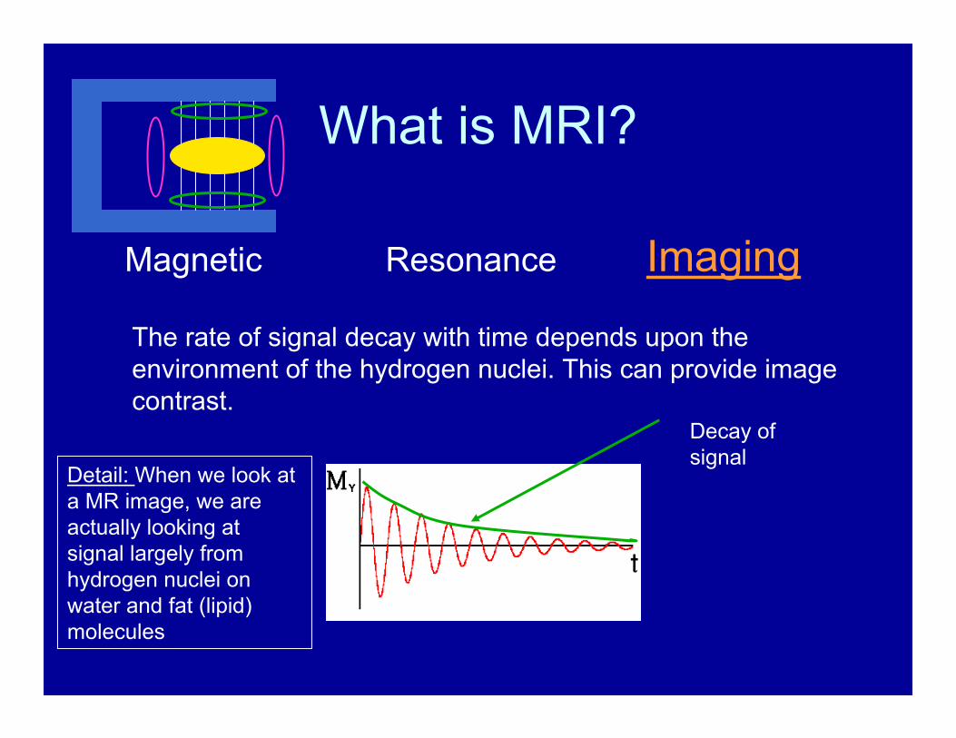
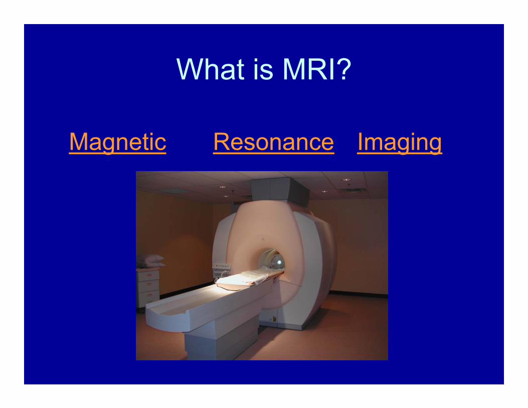
What is MRI?
Magnetic Resonance Imaging
We place our ‘sample’ in a very large magnetic field ~ 50,000 times larger than the earths field.

What is MRI?
Magnetic Resonance Imaging
In a large magnetic field hydrogen nuclei behave like little magnets. They align with the field and precess around the field at a frequency which is proportional to the magnetic field.
Precession
ωprecession = γH
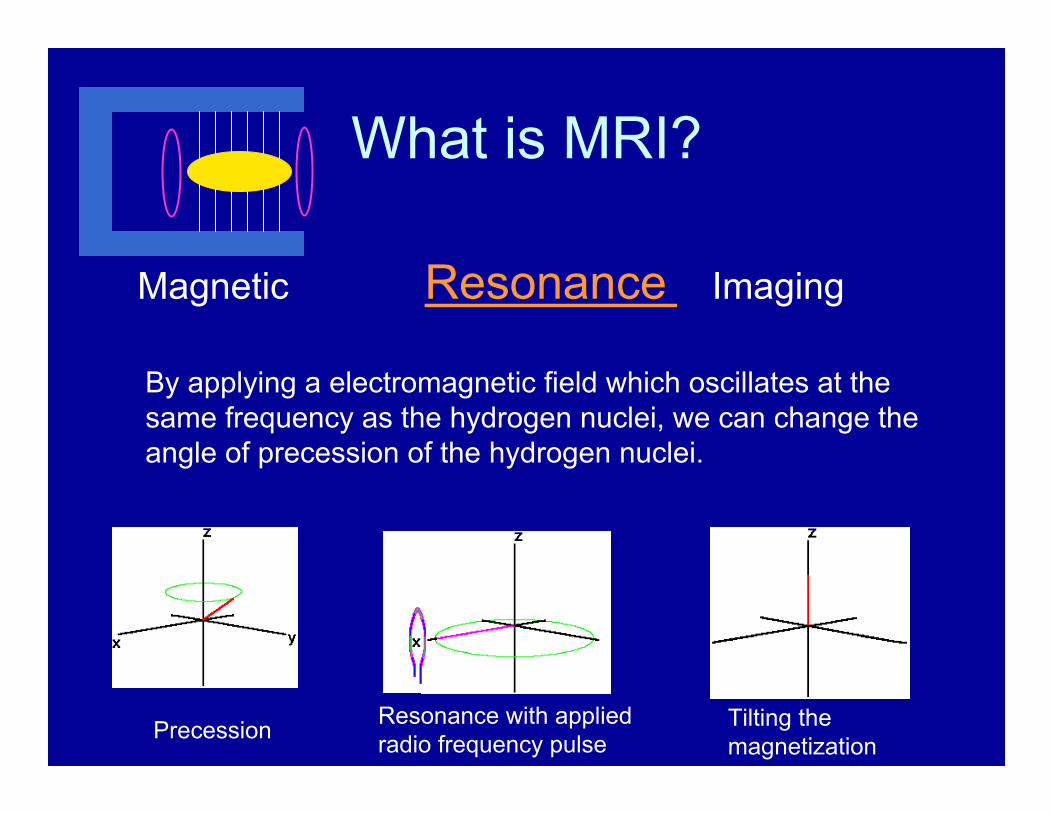
What is MRI?
Magnetic Resonance Imaging
By applying a electromagnetic field which oscillates at the same frequency as the hydrogen nuclei, we can change the angle of precession of the hydrogen nuclei.
Precession Resonance with applied radio frequency pulse
Tilting themagnetization
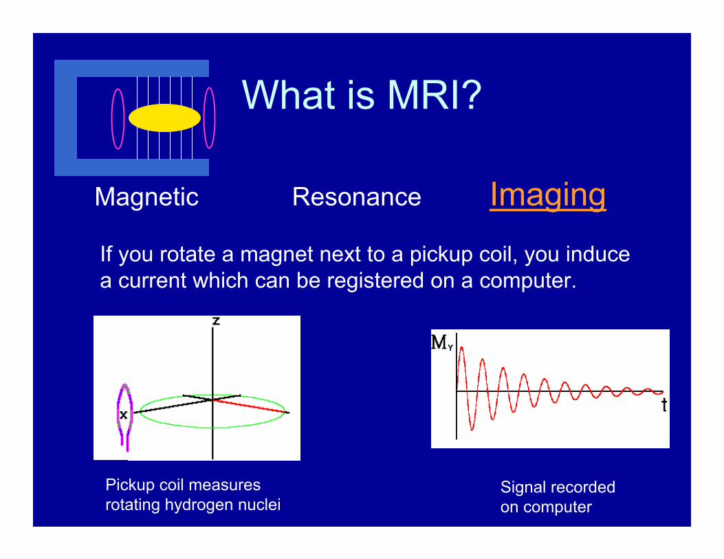
What is MRI?
Magnetic Resonance Imaging
If you rotate a magnet next to a pickup coil, you induce a current which can be registered on a computer.
Pickup coil measures rotating hydrogen nuclei
Signal recorded on computer

What is MRI?
Magnetic Resonance ImagingThe height of this signal is proportional to the number of hydrogen nuclei.
The frequency (=1/T) is proportional to the magnetic field strength.
Height
T

What is MRI?
Magnetic Resonance Imaging
By causing the magnetic field strength to change with position (using magnetic field gradient coils), we can make a magnetic resonance image.
Height
T
What is MRI?
Magnetic Resonance Imaging
The rate of signal decay with time depends upon the environment of the hydrogen nuclei. This can provide image contrast.
Decay of signal
Detail: When we look at a MR image, we are actually looking at signal largely from hydrogen nuclei on water and fat (lipid) molecules
What is MRI?
Magnetic Resonance Imaging

MRI Technology
• Four main parts of an MR Scanner are:
– Magnet– Magnetic field gradients– Radio-frequency transceiver– Computer

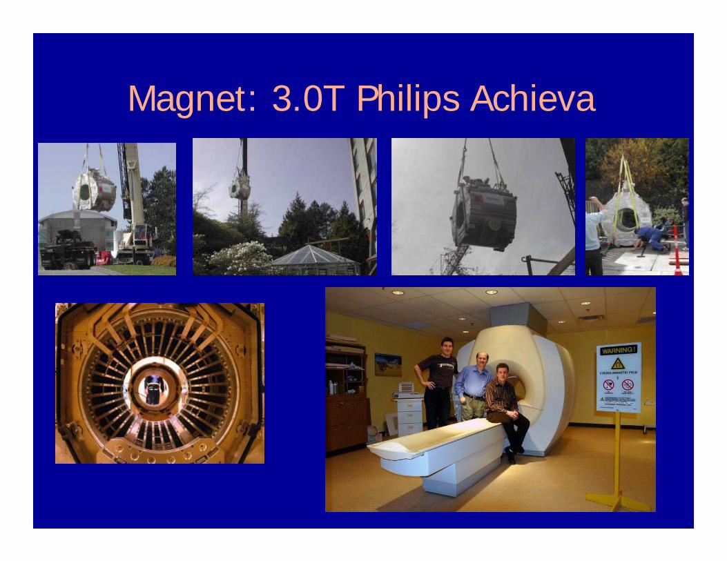
• Shortest bore (1.57m) 3.0T whole body
• Bore 60cm diameter
• Actively shielded (Two concentric magnets)
• 15 km of Niobium-Titanium wire kept at 4.2K
• 1400 litres liquid He, one fill/year
• This is a superconducting magnet
• Nausea experienced when moving in the 3.0T field
Magnet: 3.0T Philips Achieva
Magnet: 3.0T Philips Achieva

• Gradients spatially encode the MRI frequency
• There are three independent sets of gradient coils: x, y and z
• Not quiet – subjects need ear protection
• Can induce peripheral nerve stimulation
Magnetic Field Gradients

Gradients

• At 3.0 T, ωo = γB = 128 MHz
• 25 kW RF transmitter
• Up to 16 receiver channels
• Coils: Body, Head, Spine array, Cardiac, Torso, Knee, Flex L, M, S.
Radio Frequency Transeiver

Radio Frequency Transeiver
Copper room to keep out stray radio waves
Body Coil

MRIs (public) in British Columbia
• UBC • VGH (2)• St Pauls (2)• Children’s • Royal Columbian• Surrey• Victoria General (2)• Royal Jubilee
• Kelowna• Kamloops• Nanaimo• Prince George• Cranbrook, Penticton,
Trail• Abbotsford• Burnaby• White Rock

Why do MRI?1) MRI involves no ionizing radiation and is, to the best of our knowledge, harmless.
2) MRI gives rise to images with exquisite soft tissue contrast.
3) The contrast of an MR image can be altered by changing how the image is acquired.
4) MRI can detect water content changes, lesions, tumors, flowing blood, beating heart, tissue metabolites, microscopic structure, and much more.

What can we do with an MR Scanner?
1) Radiologists, cardiologists and neurologists use MR to aid in diagnosis and management of human disease.
2) Science and health professionals use MRI to research disease mechanisms and assess potential disease therapies.
3) Psychologists and psychiatrists use MRI to learn about how the brain works.

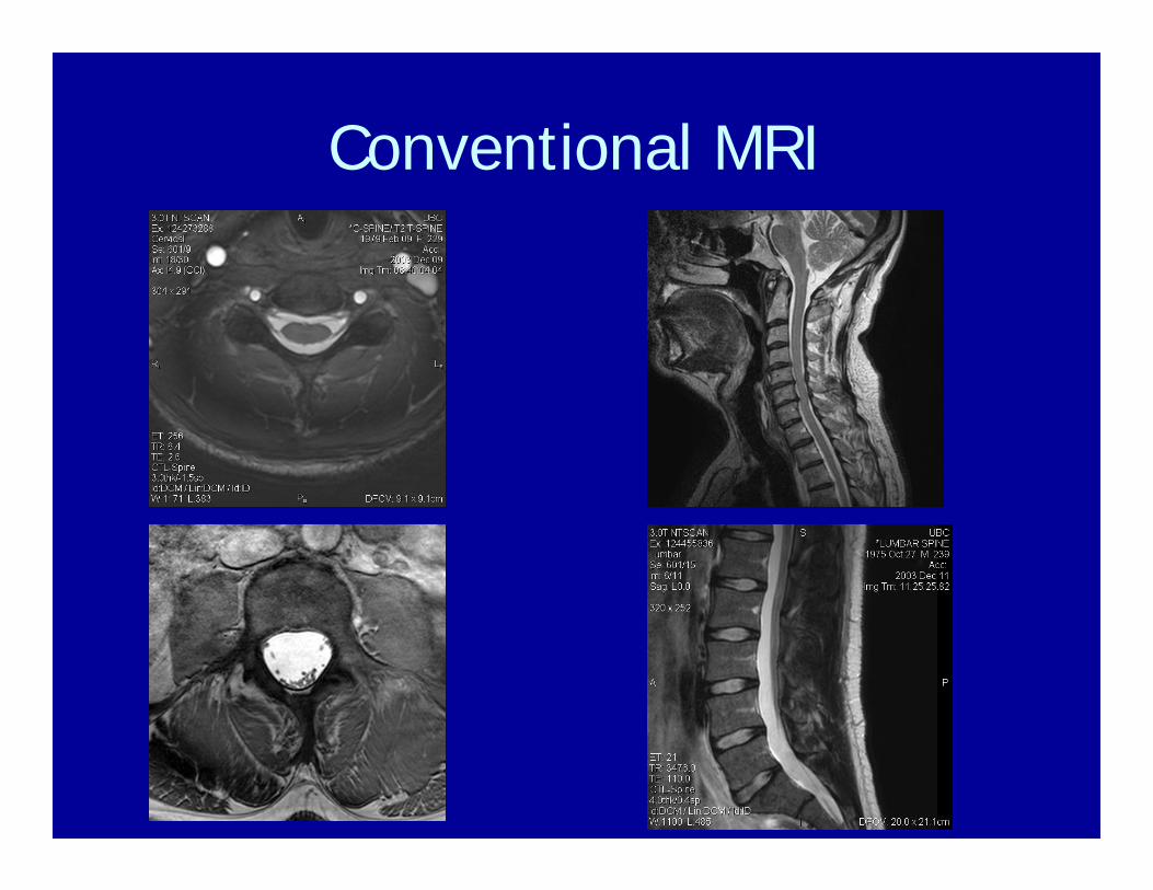
Conventional MRI
Healthy Volunteer

Conventional MRI
Patients with Multiple Sclerosis
Conventional MRI

Conventional MRI

Volumetric Imaging

Magnetic Resonance Angiography
X = Xo + Vt
Static tissue Flowing Blood

MR Angiogram from Brain
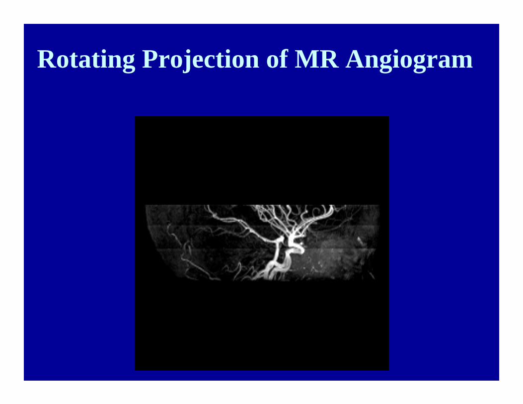
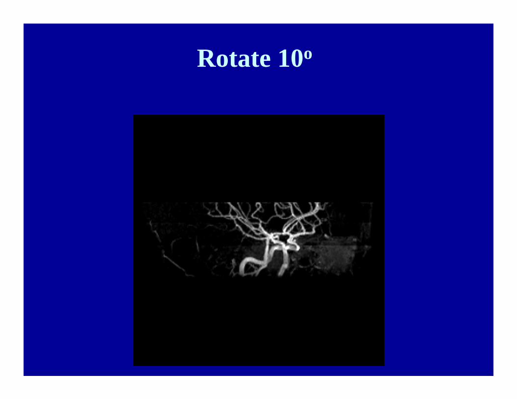
Rotating Projection of MR Angiogram

Rotate 10o

Rotate 10o
Rotate 10o

Rotate 10o

Rotate 10o

Rotate 10o

Rotate 10o
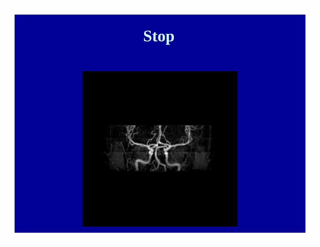
Stop

Flow Analysis of the Cerebral Aqueduct
Cerebral aqueduct

Image Across Aqueduct Plane
Aqueduct

-15
-10
-5
0
5
10
15
20
25
0 200 400 600 800
Time in Heart Cycle (msec)
Flow
(ml/m
in)
Flow Through the Cerebral Aqueduct

T = 10 msR = 13 µ
T = 60 msR = 33 µ
T = 200 msR = 60 µ
Diffusion
Diffusion is the random motion of water molecules due to their excess kinetic energy.
R
T = 1 msR = 4 µ
0
10
20
30
40
50
60
70
0 0.1 0.2 0.3 0.4 0.5
T ^(1/2) (s^(1/2))
R (m
icro
ns)
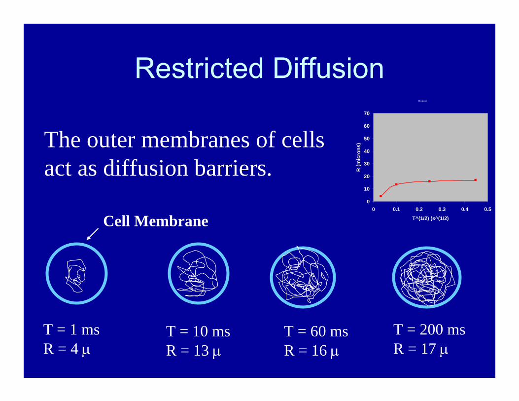
Restricted Diffusion
T = 1 msR = 4 µ
T = 10 msR = 13 µ
T = 60 msR = 16 µ
T = 200 msR = 17 µ
The outer membranes of cells act as diffusion barriers.
Cell Membrane
Distance
0
10
20
30
40
50
60
70
0 0.1 0.2 0.3 0.4 0.5
T^(1/2) (s^(1/2)
R (m
icro
ns)
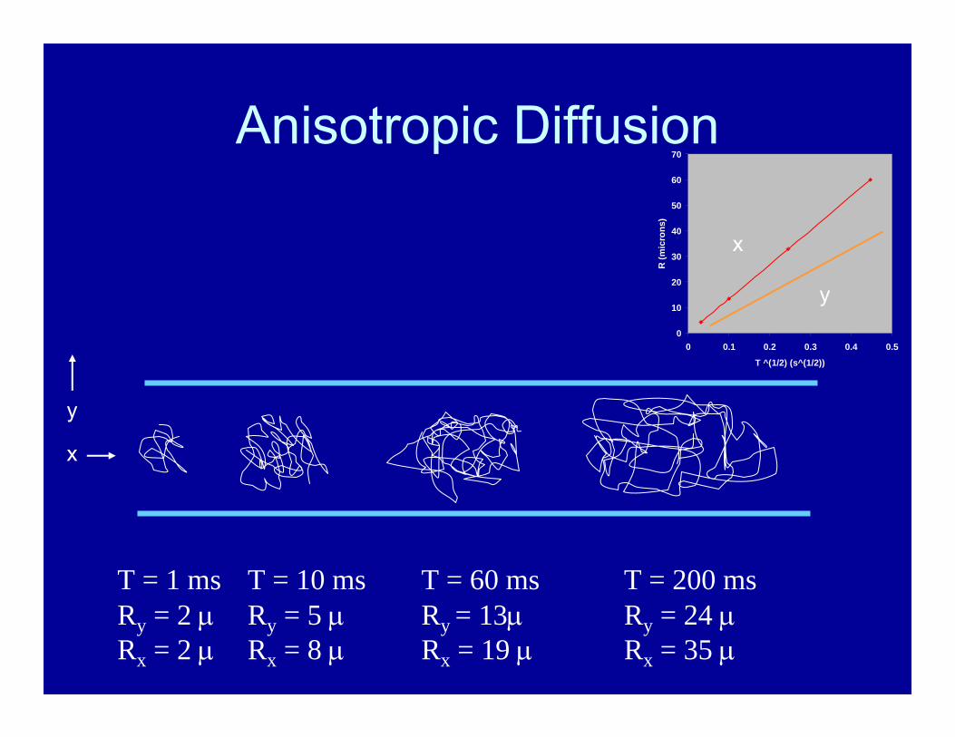
Anisotropic Diffusion
T = 1 msRy = 2 µRx = 2 µ
T = 200 msRy = 24 µRx = 35 µ
T = 10 msRy = 5 µRx = 8 µ
T = 60 msRy = 13µRx = 19 µ
0
10
20
30
40
50
60
70
0 0.1 0.2 0.3 0.4 0.5
T ^(1/2) (s^(1/2))
R (m
icro
ns)
x
y
x
y

Anisotropic Diffusion
0
10
20
30
40
50
60
70
0 0.1 0.2 0.3 0.4 0.5
T ^(1/2) (s^(1/2))
R (m
icro
ns)
xy
The white matter of brain contains many, many neuronal axons which are long tubes. Neurons cause water diffusion in brain to be anisotropic.
Using diffusion MRI, we can measure the direction of neuronal tracts in the brain.

Diffusion Tensor Imaging
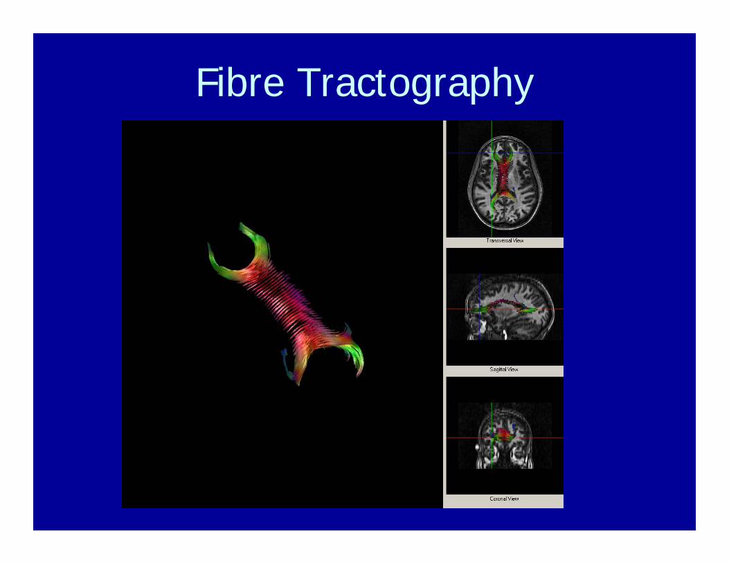
Fibre Tractography
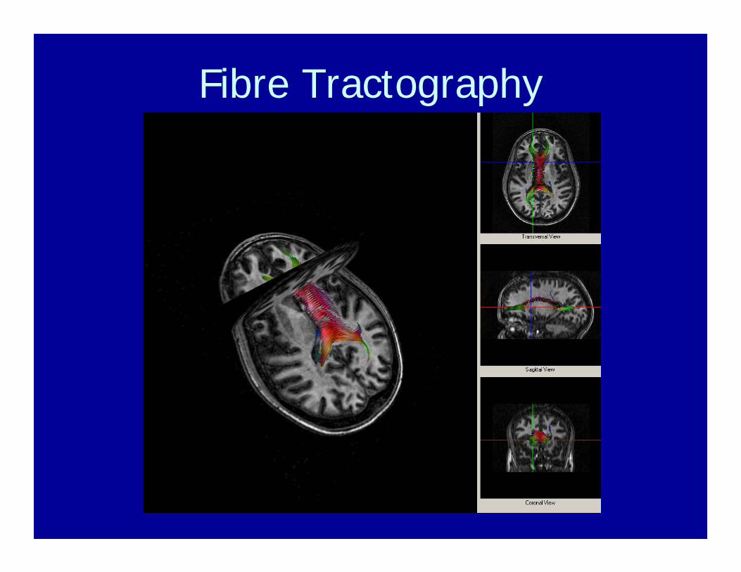
Fibre Tractography

Fibre Tractography

Functional MRI (fMRI)
When a region of brain is activated by some task:
1) Local metabolism uses some oxygen.
2) This causes an instantaneous drop in local O2concentration and also initiates increased blood flow to the region.
3) The result is a period of increased oxyhemoglobin to deoxyhemoglobin ratio.
4) fMRI produces images sensitive to this change in oxygen content.
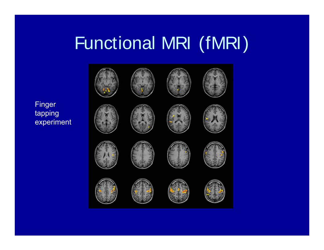
Functional MRI (fMRI)
Finger tapping experiment

Presentation Computer
Projector
Response Devices
Sense Head Coil
fMRI Equipment
Screen

fMRI Research at UBC 3T
There are 22 fMRI research projects underway on the UBC 3T magnet.
Most involve identification of brain regions used for various complex tasks.
Most of the investigators are from the Psychology, Psychiatry, Neurology and Opthalmology departments at UBC and SFU.

fMRI Research at UBC 3.0TFunctional Neuroimaging of Negative Effect in Elite SwimmersElton Ngan, Psychiatry
Abstract versus concrete thought during anagram completion: an fMRI investigationKalina Christoff, Psychology
Spatial and Temporal Aspects of Force Production in Parkinson’s Disease: Functional Magnetic Resonance Imaging StudyMartin McKeown, Neurology
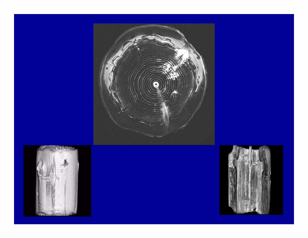
What is this?

Magnetic Resonance Spectroscopy
MRS measures signals from proton sites on several brain metabolites including:
N-acetyl-aspartate, phosphocreatine/creatine, choline, myo-inositoyl, glutamate/glutamine.
From the proton spectrum we can derive the concentrations of these metabolites.
This enables us to learn about the biochemistry of the brain.

Magnetic Resonance Spectroscopy

MR spectrum from creatine
creatine
Freq
S
MR spectrum
Peak frequency is determined by the ‘chemical shift’ of the molecular subunit (i.e. CH3 or CH2).
Peak area is determined by the concentration of contributing protons.

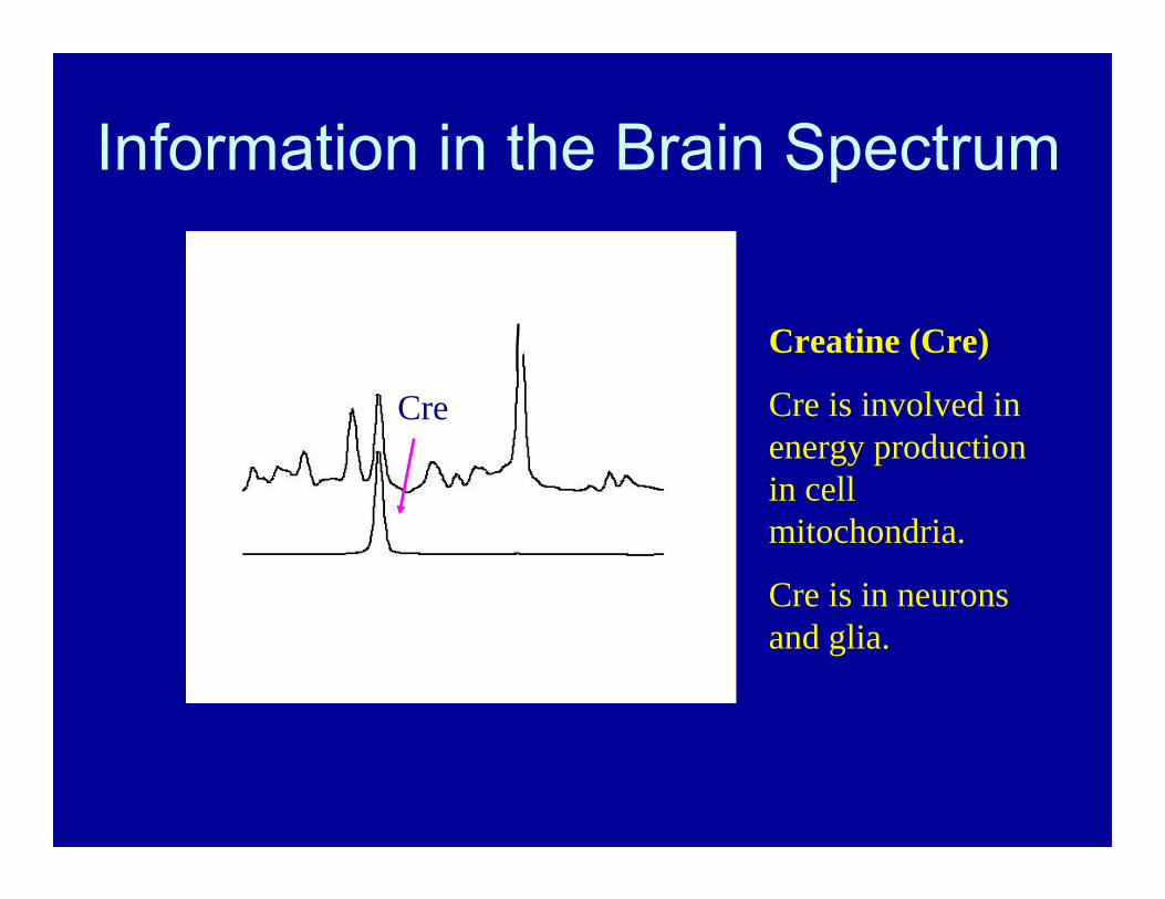
Information in the Brain Spectrum
NAA
CreCho
Ins
Spectrum composed of overlapping chemicals.
Signal area related to concentration of chemical.
NAA
CreChoIns
Creatine (Cre)
Cre is involved in energy production in cell mitochondria.
Cre is in neurons and glia.
Information in the Brain Spectrum

NAA
CreCho
Ins
Choline (Cho)
Cho takes part in membrane and neurotransmitter synthesis.
It is elevated in some tumors
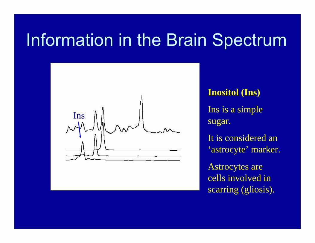
Information in the Brain Spectrum
NAA
CreChoIns
Inositol (Ins)
Ins is a simple sugar.
It is considered an ‘astrocyte’ marker.
Astrocytes are cells involved in scarring (gliosis).
Information in the Brain Spectrum

NAA
CreChoIns
N-Acetyl-Aspartate (NAA)
NAA is thought to be contained only in neurons.
“Neuronal Marker”
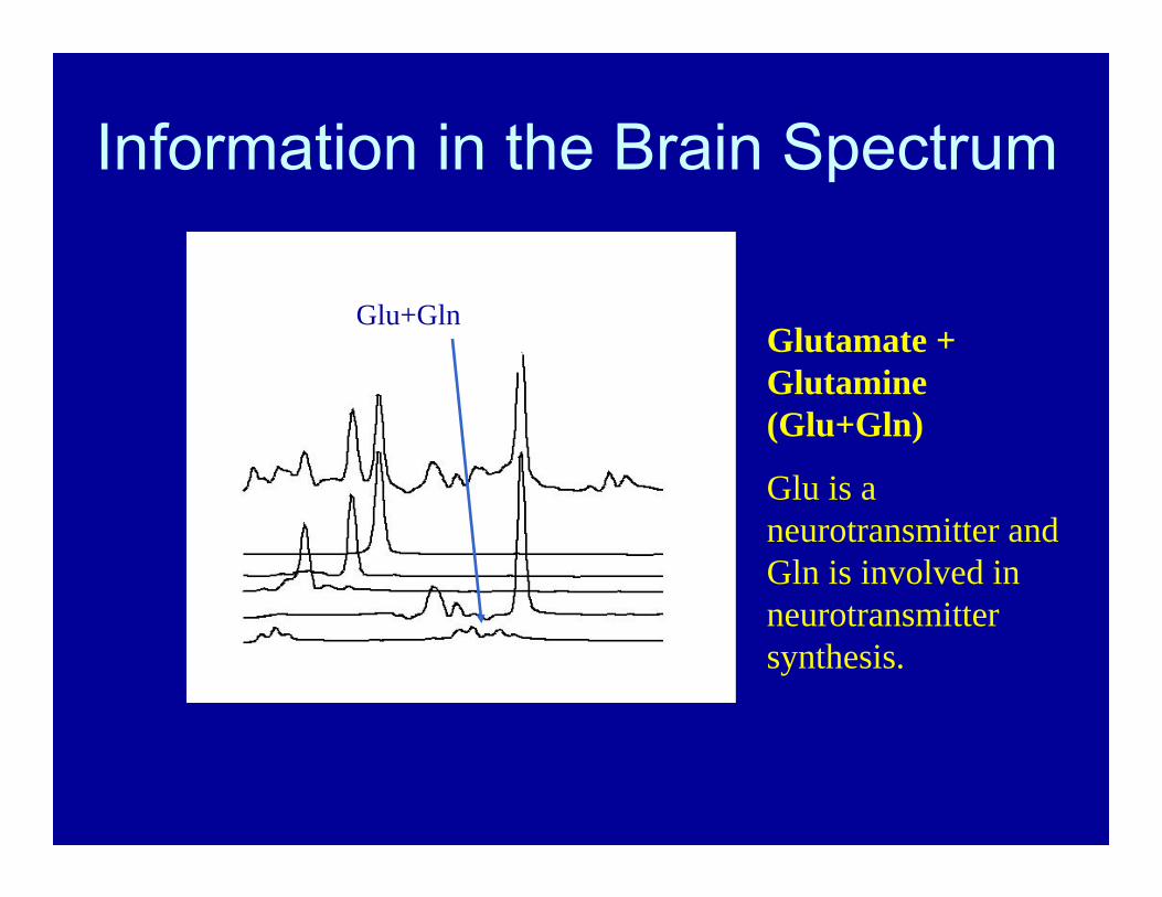
Information in the Brain Spectrum
NAA
CreChoIns
Glu+GlnGlutamate + Glutamine (Glu+Gln)
Glu is a neurotransmitter and Gln is involved in neurotransmitter synthesis.
Information in the Brain Spectrum
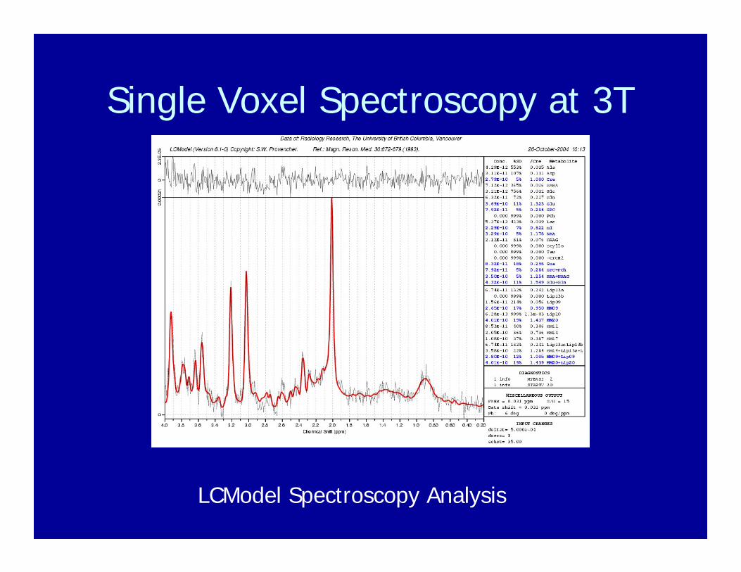
Single Voxel Spectroscopy at 3T
LCModel Spectroscopy Analysis
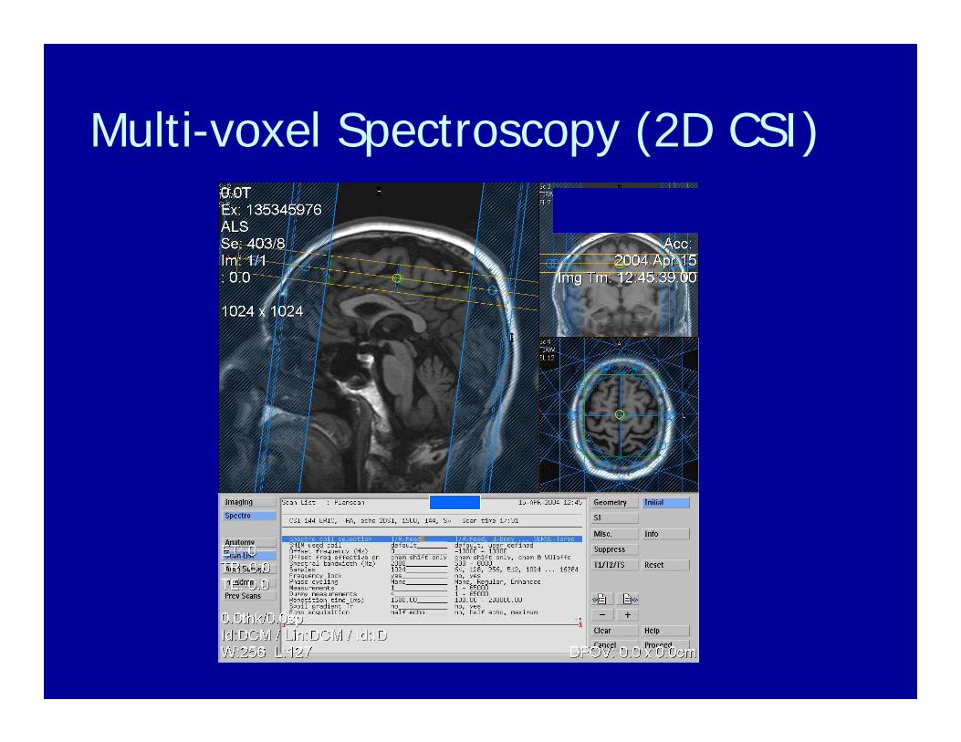
Multi-voxel Spectroscopy (2D CSI)

Multi-voxel Spectroscopy (2D CSI)
2D-SI Grid (FOV)
2D-SI Volume ofInterest
Spectra Display from ROI
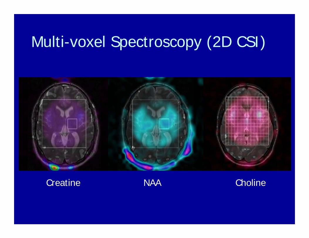
Multi-voxel Spectroscopy (2D CSI)
Creatine CholineNAA
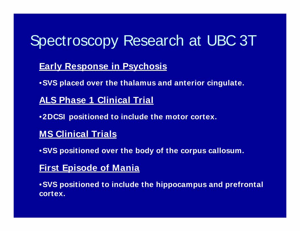
Spectroscopy Research at UBC 3T
Early Response in Psychosis
•SVS placed over the thalamus and anterior cingulate.
ALS Phase 1 Clinical Trial
•2DCSI positioned to include the motor cortex.
MS Clinical Trials
•SVS positioned over the body of the corpus callosum.
First Episode of Mania
•SVS positioned to include the hippocampus and prefrontal cortex.

Magnetic Resonance Imaging
• There are many 10’s of 1000’s of MRI’sworldwide.
• There are over 10,000 scientists worldwide using MRI for research.
• The technology of MRI is advancing very rapidly. A new MRI scanner is obsolete after 5 years
• It is a very exciting field to work in!

Who works with MRI?
• Physicists• Engineers• Chemists• Mathematicians• Biologists
• Radiologists• Neurologists• Cardiologists• Pathologists• Psychiatrists• Psychologists
The future of MRI is very bright!