Embed Size (px)
Citation preview

doi:10.1182/blood-2006-05-0194302006 108: 408-409
Alberto Mantovani Macrophage diversity and polarization: in vivo veritas
http://bloodjournal.hematologylibrary.org/content/108/2/408.full.htmlUpdated information and services can be found at:
Articles on similar topics can be found in the following Blood collections
http://bloodjournal.hematologylibrary.org/site/misc/rights.xhtml#repub_requestsInformation about reproducing this article in parts or in its entirety may be found online at:
http://bloodjournal.hematologylibrary.org/site/misc/rights.xhtml#reprintsInformation about ordering reprints may be found online at:
http://bloodjournal.hematologylibrary.org/site/subscriptions/index.xhtmlInformation about subscriptions and ASH membership may be found online at:
Copyright 2011 by The American Society of Hematology; all rights reserved.of Hematology, 2021 L St, NW, Suite 900, Washington DC 20036.Blood (print ISSN 0006-4971, online ISSN 1528-0020), is published weekly by the American Society
For personal use only. on April 29, 2014. at CAPES CONSORTIUM bloodjournal.hematologylibrary.orgFrom For personal use only. on April 29, 2014. at CAPES CONSORTIUM bloodjournal.hematologylibrary.orgFrom
on the development of methods that distin-guish human CECs derived from tumor andbone marrow. Elucidation of the origin of thepatients’ apoptotic CECs will clarify the role ofhost-versus-tumor differences in CEC re-sponses, which will have implications for thebroader significance of this study to othertypes of cancers and treatments. More exten-sive translational clinical trials that follow alarger number of patients for a longer-termfollow-up will determine whether a simpleblood test can be used to identify and monitorcancer patients who will benefit from novelantiangiogenic therapies. ■
REFERENCES
1. Kerbel RS, Kamen BA. The anti-angiogenic basis ofmetronomic chemotherapy. Nat Rev Cancer. 2004;4:423-436.
2. Colleoni M, Orlando L, Sanna G, et al. Metronomiclow-dose oral cyclophosphamide and methotrexate plusor minus thalidomide in metastatic breast cancer: antitu-mor activity and biological effects. Ann Oncol. 2006;17:232-238.
3. Shaked Y, Emmenegger U, Man S, et al. Optimal bio-logic dose of metronomic chemotherapy regimens is associ-ated with maximum antiangiogenic activity. Blood. 2005;106:3058-3061.
4. Jain RK. Normalization of tumor vasculature: an emerg-ing concept in antiangiogenic therapy. Science. 2005;307:58-62.
● ● ● IMMUNOBIOLOGY
Comment on Ghassabeh et al, page 575
Macrophage diversity and polarization:in vivo veritas----------------------------------------------------------------------------------------------------------------
Alberto Mantovani IST ITUTO CLINICO HUMANITAS, ITALY
Polarized activation of cells of the monocyte-macrophage lineage into M1 and M2cells is an operationally useful, simplified descriptor of the functional plasticity ofthese cells. Ghassabeh and colleagues now put to the test the actual in vivo validityand significance of the M1/M2 paradigm.
The microbial and cytokine milieu drivesmacrophages to express specialized and
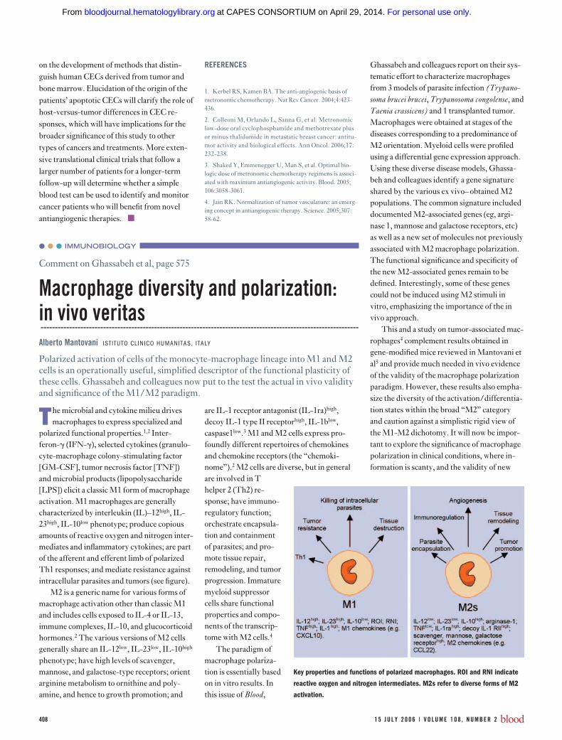
polarized functional properties.1,2 Inter-feron-� (IFN-�), selected cytokines (granulo-cyte-macrophage colony-stimulating factor[GM-CSF], tumor necrosis factor [TNF])and microbial products (lipopolysaccharide[LPS]) elicit a classic M1 form of macrophageactivation. M1 macrophages are generallycharacterized by interleukin (IL)–12high, IL-23high, IL-10low phenotype; produce copiousamounts of reactive oxygen and nitrogen inter-mediates and inflammatory cytokines; are partof the afferent and efferent limb of polarizedTh1 responses; and mediate resistance againstintracellular parasites and tumors (see figure).
M2 is a generic name for various forms ofmacrophage activation other than classic M1and includes cells exposed to IL-4 or IL-13,immune complexes, IL-10, and glucocorticoidhormones.2 The various versions of M2 cellsgenerally share an IL-12low, IL-23low, IL-10high
phenotype; have high levels of scavenger,mannose, and galactose-type receptors; orientarginine metabolism to ornithine and poly-amine, and hence to growth promotion; and
are IL-1 receptor antagonist (IL-1ra)high,decoy IL-1 type II receptorhigh, IL-1blow,caspase1low.3 M1 and M2 cells express pro-foundly different repertoires of chemokinesand chemokine receptors (the “chemoki-nome”).2 M2 cells are diverse, but in generalare involved in Thelper 2 (Th2) re-sponse; have immuno-regulatory function;orchestrate encapsula-tion and containmentof parasites; and pro-mote tissue repair,remodeling, and tumorprogression. Immaturemyeloid suppressorcells share functionalproperties and compo-nents of the transcrip-tome with M2 cells.4
The paradigm ofmacrophage polariza-tion is essentially basedon in vitro results. Inthis issue of Blood,
Ghassabeh and colleagues report on their sys-tematic effort to characterize macrophagesfrom 3 models of parasite infection (Trypano-soma brucei brucei, Trypanosoma congolense, andTaenia crassicens) and 1 transplanted tumor.Macrophages were obtained at stages of thediseases corresponding to a predominance ofM2 orientation. Myeloid cells were profiledusing a differential gene expression approach.Using these diverse disease models, Ghassa-beh and colleagues identify a gene signatureshared by the various ex vivo– obtained M2populations. The common signature includeddocumented M2-associated genes (eg, argi-nase 1, mannose and galactose receptors, etc)as well as a new set of molecules not previouslyassociated with M2 macrophage polarization.The functional significance and specificity ofthe new M2-associated genes remain to bedefined. Interestingly, some of these genescould not be induced using M2 stimuli invitro, emphasizing the importance of the invivo approach.
This and a study on tumor-associated mac-rophages4 complement results obtained ingene-modified mice reviewed in Mantovani etal5 and provide much needed in vivo evidenceof the validity of the macrophage polarizationparadigm. However, these results also empha-size the diversity of the activation/differentia-tion states within the broad “M2” categoryand caution against a simplistic rigid view ofthe M1-M2 dichotomy. It will now be impor-tant to explore the significance of macrophagepolarization in clinical conditions, where in-formation is scanty, and the validity of new
Key properties and functions of polarized macrophages. ROI and RNI indicate
reactive oxygen and nitrogen intermediates. M2s refer to diverse forms of M2
activation.
408 1 5 J U L Y 2 0 0 6 I V O L U M E 1 0 8 , N U M B E R 2 blood
For personal use only. on April 29, 2014. at CAPES CONSORTIUM bloodjournal.hematologylibrary.orgFrom

molecules as markers and, possibly, targets fortherapeutic intervention. ■
REFERENCES1. Gordon S. Alternative activation of macrophages. NatRev Immunol. 2003;3:23-35.
2. Mantovani A, Sica A, Sozzani S, Allavena P, Vecchi A,Locati M. The chemokine system in diverse forms of mac-
rophage activation and polarization. Trends Immunol.2004;25:677-686.
3. Dinarello CA. Blocking IL-1 in systemic inflammation. JExp Med. 2005;201:1355-1359.
4. Biswas SK, Gangi L, Paul S, et al. A distinct and uniquetranscriptional programme expressed by tumor-associatedmacrophages: defective NF-kB and enhanced IRF-3/STAT1 activation. Blood. 2006;107:2112-2122.
5. Mantovani A, Sica A, Locati M. Macrophage polariza-tion comes of age. Immunity. 2005;23:344-346.
● ● ● IMMUNOBIOLOGY
Comment on Cornish et al, page 600
Time to restore individual rights for IL-2and IL-15?----------------------------------------------------------------------------------------------------------------
Yutaka Tagaya NATIONAL CANCER INSTITUTE
IL-2 and IL-15 transduce distinct signals in the same cell despite their sharing ofall signaling receptor components.
The immune system uses many cytokines toaccomplish diverse functions. It is some-
times amazing that the system can avoid con-fusion, especially given that many cytokinesshare receptor and signaling components.While most cytokines share only part of thesignaling receptor components, the case ofinterleukin (IL)–2 and IL-15 seems uniquebecause they share all the signaling receptorcomponents (ie, CD122 and CD132). Each ofthese cytokines requires a private � chain(CD25 and IL-15R�) for stable binding to theCD122/132 complexes in vivo, and these �
chains have different cellular distribution,which may explain why IL-2�/� and IL-15�/� mice have totally different phenotypes.Thus, the question of whether IL-2 and IL-15transduce distinct signals at a single-cell levelhas been controversial. Ku et al1 demonstratedthat IL-2 and IL-15 display contrasting effectson the survival of memory-phenotype CD8 Tcells, which could indicate signaling differ-ences between these two cytokines. However,the basal interpretation of this study has beenchallenged recently by Boyman et al,2 whodiscovered that the anti–IL-2 antibody used inthe original study bound IL-2 in vivo but con-verted it into a “superagonist” by formingstable IL-2/anti–IL-2 complexes, rather thanneutralizing the effect of IL-2. Such superago-nistic IL-2 can expand memory phenotypeCD8 T cells even in IL-15�/� mice; thus, theaction of these complexes seems similar to thatof IL-15 in this context.2 So do these 2 factors
have distinct in vivo functions only becausethey act on different target cells? It has beensuggested that IL-15R� transduces its ownsignal upon binding by IL-15, but the processdoes not seem to operate in vivo, because IL-15�/� and IL-15R��/� mice did not mani-fest any defects that are not seen with theCD122�/� mice. Moreover, Kovanen et al3
demonstrated thatIL-2 and IL-15 (andIL-7) induce very simi-lar gene activation pro-file using cDNA mi-croarrays, basicallysuggesting that theIL-2/IL-15 receptorsdo not distinguishthese 2 cytokines func-tionally at a single-celllevel.
In this issue ofBlood, Cornish andcolleagues present el-egant and interestinginsights into this ques-tion. They demon-strated that IL-15 pri-marily activatesproliferation (and thusacts as a mitogen),whereas IL-2 activatesmore diverse pathwaysincluding prolifera-tion, amino acid up-
take, and protein synthesis (hence, IL-2 is agenuine growth factor) in ex vivo CD8 T cells.IL-2– cultured and IL-15– cultured CD8 Tcells consistently exhibited distinct morphol-ogy and expressed different cell-surface anti-gens. They also demonstrated that such differ-ences might arise from differential kinetics ofthe signaling through the Akt (PKB)–PDK1pathway (see figure). These observations donot seem to contradict the earlier microarraystudy,3 because the difference in the signalingkinetics may be translated into cellular eventsat later points that the microarray analyses didnot cover. While the mechanism underlyingthese observations needs to be investigated,Cornish et al’s study undoubtedly sheds newlight on how we interpret and design in vivoexperiments. In parallel, Dubois et al4 andother groups demonstrated that IL-15 is pre-sented to target cells in trans, which may be thedominant way IL-15 operates in vivo.5 It isreasonable to assume that trans-presentedIL-15 may signal very differently from IL-2,since such IL-15 signals inevitably includeelements coming from cell-to-cell contact.Finally, IL-15 is being considered as a clinicaltherapeutic agent as a replacement for IL-2because of its less-toxic nature, but the currentstudy poses a challenge to this concept and
Differences of IL-2 and IL-15 signaling in CD8 T cells.
blood 1 5 J U L Y 2 0 0 6 I V O L U M E 1 0 8 , N U M B E R 2 4 0 9
For personal use only. on April 29, 2014. at CAPES CONSORTIUM bloodjournal.hematologylibrary.orgFrom