Embed Size (px)
Citation preview
Low Intensity Pulsed Ultrasound (LIPUS) © Tim Watson (2014) Page 1
Low Intensity Pulsed Ultrasound (LIPUS)
The application of ultrasound energy at much lower levels than is the current clinical norm is starting to
gain ground as a therapeutic possibility. Clearly the applied energy is the same, it is the ‘dose’ which is
different – most importantly, the intensity (W cm-2) – which is MUCH lower – typically 2 or 3 times
lower than the lowest setting on most regular clinical machines, with the most common application
being at 30mW cm-2 (which is 0.03 W cm-2).
At the present time, the strongest evidence for the clinical application of this modality is in relation to
fracture healing, which is the area that this information sheet will concentrate on. It is argued – quite
reasonably – that IF it works this well on bone lesions, then it should also be effective on other soft
tissue lesions (ligament, tendon etc) but at the present time, the published research in this field is
limited.
Examples of LIPUS devices available in the UK are illustrated below :
Exogen Device (Smith & Nephew) Osteotron Device (EMS Physio)
LIPUS vs Regular Therapy Ultrasound
Ultrasound (US) is a form of MECHANICAL energy. Mechanical
vibration at increasing frequencies is known as sound energy. The
normal human sound range is from 16Hz to something approaching
15-20,000 Hz (in children and young adults). Beyond this upper
limit, the mechanical vibration is known as ULTRASOUND. The
frequencies used in therapy are typically between 1.0 and 3.0 MHz
(1MHz = 1 million cycles per second).
Sound waves are LONGITUDINAL waves consisting of areas of
COMPRESSION and RAREFACTION. Particles of a material, when
exposed to a sound wave will oscillate about a fixed point rather
than move with the wave itself. As the energy within the sound
wave is passed to the material, it will cause oscillation of the
particles of that material. Clearly any increase in the molecular
vibration in the tissue can result in heat generation, and ultrasound can be used to produce thermal

Low Intensity Pulsed Ultrasound (LIPUS) © Tim Watson (2014) Page 2
changes in the tissues, though current usage in therapy does not focus on this phenomenon (Williams
1987, Baker et al 2001, ter Haar 1999, Nussbaum 1997, Watson 2000, 2008).
In addition to thermal changes, the vibration of the tissues appears to have effects which are generally
considered to be 'non thermal' in nature, though, as with other modalities (e.g. Pulsed Shortwave)
there must be a thermal component however small.
Low Intensity Pulsed Ultrasound (LIPUS) is clearly ultrasound energy, but delivered at a much lower
intensity (W cm-2) than traditional ultrasound energy. There are other differences with the output of
LIPUS devices, but this the most obvious issue.
Whilst a typical therapy machine will offer an operating frequency choice of 1MHz or 3MHz, the LIPUS
fracture healing evidence has been generated almost exclusively at 1.5MHz. Both the Exogen and
Osteotron devices offer LIPUS at this frequency, though the Osteotron device also offers a 0.75MHz
(optional extra) probe which, it is suggested, would be effective for the more deep seated lesions (e.g.
femur). No evidence has been identified for clinical trials with LIPUS at frequencies other than 1.5MHz,
and therefore it is currently not known whether 'other' frequencies are effective, not as effective, or
possibly more effective.
BNR - inequality of the Ultrasound Beam
As the beam emerges from the treatment head, the energy across the beam profile is not 'even' - there
are areas of higher and areas a lower intensity. When the intensity is set on a therapy ultrasound
device, it would certainly not be the case that every part of the beam, even as it emerges, would
actually be at that intensity. The 'inequality' of the beam strength - or the 'beam unevenness' is
represented by the Beam Nonuniformity Ratio (or BNR). In the ideal world this value would be, or be
close to 1.0 (which means that there is equal power across the entire beam profile. In reality, most
therapy ultrasound machines will have a typical BNR of between 4 and 6 (the smaller the better). If the
BNR has a value of 5 for
example, it would mean
that the 'strongest' parts
of the beam would be at 5
x greater power than the
mean power of the beam.
One of the reasons for
needing to employ a
'moving treatment head'
application technique is to
ensure that the 'strongest' parts of the beam are not always applied to
the same part of the tissue - the treatment head movement helps to
'even out' the beam inequality.
A 'typical' beam plot can be seen in the diagram above and examples of
2 'real' beam X sectional plots from different transducers (at 3 MHz)
from the Johns et al (2007) paper are illustrated (left) .
Example of an Ultrasound Beam Plot
Low Intensity Pulsed Ultrasound (LIPUS) © Tim Watson (2014) Page 3
A recent analysis of clinical machines (Johns et al, 2007) identified that the BNR was in the range, 2.79-
5.85 at 1 MHz and ranged from 2.51 to 4.56 for the 3.3MHz devices tested.
If (as with LIPUS treatments for fractures, the treatment head needs to kept stationary for prolonged
periods (typically 20 minutes), a LOW BNR is an essential safety issue.
The LIPUS devices for fracture healing have a low BNR - the Exogen being 4.0 (max) and the Osteotron
being 3.0 or 3.5 depending on which applicator is employed.
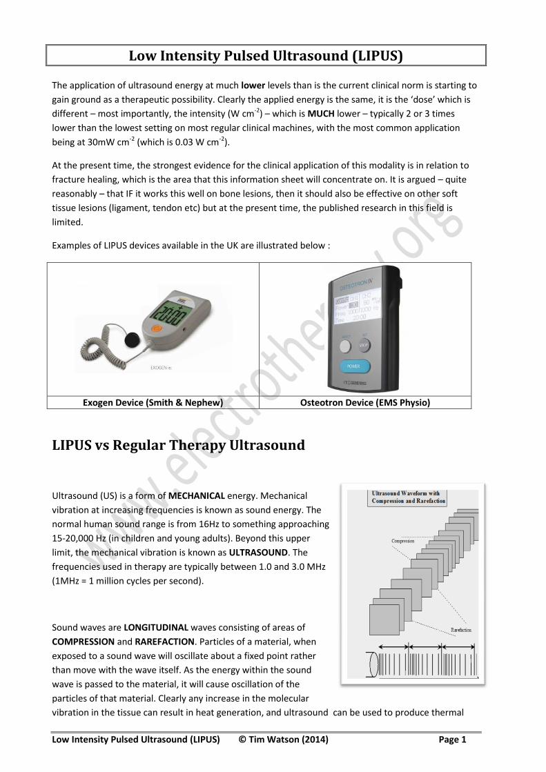
Ultrasound Pulsing
Ultrasound on standard therapy machines can be delivered in a continuous or a pulsed mode, with
pulse mode variations on many, if not all machines. LIPUS devices, having a narrow clinical application,
tend not to offer such a wide range of pulse
options.
Typical pulse ratios are 1:1 and 1:4 though others
are available. In 1:1 mode, the machine offers an
output for 2ms followed by 2ms rest. In 1:4 mode,
the 2ms output is followed by an 8ms rest period.
The adjacent diagram illustrates the effect of
varying the pulse ratio.
Until recently, the pulse duration (the time during
which the machine is on) was almost exclusively
2ms (2 thousandths of a second) with a variable off
period. Some machines now offer a variable on
time though whether this is of clinical significance has yet to be determined.
Some manufacturers describe their pulsing in terms of a percentage rather than a ratio (1:1 = 50% 1:4 =
20% etc). The pulse ratio - duty cycle percentage equivalence is shown in the table below:
Mode Pulse Ratio Duty Cycle
Continuous N/A 100%
Pulsed 1:1 50%
1:2 33%
1:3 25%
1:4 20%
1:9 10%
LIPUS machines typically deliver their ultrasound pulsed at 20% (1:4) and at 1000Hz (1kHz) - therefore
there are 1000 cycles per second, each cycle is thus 1/1000 of a second (i.e. a millisecond). In that
millisecond, there will be 20% ultrasound and 80% not ultrasound. The ultrasound 'on' cycle will
therefore be 0.2 milliseconds (200 microseconds or 200s) followed by a 'gap' of 0.8 milliseconds (or
800 s). The Osteotron device additionally offers a 100Hz pulse option.

Low Intensity Pulsed Ultrasound (LIPUS) © Tim Watson (2014) Page 4
1kHz pulsing with LIPUS devices
Ultrasound Intensity
The intensity (strength in general terms - power density to be very specific) at which ultrasound is
applied in regular clinical applications ranges from about 0.1 through to 1.0 W cm-2. Some applications
(researched and evidenced as being effective) will use intensities of up to 2.5 W cm-2, and although not
'common' is certainly deemed to be a safe application mode and can be very effective in some clinical
circumstances.
The power density clearly represents how much power is being applied (the Watts) and how
concentrated it is (the cm2).
With the LIPUS devices for fracture healing applications, as mentioned in the introduction, one of the
key differences is that the power density is much LOWER than with the traditional ultrasound
treatments. Almost all of the LIPUS research has used 0.03 W cm-2 (which is sometimes expressed as
30mW cm-2).
A typical therapy machine is not able to be set at power densities below 0.1 W cm-2 . It is not therefore
know whether a standard therapy ultrasound machine can deliver a low enough 'dose' to be effective in
this clinical area. At the moment, the available evidence would suggest that the sound energy that it
delivers would be 'too strong' for the job in hand. Whilst there have been some (limited) animal
experimentation (e.g. Warden et al 2006), this approach has yet to be formally evaluated in a human
patient clinical trial.
The Exogen device (patient, take home, portable version) offers no power density options (it is always at
30mW cm-2) whereas the Osteotron device offers additional power options at 45 and 60 mW cm-2 -
though as for as the clinical evidence goes, none can be currently identified which supports the use of
these higher dose options. It is suggested that they might / will be more effective for the deeper bone
problems - which has logic, just lacks evidence at the present time.
A recent study by Fung et al (2012) (animal model - rat femoral fracture) set out to compare a 30mW
cm-2 and a 150mW cm-2 treatment together with a control condition. The 30mW dose was shown to be
clearly advantageous over both the 150mW and the control group recovery.

Low Intensity Pulsed Ultrasound (LIPUS) © Tim Watson (2014) Page 5
Ultrasound for Fracture Healing : Mechanism of Action
A considerable amount of research has been carried out to try and identify the mechanism by which
LIPUS ultrasound applications can 'enhance' fracture repair. Necessarily, a high proportion of these
studies are based on cell, lab and animal research, but they have served to provide an ever increasing
picture of what is happening. It is suggested that this research area will continue to develop, and it is
highly likely that additional information will continue to be published for some time to some yet - which
will either add 'new pathways' to the existing ones or provide additional transduction or cytokine or
gene expression data. It is appreciated that for many therapists, this is not the most important part of
the 'story' and thus the following section will provide a summary rather than a fully explanation!
Useful summary and review papers can be found in : Claes and Willie (2007); Della Roca (2009); Jingushi
(2009); Lu et al (2009); Padilla et al (2014) and Warden (2003).
The mechanisms which have been sufficiently well evidenced to justify their inclusion are listed below
with some key references
Jungushi et al (2007) suggest that LIPUS is responsible for cell differentiation effects as a primary
mechanism of effect rather than cellular upregulation or proliferation. They identify increased matrix
synthesis, earlier expression of Type II procollagen and also prostaglandin expression and an increased
chondrocyte differentiation all being associated with LIPUS exposure. This results in an earlier callus
mass, though not an increased (volume) of callus.
Other papers do appear to provide evidence for an increase in cell upregulation and proliferation. It is
generally considered that the LIPUS energy has an effect at cell membrane level where
mechanoreceptors (integrins) respond and result in various upregulation and expressions.
COX2 (Naruse et al, 2010) expression is increased. This is essential in the PGE2 pathway (it is necessary
for PGE2 production), and both COX2 and PGE2 are known to be essential in fracture repair. Leung et al
(2004) demonstrated increased expression of VEGF, a strong angiogenic stimulator and both Naruse et
al (2010) and Sant Anna et al (2005) demonstrated increased expression of BMP2; BMP4; BMP6 and
BMP7 (linked with TGFβ) and linked to differentiation of stem cells (mesynchymal cells) into bone and
cartilage. (BMP = Bone Morphogenic Protein).
There is an increased cell division in periosteal cells in the inflammatory stage (Leung et al, 2004) and in
increased differentiation of chondrocytes triggered via a TGFβ pathway (as above) (Ebisawa et al 2004).
Upregulation of endochondral ossification (Kokubu et al, 1999, Sena et al, 2005) Increased osteoblast
differentiation (Lai et al, 2010), increased bone mineralisation (Leung et al, 2004) and increased rate of
callus remodelling (Freeman et al 2009) have all been demonstrated as being associated with LIPUS
exposure.
The Della Rocca (2009) review includes some additional information relating these and other gene
expressions to the fracture healing pathway and the Padilla et al (2014) paper provides a useful
overview of the evidence to date.
Other studies which contribute to the evidence base in this area include Nolte et al (2001) who identify
an increase in ossification activity, Ryaby et al (1991) with increased TGFβ synthesis. The increased
expression of Type II collages from the chondrocytes is linked to a TGFβ pathway (Mukai et al, 2005).
Low Intensity Pulsed Ultrasound (LIPUS) © Tim Watson (2014) Page 6
The Kokubu et al (1999) study reiterates the essential contribution made by both COX2 and PGE2 to the
fracture healing process. COX2 regulates PGE2 production, reinforced by the results obtained by Tang et
al (2006). Both Reher et al (2002) and Warden et al (2001) identify NO and pGE2 pathways as being
significantly involved in LIPUS fracture healing pathways.
This would be consistent with other proposed mechanisms of ultrasound action (ter Haar 1999) and the
relationship between the use of NSAID’s and tissue repair following injury.
Other elements described and identified include increased proliferation of periosteal cells, increased
calcitonin expression, VEGF expression and alkaline phosphatase production (Leung et al, 2004). Wang
et al (2004) argue that LIPUS exposure, resulting in increased VEGF, NO and HIF-1 (hypoxia inducible
factor 1) expression is an additional component of the stimulating pathway.
Without any further consideration of the detail of these mechanisms, it is clear that LIPUS energy,
delivered to the fracture area results in an increased expression of several critical chemical mediators,
growth factors and cytokines which have an essential role to play in the normal fracture healing
sequence. It is evidenced that the LIPUS does not change the events of fracture repair but rather
increases the expression of these various factors, and thereby stimulates the normal sequence. The
resulting increased production of collagen, differentiation of cell types and change in callus production
appears therefore to be a secondary effect as a result of the expression and upregulation functions.
Ultrasound for Fracture Healing : Clinical Issues
Numerous recent papers have identified the benefits of using therapeutic ultrasound for both normally
healing (fresh) fractures and those that demonstrate either a delayed union or non union (e.g. Mayr et
al 2000, Busse et al 2002, Warden et al 1999). Ultrasound has been historically considered to be a
contraindication is these circumstances, though the exact reason for this remains unclear. Given the
volume and quality of the published evidence, it would be entirely inappropriate for fractures to remain
on the contraindication list.
NICE Guidance :
NICE provide numerous documents (freely available from their website - listed with the references)
which identify the potential value of LIPS from both fresh fractures and those with delayed and no
union. They concentrate on the established dose (1.5MHz; pulse 200s; delivered at 20% duty cycle
(1kHz); 30 mW cm-2; 20 minutes daily, usually as a patient delivered treatment (home based) with
coupling gel as a contact medium between the treatment applicator and the skin.
Their 2010 review included a meta analysis of 1910 patients from one previous meta analysis (13
RCT's)(Busse et al, 2006) plus an additional 4 RCT's not included in the first meta analysis (Heckman et
al, 1994; Emami et al, 1999; Leung et al, 2004; Ricardo, 2006), a comparative study (Coughlin et al,
2008) and a case series (Mayr et al, 2000). Full details are provided in the NICE document together with
other research which they excluded for this work.
In their 2013 update, NICE appear to provide a paper supporting a specific machine (the Exogen device)
though they do (in the detail) point out that this is not the only device available to deliver LIPUS. The
guidance comes to the same general conclusion as the 2010 work, though there is some additional

Low Intensity Pulsed Ultrasound (LIPUS) © Tim Watson (2014) Page 7
economic analysis which is insightful. "The case for adopting the Exogen ultrasound bone healing
system to treat long bone fractures with non union (failure to heal after 9 months) is supported by the
clinical evidence which shows high rates of fracture healing. The Exogen ultrasound bone healing
system to treat long bone fractures with non union is associated with an estimated cost saving of £1164
per patient compared with current management, through avoiding surgery" (NICE 2013). The full paper
(open access) clearly provides a more detailed analysis.
The Busse et al (2006) meta analysis (13 RCT's) reported an overall reduction in mean healing time of
34% (CI 21 - 44%) for patients receiving LIPUS compared with a sham treatment. The Heckman study
(1994) involved tibial fractures, 33 patients treated with LIPUS and 34 in a sham group. They reported a
significant increased rate of healing (96 days LIPUS group, 54 days sham group). The Leung et al (2004)
study with 30 patients (16 LIPUS, 14 sham) with tibial fractures report an average time to full weight
bearing of 9.3 weeks in the treated group and 15.5 weeks in the sham group (significant difference). The
Coughlin et al (2008) study also involved 30 patients undergoing subtalar arthrodesis (15 LIPUS, 15
standard management) reported a significant difference in the number of patients healed at 9 weeks -
63% in the LIPUS group compared with 43% in the standard management group.
The Mayr et al (2000) review (case series) involved 1317 patients all of whom received LIPUS and an
89% overall healing rate, subdivided into 91% mean healing rate for the delayed unions and 86% for the
non unions.
Some of these studies are considered in further detail below. The point here is that the NICE analysis of
fracture healing rates from the available evidence is totally coincident with my own work. The NICE
analysis also includes sections on return to function, safety and infection. NICE do state that although
the data was derived from RCT's, some was of poor quality (low patient numbers, lack of blinding,
publication bias).
The NICE conclusions (phrased differently for the patient guidance and the 'medical' guidance suggests
that this treatment may provide significant benefit for patients with non union and delayed healing
fractures in whom surgical intervention may be avoided and recovery of limb function may be
accelerated. It is advised that non union and delayed healing long bone fractures, particularly of the
tibia would be most likely to benefit from this treatment. It is considered that this treatment had the
potential to be cost saving compared with standard management. Additionally, it is suggested that this
treatment may be of some benefit in patients with fresh fractures, though there were concerns with
regards the cost implications.
Clinical Trial Information
A recent systematic review and meta-analysis (Busse et al 2002) (as reported in the NICE section above)
has carefully considered the evidence in respect to the effect of low intensity pulsed ultrasound on the
time to fracture healing. They conclude that the evidence from randomised trials where the data could
be pooled (3 studies, 158 fractures) that the time to fracture healing was significantly reduced in the
ultrasound treated groups than in the control groups and the mean difference in healing time was 64
days.

Low Intensity Pulsed Ultrasound (LIPUS) © Tim Watson (2014) Page 8
Warden et al (1999) published a review paper concluded that from animal and human studies, the use
of ultrasound could accelerate the rate of fracture repair by a factor of 1.6.
Heckman et al (1994) demonstrated a 38% reduction in the healing time for tibial fractures using a
LIPUS device whilst Kristiansen et al (1997) demonstrated a 30% acceleration in healing for fractures of
the radius.
Jensen (1998) identifies the beneficial effects of ultrasound in the treatment (as opposed to the
diagnosis) of stress fractures with an overall success rate of 96%. The report fails to identify all relevant
data for consideration and must therefore be considered with some caution in terms of ‘quality
evidence’.
Mayr et al (2000) report a series of outcomes when using low intensity pulsed ultrasound for patients
with delayed unions (n=951) and non unions (n=366). The overall success rate for the delayed unions
was 91% for the delayed and 86% for the non unions.
The authors undertook an interesting stratified analysis of their patients, and identified that those who
were using non steroidal anti inflammatory drugs, calcium channel blockers or steroids had a less
favourable outcome, a finding that could be considered to be consistent with several research
publications that have tried to identify the mechanism by which the ultrasound could bring about
fracture healing acceleration and other wider research concerning the adverse influence of NSAID’s on
tissue repair (e.g. Tsai et al 2004, Evans & Butcher2004).
A more recent paper (Rutten et al 2007) demonstrated a 73% union rate in their group of tibial non
unions (n=71 patients) which is clearly much better than the most optimistic spontaneous healing rate
in this group (usually cited at between 5 and 30%).
The use of such low doses has been shown to result in non significant increases in tissue temperature.
Using higher ultrasound doses could have an adverse effect on the fracture healing process and the
low intensity pulsed system is considered to be effective and safe for this patient group. Reher et al
(1997) demonstrated a stimulative effect at low dose (0.1 W cm-2) whilst an inhibitory effect at a higher
dose (1 – 2 W cm-2). Chang et al (2002) demonstrated that the effect of low intensity pulsed ultrasound
in these circumstances was achieved by non thermal mechanisms rather than as a phenomenon
secondary to thermal effects.
Both Tis et al (2002) and Sakurakichi et al (2004) have evaluated the use of ultrasound as a component
of treatment (in an animal model) during distraction osteogenesis, and both have demonstrated
significant benefits. Cook et al (2001) have demonstrated similar benefits following spinal fusion surgery
and Tanzer et al (2001) have shown that the use of ultrasound in combination with porous
intramedullary implants is also beneficial. There are many other studies concerning the use of US and
bone repair, but essentially the published work shows a consistent benefit, and the use of low intensity
pulsed ultrasound for patients with bone related disorders, including normally healing fractures, stress
fractures, delayed and non unions and as a post surgical intervention should be considered positively.
One study (Schortinghuis et al 2004) that employed the SAFHS ultrasound system yet failed to
demonstrate a significant effect (following deliberate bone injury – rat model) is probably related to the
additional inclusion of a PTFE membrane – a GoreTex® like material). This would almost certainly not
enable adequate ultrasound energy transmission due to the porous nature of the material, and the
consequent air trapping, leading to ultrasound energy reflection.
Low Intensity Pulsed Ultrasound (LIPUS) © Tim Watson (2014) Page 9
The Warden et al (1999) paper provides a useful review and another useful review of this field can be
found in Pounder and Harrison (2008).
Summary and Conclusion
There is good lab, cell, animal and clinical (RCT and other) evidence to support the use of LIPUS in
patients with fractures. It has demonstrated benefit for fresh fractures, those with delayed healing and
those with established non union. In current clinical practice, it is most commonly employed for those
with fracture healing problems (though in elite sport for example, it is routinely used on most, if not all
fractures given that speed of healing and rapid return to sport is a time critical activity).
The intervention is supported by the NICE guidance, and thus would constitute a recognised 'evidence
based' treatment. It is not routinely incorporated into therapy practice, though it is suggested that this
position should change in the near future. The treatment need not involve ' therapy time' beyond
setting up the treatment and teaching the patient how to manage the device. The treatment is best
delivered using a home based, patient delivery system. The effective treatment dose is known and well
established (summarised as 1.5MHz; 0.03 W cm-2; 20% duty cycle at 1kHz; 20 minutes; daily).
There is currently not enough evidence to support the use of a 'regular' therapy ultrasound machine to
deliver this treatment. Not only are most therapy machines completely unable to deliver the evidenced
therapy, the treatment needs to be delivered on a daily basis, and this therefore may be an ineffective
use of a therapy machine which is 'in demand' in a department or clinic.
References :
Aiyegbusi, A., F. Duru, et al. (2012). "The healing of acute tendon injuries: effects of intrasound therapy
and low intensity pulsed ultrasound." Journal of Bodywork and Movement Therapies 16(4): 524-524.
Busse, J. W., M. Bhandari, et al. (2002). "The effect of low-intensity pulsed ultrasound therapy on time
to fracture healing: a meta-analysis." CMAJ 166(4): 437-41.
Chang, W. H., J. S. Sun, et al. (2002). "Study of thermal effects of ultrasound stimulation on fracture
healing." Bioelectromagnetics 23(4): 256-63.
Cook, S. D., S. L. Salkeld, et al. (2001). "Low-intensity pulsed ultrasound improves spinal fusion." The
Spine Journal 1: 246-254.
Evans, C. E. and C. Butcher (2004). Journal of Bone and Joint Surgery 86-B(3): 444-449.
Heckman, J. D., J. P. Ryaby, et al. (1994). "Acceleration of tibial fracture-healing by non-invasive, low-
intensity pulsed ultrasound." J Bone Joint Surg Am 76(1): 26-34.
Jensen, J. E. (1998). "Stress fracture in the world class athlete: a case study." Med Sci Sports Exerc 30(6):
783-7.
Johns, L. D., S. J. Straub, et al. (2007). "Analysis of effective radiating area, power, intensity, and field
characteristics of ultrasound transducers." Arch Phys Med Rehabil 88(1): 124-129.

Low Intensity Pulsed Ultrasound (LIPUS) © Tim Watson (2014) Page 10
Kinami, Y., T. Noda, et al. (2013). "Efficacy of low-intensity pulsed ultrasound treatment for surgically
managed fresh diaphyseal fractures of the lower extremity: multi-center retrospective cohort study." J
Orthop Sci 18(3): 410-418.
Kristiansen, T. K., J. P. Ryaby, et al. (1997). "Accelerated healing of distal radial fractures with the use of
specific, low-intensity ultrasound. A multicenter, prospective, randomized, double-blind, placebo-
controlled study." J Bone Joint Surg Am 79(7): 961-73.
Lerner, A., H. Stein, et al. (2004). "Compound high-energy limb fractures with delayed union: our
experience with adjuvant ultrasound stimulation (exogen)." Ultrasonics 42: 915-917.
Mayr, E., V. Frankel, et al. (2000). "Ultrasound--an alternative healing method for nonunions?" Arch
Orthop Trauma Surg 120(1-2): 1-8.
Pounder, N. M. and A. J. Harrison (2008). "Low intensity pulsed ultrasound for fracture healing: a review
of the clinical evidence and the associated biological mechanism of action." Ultrasonics 48(4): 330-8.
Reher, P., N. I. Elbeshir el, et al. (1997). "The stimulation of bone formation in vitro by therapeutic
ultrasound." Ultrasound Med Biol 23(8): 1251-8.
Reher, P., M. Harris, et al. (2002). "Ultrasound stimulates nitric oxide and prostaglandin E2 production
by human osteoblasts." Bone 31(1): 236-41.
Rutten, S., P. A. Nolte, et al. (2007). "Use of low-intensity pulsed ultrasound for posttraumatic
nonunions of the tibia: a review of patients treated in the Netherlands." J Trauma 62(4): 902-8.
Sakurakichi, K., H. Tsuchiya, et al. (2004). "Effects of timing of low-intensity pulsed ultrasound on
distraction osteogenesis." J Orthop Res 22: 395-403.
Schortinghuis, J., J. L. Rubenb, et al. (2004). "Therapeutic ultrasound to stimulate osteoconduction A
placebo controlled single blind study using e-PTFE membranes in rats." Archives of Oral Biology 49: 413-
420.
Tanzer, M., S. Kantor, et al. (2001). "Enhancement of bone growth into porous intramedullary implant
using non-invasive low intensity ultrasound." J Orthop Res 19: 195-199.
ter Haar, G. (1999). "Therapeutic Ultrsound." Eur J Ultrasound 9: 3-9.
Tis, J. E., R. H. Meffert, et al. (2002). "The effect of low intensity pulsed ultrasound applied to rabbit
tibiae during the consolidation phase of distraction osteogenesis." J Orthop Res 20: 793-800.
Tsai, W.-C., F.-T. Tang, et al. (2004). "Ibuprofen inhibition of tendon cell proliferation and upregulation
of the cyclin kinase inhibitor p21CIP1." Journal of Orthopaedic Research 22(3): 586-591.
Warden, S., K. Bennell, et al. (1999). "Can conventional therapeutic ultrasound units be used to
accelerate fracture repair?" Phys Ther Rev 4: 117-126.
Warden, S. J., J. M. Favaloro, et al. (2001). "Low-intensity pulsed ultrasound stimulates a bone-forming
response in UMR-106 cells." Biochem Biophys Res Commun 286(3): 443-50.
Low Intensity Pulsed Ultrasound (LIPUS) © Tim Watson (2014) Page 11
LIPUS Mechanisms
Claes, L. and B. Willie (2007). "The enhancement of bone regeneration by ultrasound." Prog Biophys Mol
Biol 93(1-3): 384-398.
Della Rocca, G. J. (2009). "The science of ultrasound therapy for fracture healing." Indian J Orthop 43(2):
121-126.
Ebisawa, K., K. Hata et al (2004). "Ultrasound enhances transforming growth factor beta-mediated
chondrocyte differentiation of human mesenchymal stem cells." Tissue Eng 10(5-6): 921-929.
Freeman, T. A., P. Patel et al (2009). "Micro-CT analysis with multiple thresholds allows detection of
bone formation and resorption during ultrasound-treated fracture healing." J Orthop Res 27(5): 673-
679.
Fung, C. H., W. H. Cheung, et al. (2012). "Effects of Different Therapeutic Ultrasound Intensities on
Fracture Healing in Rats." Ultrasound Med Biol 38(5): 745-752.
Jingushi, S., K. Mizuno et al (2007). "Low-intensity pulsed ultrasound treatment for postoperative
delayed union or nonunion of long bone fractures." J Orthop Sci 12(1): 35-41.
Jingushi, S. (2009). "[Bone fracture and the healing mechanisms. Fracture treatment by low-intensity
pulsed ultrasound]." Clin Calcium 19(5): 704-708.
Kokubu, T., N. Matsui et al (1999). "Low intensity pulsed ultrasound exposure increases prostaglandin
E2 production via the induction of cyclooxygenase-2 mRNA in mouse osteoblasts." Biochem Biophys Res
Commun 256(2): 284-287.
Lai, C. H., S. C. Chen et al (2010). "Effects of low-intensity pulsed ultrasound, dexamethasone/TGF-beta1
and/or BMP-2 on the transcriptional expression of genes in human mesenchymal stem cells:
chondrogenic vs. osteogenic differentiation." Ultrasound Med Biol 36(6): 1022-1033.
Leung, K. S., W. H. Cheung et al (2004). "Low intensity pulsed ultrasound stimulates osteogenic activity
of human periosteal cells." Clin. Orthop. Rel. Res.(418): 253-259.
Lu, H., L. Qin et al (2009). "Identification of genes responsive to low-intensity pulsed ultrasound
stimulations." Biochem Biophys Res Commun 378(3): 569-573.
Naruse, K., Y. Mikuni-Takagaki et al (2009). "Therapeutic ultrasound induces periosteal ossification
without apparent changes in cartilage." Connect Tissue Res 50(1): 55-63.
Naruse, K., H. Sekiya et al (2010). "Prolonged endochondral bone healing in senescence is shortened by
low-intensity pulsed ultrasound in a manner dependent on COX-2." Ultrasound Med Biol 36(7): 1098-
1108.
Nolte, P. A., J. Klein-Nulend et al (2001). "Low intensity ultrasound stimulates endochondral assification
in vitro." J Orthop Res 19: 301-307.
Padilla, F., R. Puts, et al. (2014). "Stimulation of bone repair with ultrasound: a review of the possible
mechanic effects." Ultrasonics 54(5): 1125-1145.
Ryaby, J. T., J. Mathew et al (1991). Electromagnetics in Biology and Medicine. C. T. Brighton and S. R.
Pollack. San Francisco, San Francisco Press.
Low Intensity Pulsed Ultrasound (LIPUS) © Tim Watson (2014) Page 12
Sant'Anna, E. F., R. M. Leven et al (2005). "Effect of low intensity pulsed ultrasound and BMP-2 on rat
bone marrow stromal cell gene expression." J Orthop Res 23(3): 646-652.
Sena, K., R. M. Leven et al (2005). "Early gene response to low-intensity pulsed ultrasound in rat
osteoblastic cells" Ultrasound in Medicine & Biology 31(5): 703-708.
Tang, C. H., R. S. Yang et al (2006). "Ultrasound stimulates cyclooxygenase-2 expression and increases
bone formation through integrin, focal adhesion kinase, phosphatidylinositol 3-kinase, and Akt pathway
in osteoblasts." Mol Pharmacol 69(6): 2047-2057.
Wang, F. S., Y. R. Kuo et al (2004). "Nitric oxide mediates ultrasound-induced hypoxia-inducible factor-
1alpha activation and vascular endothelial growth factor-A expression in human osteoblasts." Bone
35(1): 114-123.
Warden, S. J. (2003). "A new direction for ultrasound therapy in sports medicine." Sports Med 33(2): 95-
107.
Warden, S. J., R. K. Fuchs et al (2006). "Ultrasound produced by a conventional therapeutic ultrasound
unit accelerates fracture repair." Physical Therapy 86(8): 1118-1127.
Web Resources :
NICE LIPUS data
There are several NICE documents available with regards the use of LIPUS for fracture healing. This page
will make a useful start point, and other documents can be found via the links from here :
2010 : http://publications.nice.org.uk/low-intensity-pulsed-ultrasound-to-promote-fracture-healing-ipg374 2013 http://guidance.nice.org.uk/MTG12/Guidance/pdf/English
LIPUS Manufacturer and Distributor pages
Exogen (Smith and Nephew) :
global.smith-nephew.com/master/EXOGEN_ULTRASOUND_BONE_HEALING_SYSTEM.htm
Osteotron (EMS Physio) :
www.emsphysio.co.uk/124_osteotron-iv.htm
Updated LIPUS information on :: www.electrotherapy.org/modality/ultrasound-for-fracture-healing


![Clinical Study High Intensity Focused Ultrasound versus ... · Clinical Study High Intensity Focused Ultrasound versus ... Nazareno Suardi ... ca-tion system [ ]](https://img.dokumen.tips/doc/110x75/5af97c097f8b9aff288d3dc0/clinical-study-high-intensity-focused-ultrasound-versus-study-high-intensity.jpg)