Embed Size (px)
Citation preview
Research ArticleLow Dose of Lipopolysaccharide Pretreatment PreventingSubsequent Endotoxin-Induced Uveitis Is Associated withPI3K/AKT Pathway
Nan Zhang, Shuo Yu, Xinli Liu, and Hong Lu
Department of Ophthalmology, Beijing Chao Yang Hospital, Capital Medical University, No. 8 South Gongren Tiyuchang Road,Chao Yang District, Beijing 100020, China
Correspondence should be addressed to Hong Lu; [email protected]
Received 20 January 2017; Revised 29 May 2017; Accepted 1 June 2017; Published 18 July 2017
Academic Editor: Mario Clerici
Copyright © 2017 Nan Zhang et al. This is an open access article distributed under the Creative Commons AttributionLicense, which permits unrestricted use, distribution, and reproduction in any medium, provided the original work isproperly cited.
Purpose. To explore the effects of LPS pretreatment on endotoxin-induced uveitis and PI3K/AKT pathway. Methods. Endotoxin-induced uveitis was induced by a single subcutaneous injection of 200 μg LPS. For the endotoxin tolerance group, induction ofEIU was preceded by daily subcutaneous injection of 0.1mg/kg LPS for five days. Clinical scores were graded at 24 h after EIUunder a slit lamp microscope. HE stain was performed to observe the histopathology. Aqueous humor TNF-α concentration wasquantified with enzyme-linked immunosorbent assay. The expressions of PI3K and AKT were detected through Western blotanalyses, and the activation of AKT was detected through immunofluorescence study. Results. Endotoxin tolerance producedsuppressive effects by significantly reducing the inflammatory reaction of anterior segment of the rats as measured by slit lampand histopathology. Low dose of LPS pretreatment significantly reduced TNF-α concentrations and the expressions of PI3K andAKT. Furthermore, the activation of AKT was also inhibited. Conclusions. LPS pretreatment can ameliorate endotoxin-induceduveitis in rats. This protection of endotoxin tolerance against EIU is associated with PI3K/AKT pathway by reducing level ofTNF-α in the aqueous humor.
1. Introduction
Uveitis is one of the world’s major sight-threatening diseases,which mainly affects the iris, ciliary body, and choroid [1].Lipopolysaccharide (LPS), which is a kind of pathogen-associated molecular patterns (PAMPs), activates the innateimmune system through toll-like receptor 4 (TLR4) whichis a member of innate immune recognition receptors(PRRs) [2]. Substantial clinical and experimental evidencessupport the role of LPS in the pathogenesis of immune-mediated, noninfectious acute anterior uveitis (AAU), par-ticularly human leukocyte antigen- (HLA-) B27-associatedAAU [3]. In terms of LPS-induced inflammatory response,there is an important protective mechanism, which is calledendotoxin tolerance that the body or cells preexposed
to low dose of LPS are refractory to subsequent LPSchallenge [4]. Elucidating the effect of endotoxin toleranceon ocular inflammation and the underlying mechanismare of great importance for the prevention and treatmentof uveitis.
Endotoxin-induced uveitis (EIU) with the injection ofLPS into certain susceptible strains of rodents inducing anacute and preferential inflammation of the iris and ciliarybody is a well-established animal model that closely resem-bles AAU in humans [5]. The inflammation of the cellularinfiltration and protein leakage into the anterior chamber ofthe eye reaches a maximum 24h after LPS injection [6].Elevated expression of cytokines such as TNF-α and IL-6has been observed to be concomitant with maximum EIU[6, 7]. Cytokine levels, especially those of TNF-α, can be
HindawiJournal of Immunology ResearchVolume 2017, Article ID 1273940, 7 pageshttps://doi.org/10.1155/2017/1273940
used as a marker for monitoring the inflammation and dis-ease progression.
As is known to us, LPS could activate the PI3K/AKTpathway, which regulates the expressions of cytokine andchemokine, such as TNF-α [8–10], moreover, which isinvolved in endotoxin tolerance [11]. Furthermore, a studyof gene expression microarray analysis revealed that PI3Kand AKT play critical roles in HLA-B27-associated AA[12]. We sought to explore the effect of endotoxin toleranceon endotoxin-induced uveitis and evaluate whether endo-toxin tolerance could modulate the PI3K/AKT pathway.
2. Materials and Methods
2.1. Animals. Inbred male pathogen-free Wistar rats (8–10weeks old, weighing 180–200 g) were obtained from the VitalRiver Laboratory Animal Technology Co. Ltd. (Beijing,China) and maintained in an air-conditioned room with12 h light/12 h dark cycles. Food and water were suppliedad libitum. Experiments were conducted in accordance withthe Institute for Laboratory Animal Research guidelines(Guide for the Care and Use of Laboratory Animals).
2.2. Reagents. Lipopolysaccharide (V. cholerae, classical Bio-type, serotype Ogawa) was provided by the Lanzhou Biolog-ical Product Research. Rabbit polyclonal PI3-kinase p85αantibody and rabbit polyclonal AKT 1/2/3 antibody werepurchased from Santa Cruz Biotechnology, CA, USA. Affini-Pure Fab Fragment Donkey Anti-Rabbit IgG (H+L), AlexaFluor® 488 were purchased from Jackson ImmunoResearchLaboratories, Inc., PA. ReverTra Ace qPCR RT Master Mixand SYBR Green Realtime PCR Master Mix were purchasedfrom Toyobo (Shanghai) Biotech, Co., Ltd. Rat TNF-α ELISAkit was purchased from Cusabio.
2.3. Animal Model. Endotoxin-induced uveitis (EIU) wasinduced by a single subcutaneous injection of 200μg LPS dis-solved in 0.1ml sterile saline (1mg/kg) as previouslydescribed [13]. All animals were randomly divided intothree groups: normal control (NC) group, endotoxin toler-ance (ET) group, and endotoxin-induced uveitis (EIU)group. In the ET group, endotoxin tolerance was inducedby daily subcutaneous injection of 0.1mg/kg LPS for fivedays [14]. The other two groups of rats were treated withsterilized saline in the same manner as pretreatment. Onday 6, the animals in the ET and EIU groups received asingle subcutaneous injection of 200μg LPS to induceEIU. The NC group received subcutaneous injection of0.1ml sterile saline.
2.4. Clinical Manifestations Scoring. Clinical features of ocu-lar inflammation were evaluated in both eyes at 24 h afterinjection of 200μg LPS or vehicle under a slit lamp micro-scope. Inflammatory signs were recorded. The severity ofuveitis was graded according to the scoring system inTable 1 by two observers who were blinded to the treatmentgroups [15, 16].
2.5. Enzyme-Linked Immunosorbent Assay (ELISA). Afterclinical evaluation, an aqueous humor sample was collected
from both eyes of all rats using a 30-gauge needle attachedto a 1mL syringe under a microscope. TNF-α levels in theaqueous humor were assessed with rat TNF-α ELISA kit.All measurements were conducted according to the manu-facturer’s instructions.
2.6. Histopathological Examination. For histopathologicanalysis, the rats were killed 24hours after the last injectionof LPS or vehicle; their intact eyes were enucleated and placedin 10% neutral buffered formalin solution for 24h. The eyespecimens were dehydrated in a graded ethanol series andembedded in paraffin. Sagittal sections (4-5μm thick), cutnear the optic nerve head, were stained with hematoxylinand eosin. As previously described [16, 17], anterior chambertissues were scored for severity of inflammation as follows:grade 0=normal tissue; grade 1+=dilated iris vessels andthickened iris stroma with exudate, protein, and/or a fewscattered inflammatory cells in the anterior chamber; grade2+= infiltration of inflammatory cells into the stroma of theiris and/or ciliary body with a moderate number of inflam-matory cells within the anterior chamber; grade 3+=heavyinfiltration of inflammatory cells within the iris stroma, cili-ary body, and the anterior chamber; and grade 4+=heavyexudation of cells, dense protein aggregation in the anteriorchamber, and in inflammatory cell deposits on the cornealendothelium. Histopathologic analyses were performed by asingle pathologist blinded to treatment history.
2.7. Western Blot. Each ICB sample was harvested, rinsedwith PBS, and lysed. A total of 40μg of protein per samplewas separated by SDS-polyacrylamide gel electrophoresis,incubated with anti-PI3-kinase p85α (Santa Cruz Biotech-nology) at a dilution of 1 : 1000 or anti-AKT1/2/3 (SantaCruz Biotechnology) at a dilution of 1 : 500. Signal wasvisualized after secondary antibody staining (donkey anti-rabbit IgG antibody, 1 : 4000). All values were normalized
Table 1: Scoring system for clinical evaluation of uveitis.
Clinical signs Grade of uveitis (score)
Iris hyperemia
Absent 0
Mild 1
Moderate 2
Severe 3
Pupil
Normal 0
Miosis 1
Exudate in anterior chamber
Absent 0
Small 1
Large 2
Hypopyon
Absent 0
Present 1
Maximum possible score 7
2 Journal of Immunology Research
with β-actin as loading control. Each sample was collectedfrom two rats (4 eyes).
2.8. Immunofluorescence. Eye specimens were fixed in 4%paraformaldehyde for 1-2 h after being enucleated. The ICBwas dissected into four segments; each prepared tissue waspermeabilized with 0.3% Triton X-100 for 30min at roomtemperature and then washed three times with PBS. Afterblocking nonspecific binding with 3% bovine serum albumin(BSA)/PBS, the tissues were incubated with rabbit polyclonalPI3-kinase p85α antibody (1 : 100 in 3% BSA/PBS) and rabbitpolyclonal AKT 1/2/3 antibody (1 : 100 in 3% BSA/PBS),respectively, overnight at 4°C. Excessive antibodies wereremoved by washing with PBS, three times. The tissues wereincubated with AffiniPure Fab Fragment Donkey Anti-Rabbit IgG (H+L), Alexa Fluor 488 (1 : 200 in PBS), for 2 h,protected from light at room temperature. After three washeswith PBS for 10min each, the slides were mounted in Anti-fade Solution. The negative controls were included replacingthe first or second primary antibody or both antibodies withspecies- and isotype-matched irrelevant antibodies. Blankcontrols were included replacing the first or second primaryantibody with PBS.
The slides were examined under a fluorescence micro-scope (Leica DM-4000B; Leica, Wetzlar, Germany). Theimages were captured, and the pairs of images were
superimposed for colocalization analysis using image-management software (Adobe Photoshop CS3. 10.0; AdobeSystems, Mountain View, CA).
2.9. Statistical Analysis. Quantitative data were analyzed withone-way analysis of variance (ANOVA) (SPSS 19.0; SPSSInc., Chicago, IL). Values of p < 0 05 were considered statis-tically significant.
3. Results
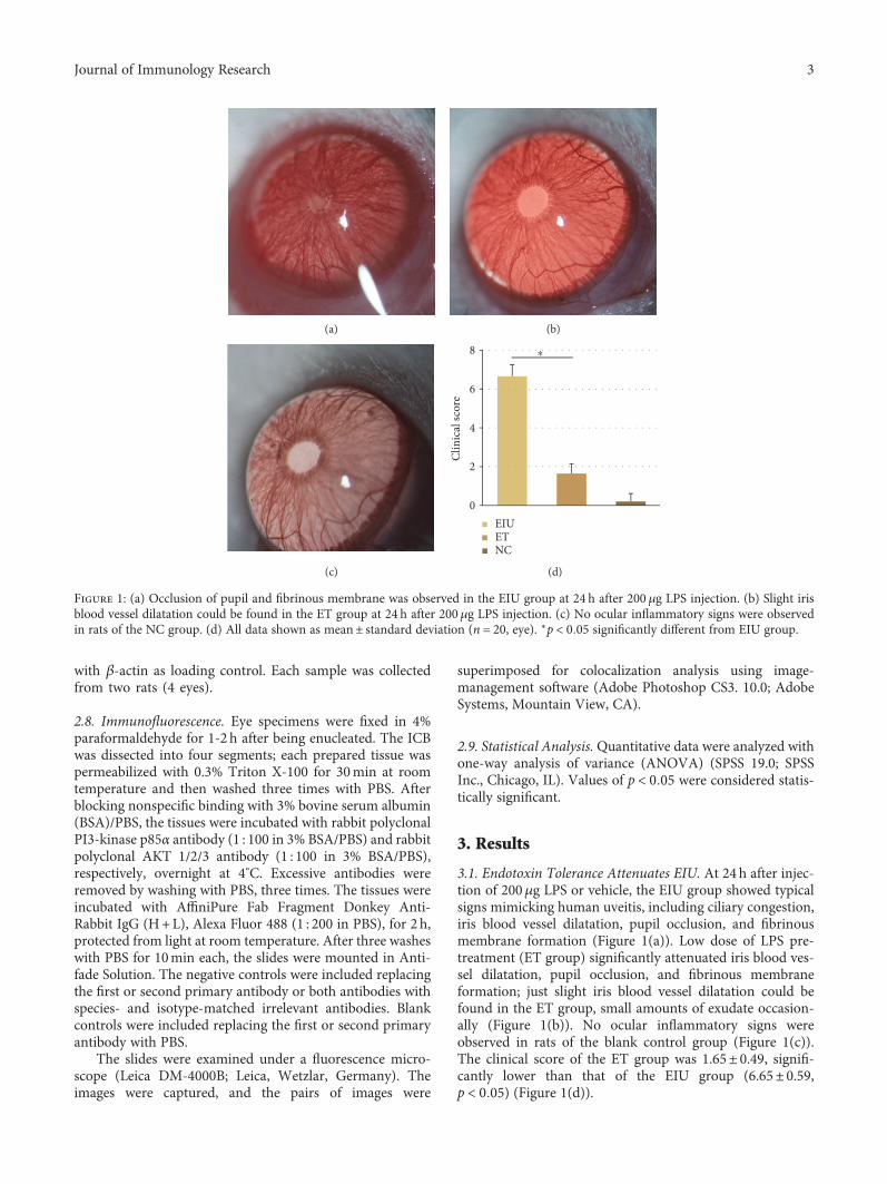
3.1. Endotoxin Tolerance Attenuates EIU. At 24 h after injec-tion of 200μg LPS or vehicle, the EIU group showed typicalsigns mimicking human uveitis, including ciliary congestion,iris blood vessel dilatation, pupil occlusion, and fibrinousmembrane formation (Figure 1(a)). Low dose of LPS pre-treatment (ET group) significantly attenuated iris blood ves-sel dilatation, pupil occlusion, and fibrinous membraneformation; just slight iris blood vessel dilatation could befound in the ET group, small amounts of exudate occasion-ally (Figure 1(b)). No ocular inflammatory signs wereobserved in rats of the blank control group (Figure 1(c)).The clinical score of the ET group was 1.65± 0.49, signifi-cantly lower than that of the EIU group (6.65± 0.59,p < 0 05) (Figure 1(d)).
(a) (b)
(c)
0
2
4
6
8 ⁎
Clin
ical
scor
e
EIUETNC
(d)
Figure 1: (a) Occlusion of pupil and fibrinous membrane was observed in the EIU group at 24 h after 200 μg LPS injection. (b) Slight irisblood vessel dilatation could be found in the ET group at 24 h after 200μg LPS injection. (c) No ocular inflammatory signs were observedin rats of the NC group. (d) All data shown as mean± standard deviation (n = 20, eye). ∗p < 0 05 significantly different from EIU group.
3Journal of Immunology Research
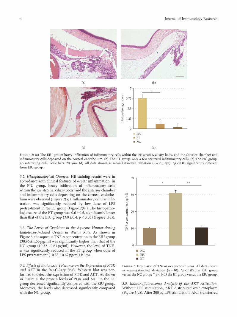
3.2. Histopathological Changes. HE staining results were inaccordance with clinical features of ocular inflammation. Inthe EIU group, heavy infiltration of inflammatory cellswithin the iris stroma, ciliary body, and the anterior chamberand inflammatory cells depositing on the corneal endothe-lium were observed (Figure 2(a)). Inflammatory cellular infil-tration was significantly reduced by low dose of LPSpretreatment in the ET group (Figure 2(b)). The histopatho-logic score of the ET group was 0.6± 0.5, significantly lowerthan that of the EIU group (3.8± 0.4, p < 0 05) (Figure 1(d)).
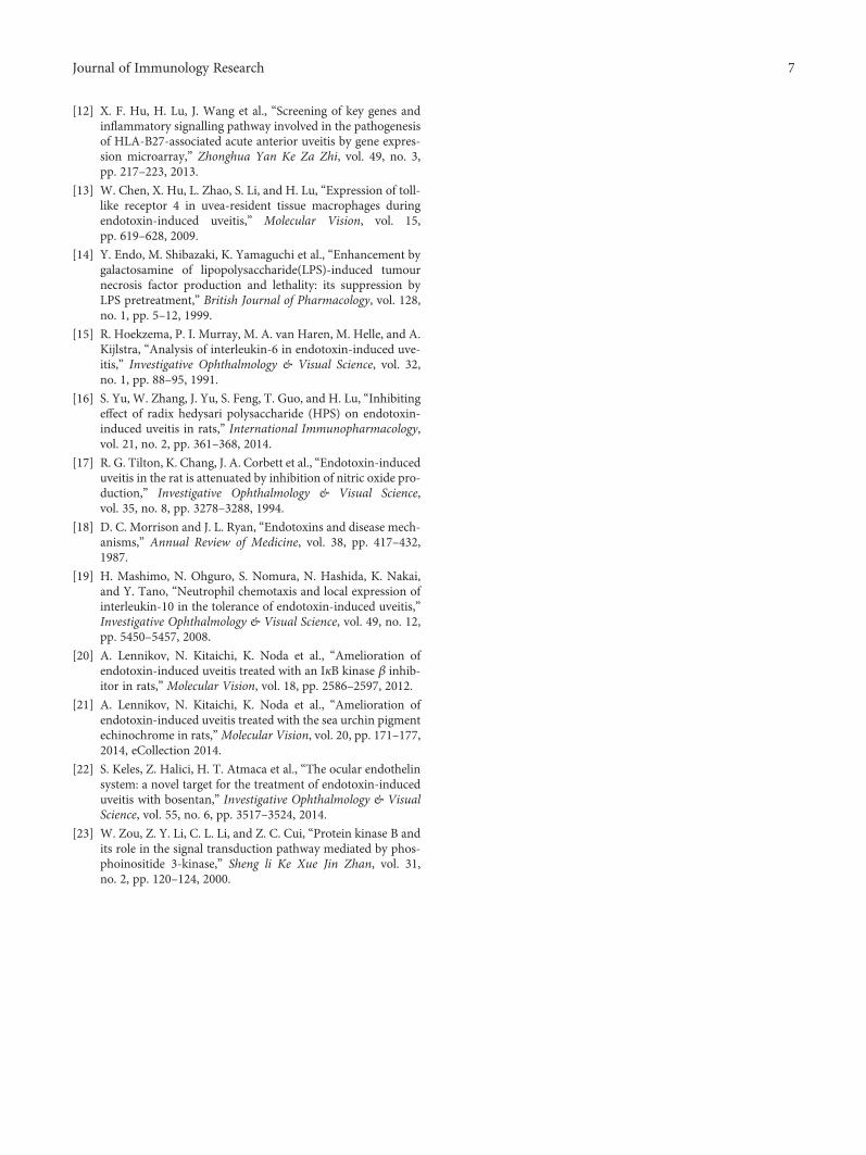
3.3. The Levels of Cytokines in the Aqueous Humor duringEndotoxin-Induced Uveitis in Wistar Rats. As shown inFigure 3, the aqueous TNF-α concentration in the EIU group(30.96± 1.55 pg/ml) was significantly higher than that of theNC group (10.32± 0.61 pg/ml). However, the level of TNF-α was significantly reduced in the ET group when dose ofLPS pretreatment (10.58± 0.67 pg/ml) is low.
3.4. Effects of Endotoxin Tolerance on the Expression of PI3Kand AKT in the Iris-Ciliary Body. Western blot was per-formed to detect the expression of PI3K and AKT. As shownin Figure 4, the protein levels of PI3K and AKT in the ETgroup decreased significantly compared with the EIU group.Moreover, the levels also decreased significantly comparedwith the NC group.
3.5. Immunofluorescence Analysis of the AKT Activation.Without LPS stimulation, AKT distributed over cytoplasm(Figure 5(a)). After 200μg LPS stimulation, AKT transferred
(a) (b)
(c)
0
1.25
2.5
3.75
5
Hist
opat
holo
gic
scor
e
EIUETNC
⁎
(d)
Figure 2: (a) The EIU group: heavy infiltration of inflammatory cells within the iris stroma, ciliary body, and the anterior chamber andinflammatory cells deposited on the corneal endothelium. (b) The ET group: only a few scattered inflammatory cells. (c) The NC group:no infiltrating cells. Scale bars: 200 μm. (d) All data shown as mean± standard deviation (n = 20, eye). ∗p < 0 05 significantly differentfrom EIU group.
0
10
20
30
40
TNF-�훼
con
cent
ratio
n (p
g/m
l)
NCEIUET
⁎ ⁎⁎
Figure 3: Expression of TNF-α in aqueous humor. All data shownas mean± standard deviation (n = 10). ∗p < 0 05 the EIU groupversus the NC group; ∗∗p < 0 05 the ET group versus the EIU group.
4 Journal of Immunology Research

from cytoplasm to cell membrane, representing activation ofAKT (Figure 5(b)). However, low dose of LPS pretreatmentinhibited translocation of AKT partially (Figure 5(c)).
4. Discussion
As a potent inflammatory stimulant, high doses of LPS leadto systemic inflammatory response syndrome and death
[18]. However, if the body was given a small dose of LPSstimulation in advance, the body showed low or no responseto the subsequent large dose or lethal dose of LPS stimula-tion. This important protective mechanism is called endo-toxin tolerance [4].
To elucidate the effect of endotoxin tolerance on ocularinflammation, endotoxin-induced uveitis with that a well-established animal model of AAU was used in this study.
0
0.15
0.3
0.45
0.6
Opt
ical
den
sity
NCETEIU
NCETEIU
NC ET EIU
PI3-kinase p85�훼/�훽-actin
0
0.15
0.3
0.45
0.6
Opt
ical
den
sity
Akt 1/2/3/�훽-actin
⁎⁎
⁎
PI3-kinase p85�훼
Akt 1/2/3
�훽-Actin
⁎⁎
⁎
Figure 4: Western blot analysis and protein levels of PI3K and AKT. ∗p < 0 05 the ET group versus the EIU group; ∗∗p < 0 05 the ET groupversus the NC group. Data represent the mean± SD from ten independent experiments.
NC
(a)
EIU
(b)
ET
(c)
Figure 5: In the NC group, AKT distributed over cytoplasm (a). In the EIU group, AKT transferred from cytoplasm to cell membrane (b). Inthe ET group, translocation of AKT was inhibited partially (c). Scale bars: 50 μm.
5Journal of Immunology Research

The Wistar rats in the EIU group showed typical signs mim-icking human uveitis, including ciliary congestion, iris bloodvessel dilatation, pupil occlusion, and fibrinous membraneformation. However, the inflammatory reaction of anteriorsegment of the rats significantly decreased in the ET groupas measured by slit lamp assessment and histopathology.These results demonstrate that endotoxin tolerance whichwas induced by low dose of LPS pretreatment could success-fully suppress the ocular inflammation in the EIU model.
Further, these findings suggest that endotoxin toleranceis of great potential in the treatment and prevention of uve-itis. However, the molecular mechanisms underlying theinduction of endotoxin tolerance are not yet fully under-stood. Hoekzema et al. [15] demonstrated that repeatedinjection of endotoxin resulted in no detectable IL-6 in theaqueous humor and the absence of uveitis. Mashimo et al.[19] indicated that continuous high expression of IL-10 inthe eye plays significant role in the mechanism of endotoxintolerance in a rat model of EIU. In this study, TNF-α levels inthe aqueous humor during endotoxin-induced uveitis in ratswere investigated to further clarify the anti-inflammatoryeffect of endotoxin tolerance and explore the role of cytokinesin this protective mechanism. Consistent with the literatures[6, 20–22], elevated TNF-α was observed in the EIU group,and the TNF-α levels were significantly reduced by low doseof LPS pretreatment in ET group. This result strongly con-firmed the anti-inflammatory effect of endotoxin tolerance,which is consistent with the slit lamp assessment and histo-pathology. Furthermore, the result suggests that TNF-α aswell as IL-6 and IL-10 is closely involved in the protectivemechanism of endotoxin tolerance. However, the upstreammechanism remains unclear. As mentioned earlier, thePI3K/AKT pathway plays an important role in regulatingLPS-induced cytokine such as TNF-α expression. Therefore,we next evaluated whether endotoxin tolerance could modu-late the PI3K/AKT pathway.
Similar to many protein kinases, AKT has a specific AH/PH domain which mediates the interaction between signalingmolecules. PI3K generates phosphoinositide-3,4,5-triphos-phate (PIP3) that can bind to the AH/PH domain of AKT inresponse to upstream signals, resulting in AKT translocatingto the plasmamembrane and being activated [23]. Thus, boththe expressions of PI3K and AKT in ICB of the EIU rats andactivation of AKT were detected. In this work, the proteinlevels ofPI3KandAKTin theETgroupdecreased significantlycompared with the EIU group, which were associated with theinflammatory outcome of the rats. Moreover, the levels in theET group were even lower than those in the NC group, whichindicated that the expressionofPI3KandAKTwas suppressedwhendoseofLPSpretreatment is low. Furthermore, accordingto the immunofluorescence, the activation of AKT was alsosuppressed in the ET group. Taken together, both low expres-sions of PI3K/AKT and inhibition of AKT activation contrib-ute to the induction of endotoxin tolerance.
In summary, the present study demonstrates that lowdoseof LPS pretreatment could prevent subsequent endotoxin-induced uveitis by reducing the expression of TNF-α in theaqueous humor, and this protective effect of endotoxin toler-ance is associated with PI3K/AKT pathway. Furthermore, in
virtue of the critical roles of PI3K and AKT in HLA-B27-associated acute anterior uveitis, endotoxin tolerance holdspromise for the prevention and treatment of this sight-threatening diseases.
Conflicts of Interest
The authors declare that they have no conflicts of interest.
Acknowledgments
This study was supported by the National Nature ScienceFund of China (Grant nos. 81471575 and 81273246).
References
[1] J. H. Chang and D. Wakefield, “Uveitis: a global perspective,”Ocular Immunology and Inflammation, vol. 10, no. 4,pp. 263–279, 2002.
[2] J. H. Chang, P. J. McCluskey, and D. Wakefield, “Toll-likereceptors in ocular immunity and the immunopathogenesisof inflammatory eye disease,” The British Journal of Ophthal-mology, vol. 90, no. 1, pp. 103–108, 2006.
[3] J. H. Chang, P. J. McCluskey, and D. Wakefield, “Acute ante-rior uveitis and HLA-B27,” Survey of Ophthalmology, vol. 50,pp. 364–388, 2005.
[4] S. K. Biswas and E. Lopez-Collazo, “Endotoxin tolerance: newmechanisms, molecules and clinical significance,” Trends inImmunology, vol. 30, no. 10, pp. 475–487, 2009.
[5] J. T. Rosenbaum, H. O. McDevitt, R. B. Guss, and P. R. Egbert,“Endotoxin-induced uveitis in rats as a model for human dis-ease,” Nature, vol. 286, no. 5773, pp. 611–613, 1980.
[6] A. F. De Vos, M. A. van Haren, C. Verhagen, R. Hoekzema,and A. Kijlstra, “Kinetics of intraocular tumor necrosis factorand interleukin-6 in endotoxin-induced uveitis in the rat,”Investigative Ophthalmology & Visual Science, vol. 35,pp. 1100–1106, 1994.
[7] S. R. Planck, X. N. Huang, J. E. Robertson, and J. T.Rosenbaum, “Cytokine mRNA levels in rat ocular tissuesafter systemic endotoxin treatment,” Investigative Ophthal-mology & Visual Science, vol. 35, pp. 924–930, 1994.
[8] M. Ojaniemi, V. Glumoff, K. Harju, M. Liljeroos, K. Vuori, andM. Hallman, “Phosphatidylinositol 3-kinase is involved inToll-like receptor 4-mediated cytokine expression in mousemacrophages,” European Journal of Immunology, vol. 33,no. 3, pp. 597–605, 2003.
[9] M. M. Monick, A. B. Carter, P. K. Robeff, D. M. Flaherty,M.W. Peterson, andG.W.Hunninghake, “Lipopolysaccharideactivates Akt in human alveolar macrophages resulting innuclear accumulation and transcriptional activity of beta-catenin,” Journal of Immunology, vol. 166, no. 7, pp. 4713–4720, 2001.
[10] B. W. Jones, K. A. Heldwein, T. K. Means, J. J. Saukkonen, andM. J. Fenton, “Differential roles of Toll-like receptors in theelicitation of proinflammatory responses by macrophages,”Annals of the Rheumatic Diseases, vol. 60, Supplement 3,pp. iii6–iii12, 2001.
[11] Q. Yang, S. E. Calvano, S. F. Lowry, and I. P. Androulakis, “Adual negative regulationmodel of Toll-like receptor 4 signalingfor endotoxin preconditioning in human endotoxemia,”Mathematical Biosciences, vol. 232, no. 2, pp. 151–163, 2011.
6 Journal of Immunology Research
[12] X. F. Hu, H. Lu, J. Wang et al., “Screening of key genes andinflammatory signalling pathway involved in the pathogenesisof HLA-B27-associated acute anterior uveitis by gene expres-sion microarray,” Zhonghua Yan Ke Za Zhi, vol. 49, no. 3,pp. 217–223, 2013.
[13] W. Chen, X. Hu, L. Zhao, S. Li, and H. Lu, “Expression of toll-like receptor 4 in uvea-resident tissue macrophages duringendotoxin-induced uveitis,” Molecular Vision, vol. 15,pp. 619–628, 2009.
[14] Y. Endo, M. Shibazaki, K. Yamaguchi et al., “Enhancement bygalactosamine of lipopolysaccharide(LPS)-induced tumournecrosis factor production and lethality: its suppression byLPS pretreatment,” British Journal of Pharmacology, vol. 128,no. 1, pp. 5–12, 1999.
[15] R. Hoekzema, P. I. Murray, M. A. van Haren, M. Helle, and A.Kijlstra, “Analysis of interleukin-6 in endotoxin-induced uve-itis,” Investigative Ophthalmology & Visual Science, vol. 32,no. 1, pp. 88–95, 1991.
[16] S. Yu, W. Zhang, J. Yu, S. Feng, T. Guo, and H. Lu, “Inhibitingeffect of radix hedysari polysaccharide (HPS) on endotoxin-induced uveitis in rats,” International Immunopharmacology,vol. 21, no. 2, pp. 361–368, 2014.
[17] R. G. Tilton, K. Chang, J. A. Corbett et al., “Endotoxin-induceduveitis in the rat is attenuated by inhibition of nitric oxide pro-duction,” Investigative Ophthalmology & Visual Science,vol. 35, no. 8, pp. 3278–3288, 1994.
[18] D. C. Morrison and J. L. Ryan, “Endotoxins and disease mech-anisms,” Annual Review of Medicine, vol. 38, pp. 417–432,1987.
[19] H. Mashimo, N. Ohguro, S. Nomura, N. Hashida, K. Nakai,and Y. Tano, “Neutrophil chemotaxis and local expression ofinterleukin-10 in the tolerance of endotoxin-induced uveitis,”Investigative Ophthalmology & Visual Science, vol. 49, no. 12,pp. 5450–5457, 2008.
[20] A. Lennikov, N. Kitaichi, K. Noda et al., “Amelioration ofendotoxin-induced uveitis treated with an IκB kinase β inhib-itor in rats,” Molecular Vision, vol. 18, pp. 2586–2597, 2012.
[21] A. Lennikov, N. Kitaichi, K. Noda et al., “Amelioration ofendotoxin-induced uveitis treated with the sea urchin pigmentechinochrome in rats,”Molecular Vision, vol. 20, pp. 171–177,2014, eCollection 2014.
[22] S. Keles, Z. Halici, H. T. Atmaca et al., “The ocular endothelinsystem: a novel target for the treatment of endotoxin-induceduveitis with bosentan,” Investigative Ophthalmology & VisualScience, vol. 55, no. 6, pp. 3517–3524, 2014.
[23] W. Zou, Z. Y. Li, C. L. Li, and Z. C. Cui, “Protein kinase B andits role in the signal transduction pathway mediated by phos-phoinositide 3-kinase,” Sheng li Ke Xue Jin Zhan, vol. 31,no. 2, pp. 120–124, 2000.
7Journal of Immunology Research

Submit your manuscripts athttps://www.hindawi.com
Stem CellsInternational
Hindawi Publishing Corporationhttp://www.hindawi.com Volume 2014
Hindawi Publishing Corporationhttp://www.hindawi.com Volume 2014
MEDIATORSINFLAMMATION
of
Hindawi Publishing Corporationhttp://www.hindawi.com Volume 2014
Behavioural Neurology
EndocrinologyInternational Journal of
Hindawi Publishing Corporationhttp://www.hindawi.com Volume 2014
Hindawi Publishing Corporationhttp://www.hindawi.com Volume 2014
Disease Markers
Hindawi Publishing Corporationhttp://www.hindawi.com Volume 2014
BioMed Research International
OncologyJournal of
Hindawi Publishing Corporationhttp://www.hindawi.com Volume 2014
Hindawi Publishing Corporationhttp://www.hindawi.com Volume 2014
Oxidative Medicine and Cellular Longevity
Hindawi Publishing Corporationhttp://www.hindawi.com Volume 2014
PPAR Research
The Scientific World JournalHindawi Publishing Corporation http://www.hindawi.com Volume 2014
Immunology ResearchHindawi Publishing Corporationhttp://www.hindawi.com Volume 2014
Journal of
ObesityJournal of
Hindawi Publishing Corporationhttp://www.hindawi.com Volume 2014
Hindawi Publishing Corporationhttp://www.hindawi.com Volume 2014
Computational and Mathematical Methods in Medicine
OphthalmologyJournal of
Hindawi Publishing Corporationhttp://www.hindawi.com Volume 2014
Diabetes ResearchJournal of
Hindawi Publishing Corporationhttp://www.hindawi.com Volume 2014
Hindawi Publishing Corporationhttp://www.hindawi.com Volume 2014
Research and TreatmentAIDS
Hindawi Publishing Corporationhttp://www.hindawi.com Volume 2014
Gastroenterology Research and Practice
Hindawi Publishing Corporationhttp://www.hindawi.com Volume 2014
Parkinson’s Disease
Evidence-Based Complementary and Alternative Medicine
Volume 2014Hindawi Publishing Corporationhttp://www.hindawi.com